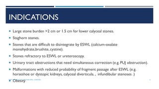
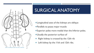
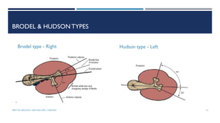
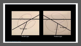
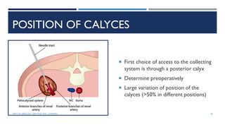
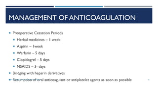
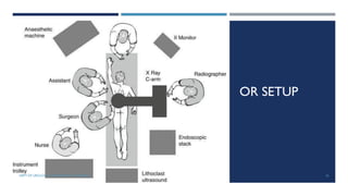
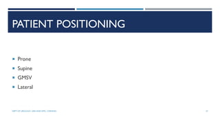
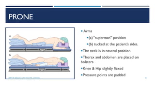
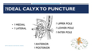
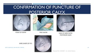
Dept of Urology
Govt Royapettah Hospital and Kilpauk Medical College
Chennai
1
MODERATORS:
Professors:
 Prof. Dr. G. Sivasankar, M.S., M.Ch.,
 Prof. Dr.A. Senthilvel, M.S., M.Ch.,
Asst Professors:
 Dr. J. Sivabalan, M.S., M.Ch.,
 Dr. R. Bhargavi, M.S., M.Ch.,
 Dr. S. Raju,M.S., M.Ch.,
 Dr. K. Muthurathinam, M.S., M.Ch.,
 Dr. D.Tamilselvan, M.S., M.Ch.,
 Dr. K. Senthilkumar, M.S., M.Ch.
DEPT OF UROLOGY, GRH AND KMC, CHENNAI. 2
INTRODUCTION
 Fernstrom and Johansson first reported the technique of establishing a percutaneous
track specifically to remove a stone in 1976 .`
 PNL as a routinely used technique to treat patients with large or complex calculi
(Alken et al, 1981 ;Wickham and Kellett, 1981 ; Segura et al, 1982 ; Clayman et al,
1984)
3
DEPT OF UROLOGY, GRH AND KMC, CHENNAI.
INDICATIONS
 Large stone burden >2 cm or 1.5 cm for lower calyceal stones.
 Staghorn stones.
 Stones that are difficult to disintegrate by ESWL (calcium-oxalate
monohydrate,brushite,cystine).
 Stones refractory to ESWL or ureteroscopy.
 Urinary tract obstructions that need simultaneous correction (e.g. PUJ obstruction).
 Malformations with reduced probability of fragment passage after ESWL (e.g.
horseshoe or dystopic kidneys, calyceal diverticula. , infundibular stenoses .)
 Obesity 4
DEPT OF UROLOGY, GRH AND KMC, CHENNAI.
CONTRAINDICATIONS
 Uncorrected coagulopathy
 Untreated UTI
 Tumour in the presumptive access tract area
 Potential malignant kidney tumour
 Pregnancy
 Within 12 months of drug eluting stent / within 3 months of bare metal stent
Patients with bleeding diathesis or receiving anticoagulant therapy
must be monitored carefully pre- and post-operatively.
5
DEPT OF UROLOGY, GRH AND KMC, CHENNAI.
INVESTIGATIONS
 A complete blood count
 Serum electrolytes
 Renal function tests
 Urine culture is mandatory for all patients
 Perioperative antibiotics can be appropriately tailored to culture-specific organisms
 Typing and screening of the patient's blood
 Imaging – Xray KUB, IVU / CECT KUB with 3D reconstruction
6
DEPT OF UROLOGY, GRH AND KMC, CHENNAI.
CONSENT
 For multiple access
 Bleeding & blood transfusion
 Staged / Repeat endoscopic procedures
 Need for auxillary treatment – ESWL,Angioembolisation
 Renal function loss & late complications
7
DEPT OF UROLOGY, GRH AND KMC, CHENNAI.
ANATOMIC CONSIDERATIONS
 Familiarity with basic renal anatomy is essential for access to be obtained safely
 Vascular Anatomy
 Pelvicalyceal Anatomy
8
DEPT OF UROLOGY, GRH AND KMC, CHENNAI.
SURGICAL ANATOMY
Longitudinal axes of the kidneys are oblique
Parallels to psoas major muscle
Superior poles more medial than the Inferior poles.
Usually the posterior surface of
 Right kidney is crossed by the 12th rib
 Left kidney by the 11th and 12th ribs.
9
DEPT OF UROLOGY, GRH AND KMC, CHENNAI.
TRANSVERSE SECTION
Renal frontal axis angles 30-50 degree
to the frontal axis of the body
10
DEPT OF UROLOGY, GRH AND KMC, CHENNAI.
INTRARENAL ARTERIES
InterlobarA
InterlobularA
ArcuateArtery
Afferent arteriole of the glomerulus
11
DEPT OF UROLOGY, GRH AND KMC, CHENNAI.
12
DEPT OF UROLOGY, GRH AND KMC, CHENNAI.
BRODEL’S PLANE
 Ideal point of renal entry.
 The avascular field between the
anterior and posterior divisions,
known as Brödel's bloodless line
 Because of the orientation of the
kidney in the body, entry through a
posterior calyx usually traverses this
line
13
DEPT OF UROLOGY, GRH AND KMC, CHENNAI.
BRODEL & HUDSONTYPES
Brodel type - Right Hudson type - Left
14
DEPT OF UROLOGY, GRH AND KMC, CHENNAI.
20 degrees
70 degrees
Brodel type Hudson type
15
DEPT OF UROLOGY, GRH AND KMC, CHENNAI.
INTRARENAL
VEINS
Stellate veins arcuate veins
interlobar veins  RV, renal
vein.
Three orders of arcades: 1,
first‐order arcade; 2, second‐order
arcade; 3, third‐order arcade
16
DEPT OF UROLOGY, GRH AND KMC, CHENNAI.
PELVICALYCEAL SYSTEM
 Pyramid  collecting duct  papillary
ducts  renal papilla  minor calyx
 major calyx / infundibulum  renal
pelvis
 Minor calyces: 5 to 14 (mean, 8)
17
DEPT OF UROLOGY, GRH AND KMC, CHENNAI.
SIMPLE / COMPOUND CALYX
 Single / Simple calyx drains only one
papilla
 Compound calyx drains two or three
papillae
 The polar calyces are often compound,
markedly in the superior pole
18
DEPT OF UROLOGY, GRH AND KMC, CHENNAI.
CLASSIFICATION OF PELVI CALYCEAL SYSTEM
 Sampaio Classification - based on superior pole, inferior pole, and kidney midzone
(hilar) calyceal drainage.
19
DEPT OF UROLOGY, GRH AND KMC, CHENNAI.
GROUP A – 62.2%
 Midzone calyceal drainage dependent on superior/inferior calyceal groups
 Type A‐I (45%): the kidney midzone is drained by minor calyces that are dependent
on the superior and/or inferior calyceal group
 Type A‐II (17.2%): the kidney midzone is drained simultaneously by crossed calyces,
one draining into the superior calyceal group and the other draining into the
inferior calyceal group
20
DEPT OF UROLOGY, GRH AND KMC, CHENNAI.
Type A 1 Type A 1I
Interpelvioca
lyceal space
21
DEPT OF UROLOGY, GRH AND KMC, CHENNAI.
IMPORTANCE OF IPC SPACE
 Detection of an inter-pelviocalyceal region on the pyelograms is an indirect sign of
crossed calyces in the kidney midzone.
 Crosses calyces  Inferior calyceal group – ventral in 87.5%
 Even when radiographically the calyx draining into the inferior group was apparently
in the dorsal position, its ventral position is verified on the endocast
22
DEPT OF UROLOGY, GRH AND KMC, CHENNAI.
TYPE B (37.8%)
 Midzone (hilar) calyceal drainage independent of both the superior and inferior
calyceal groups
 Type B‐I (21.4%): the kidney midzone is drained by a major calyceal group
 Type B‐II (16.4%): the kidney midzone is drained by minor calyces (one to four)
entering directly into the renal pelvis
23
DEPT OF UROLOGY, GRH AND KMC, CHENNAI.
TYPE B
24
DEPT OF UROLOGY, GRH AND KMC, CHENNAI.
FAVOURABLE
CALYX
 Unfavourable - long
and thin calyceal
infundibulum
 Favourable - short and
thick calyceal
infundibula
Favourable Unfavourable 25
DEPT OF UROLOGY, GRH AND KMC, CHENNAI.
POSITION OF CALYCES
 First choice of access to the collecting
system is through a posterior calyx
 Determine preoperatively
 Large variation of position of the
calyces (>50% in different positions)
26
DEPT OF UROLOGY, GRH AND KMC, CHENNAI.
POSITION OF THE CALYCES RELATIVE TO THE
POLAR REGIONS
 Superior pole was drained by a midline calyceal infundibulum in 98.6%
 Midzone (hilar) was drained by paired calyces that were arranged in two rows
(anterior and posterior) in 95.7%
 Inferior pole was drained by -
 paired calyces arranged in two rows (57.9%)
 single midline calyceal infundibulum (42.1%)
27
DEPT OF UROLOGY, GRH AND KMC, CHENNAI.
INFERIOR POLE – SINGLE INFUNDIBULUM
INFERIOR POLE – PAIRED CALYCES
28
DEPT OF UROLOGY, GRH AND KMC, CHENNAI.
ANTERIOR CALYCES – LATERAL & PERIPHERAL
29
DEPT OF UROLOGY, GRH AND KMC, CHENNAI.
POSTERIOR CALYCES – LATERALAND PERIPHERAL
30
DEPT OF UROLOGY, GRH AND KMC, CHENNAI.
The calyces in the
anterior plane
(arrows) are
located alternately
relative to the
lateral margin of
the kidney
In one
region they are
more lateral and in
another they are
more medial.
ALTERNATIVELY LOCATED CALYCES
31
DEPT OF UROLOGY, GRH AND KMC, CHENNAI.
PERPENDICULAR
MINOR CALYCES
 Inferior pole is drained
by paired calyces in
57.9%
 This anatomic detail
must be kept in mind,
both to plan and
perform the intrarenal
access and endoscopic
procedures in the
inferior pole
32
DEPT OF UROLOGY, GRH AND KMC, CHENNAI.
PCNLTECNIQUE
33
DEPT OF UROLOGY, GRH AND KMC, CHENNAI.
PERIPROCEDURAL ANTIBIOTICS
 Urine culture & therapeutic course to sterilise the urine
 AUA recommendation – Periprocedural antibiotics for all cases of percutaneous
renal surgery
 Antimicrobial
 Coverage – E.coli, Proteus, Klebsiella, Enterococcus;Skin – Staph.Aureus, coag
negative Staph., group A Streptococcus
 First & 2nd generation Cephalosporins,Amino glycosides/Aztreonam +
Metronidazone/clindamycin;Ampicillin/sulbactum; Fluroquinolone
 Immediate perioperative period <24 hrs before surgery & short course at the
time of nephrostomy removal
34
DEPT OF UROLOGY, GRH AND KMC, CHENNAI.
MANAGEMENT OF ANTICOAGULATION
 Preoperative Cessation Periods
 Herbal medicines – 1 week
 Aspirin – 1week
 Warfarin – 5 days
 Clopidogrel – 5 days
 NSAIDS – 3- days
 Bridging with heparin derivatives
 Resumption of oral anticoagulant or antiplatelet agents as soon as possible 35
DEPT OF UROLOGY, GRH AND KMC, CHENNAI.
OR SETUP
36
DEPT OF UROLOGY, GRH AND KMC, CHENNAI.
PATIENT POSITIONING
 Prone
 Supine
 GMSV
 Lateral
37
DEPT OF UROLOGY, GRH AND KMC, CHENNAI.
PRONE
 Arms
(a) “superman” position
(b) tucked at the patient’s sides.
The neck is in neutral position
Thorax and abdomen are placed on
bolsters
Knee & Hip slightly flexed
Pressure points are padded
38
DEPT OF UROLOGY, GRH AND KMC, CHENNAI.
PRONE POSITION
 Accidental extubation and kinking of the endotracheal tube during positioning
 Hampered ventilation – Decreased pulmonary capacity
 Altered circulation – decrease in cardiac index
 Torsion of the neck
 Pressure and transient ischemia of the eyeballs
 Overstretching, and pressure injuries of the peripheral plexuses
39
DEPT OF UROLOGY, GRH AND KMC, CHENNAI.
PRONE POSITION
40
DEPT OF UROLOGY, GRH AND KMC, CHENNAI.
SUPINE
 Dr. Jose GabrielValdivia Uria
 Supine position with an air bag under the flank
 MODIFICATIONS:
 Supine modified - modified supine
 Galdakao modified supineValdivia
 Semisupine & supine‐oblique positions
41
DEPT OF UROLOGY, GRH AND KMC, CHENNAI.
GMSV
 Galdakao‐modified supineValdivia (GMSV) position
 Combination of the supineValdivia position with a modified lithotomic arrangement
of the lower limbs.
42
DEPT OF UROLOGY, GRH AND KMC, CHENNAI.
Flank to be operated near to the border
Elevated 20–30
Extended,and only slightly abducted
Contralateral one is lifted, flexed,and well abducted
Arm on the side to be operated is bent at the thorax
Contralateral one lies abducted less than 90
43
DEPT OF UROLOGY, GRH AND KMC, CHENNAI.
SUPINE POSITIONS - ADVANTAGES
 Easy simultaneous combined antegrade and retrograde approach to the upper
urinary tract for stone treatment with both rigid and flexible endoscopes
 Optimal cardiovascular and airway control
44
DEPT OF UROLOGY, GRH AND KMC, CHENNAI.
SUPINE POSITIONS - DISADVANTAGES
 Unfamiliar
 Reduced pressure in collecting system
 Less room for visualisation and manipulation
 Upper pole calyceal access is more difficult
 Longer percutaneous tract length
45
DEPT OF UROLOGY, GRH AND KMC, CHENNAI.
LATERAL DECUBITUS POSITION
 Less commonly used
 Allows simultaneous access to anterior & posterior calyces
 Useful for morbidly obese & spinal deformity
46
DEPT OF UROLOGY, GRH AND KMC, CHENNAI.
INTRARENAL ACCESS
 ? PELVIS
 ? INFUNDIBULUM
 ? CALYX
47
DEPT OF UROLOGY, GRH AND KMC, CHENNAI.
ACCESSTHROUGH PELVIS
 SHOULD NEVER BE PERFORMED
 Difficult to reintroduce during the
operative maneuvers
 Unnecessary risk of injuring a
retropelvic vessel
 Nephrostomy tube inserted at this
site is easily dislodged
48
DEPT OF UROLOGY, GRH AND KMC, CHENNAI.
ACCESSTHROUGH INFUNDIBULUM
 Puncture through an infundibulum (in any region of the kidney) presents clear
hazards
49
DEPT OF UROLOGY, GRH AND KMC, CHENNAI.
UPPER POLE INFUNDIBULUM
 Most dangerous
 Surrounded almost completely by large vessels
 Infundibular arteries and veins course parallel to the anterior and posterior aspects
of the upper pole infundibulum.
 Injury to an interlobar (infundibular) vessel – 67%
 Injured vessel was an artery in 26%
 Most serious vascular accident - posterior segmental artery
 Crossed by and is related to the posterior surface of the upper
infundibulum in 57% 50
DEPT OF UROLOGY, GRH AND KMC, CHENNAI.
UPPER POLE PUNCTURE THROUGH INFUNDIBULUM
Upper infundibulum almost completely
encircled by infundibular arteries and
veins.
This anatomic arrangement makes
upper pole infundibular puncture
especially dangerous
51
DEPT OF UROLOGY, GRH AND KMC, CHENNAI.
UPPER POLE PUNCTURE THROUGH INFUNDIBULUM
Posterior segmental artery (retropelvic artery) crossing
the posterior surface of the upper infundibulum (arrow).
May supply up to 50% of the renal parenchyma
Injury to it may result in significant loss of
functioning
renal tissue, as well as causing hemorrhage
52
DEPT OF UROLOGY, GRH AND KMC, CHENNAI.
ACCESSTHROUGH IP INFUNDIBULUM
 Arterial lesion in 23% of the kidneys studied.
 Most commonly injured vessel – Middle branch of posterior segmental artery
 Through and through perforation of the collecting system
53
DEPT OF UROLOGY, GRH AND KMC, CHENNAI.
THROUGH &THROUGH PERFORATION
 Marked hemorrhage may occur as a
result of an anterior
through‐and‐through perforation.
 Effective tamponade of injured
anterior vessels is difficult because
they lie distantly in the nephrostomy
tract
54
DEPT OF UROLOGY, GRH AND KMC, CHENNAI.
ACCESSTHROUGH LP INFUNDIBULUM
 Posterior aspect of the lower pole infundibulum is widely presumed to be free of
arteries.
 Considered to be a safe region through which to gain access to the collecting system
and to place a nephrostomy tube.
 However
 About 38% - an infundibular artery was found in this region
 Arterial injury – around 13%
55
DEPT OF UROLOGY, GRH AND KMC, CHENNAI.
ACCESS THROUGH LP INFUNDIBULUM
Large venous anastomoses
Puncture through the lower pole
infundibulum risks injury to a venous
arcade
A venous lesion usually heals
spontaneously, but consequent
hemorrhage may be problematic
during the procedure.
56
DEPT OF UROLOGY, GRH AND KMC, CHENNAI.
ACCESSTHROUGH INFUNDIBULUM
 Not a safe route
 Poses an important risk of significant bleeding from interlobar (infundibular) vessels
 Through and‐through (two‐wall) puncture of the collecting system
 Infundibular access is feasible in some circumstances and must be considered in
specific situations (e.G. Some difficult anatomic cases)
 Must evaluate the risk of an arterial lesion, primarily in the superior pole and in the
mid kidney
57
DEPT OF UROLOGY, GRH AND KMC, CHENNAI.
ACCESS THROUGH A CALYCEAL FORNIX
 Safe and should be the site chosen by
the operator
 Even in the superior pole, intrarenal
puncture through a calyceal fornix is
harmless
 Injury is always to a peripheric vessel,
such as a small venous arcade
58
DEPT OF UROLOGY, GRH AND KMC, CHENNAI.
59
DEPT OF UROLOGY, GRH AND KMC, CHENNAI.
?IDEAL CALYXTO PUNCTURE
? UPPER POLE
? LOWER POLE
? INTER POLE
? ANTERIOR
? POSTERIOR
• ? MEDIAL
• ? LATERAL
60
DEPT OF UROLOGY, GRH AND KMC, CHENNAI.
IDEAL CALYX
 Careful appreciation of the collecting system anatomy and the position of stone/ s
within it.
 Should allow easy and maximal visualization of the pelvis/upper ureter and as many of
the calyces as possible.
 At the end of the PCNL all or the maximum amount of the stone should have been
retrieved without losing fragments down the ureter.
61
DEPT OF UROLOGY, GRH AND KMC, CHENNAI.
IDEAL CALYX
 Easier to access endoscopically a polar region drained by a single infundibulum, which
usually has suitable diameter, rather than a polar region drained by paired calyces.
 For best access to the pelvic-ureteric junction (PUJ) one should choose a pole whose
calyx forms an angle of 90° or more with the PUJ
 Select a pole for puncture which provides the straightest path along the stone axis
62
DEPT OF UROLOGY, GRH AND KMC, CHENNAI.
IDEAL CALYX
 A lower pole, postero-Iateral puncture of the centre of the calyx is theoretically the
safest.
 The upper pole is more posterior and allows for easier navigation but has to be
approached with due care.
63
DEPT OF UROLOGY, GRH AND KMC, CHENNAI.
POSTERIOR CALYCEAL PUNCTURE
Closer to the skin surface in the prone
position.
Lies in Brodel’s avascular plane
Route from a posterior to an adjacent
anterior calyx or the renal pelvis is more
or less in a straight line forward.
Easier to negotiate a wire out of the
calyx and into the ureter
64
DEPT OF UROLOGY, GRH AND KMC, CHENNAI.
TO IDENTIFY A POSTERIOR CALYX
 Air pyelogram  Posterior calyces are
opacified
 Along the renal axis – End on view –
calyx appears short and wider
 Away from the calyx – lateral view –
appears longer
65
DEPT OF UROLOGY, GRH AND KMC, CHENNAI.
RENAL ACCESS
Posterior calyces allow access to anterior
calyces
Anterior calyceal entry poorer for intra renal
navigation
66
DEPT OF UROLOGY, GRH AND KMC, CHENNAI.
UPPER POLE PUNCTURES
 More posterior
 Entry is easier
 Navigation down to the PUJ simple and even the upper half of the ureter is accessible
as the navigation route is more or less 'downhill’.
 Disadvantages
 Intercostal puncture is often necessary with the risk of pleural or intercostal
artery damage
 Possibility of puncturing the posterior division of the renal artery,
 Postoperative pain from pleural and intercostal muscle irritation 67
DEPT OF UROLOGY, GRH AND KMC, CHENNAI.
RENAL ACCESS
Upper pole – allows deep access of PUJ / Upper ureter; Injury to
posterior division artery,pleura
LOWER POLE – Ideal and most commonly used
INTERPOLE – Rarely used;AdditionalTract / Anatomic abnormalities
68
DEPT OF UROLOGY, GRH AND KMC, CHENNAI.
RENAL ACCESS
 Lateral or Medial?
 Lateral – As it traverses the Brodel’s
avascular plane
69
DEPT OF UROLOGY, GRH AND KMC, CHENNAI.
SAFEST SITE OF INTRARENAL ACCESS
CENTRE OFTHE CALYCEAL FORNIX
70
DEPT OF UROLOGY, GRH AND KMC, CHENNAI.
IDEAL CALYX
 Posterior
 Inferior pole
 Lateral
71
DEPT OF UROLOGY, GRH AND KMC, CHENNAI.
IDEAL PUNCTURE
 Performed from a posterolateral position
 Performed through the renal parenchyma thick enough to
maintain a stable path
 Toward the center of the calyx posterolaterally
 Toward the center of the renal pelvis
As a result of these four conditions, the trajectory does
not damage any major blood vessels.
72
DEPT OF UROLOGY, GRH AND KMC, CHENNAI.
PRE REQUISITE
 0.035 inchTeflon-coated guidewire / Terumo glidewire into the upper collecting
system.
 When the guidewire is in position, 5/6 Fr open end ureteral catheter passed into the
collecting system.
 Rigid cystoscope (with the patient in lithotomy position)
 Flexible cystoscope (with the patient prone)
73
DEPT OF UROLOGY, GRH AND KMC, CHENNAI.
RETROGRADE PYELOGRAM
 Retrograde pyelogram
 Ureteral occlusion balloon / open end ureteric catheter is then inserted over a
guidewire & positioned at the ureteropelvic junction
 Ureteral catheter / occlusion balloon is secured externally to a foley catheter
 Using a tegaderm™ (3 m) to allow for easy separation
 Tied with 2‐0 silk to secure them in place.
 And wrapped in sterile towels where is exits from the urethra
 Allowing for sterile insertion of a double J stent on a tether in a retrograde fashion
at the end of the procedure 74
DEPT OF UROLOGY, GRH AND KMC, CHENNAI.
 A three‐way valve is attached and connected to a 10 ml syringe and intravenous
tubing leading to a bottle of contrast.
 This allows aspiration and infusion of additional contrast as needed during the
case..
 Flow of contrast under gravity distends the collecting system, providing a larger
target for access.
75
DEPT OF UROLOGY, GRH AND KMC, CHENNAI.
INTRA RENAL ACCESS
 Fluroscopic
 Ultrasound guided
 CT guided
 Laparoscopic guided
76
DEPT OF UROLOGY, GRH AND KMC, CHENNAI.
FLUROSCOPIC ACCESSTECHNIQUES
 Bull’s eye technique
 Triangulation technique
 Hybrid technique
77
DEPT OF UROLOGY, GRH AND KMC, CHENNAI.
BASIC C – ARM
POSITIONING
78
DEPT OF UROLOGY, GRH AND KMC, CHENNAI.
C ARM AT 90
DEGREE
79
DEPT OF UROLOGY, GRH AND KMC, CHENNAI.
C ARM AT 30
DEGREE
TOWARDSTHE
SURGEON
80
DEPT OF UROLOGY, GRH AND KMC, CHENNAI.
C ARM AT 30
DEGREE
TOWARDSTHE
OPERATOR
81
DEPT OF UROLOGY, GRH AND KMC, CHENNAI.
C ARM AT 30
DEGREE
CEPHALAD
82
DEPT OF UROLOGY, GRH AND KMC, CHENNAI.
C ARM AT 30
DEGREE
CAUDAD
83
DEPT OF UROLOGY, GRH AND KMC, CHENNAI.
PCNL ACCESS
TECHNIQUES
84
DEPT OF UROLOGY, GRH AND KMC, CHENNAI.
TECHNIQUES FOR RENAL ACCESS
 ANTEGRADE
 RETROGRADE
85
DEPT OF UROLOGY, GRH AND KMC, CHENNAI.
INITIAL PUNCTURE NEEDLE
 The standard choices for the needle are
 21-gauge needle (relatively minor injury)
through which is passed a 0.018-inch
guidewire
 18-gauge needle through which is passed a
standard 0.035-inch guidewire.
 Both needles have a blunt sheath, a sharp
obturator
86
DEPT OF UROLOGY, GRH AND KMC, CHENNAI.
ANTEGRADE ACCESS
 Site of entry
 Angle of entry
 Depth of the puncture
87
DEPT OF UROLOGY, GRH AND KMC, CHENNAI.
SITE OF SKIN ENTRY
 Depends on the fluoroscopic technique used
 Bull’s eye – direct under fluoroscopy
 Triangulation technique – puncture along the stone axis i.e in alignment with the
infundibulum
 Hybrid technique – Mathematically calculated
88
DEPT OF UROLOGY, GRH AND KMC, CHENNAI.
SITE OF SKIN ENTRY
 Reference lines
 Posterior axillary line
 Costal margin
 Iliac crest
 Mid scapular line
89
DEPT OF UROLOGY, GRH AND KMC, CHENNAI.
SKIN ENTRY
 Too medial or lateral – tract of variable length and angle of entry
 Medial to posterior axillary line – avoid injury to colon
 Too medial – traverse paraspinal muscles  increased postoperative pain, direct
puncture to pelvis
 Too close to the rib – intercostal nerve & vessel injury, pleural injury
90
DEPT OF UROLOGY, GRH AND KMC, CHENNAI.
BULL’S EYE TECHNIQUE
 Site of skin entry – Directly towards the target calyx
 Angle – Angle at which the needle forms bull’s eye
 Depth – at 0 degrees
91
DEPT OF UROLOGY, GRH AND KMC, CHENNAI.
92
DEPT OF UROLOGY, GRH AND KMC, CHENNAI.
TRIANGULATION TECHNIQUE
 Skin puncture – Along the stone axis in alignment with the infundibulum
 Withdraw the needle laterally (around 4 cms) along its axis
 Angle – 30-45 degree from frontal plane
 Depth – 30 degrees CC/towards the surgeon
 Fluroscopic view
 Mediolateral axis – 0 degree
 Depth - 30 degrees
93
DEPT OF UROLOGY, GRH AND KMC, CHENNAI.
94
DEPT OF UROLOGY, GRH AND KMC, CHENNAI.
HYBRID
TECHNIQUE
SITE OF SKIN
ENTRY
95
DEPT OF UROLOGY, GRH AND KMC, CHENNAI.
ANGLE OF PUNCTURE
 Angle of puncture – 30 degrees at
Point B
96
DEPT OF UROLOGY, GRH AND KMC, CHENNAI.
DEPTH OF THE PUNCTURE
 Calculated mathematically
 One side of triangle – AB
 One angle – 90 degree with Carm at 0 degree
 Another angle – Measured using the protractor at point
B
B A
C 97
DEPT OF UROLOGY, GRH AND KMC, CHENNAI.
UNIVERSAL LAW OF SINES
C
A
B
98
DEPT OF UROLOGY, GRH AND KMC, CHENNAI.
99
DEPT OF UROLOGY, GRH AND KMC, CHENNAI.
RETROGRADE ACCESS
 Indications
 Surgeon has limited experience with antegrade percutaneous
 Morbid obesity
 Hypermobile or abnormally situated kidney
100
DEPT OF UROLOGY, GRH AND KMC, CHENNAI.
LAWSON RG NEPHROSTOMY WIRE PUNCTURE SET
 7-Fr Torcon catheter (actively deflectable from 0 to
140 degrees)
 3-Fr polytetrafluoroethylene (PTFE) sheath containing
the 0.017-inch stainless steel puncture wire
Cook Urological,Spencer,IN
101
DEPT OF UROLOGY, GRH AND KMC, CHENNAI.
LAWSON RG NEPHROSTOMY WIRE PUNCTURE SET
 Torcon catheter passed through the guide wire and
into the desired calyx
 Insert the puncture wire through the Torcon catheter.
 Advance the puncture wire through the kidney and
body wall under fluoroscopic control
102
DEPT OF UROLOGY, GRH AND KMC, CHENNAI.
LAWSON RG NEPHROSTOMY WIRE PUNCTURE SET
 Make a small skin incision and grasp the wire
externally.
 Use the fascial dilators in an antegrade fashion until
theTorcon catheter can be advanced through the
tract.
 Once the end of the catheter exits the skin, exchange
the puncture wire for a standard 0.035 inch guidewire,
thus attaining through-and-through access.
103
DEPT OF UROLOGY, GRH AND KMC, CHENNAI.
CONFIRMATION OF PUNCTURE OF
POSTERIOR CALYX
104
DEPT OF UROLOGY, GRH AND KMC, CHENNAI.
PASSAGE OF GLIDE /GUIDE WIRE
 0.035 inch GW initially hydrophilic  replaced with 0.035 zebra or Amplatz super
stiff GW
105
DEPT OF UROLOGY, GRH AND KMC, CHENNAI.
SECURING THE INTRARENAL ACCESS
 Stiff,straight wire path makes dilatation much easier.
 Time spent in securing a stiff wire is always rewarded.
 The hydrophilic wire should be exchanged for an amplatz super-stiff wire.
 Rigid wire will orientate the calyx, infundibulum, the renal pelvis and the ureter
into a straight path without acute angles.
 Avoids kinking of the wire during dilatation.
 A stiff wire down the ureter and curled in the bladder is the most secure for
dilatation
106
DEPT OF UROLOGY, GRH AND KMC, CHENNAI.
SECURING THE INTRARENAL ACCESS
107
DEPT OF UROLOGY, GRH AND KMC, CHENNAI.
INCISING SKIN & FASCIA
 Using the knife along the needle under fluoroscopic control
 Incised at two planes at right angles to each other
 Can also use 18G coaxial fascial incising needle
 Care ! – subcostal / intercostal neurovascular bundle on the inferior rib margin
108
DEPT OF UROLOGY, GRH AND KMC, CHENNAI.
GW & SAFETY WIRE
 Pass the GW all the way to the bladder
 Placement of second safety GW to access the tract in event of inadvertent slipping
out of working GW
 Safety GW – introduced alongside the initial wire using a dual lumen catheter or
8/10 Fr coaxial dilator of the dilatation canula
109
DEPT OF UROLOGY, GRH AND KMC, CHENNAI.
TRACK DILATATION
 Tract size – 24 – 30 in most cases
 Dilators
 Sequential teflon
 Single step
 Telescopic Alken’s metal dilators
 Amplatz dilators
 Balloon dilator
110
DEPT OF UROLOGY, GRH AND KMC, CHENNAI.
111
DEPT OF UROLOGY, GRH AND KMC, CHENNAI.
TRACK DILATATION
 Adequate skin incision for safe dilatation
 Every step monitored on fluoroscopy
 Collecting system should be kept distended
 Tract should be dilated only till the minor calyx
 If the infundibulum is narrow do not advance the dilators/ sheath across the
infundibulum
 Dilate using a rotatory motion with slow advancement
 Do not push a dilator over a kinked guidewire - tear the collecting system 112
DEPT OF UROLOGY, GRH AND KMC, CHENNAI.
AMPLATZ SHEATH
 Maintains the tract
 Tamponade of the tract & reduces bleeding
 Beveled end uses to tamponade a part of renal parenchyma
 Protects parenchyma from injury by the instruments used
 Maintains low pressure system – reduces fluid intravasation, sepsis in infected calculi
113
DEPT OF UROLOGY, GRH AND KMC, CHENNAI.
TROUBLE SHOOTING
IN PCNL
114
DEPT OF UROLOGY, GRH AND KMC, CHENNAI.
FAILURETO OPACIFY THE SYSTEM
CAUSE REMEDY
Ureteric catheter slipping out while positioning Fix to the per urethral catheter
Placing GW across the calculus
Tightly impacted calculus preventing passage of
contrast
Head low position
Diluting the contrast
USG guided puncture & opacify the
system
115
DEPT OF UROLOGY, GRH AND KMC, CHENNAI.
EXTRAVASATION OF CONTRAST
CAUSE REMEDY
Large volume of contrast instilled at high
pressure
Inject small amount of diluted contrast
slowly with ureteric catheter in pelvis
Extravasation through improperly placed
ureteric catheter (large impacted calculus
with infection)
1. Give diuretic & wait for 15 mins
2. Use concentrated contrast to identify
PCS
3. USG guided access
4. Air pyelogram
5. Ureteroscopically assisted percutaneous
access
6. Angled tip angiographic catheter
7. Rarely – stage the procedure & re-
attempt after 48 hrs
Non-satisfactory first attempt at puncture
116
DEPT OF UROLOGY, GRH AND KMC, CHENNAI.
INABILITY TO PUNCTURE
Cause Remedy
Inexperience & Incorrect choice for
puncture
Presence of more experienced surgeon
Non dilated system PCS adequately filled & distended
Continuously flush saline in ureteric
catheter
Add a drop of methylene blue / betadiene
to the contrast
Use of 21 gause needle for initial puncture
USG / CT guidance
117
DEPT OF UROLOGY, GRH AND KMC, CHENNAI.
BLOOD ATTHE TIP OF THE NEEDLE
CAUSE REMEDY
• Needle at blood vessel or renal
parenchyma
• Ensure proper posterolateral calyceal
puncture
• Adjust the depth of the needle –
withdraw outside the parenchyma
• Multiple attempts • Use of 21 G needle
• Flush the ureteric catheter with saline –
clears  PCS access confirmed
118
DEPT OF UROLOGY, GRH AND KMC, CHENNAI.
MULTIPLE PUNCTURES?
 Large & complex stones & staghorn calculi
 Percutaneous calyceal lavage to flush the calculi to be picked through primary tract
 Flexible nephroscopy with laser
 Ist tract – most stone bulk removed
 Accessory tract – mini PCNL tracts for peripheral small calculi
 Upper calyx advantageous – direct access to upper calyx, pelvis, lower calyx, upper
ureter
 If a second tract is anticipated – place the guide wires in the calyces where the
second tract is expected before dilatation of primary tract 119
DEPT OF UROLOGY, GRH AND KMC, CHENNAI.
TRACT DILATATION
 Dense scar tissue – Collings knife or plasma vaporization for tract making
 Largest Amplatz dilator without initial small dilators – rapid, easy & less blood loss
 Radially expanding single step dilator– advantage of not removing the needle;
dilatation over rigid system
120
DEPT OF UROLOGY, GRH AND KMC, CHENNAI.
KINK IN GUIDEWIRE
CAUSE REMEDY
Forceful dilatation in wrong direction / against
resistance
Placement of guide rod
Dilatation in correct direction & adequate force
• 2/3 of progress by rotational screwing
movements
• 1/3 by force
Usually kinks at thoracolumbar fascia Fascia to be incised well before starting dilatation
Exchange for a new wire
Advance the kink down the ureter / pull the kink
externally
Use of super stiff wire for dilatation
121
DEPT OF UROLOGY, GRH AND KMC, CHENNAI.
LOSS OFTRACT
CAUSE REMEDY
Slipping of guide wire before dilatation Adequately park the guide wire in the collecting system
Use of safety guide wire
Use of super stiff wire
If the amplatz sheath not held properly Follow the GW with Nephroscope till it is positioned in
PCS
AdvanceAmplatz sheath over the nephroscope
Under dilatation Flushing saline during dilatation
Over dilatation (traversing opposite wall of PCS) No forceful dilatation
Dilatation till the calyx & not till the calculus
Withdraw the sheath to get back in PCS
Large perforation/significant bleeding  abandon & place
large bore nephrostomy tube;stage after 3-4 days
122
DEPT OF UROLOGY, GRH AND KMC, CHENNAI.
MOBILE KIDNEY
 Place a bolster underneath the patient to fix the kidney.
 Use a stiff wire.
 Use a balloon dilator
123
DEPT OF UROLOGY, GRH AND KMC, CHENNAI.
STAGHORN / TIGHT SYSTEM
 Use the ureteric catheter to distend and a hydrophilic wire.
 If wire will not advance past the stone, coil tip in the calyx if possible and dilate with
care
124
DEPT OF UROLOGY, GRH AND KMC, CHENNAI.
STONE TIGHTLY WITHIN CALYX
 Use the ureteric catheter to distend and a hydrophilic wire.
 If wire will not advance past the stone, coil tip in the calyx if possible and dilate with
care
 Puncture another calyx and approach the stone internally
125
DEPT OF UROLOGY, GRH AND KMC, CHENNAI.
PELVIC / INFUNDIBULAR TEAR
 There is a danger of absorption or retroperitoneal collection of irrigant.
 Procedure can continue with care but proper post-operative drainage should be
ensured
126
DEPT OF UROLOGY, GRH AND KMC, CHENNAI.
BRISK BLEEDING
 Place a balloon and tamponade the track.
 If bleeding continues consider open surgical repair or embolisation
127
DEPT OF UROLOGY, GRH AND KMC, CHENNAI.
HORSESHOE KIDNEY
 Under-rotated and malascended kidney, with numerous accessory arteries.
 Relatively immobile,which hinders intrarenal navigation
 Pre-operative 3D CT is of particular help here for access planning.
 As a general rule the upper pole, medial calyces are preferred,and the use of double-
contrast pyelography is very useful in identifying the most posterior calyces.
 Long sheaths may be necessary as the horseshoe is usually more deeply located
128
DEPT OF UROLOGY, GRH AND KMC, CHENNAI.
MAL-ROTATED KIDNEYS
 Mal-rotated kidneys: a kidney may be over- or under-rotated with the posterior
calyces facing medially or laterally.
 There may also be anomalous vessels present.
 CT scan with 3D volume reconstruction - Helps to clarify the calyceal anatomy and
its relationship with the vessels
129
DEPT OF UROLOGY, GRH AND KMC, CHENNAI.
CALYCEAL DIVERTICULUM
 Calyceal diverticulum: most are stone packed and in the upper pole.
 Needle can be readily targeted onto the stone
 Wire will either not enter the diverticulum, because of lack of space, or if it does it
cannot be manipulated across the tight calyceal neck and down the ureter
 Firm retrograde injection can help to distend both the neck and the diverticulum
 If the wire could not be passed through the neck, then it should be coiled firmly
within the diverticulum and dilatation carried out with the utmost care and slowly
130
DEPT OF UROLOGY, GRH AND KMC, CHENNAI.
BIFID / DUPLEX SYSTEM
 The importance here is to recognize these anomalies and that navigation will be
restricted.
 Such systems are also overall 'small' and prone to calyceal tearing during dilatation.
 Entry should be directly onto the stone-bearing calyx
131
DEPT OF UROLOGY, GRH AND KMC, CHENNAI.
PELVIC / THORACIC KIDNEYS
 Pre-operative CT is a must
 Percutaneous entry may require CT guidance or laparoscopic assistance to move
away interposed bowel loops or lungs.
132
DEPT OF UROLOGY, GRH AND KMC, CHENNAI.
COLONIC INJURY
 1%
 Retrorenal colon
 More common on left side
 Risk factors: thin, elderly, dilated colon, prior colon surgery or disease, horseshoe
kidney
 Prevention: Preoperative CT,Awareness of colonic gas bubble on fluoroscopy,USG
guided punctures
 Management :Abandon & place NT in colon and RP drain
133
DEPT OF UROLOGY, GRH AND KMC, CHENNAI.
LIVER & SPLENIC INJURY
 With supracostal access if above 10 th rib in normal individual
 Hepatomegaly / splenomegaly – preoperative CT to decide a safe access, CT guided
access
134
DEPT OF UROLOGY, GRH AND KMC, CHENNAI.
PLEURAL INJURY
 Asssociated with supracostal access
 12th rib access – 4%
 11th rib access – 20%
135
DEPT OF UROLOGY, GRH AND KMC, CHENNAI.
PLEURAL INJURY
 Parietal pleura crosses the 12th rib
 Medial half covered by pleura
 Mid scapular line – Parietal pleura
at 12th &Visceral Pleura at 10th rib
 Both rise cranially and laterally on
ribs
 Further rise in deep expiration
 Tracts below 11th rib made lateral
to mid scapular line 136
DEPT OF UROLOGY, GRH AND KMC, CHENNAI.
PLEURAL INJURY
 Make the tract lateral to mid scapular line
 Stay below 10th rib
 Perform puncture in deep expiration
 Minimize size of the tract as possible
 Check Costo-phrenic angle at the end of
supra costal access
 Pleural fluid collection if occurs – Chest
drain at the end of the procedure
 Thoracoscopically guided access superior
to 10th rib
137
DEPT OF UROLOGY, GRH AND KMC, CHENNAI.
EXIT STRATEGIES IN PCNL
 Externalised nephrostomy tube
 Nephroureteral stent
 Tubeless with ureteral stent
 No drainage tube
138
DEPT OF UROLOGY, GRH AND KMC, CHENNAI.
NEPHROSTOMY TUBES
 Foley and council catheters
 Malecot / malecot re entry catheter
 Cope catheter with retention string
 Nephroureteral stent
139
DEPT OF UROLOGY, GRH AND KMC, CHENNAI.
NEPHROSTOMY TUBES
 Advantages
 Good drainage
 ?Tamponade the tract & reduce haemorrhage
 Maintains percutaneous access for additional procedures
 Disadvantages
 Pain
 Urinary leak
Use smaller calibre tubes
140
DEPT OF UROLOGY, GRH AND KMC, CHENNAI.
URETERAL INTUBATION
 Greatest control & assurance of drainage
 Only be used when needed
 Morbidly obese
 Ureteral obstruction
 Ureteral injury
141
DEPT OF UROLOGY, GRH AND KMC, CHENNAI.
TUBELESS WITH URETERAL STENT
 Advantages
 Decreased pain & analgesic use
 Shorter hospital stay
 Decreased cost
 Should not be advocated in
 Significant bleeding
 Perforation of PCS
 Second percutaneous procedure anticipated 142
DEPT OF UROLOGY, GRH AND KMC, CHENNAI.
TOTALLY TUBELESS
 Selected patients with low volume stones
 Atraumatic single access
 No haemorrhage/perforation/obstruction
143
DEPT OF UROLOGY, GRH AND KMC, CHENNAI.
ADJUNCTS TO DRAINAGE WITHOUT
NEPHROSTOMY TUBE IN BLEEDING FROM TRACT
 Placing a fascial suture
 Direct monopolar cauterisation of the tract
 Cryotreatment of tract
 Insertion/instillation of hemostatic agents – surgicel, gelatin sponge/granules, fibrin
glue, collage matrix coated with fibrin glue
 Systemic enhancements to hemostasis – oral tranexamic acid
144
DEPT OF UROLOGY, GRH AND KMC, CHENNAI.
COMPLICATIONS
 Acute haemorrhage – 0.5 – 4% transfusion rate
 Delayed haemorrhage – 1% - AV fistula, arterial pseudoaneurysm
 Collecting system injury
 Visceral injury – colon, small bowel, hepatic & splenic injuries
 Pleural injury
 Postoperative fever – 15 -30%
 Sepsis – 0.5-2.5%
 Loss of renal function – 1.6%; negligible long term loss 145
DEPT OF UROLOGY, GRH AND KMC, CHENNAI.
COMPLICATIONS
 Extravasation of large amount of saline  respiratory distress, cardiac failure from
volume overload
 Venous gas embolism – air pyelography
 DVT – early ambulation; no prophylaxis for PCNL
 Collecting system obstruction – ureteral edema, clot retention
 Death – extremely rare ; reported in underlying cardiovascular conditions
146
DEPT OF UROLOGY, GRH AND KMC, CHENNAI.
THANKYOU
147
DEPT OF UROLOGY, GRH AND KMC, CHENNAI.

Urolithiasis management- pcnl

  • 1.
    Dept of Urology GovtRoyapettah Hospital and Kilpauk Medical College Chennai 1
  • 2.
    MODERATORS: Professors:  Prof. Dr.G. Sivasankar, M.S., M.Ch.,  Prof. Dr.A. Senthilvel, M.S., M.Ch., Asst Professors:  Dr. J. Sivabalan, M.S., M.Ch.,  Dr. R. Bhargavi, M.S., M.Ch.,  Dr. S. Raju,M.S., M.Ch.,  Dr. K. Muthurathinam, M.S., M.Ch.,  Dr. D.Tamilselvan, M.S., M.Ch.,  Dr. K. Senthilkumar, M.S., M.Ch. DEPT OF UROLOGY, GRH AND KMC, CHENNAI. 2
  • 3.
    INTRODUCTION  Fernstrom andJohansson first reported the technique of establishing a percutaneous track specifically to remove a stone in 1976 .`  PNL as a routinely used technique to treat patients with large or complex calculi (Alken et al, 1981 ;Wickham and Kellett, 1981 ; Segura et al, 1982 ; Clayman et al, 1984) 3 DEPT OF UROLOGY, GRH AND KMC, CHENNAI.
  • 4.
    INDICATIONS  Large stoneburden >2 cm or 1.5 cm for lower calyceal stones.  Staghorn stones.  Stones that are difficult to disintegrate by ESWL (calcium-oxalate monohydrate,brushite,cystine).  Stones refractory to ESWL or ureteroscopy.  Urinary tract obstructions that need simultaneous correction (e.g. PUJ obstruction).  Malformations with reduced probability of fragment passage after ESWL (e.g. horseshoe or dystopic kidneys, calyceal diverticula. , infundibular stenoses .)  Obesity 4 DEPT OF UROLOGY, GRH AND KMC, CHENNAI.
  • 5.
    CONTRAINDICATIONS  Uncorrected coagulopathy Untreated UTI  Tumour in the presumptive access tract area  Potential malignant kidney tumour  Pregnancy  Within 12 months of drug eluting stent / within 3 months of bare metal stent Patients with bleeding diathesis or receiving anticoagulant therapy must be monitored carefully pre- and post-operatively. 5 DEPT OF UROLOGY, GRH AND KMC, CHENNAI.
  • 6.
    INVESTIGATIONS  A completeblood count  Serum electrolytes  Renal function tests  Urine culture is mandatory for all patients  Perioperative antibiotics can be appropriately tailored to culture-specific organisms  Typing and screening of the patient's blood  Imaging – Xray KUB, IVU / CECT KUB with 3D reconstruction 6 DEPT OF UROLOGY, GRH AND KMC, CHENNAI.
  • 7.
    CONSENT  For multipleaccess  Bleeding & blood transfusion  Staged / Repeat endoscopic procedures  Need for auxillary treatment – ESWL,Angioembolisation  Renal function loss & late complications 7 DEPT OF UROLOGY, GRH AND KMC, CHENNAI.
  • 8.
    ANATOMIC CONSIDERATIONS  Familiaritywith basic renal anatomy is essential for access to be obtained safely  Vascular Anatomy  Pelvicalyceal Anatomy 8 DEPT OF UROLOGY, GRH AND KMC, CHENNAI.
  • 9.
    SURGICAL ANATOMY Longitudinal axesof the kidneys are oblique Parallels to psoas major muscle Superior poles more medial than the Inferior poles. Usually the posterior surface of  Right kidney is crossed by the 12th rib  Left kidney by the 11th and 12th ribs. 9 DEPT OF UROLOGY, GRH AND KMC, CHENNAI.
  • 10.
    TRANSVERSE SECTION Renal frontalaxis angles 30-50 degree to the frontal axis of the body 10 DEPT OF UROLOGY, GRH AND KMC, CHENNAI.
  • 11.
    INTRARENAL ARTERIES InterlobarA InterlobularA ArcuateArtery Afferent arterioleof the glomerulus 11 DEPT OF UROLOGY, GRH AND KMC, CHENNAI.
  • 12.
    12 DEPT OF UROLOGY,GRH AND KMC, CHENNAI.
  • 13.
    BRODEL’S PLANE  Idealpoint of renal entry.  The avascular field between the anterior and posterior divisions, known as Brödel's bloodless line  Because of the orientation of the kidney in the body, entry through a posterior calyx usually traverses this line 13 DEPT OF UROLOGY, GRH AND KMC, CHENNAI.
  • 14.
    BRODEL & HUDSONTYPES Brodeltype - Right Hudson type - Left 14 DEPT OF UROLOGY, GRH AND KMC, CHENNAI.
  • 15.
    20 degrees 70 degrees Brodeltype Hudson type 15 DEPT OF UROLOGY, GRH AND KMC, CHENNAI.
  • 16.
    INTRARENAL VEINS Stellate veins arcuateveins interlobar veins  RV, renal vein. Three orders of arcades: 1, first‐order arcade; 2, second‐order arcade; 3, third‐order arcade 16 DEPT OF UROLOGY, GRH AND KMC, CHENNAI.
  • 17.
    PELVICALYCEAL SYSTEM  Pyramid collecting duct  papillary ducts  renal papilla  minor calyx  major calyx / infundibulum  renal pelvis  Minor calyces: 5 to 14 (mean, 8) 17 DEPT OF UROLOGY, GRH AND KMC, CHENNAI.
  • 18.
    SIMPLE / COMPOUNDCALYX  Single / Simple calyx drains only one papilla  Compound calyx drains two or three papillae  The polar calyces are often compound, markedly in the superior pole 18 DEPT OF UROLOGY, GRH AND KMC, CHENNAI.
  • 19.
    CLASSIFICATION OF PELVICALYCEAL SYSTEM  Sampaio Classification - based on superior pole, inferior pole, and kidney midzone (hilar) calyceal drainage. 19 DEPT OF UROLOGY, GRH AND KMC, CHENNAI.
  • 20.
    GROUP A –62.2%  Midzone calyceal drainage dependent on superior/inferior calyceal groups  Type A‐I (45%): the kidney midzone is drained by minor calyces that are dependent on the superior and/or inferior calyceal group  Type A‐II (17.2%): the kidney midzone is drained simultaneously by crossed calyces, one draining into the superior calyceal group and the other draining into the inferior calyceal group 20 DEPT OF UROLOGY, GRH AND KMC, CHENNAI.
  • 21.
    Type A 1Type A 1I Interpelvioca lyceal space 21 DEPT OF UROLOGY, GRH AND KMC, CHENNAI.
  • 22.
    IMPORTANCE OF IPCSPACE  Detection of an inter-pelviocalyceal region on the pyelograms is an indirect sign of crossed calyces in the kidney midzone.  Crosses calyces  Inferior calyceal group – ventral in 87.5%  Even when radiographically the calyx draining into the inferior group was apparently in the dorsal position, its ventral position is verified on the endocast 22 DEPT OF UROLOGY, GRH AND KMC, CHENNAI.
  • 23.
    TYPE B (37.8%) Midzone (hilar) calyceal drainage independent of both the superior and inferior calyceal groups  Type B‐I (21.4%): the kidney midzone is drained by a major calyceal group  Type B‐II (16.4%): the kidney midzone is drained by minor calyces (one to four) entering directly into the renal pelvis 23 DEPT OF UROLOGY, GRH AND KMC, CHENNAI.
  • 24.
    TYPE B 24 DEPT OFUROLOGY, GRH AND KMC, CHENNAI.
  • 25.
    FAVOURABLE CALYX  Unfavourable -long and thin calyceal infundibulum  Favourable - short and thick calyceal infundibula Favourable Unfavourable 25 DEPT OF UROLOGY, GRH AND KMC, CHENNAI.
  • 26.
    POSITION OF CALYCES First choice of access to the collecting system is through a posterior calyx  Determine preoperatively  Large variation of position of the calyces (>50% in different positions) 26 DEPT OF UROLOGY, GRH AND KMC, CHENNAI.
  • 27.
    POSITION OF THECALYCES RELATIVE TO THE POLAR REGIONS  Superior pole was drained by a midline calyceal infundibulum in 98.6%  Midzone (hilar) was drained by paired calyces that were arranged in two rows (anterior and posterior) in 95.7%  Inferior pole was drained by -  paired calyces arranged in two rows (57.9%)  single midline calyceal infundibulum (42.1%) 27 DEPT OF UROLOGY, GRH AND KMC, CHENNAI.
  • 28.
    INFERIOR POLE –SINGLE INFUNDIBULUM INFERIOR POLE – PAIRED CALYCES 28 DEPT OF UROLOGY, GRH AND KMC, CHENNAI.
  • 29.
    ANTERIOR CALYCES –LATERAL & PERIPHERAL 29 DEPT OF UROLOGY, GRH AND KMC, CHENNAI.
  • 30.
    POSTERIOR CALYCES –LATERALAND PERIPHERAL 30 DEPT OF UROLOGY, GRH AND KMC, CHENNAI.
  • 31.
    The calyces inthe anterior plane (arrows) are located alternately relative to the lateral margin of the kidney In one region they are more lateral and in another they are more medial. ALTERNATIVELY LOCATED CALYCES 31 DEPT OF UROLOGY, GRH AND KMC, CHENNAI.
  • 32.
    PERPENDICULAR MINOR CALYCES  Inferiorpole is drained by paired calyces in 57.9%  This anatomic detail must be kept in mind, both to plan and perform the intrarenal access and endoscopic procedures in the inferior pole 32 DEPT OF UROLOGY, GRH AND KMC, CHENNAI.
  • 33.
    PCNLTECNIQUE 33 DEPT OF UROLOGY,GRH AND KMC, CHENNAI.
  • 34.
    PERIPROCEDURAL ANTIBIOTICS  Urineculture & therapeutic course to sterilise the urine  AUA recommendation – Periprocedural antibiotics for all cases of percutaneous renal surgery  Antimicrobial  Coverage – E.coli, Proteus, Klebsiella, Enterococcus;Skin – Staph.Aureus, coag negative Staph., group A Streptococcus  First & 2nd generation Cephalosporins,Amino glycosides/Aztreonam + Metronidazone/clindamycin;Ampicillin/sulbactum; Fluroquinolone  Immediate perioperative period <24 hrs before surgery & short course at the time of nephrostomy removal 34 DEPT OF UROLOGY, GRH AND KMC, CHENNAI.
  • 35.
    MANAGEMENT OF ANTICOAGULATION Preoperative Cessation Periods  Herbal medicines – 1 week  Aspirin – 1week  Warfarin – 5 days  Clopidogrel – 5 days  NSAIDS – 3- days  Bridging with heparin derivatives  Resumption of oral anticoagulant or antiplatelet agents as soon as possible 35 DEPT OF UROLOGY, GRH AND KMC, CHENNAI.
  • 36.
    OR SETUP 36 DEPT OFUROLOGY, GRH AND KMC, CHENNAI.
  • 37.
    PATIENT POSITIONING  Prone Supine  GMSV  Lateral 37 DEPT OF UROLOGY, GRH AND KMC, CHENNAI.
  • 38.
    PRONE  Arms (a) “superman”position (b) tucked at the patient’s sides. The neck is in neutral position Thorax and abdomen are placed on bolsters Knee & Hip slightly flexed Pressure points are padded 38 DEPT OF UROLOGY, GRH AND KMC, CHENNAI.
  • 39.
    PRONE POSITION  Accidentalextubation and kinking of the endotracheal tube during positioning  Hampered ventilation – Decreased pulmonary capacity  Altered circulation – decrease in cardiac index  Torsion of the neck  Pressure and transient ischemia of the eyeballs  Overstretching, and pressure injuries of the peripheral plexuses 39 DEPT OF UROLOGY, GRH AND KMC, CHENNAI.
  • 40.
    PRONE POSITION 40 DEPT OFUROLOGY, GRH AND KMC, CHENNAI.
  • 41.
    SUPINE  Dr. JoseGabrielValdivia Uria  Supine position with an air bag under the flank  MODIFICATIONS:  Supine modified - modified supine  Galdakao modified supineValdivia  Semisupine & supine‐oblique positions 41 DEPT OF UROLOGY, GRH AND KMC, CHENNAI.
  • 42.
    GMSV  Galdakao‐modified supineValdivia(GMSV) position  Combination of the supineValdivia position with a modified lithotomic arrangement of the lower limbs. 42 DEPT OF UROLOGY, GRH AND KMC, CHENNAI.
  • 43.
    Flank to beoperated near to the border Elevated 20–30 Extended,and only slightly abducted Contralateral one is lifted, flexed,and well abducted Arm on the side to be operated is bent at the thorax Contralateral one lies abducted less than 90 43 DEPT OF UROLOGY, GRH AND KMC, CHENNAI.
  • 44.
    SUPINE POSITIONS -ADVANTAGES  Easy simultaneous combined antegrade and retrograde approach to the upper urinary tract for stone treatment with both rigid and flexible endoscopes  Optimal cardiovascular and airway control 44 DEPT OF UROLOGY, GRH AND KMC, CHENNAI.
  • 45.
    SUPINE POSITIONS -DISADVANTAGES  Unfamiliar  Reduced pressure in collecting system  Less room for visualisation and manipulation  Upper pole calyceal access is more difficult  Longer percutaneous tract length 45 DEPT OF UROLOGY, GRH AND KMC, CHENNAI.
  • 46.
    LATERAL DECUBITUS POSITION Less commonly used  Allows simultaneous access to anterior & posterior calyces  Useful for morbidly obese & spinal deformity 46 DEPT OF UROLOGY, GRH AND KMC, CHENNAI.
  • 47.
    INTRARENAL ACCESS  ?PELVIS  ? INFUNDIBULUM  ? CALYX 47 DEPT OF UROLOGY, GRH AND KMC, CHENNAI.
  • 48.
    ACCESSTHROUGH PELVIS  SHOULDNEVER BE PERFORMED  Difficult to reintroduce during the operative maneuvers  Unnecessary risk of injuring a retropelvic vessel  Nephrostomy tube inserted at this site is easily dislodged 48 DEPT OF UROLOGY, GRH AND KMC, CHENNAI.
  • 49.
    ACCESSTHROUGH INFUNDIBULUM  Puncturethrough an infundibulum (in any region of the kidney) presents clear hazards 49 DEPT OF UROLOGY, GRH AND KMC, CHENNAI.
  • 50.
    UPPER POLE INFUNDIBULUM Most dangerous  Surrounded almost completely by large vessels  Infundibular arteries and veins course parallel to the anterior and posterior aspects of the upper pole infundibulum.  Injury to an interlobar (infundibular) vessel – 67%  Injured vessel was an artery in 26%  Most serious vascular accident - posterior segmental artery  Crossed by and is related to the posterior surface of the upper infundibulum in 57% 50 DEPT OF UROLOGY, GRH AND KMC, CHENNAI.
  • 51.
    UPPER POLE PUNCTURETHROUGH INFUNDIBULUM Upper infundibulum almost completely encircled by infundibular arteries and veins. This anatomic arrangement makes upper pole infundibular puncture especially dangerous 51 DEPT OF UROLOGY, GRH AND KMC, CHENNAI.
  • 52.
    UPPER POLE PUNCTURETHROUGH INFUNDIBULUM Posterior segmental artery (retropelvic artery) crossing the posterior surface of the upper infundibulum (arrow). May supply up to 50% of the renal parenchyma Injury to it may result in significant loss of functioning renal tissue, as well as causing hemorrhage 52 DEPT OF UROLOGY, GRH AND KMC, CHENNAI.
  • 53.
    ACCESSTHROUGH IP INFUNDIBULUM Arterial lesion in 23% of the kidneys studied.  Most commonly injured vessel – Middle branch of posterior segmental artery  Through and through perforation of the collecting system 53 DEPT OF UROLOGY, GRH AND KMC, CHENNAI.
  • 54.
    THROUGH &THROUGH PERFORATION Marked hemorrhage may occur as a result of an anterior through‐and‐through perforation.  Effective tamponade of injured anterior vessels is difficult because they lie distantly in the nephrostomy tract 54 DEPT OF UROLOGY, GRH AND KMC, CHENNAI.
  • 55.
    ACCESSTHROUGH LP INFUNDIBULUM Posterior aspect of the lower pole infundibulum is widely presumed to be free of arteries.  Considered to be a safe region through which to gain access to the collecting system and to place a nephrostomy tube.  However  About 38% - an infundibular artery was found in this region  Arterial injury – around 13% 55 DEPT OF UROLOGY, GRH AND KMC, CHENNAI.
  • 56.
    ACCESS THROUGH LPINFUNDIBULUM Large venous anastomoses Puncture through the lower pole infundibulum risks injury to a venous arcade A venous lesion usually heals spontaneously, but consequent hemorrhage may be problematic during the procedure. 56 DEPT OF UROLOGY, GRH AND KMC, CHENNAI.
  • 57.
    ACCESSTHROUGH INFUNDIBULUM  Nota safe route  Poses an important risk of significant bleeding from interlobar (infundibular) vessels  Through and‐through (two‐wall) puncture of the collecting system  Infundibular access is feasible in some circumstances and must be considered in specific situations (e.G. Some difficult anatomic cases)  Must evaluate the risk of an arterial lesion, primarily in the superior pole and in the mid kidney 57 DEPT OF UROLOGY, GRH AND KMC, CHENNAI.
  • 58.
    ACCESS THROUGH ACALYCEAL FORNIX  Safe and should be the site chosen by the operator  Even in the superior pole, intrarenal puncture through a calyceal fornix is harmless  Injury is always to a peripheric vessel, such as a small venous arcade 58 DEPT OF UROLOGY, GRH AND KMC, CHENNAI.
  • 59.
    59 DEPT OF UROLOGY,GRH AND KMC, CHENNAI.
  • 60.
    ?IDEAL CALYXTO PUNCTURE ?UPPER POLE ? LOWER POLE ? INTER POLE ? ANTERIOR ? POSTERIOR • ? MEDIAL • ? LATERAL 60 DEPT OF UROLOGY, GRH AND KMC, CHENNAI.
  • 61.
    IDEAL CALYX  Carefulappreciation of the collecting system anatomy and the position of stone/ s within it.  Should allow easy and maximal visualization of the pelvis/upper ureter and as many of the calyces as possible.  At the end of the PCNL all or the maximum amount of the stone should have been retrieved without losing fragments down the ureter. 61 DEPT OF UROLOGY, GRH AND KMC, CHENNAI.
  • 62.
    IDEAL CALYX  Easierto access endoscopically a polar region drained by a single infundibulum, which usually has suitable diameter, rather than a polar region drained by paired calyces.  For best access to the pelvic-ureteric junction (PUJ) one should choose a pole whose calyx forms an angle of 90° or more with the PUJ  Select a pole for puncture which provides the straightest path along the stone axis 62 DEPT OF UROLOGY, GRH AND KMC, CHENNAI.
  • 63.
    IDEAL CALYX  Alower pole, postero-Iateral puncture of the centre of the calyx is theoretically the safest.  The upper pole is more posterior and allows for easier navigation but has to be approached with due care. 63 DEPT OF UROLOGY, GRH AND KMC, CHENNAI.
  • 64.
    POSTERIOR CALYCEAL PUNCTURE Closerto the skin surface in the prone position. Lies in Brodel’s avascular plane Route from a posterior to an adjacent anterior calyx or the renal pelvis is more or less in a straight line forward. Easier to negotiate a wire out of the calyx and into the ureter 64 DEPT OF UROLOGY, GRH AND KMC, CHENNAI.
  • 65.
    TO IDENTIFY APOSTERIOR CALYX  Air pyelogram  Posterior calyces are opacified  Along the renal axis – End on view – calyx appears short and wider  Away from the calyx – lateral view – appears longer 65 DEPT OF UROLOGY, GRH AND KMC, CHENNAI.
  • 66.
    RENAL ACCESS Posterior calycesallow access to anterior calyces Anterior calyceal entry poorer for intra renal navigation 66 DEPT OF UROLOGY, GRH AND KMC, CHENNAI.
  • 67.
    UPPER POLE PUNCTURES More posterior  Entry is easier  Navigation down to the PUJ simple and even the upper half of the ureter is accessible as the navigation route is more or less 'downhill’.  Disadvantages  Intercostal puncture is often necessary with the risk of pleural or intercostal artery damage  Possibility of puncturing the posterior division of the renal artery,  Postoperative pain from pleural and intercostal muscle irritation 67 DEPT OF UROLOGY, GRH AND KMC, CHENNAI.
  • 68.
    RENAL ACCESS Upper pole– allows deep access of PUJ / Upper ureter; Injury to posterior division artery,pleura LOWER POLE – Ideal and most commonly used INTERPOLE – Rarely used;AdditionalTract / Anatomic abnormalities 68 DEPT OF UROLOGY, GRH AND KMC, CHENNAI.
  • 69.
    RENAL ACCESS  Lateralor Medial?  Lateral – As it traverses the Brodel’s avascular plane 69 DEPT OF UROLOGY, GRH AND KMC, CHENNAI.
  • 70.
    SAFEST SITE OFINTRARENAL ACCESS CENTRE OFTHE CALYCEAL FORNIX 70 DEPT OF UROLOGY, GRH AND KMC, CHENNAI.
  • 71.
    IDEAL CALYX  Posterior Inferior pole  Lateral 71 DEPT OF UROLOGY, GRH AND KMC, CHENNAI.
  • 72.
    IDEAL PUNCTURE  Performedfrom a posterolateral position  Performed through the renal parenchyma thick enough to maintain a stable path  Toward the center of the calyx posterolaterally  Toward the center of the renal pelvis As a result of these four conditions, the trajectory does not damage any major blood vessels. 72 DEPT OF UROLOGY, GRH AND KMC, CHENNAI.
  • 73.
    PRE REQUISITE  0.035inchTeflon-coated guidewire / Terumo glidewire into the upper collecting system.  When the guidewire is in position, 5/6 Fr open end ureteral catheter passed into the collecting system.  Rigid cystoscope (with the patient in lithotomy position)  Flexible cystoscope (with the patient prone) 73 DEPT OF UROLOGY, GRH AND KMC, CHENNAI.
  • 74.
    RETROGRADE PYELOGRAM  Retrogradepyelogram  Ureteral occlusion balloon / open end ureteric catheter is then inserted over a guidewire & positioned at the ureteropelvic junction  Ureteral catheter / occlusion balloon is secured externally to a foley catheter  Using a tegaderm™ (3 m) to allow for easy separation  Tied with 2‐0 silk to secure them in place.  And wrapped in sterile towels where is exits from the urethra  Allowing for sterile insertion of a double J stent on a tether in a retrograde fashion at the end of the procedure 74 DEPT OF UROLOGY, GRH AND KMC, CHENNAI.
  • 75.
     A three‐wayvalve is attached and connected to a 10 ml syringe and intravenous tubing leading to a bottle of contrast.  This allows aspiration and infusion of additional contrast as needed during the case..  Flow of contrast under gravity distends the collecting system, providing a larger target for access. 75 DEPT OF UROLOGY, GRH AND KMC, CHENNAI.
  • 76.
    INTRA RENAL ACCESS Fluroscopic  Ultrasound guided  CT guided  Laparoscopic guided 76 DEPT OF UROLOGY, GRH AND KMC, CHENNAI.
  • 77.
    FLUROSCOPIC ACCESSTECHNIQUES  Bull’seye technique  Triangulation technique  Hybrid technique 77 DEPT OF UROLOGY, GRH AND KMC, CHENNAI.
  • 78.
    BASIC C –ARM POSITIONING 78 DEPT OF UROLOGY, GRH AND KMC, CHENNAI.
  • 79.
    C ARM AT90 DEGREE 79 DEPT OF UROLOGY, GRH AND KMC, CHENNAI.
  • 80.
    C ARM AT30 DEGREE TOWARDSTHE SURGEON 80 DEPT OF UROLOGY, GRH AND KMC, CHENNAI.
  • 81.
    C ARM AT30 DEGREE TOWARDSTHE OPERATOR 81 DEPT OF UROLOGY, GRH AND KMC, CHENNAI.
  • 82.
    C ARM AT30 DEGREE CEPHALAD 82 DEPT OF UROLOGY, GRH AND KMC, CHENNAI.
  • 83.
    C ARM AT30 DEGREE CAUDAD 83 DEPT OF UROLOGY, GRH AND KMC, CHENNAI.
  • 84.
    PCNL ACCESS TECHNIQUES 84 DEPT OFUROLOGY, GRH AND KMC, CHENNAI.
  • 85.
    TECHNIQUES FOR RENALACCESS  ANTEGRADE  RETROGRADE 85 DEPT OF UROLOGY, GRH AND KMC, CHENNAI.
  • 86.
    INITIAL PUNCTURE NEEDLE The standard choices for the needle are  21-gauge needle (relatively minor injury) through which is passed a 0.018-inch guidewire  18-gauge needle through which is passed a standard 0.035-inch guidewire.  Both needles have a blunt sheath, a sharp obturator 86 DEPT OF UROLOGY, GRH AND KMC, CHENNAI.
  • 87.
    ANTEGRADE ACCESS  Siteof entry  Angle of entry  Depth of the puncture 87 DEPT OF UROLOGY, GRH AND KMC, CHENNAI.
  • 88.
    SITE OF SKINENTRY  Depends on the fluoroscopic technique used  Bull’s eye – direct under fluoroscopy  Triangulation technique – puncture along the stone axis i.e in alignment with the infundibulum  Hybrid technique – Mathematically calculated 88 DEPT OF UROLOGY, GRH AND KMC, CHENNAI.
  • 89.
    SITE OF SKINENTRY  Reference lines  Posterior axillary line  Costal margin  Iliac crest  Mid scapular line 89 DEPT OF UROLOGY, GRH AND KMC, CHENNAI.
  • 90.
    SKIN ENTRY  Toomedial or lateral – tract of variable length and angle of entry  Medial to posterior axillary line – avoid injury to colon  Too medial – traverse paraspinal muscles  increased postoperative pain, direct puncture to pelvis  Too close to the rib – intercostal nerve & vessel injury, pleural injury 90 DEPT OF UROLOGY, GRH AND KMC, CHENNAI.
  • 91.
    BULL’S EYE TECHNIQUE Site of skin entry – Directly towards the target calyx  Angle – Angle at which the needle forms bull’s eye  Depth – at 0 degrees 91 DEPT OF UROLOGY, GRH AND KMC, CHENNAI.
  • 92.
    92 DEPT OF UROLOGY,GRH AND KMC, CHENNAI.
  • 93.
    TRIANGULATION TECHNIQUE  Skinpuncture – Along the stone axis in alignment with the infundibulum  Withdraw the needle laterally (around 4 cms) along its axis  Angle – 30-45 degree from frontal plane  Depth – 30 degrees CC/towards the surgeon  Fluroscopic view  Mediolateral axis – 0 degree  Depth - 30 degrees 93 DEPT OF UROLOGY, GRH AND KMC, CHENNAI.
  • 94.
    94 DEPT OF UROLOGY,GRH AND KMC, CHENNAI.
  • 95.
    HYBRID TECHNIQUE SITE OF SKIN ENTRY 95 DEPTOF UROLOGY, GRH AND KMC, CHENNAI.
  • 96.
    ANGLE OF PUNCTURE Angle of puncture – 30 degrees at Point B 96 DEPT OF UROLOGY, GRH AND KMC, CHENNAI.
  • 97.
    DEPTH OF THEPUNCTURE  Calculated mathematically  One side of triangle – AB  One angle – 90 degree with Carm at 0 degree  Another angle – Measured using the protractor at point B B A C 97 DEPT OF UROLOGY, GRH AND KMC, CHENNAI.
  • 98.
    UNIVERSAL LAW OFSINES C A B 98 DEPT OF UROLOGY, GRH AND KMC, CHENNAI.
  • 99.
    99 DEPT OF UROLOGY,GRH AND KMC, CHENNAI.
  • 100.
    RETROGRADE ACCESS  Indications Surgeon has limited experience with antegrade percutaneous  Morbid obesity  Hypermobile or abnormally situated kidney 100 DEPT OF UROLOGY, GRH AND KMC, CHENNAI.
  • 101.
    LAWSON RG NEPHROSTOMYWIRE PUNCTURE SET  7-Fr Torcon catheter (actively deflectable from 0 to 140 degrees)  3-Fr polytetrafluoroethylene (PTFE) sheath containing the 0.017-inch stainless steel puncture wire Cook Urological,Spencer,IN 101 DEPT OF UROLOGY, GRH AND KMC, CHENNAI.
  • 102.
    LAWSON RG NEPHROSTOMYWIRE PUNCTURE SET  Torcon catheter passed through the guide wire and into the desired calyx  Insert the puncture wire through the Torcon catheter.  Advance the puncture wire through the kidney and body wall under fluoroscopic control 102 DEPT OF UROLOGY, GRH AND KMC, CHENNAI.
  • 103.
    LAWSON RG NEPHROSTOMYWIRE PUNCTURE SET  Make a small skin incision and grasp the wire externally.  Use the fascial dilators in an antegrade fashion until theTorcon catheter can be advanced through the tract.  Once the end of the catheter exits the skin, exchange the puncture wire for a standard 0.035 inch guidewire, thus attaining through-and-through access. 103 DEPT OF UROLOGY, GRH AND KMC, CHENNAI.
  • 104.
    CONFIRMATION OF PUNCTUREOF POSTERIOR CALYX 104 DEPT OF UROLOGY, GRH AND KMC, CHENNAI.
  • 105.
    PASSAGE OF GLIDE/GUIDE WIRE  0.035 inch GW initially hydrophilic  replaced with 0.035 zebra or Amplatz super stiff GW 105 DEPT OF UROLOGY, GRH AND KMC, CHENNAI.
  • 106.
    SECURING THE INTRARENALACCESS  Stiff,straight wire path makes dilatation much easier.  Time spent in securing a stiff wire is always rewarded.  The hydrophilic wire should be exchanged for an amplatz super-stiff wire.  Rigid wire will orientate the calyx, infundibulum, the renal pelvis and the ureter into a straight path without acute angles.  Avoids kinking of the wire during dilatation.  A stiff wire down the ureter and curled in the bladder is the most secure for dilatation 106 DEPT OF UROLOGY, GRH AND KMC, CHENNAI.
  • 107.
    SECURING THE INTRARENALACCESS 107 DEPT OF UROLOGY, GRH AND KMC, CHENNAI.
  • 108.
    INCISING SKIN &FASCIA  Using the knife along the needle under fluoroscopic control  Incised at two planes at right angles to each other  Can also use 18G coaxial fascial incising needle  Care ! – subcostal / intercostal neurovascular bundle on the inferior rib margin 108 DEPT OF UROLOGY, GRH AND KMC, CHENNAI.
  • 109.
    GW & SAFETYWIRE  Pass the GW all the way to the bladder  Placement of second safety GW to access the tract in event of inadvertent slipping out of working GW  Safety GW – introduced alongside the initial wire using a dual lumen catheter or 8/10 Fr coaxial dilator of the dilatation canula 109 DEPT OF UROLOGY, GRH AND KMC, CHENNAI.
  • 110.
    TRACK DILATATION  Tractsize – 24 – 30 in most cases  Dilators  Sequential teflon  Single step  Telescopic Alken’s metal dilators  Amplatz dilators  Balloon dilator 110 DEPT OF UROLOGY, GRH AND KMC, CHENNAI.
  • 111.
    111 DEPT OF UROLOGY,GRH AND KMC, CHENNAI.
  • 112.
    TRACK DILATATION  Adequateskin incision for safe dilatation  Every step monitored on fluoroscopy  Collecting system should be kept distended  Tract should be dilated only till the minor calyx  If the infundibulum is narrow do not advance the dilators/ sheath across the infundibulum  Dilate using a rotatory motion with slow advancement  Do not push a dilator over a kinked guidewire - tear the collecting system 112 DEPT OF UROLOGY, GRH AND KMC, CHENNAI.
  • 113.
    AMPLATZ SHEATH  Maintainsthe tract  Tamponade of the tract & reduces bleeding  Beveled end uses to tamponade a part of renal parenchyma  Protects parenchyma from injury by the instruments used  Maintains low pressure system – reduces fluid intravasation, sepsis in infected calculi 113 DEPT OF UROLOGY, GRH AND KMC, CHENNAI.
  • 114.
    TROUBLE SHOOTING IN PCNL 114 DEPTOF UROLOGY, GRH AND KMC, CHENNAI.
  • 115.
    FAILURETO OPACIFY THESYSTEM CAUSE REMEDY Ureteric catheter slipping out while positioning Fix to the per urethral catheter Placing GW across the calculus Tightly impacted calculus preventing passage of contrast Head low position Diluting the contrast USG guided puncture & opacify the system 115 DEPT OF UROLOGY, GRH AND KMC, CHENNAI.
  • 116.
    EXTRAVASATION OF CONTRAST CAUSEREMEDY Large volume of contrast instilled at high pressure Inject small amount of diluted contrast slowly with ureteric catheter in pelvis Extravasation through improperly placed ureteric catheter (large impacted calculus with infection) 1. Give diuretic & wait for 15 mins 2. Use concentrated contrast to identify PCS 3. USG guided access 4. Air pyelogram 5. Ureteroscopically assisted percutaneous access 6. Angled tip angiographic catheter 7. Rarely – stage the procedure & re- attempt after 48 hrs Non-satisfactory first attempt at puncture 116 DEPT OF UROLOGY, GRH AND KMC, CHENNAI.
  • 117.
    INABILITY TO PUNCTURE CauseRemedy Inexperience & Incorrect choice for puncture Presence of more experienced surgeon Non dilated system PCS adequately filled & distended Continuously flush saline in ureteric catheter Add a drop of methylene blue / betadiene to the contrast Use of 21 gause needle for initial puncture USG / CT guidance 117 DEPT OF UROLOGY, GRH AND KMC, CHENNAI.
  • 118.
    BLOOD ATTHE TIPOF THE NEEDLE CAUSE REMEDY • Needle at blood vessel or renal parenchyma • Ensure proper posterolateral calyceal puncture • Adjust the depth of the needle – withdraw outside the parenchyma • Multiple attempts • Use of 21 G needle • Flush the ureteric catheter with saline – clears  PCS access confirmed 118 DEPT OF UROLOGY, GRH AND KMC, CHENNAI.
  • 119.
    MULTIPLE PUNCTURES?  Large& complex stones & staghorn calculi  Percutaneous calyceal lavage to flush the calculi to be picked through primary tract  Flexible nephroscopy with laser  Ist tract – most stone bulk removed  Accessory tract – mini PCNL tracts for peripheral small calculi  Upper calyx advantageous – direct access to upper calyx, pelvis, lower calyx, upper ureter  If a second tract is anticipated – place the guide wires in the calyces where the second tract is expected before dilatation of primary tract 119 DEPT OF UROLOGY, GRH AND KMC, CHENNAI.
  • 120.
    TRACT DILATATION  Densescar tissue – Collings knife or plasma vaporization for tract making  Largest Amplatz dilator without initial small dilators – rapid, easy & less blood loss  Radially expanding single step dilator– advantage of not removing the needle; dilatation over rigid system 120 DEPT OF UROLOGY, GRH AND KMC, CHENNAI.
  • 121.
    KINK IN GUIDEWIRE CAUSEREMEDY Forceful dilatation in wrong direction / against resistance Placement of guide rod Dilatation in correct direction & adequate force • 2/3 of progress by rotational screwing movements • 1/3 by force Usually kinks at thoracolumbar fascia Fascia to be incised well before starting dilatation Exchange for a new wire Advance the kink down the ureter / pull the kink externally Use of super stiff wire for dilatation 121 DEPT OF UROLOGY, GRH AND KMC, CHENNAI.
  • 122.
    LOSS OFTRACT CAUSE REMEDY Slippingof guide wire before dilatation Adequately park the guide wire in the collecting system Use of safety guide wire Use of super stiff wire If the amplatz sheath not held properly Follow the GW with Nephroscope till it is positioned in PCS AdvanceAmplatz sheath over the nephroscope Under dilatation Flushing saline during dilatation Over dilatation (traversing opposite wall of PCS) No forceful dilatation Dilatation till the calyx & not till the calculus Withdraw the sheath to get back in PCS Large perforation/significant bleeding  abandon & place large bore nephrostomy tube;stage after 3-4 days 122 DEPT OF UROLOGY, GRH AND KMC, CHENNAI.
  • 123.
    MOBILE KIDNEY  Placea bolster underneath the patient to fix the kidney.  Use a stiff wire.  Use a balloon dilator 123 DEPT OF UROLOGY, GRH AND KMC, CHENNAI.
  • 124.
    STAGHORN / TIGHTSYSTEM  Use the ureteric catheter to distend and a hydrophilic wire.  If wire will not advance past the stone, coil tip in the calyx if possible and dilate with care 124 DEPT OF UROLOGY, GRH AND KMC, CHENNAI.
  • 125.
    STONE TIGHTLY WITHINCALYX  Use the ureteric catheter to distend and a hydrophilic wire.  If wire will not advance past the stone, coil tip in the calyx if possible and dilate with care  Puncture another calyx and approach the stone internally 125 DEPT OF UROLOGY, GRH AND KMC, CHENNAI.
  • 126.
    PELVIC / INFUNDIBULARTEAR  There is a danger of absorption or retroperitoneal collection of irrigant.  Procedure can continue with care but proper post-operative drainage should be ensured 126 DEPT OF UROLOGY, GRH AND KMC, CHENNAI.
  • 127.
    BRISK BLEEDING  Placea balloon and tamponade the track.  If bleeding continues consider open surgical repair or embolisation 127 DEPT OF UROLOGY, GRH AND KMC, CHENNAI.
  • 128.
    HORSESHOE KIDNEY  Under-rotatedand malascended kidney, with numerous accessory arteries.  Relatively immobile,which hinders intrarenal navigation  Pre-operative 3D CT is of particular help here for access planning.  As a general rule the upper pole, medial calyces are preferred,and the use of double- contrast pyelography is very useful in identifying the most posterior calyces.  Long sheaths may be necessary as the horseshoe is usually more deeply located 128 DEPT OF UROLOGY, GRH AND KMC, CHENNAI.
  • 129.
    MAL-ROTATED KIDNEYS  Mal-rotatedkidneys: a kidney may be over- or under-rotated with the posterior calyces facing medially or laterally.  There may also be anomalous vessels present.  CT scan with 3D volume reconstruction - Helps to clarify the calyceal anatomy and its relationship with the vessels 129 DEPT OF UROLOGY, GRH AND KMC, CHENNAI.
  • 130.
    CALYCEAL DIVERTICULUM  Calycealdiverticulum: most are stone packed and in the upper pole.  Needle can be readily targeted onto the stone  Wire will either not enter the diverticulum, because of lack of space, or if it does it cannot be manipulated across the tight calyceal neck and down the ureter  Firm retrograde injection can help to distend both the neck and the diverticulum  If the wire could not be passed through the neck, then it should be coiled firmly within the diverticulum and dilatation carried out with the utmost care and slowly 130 DEPT OF UROLOGY, GRH AND KMC, CHENNAI.
  • 131.
    BIFID / DUPLEXSYSTEM  The importance here is to recognize these anomalies and that navigation will be restricted.  Such systems are also overall 'small' and prone to calyceal tearing during dilatation.  Entry should be directly onto the stone-bearing calyx 131 DEPT OF UROLOGY, GRH AND KMC, CHENNAI.
  • 132.
    PELVIC / THORACICKIDNEYS  Pre-operative CT is a must  Percutaneous entry may require CT guidance or laparoscopic assistance to move away interposed bowel loops or lungs. 132 DEPT OF UROLOGY, GRH AND KMC, CHENNAI.
  • 133.
    COLONIC INJURY  1% Retrorenal colon  More common on left side  Risk factors: thin, elderly, dilated colon, prior colon surgery or disease, horseshoe kidney  Prevention: Preoperative CT,Awareness of colonic gas bubble on fluoroscopy,USG guided punctures  Management :Abandon & place NT in colon and RP drain 133 DEPT OF UROLOGY, GRH AND KMC, CHENNAI.
  • 134.
    LIVER & SPLENICINJURY  With supracostal access if above 10 th rib in normal individual  Hepatomegaly / splenomegaly – preoperative CT to decide a safe access, CT guided access 134 DEPT OF UROLOGY, GRH AND KMC, CHENNAI.
  • 135.
    PLEURAL INJURY  Asssociatedwith supracostal access  12th rib access – 4%  11th rib access – 20% 135 DEPT OF UROLOGY, GRH AND KMC, CHENNAI.
  • 136.
    PLEURAL INJURY  Parietalpleura crosses the 12th rib  Medial half covered by pleura  Mid scapular line – Parietal pleura at 12th &Visceral Pleura at 10th rib  Both rise cranially and laterally on ribs  Further rise in deep expiration  Tracts below 11th rib made lateral to mid scapular line 136 DEPT OF UROLOGY, GRH AND KMC, CHENNAI.
  • 137.
    PLEURAL INJURY  Makethe tract lateral to mid scapular line  Stay below 10th rib  Perform puncture in deep expiration  Minimize size of the tract as possible  Check Costo-phrenic angle at the end of supra costal access  Pleural fluid collection if occurs – Chest drain at the end of the procedure  Thoracoscopically guided access superior to 10th rib 137 DEPT OF UROLOGY, GRH AND KMC, CHENNAI.
  • 138.
    EXIT STRATEGIES INPCNL  Externalised nephrostomy tube  Nephroureteral stent  Tubeless with ureteral stent  No drainage tube 138 DEPT OF UROLOGY, GRH AND KMC, CHENNAI.
  • 139.
    NEPHROSTOMY TUBES  Foleyand council catheters  Malecot / malecot re entry catheter  Cope catheter with retention string  Nephroureteral stent 139 DEPT OF UROLOGY, GRH AND KMC, CHENNAI.
  • 140.
    NEPHROSTOMY TUBES  Advantages Good drainage  ?Tamponade the tract & reduce haemorrhage  Maintains percutaneous access for additional procedures  Disadvantages  Pain  Urinary leak Use smaller calibre tubes 140 DEPT OF UROLOGY, GRH AND KMC, CHENNAI.
  • 141.
    URETERAL INTUBATION  Greatestcontrol & assurance of drainage  Only be used when needed  Morbidly obese  Ureteral obstruction  Ureteral injury 141 DEPT OF UROLOGY, GRH AND KMC, CHENNAI.
  • 142.
    TUBELESS WITH URETERALSTENT  Advantages  Decreased pain & analgesic use  Shorter hospital stay  Decreased cost  Should not be advocated in  Significant bleeding  Perforation of PCS  Second percutaneous procedure anticipated 142 DEPT OF UROLOGY, GRH AND KMC, CHENNAI.
  • 143.
    TOTALLY TUBELESS  Selectedpatients with low volume stones  Atraumatic single access  No haemorrhage/perforation/obstruction 143 DEPT OF UROLOGY, GRH AND KMC, CHENNAI.
  • 144.
    ADJUNCTS TO DRAINAGEWITHOUT NEPHROSTOMY TUBE IN BLEEDING FROM TRACT  Placing a fascial suture  Direct monopolar cauterisation of the tract  Cryotreatment of tract  Insertion/instillation of hemostatic agents – surgicel, gelatin sponge/granules, fibrin glue, collage matrix coated with fibrin glue  Systemic enhancements to hemostasis – oral tranexamic acid 144 DEPT OF UROLOGY, GRH AND KMC, CHENNAI.
  • 145.
    COMPLICATIONS  Acute haemorrhage– 0.5 – 4% transfusion rate  Delayed haemorrhage – 1% - AV fistula, arterial pseudoaneurysm  Collecting system injury  Visceral injury – colon, small bowel, hepatic & splenic injuries  Pleural injury  Postoperative fever – 15 -30%  Sepsis – 0.5-2.5%  Loss of renal function – 1.6%; negligible long term loss 145 DEPT OF UROLOGY, GRH AND KMC, CHENNAI.
  • 146.
    COMPLICATIONS  Extravasation oflarge amount of saline  respiratory distress, cardiac failure from volume overload  Venous gas embolism – air pyelography  DVT – early ambulation; no prophylaxis for PCNL  Collecting system obstruction – ureteral edema, clot retention  Death – extremely rare ; reported in underlying cardiovascular conditions 146 DEPT OF UROLOGY, GRH AND KMC, CHENNAI.
  • 147.
    THANKYOU 147 DEPT OF UROLOGY,GRH AND KMC, CHENNAI.