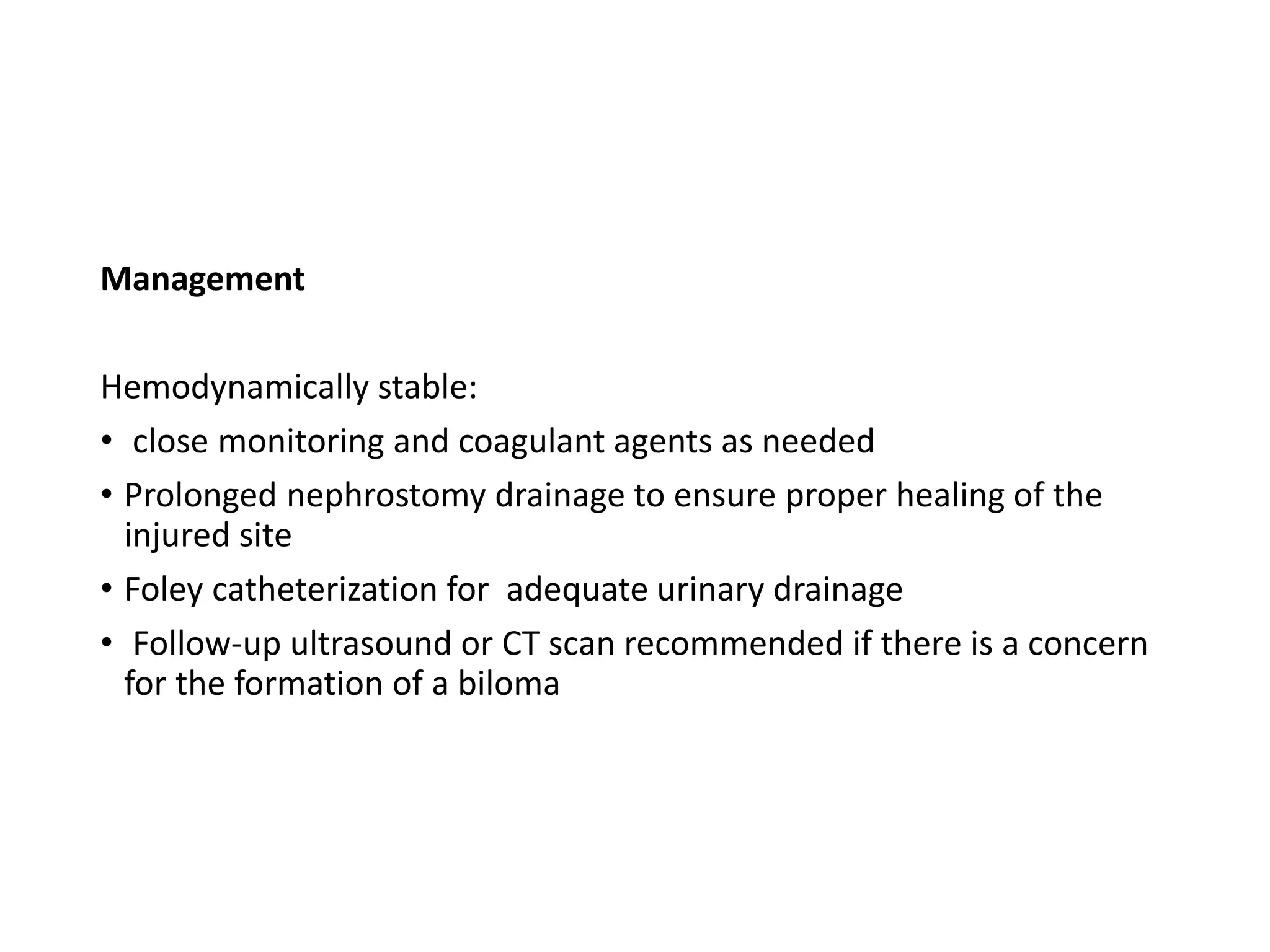
POST PCNL COMPLICATIONS AND
MANAGEMENT
Dr Shambhavi Sharma
MS Resident
PAHS
Moderator :
Ass.prof Dr Samir Shrestha
INTRODUCTION
• Percutaneous nephrolithotomy (PNL) is accepted as the
procedure of choice for the treatment of large or complex
renal calculi
AUA guideline on management of staghorn calculi: diagnosis and treatment recommendations.Preminger
Gm et al.J Urol. 2005 Jun; 173(6):1991-2000.
OPERATIVE STEPS
• Insertion of ureteric catheter (allowing dilation of the
collecting system with saline and contrast media)
• Positioning of the patient
• Puncture of calyx and access
• Serial Dilatation of access tract
• Amplatz sheath
• Introduction of nephroscope
• Stone fragmentation and retrieval
COMPLICATIONS
During Positioning
Neuromusculoskeletal Complications
associated with prone positioning
related to the head and neck region
• Ocular injury resulting in visual loss
• Facial nerve injury
• Necrosis over facial bones,tip of the nose,
• Cerebrovascular accident due to carotid or vertebrobasilar
artery dissection
• Careful padding of the head, in a neutral and nonextended
position,
• Malpositioning of the extremities -- peripheral nerve injury
• Shoulder and elbow should not be abducted more than 90
degrees, so as to prevent brachial plexopathy
• Generous padding at the elbow and forearm reduces the risk of
nerve compression
COMPLICATIONS
•During access/puncture of calyx
1. Pleural Injury
• Hydrothorax, pneumothorax-when
access made above 11th rib
(1.8-8%)
• Lung injury
• Nephropleural fistula (urinothorax)-
rare
• incidence of pleural complications
with punctures above the 12th rib
considered acceptable risk
• if provides optimal access to
the upper urinary tract
Diagnosis :
• Intraoperative:
• Monitoring airway pressure
• ETCO2 and oxygen saturation
• Postoperative :
• Chest fluoroscopy during or at the conclusion of the procedure
• Chest radiography
• If hydrothorax is noted intraoperatively:
• insert a small- caliber (8-Fr to 12-Fr) Cope nephrostomy tube as
the thoracostomy
• Large-bore thoracostomy tube -for lung injury
2. Acute hemorrhage
Most common significant complication(6.5-8.5%)
• Factors associated with hemorrhage
 multiple access
 increasing tract size
 Guys Stone score >3
 Prolonged operative time >83 min
 Renal pelvic perforation
 Previous ipsilateral surgery
Shakhawan H.A. Said,Arab J Urol 2017 Mar; 15(1): 24–29.
Hemorrhage can occur during :
• during needle passage
• tract dilatation
• nephrostomy
Technical errors predisposing to hemorrhage:
• Infundibular entry risks injury to interlobar (infundibular)
arteries
• Entry into wrong calyx resulting into overly aggressive
torquing of the sheath and rigid endoscope
• Misplacement of nephrostomy tube
Ideal access:
• one that enters a posterior calyx
at the fornix
• second most inferior calyx seen on
retrograde pyelography is typically
posteriorly oriented
• ideal for initial access for most
patients
• Most straight path to calyx with
highest stone burden
• Dilated calyx
To minimize the unfavorable impact of multiple
accesses on bleeding:
• Flexible nephroscopy
• Holmium laser lithotripsy,
• Improved grasping devices and baskets
Akman T.et al. J Endourol. 2011;25:327–333
Identification of source of bleeding
• Most hemorrhage occurs from the renal parenchyma, most
cases not significant
• noticeable bleeding from the tract after sheath removal
following an otherwise unremarkable procedure
Management
• Insert and occlude a nephrostomy tube
• Apply pressure to the incision
• Let the collecting system clot off
• Nephrostomy tubes should not be irrigated the day or evening
of the procedure if not draining
• By the next morning, it is safe to gently irrigate the tube
because hemostasis is more certain
If severe hemorrhage occurs following
sheath removal refractory to the
hemostatic measure:
Kaye Nephrostomy Tamponade Balloon
considered
• 15-cm long
• 36 F balloon which surrounds the
length of the tube
• Tamponade the nephrostomy
tract
• Simultaneous drainage of tract
• Intraoperative hemorrhage from an injured vein or artery
within the collecting system
• mandates cessation of the procedure if vision is lost
• If Venous bleeding :
• Place nephrostomy tube
• let the collecting system clot off
• If small arterial injury
• fulguration under direct vision
Significant arterial hemorrhage (drop in Hb by 2gm/dl with 4
or more transfusions)
• If the bleeding from the nephrostomy tube continues or
gross hematuria with acute urine retention occurs
• Blood transfusion plus fluid resuscitation
• Foley catheterization with urinary bladder irrigation
• IV administration of mannitol (hemodynamically stable
patients )
• lead to rapid forced diuresis and swelling of the kidney within the capsule,
which may enhance tract tamponade
In case of failure of these
maneuvers:
Color duplex sonography/
CT/ MR angiography or
renal angiography
• Selective renal artery
embolization
Misplacement of nephrostomy tube
At the stage of access tract dilation.
Diagnosis:
massive hemorrhage necessitating immediate placement of a
nephrostomy tube and abortion of the procedure
Management:
hemodynamically stable:
• managed conservatively with strict bed rest,
• intravenous antibiotics,
• CT or fluoroscopy-guided nephrostomy tube withdrawal
• (in the operating room with the vascular team ready to intervene if
needed)
• Open surgery can be used as an alternative treatment
3.postoperative Hemorrhage
Can occur during :
 with the nephrostomy tube in place early
 at time of tube removal
 after discharge from the hospital(1-3weeks)
• Delayed hemorrhage due to arteriovenous fistulas or
arterial pseudoaneurysms(more common)
• formed by a high-pressure leak from a lacerated artery
• leak transmitted through the tract into a lower
resistance system, such as a vein or a connective tissue
space
• As late as 13 weeks after a percutaneous nephrolihotomy
• Continuous bleeding -arteriovenous fistula
• Intermittent bleeding-arterial pseudoaneurysm
• Selective angio-embolization
Other options :
• endovascular placement of a covered stent to occlude the site
of arterial injury
• USG guided percutaneous puncture of an arterial
pseudoaneurysm
• Injection of thrombin or fibrin tissue adhesive
Anil kumar et al. Ijcmr.2016;3:2454-7379
•In case of failure of these maneuvers:
•Partial nephrectomy
3. Collecting System Injury
(7.2%)
• Tears in the infundibulum
• Renal pelvic perforation:
• occur during initial access or during dilation
• Pushing on a renal pelvic stone too hard during lithotripsy
• Collapse of a previously distended renal pelvis is a usual sign
if the perforation is not visualized directly at first
• avoided by using a J guide wire with a soft and curved tip
• Perforation can lead to :
• Retroperitoneal extravasation
• Intraperitoneal extravasation
Retroperitoneal extravasation :
• Noted by medial displacement of
the kidney during fluoroscopy
• Direct visualization of perinephric
structures or fat
• abnormal hemodynamic
parameters,
• decrease in irrigation fluid drainage
• Postoperative enhanced CT may
reveal signs of urine leakage
• Minor perforations
• No intervention required
• Significant perforations
• Termination of the procedure and nephrostomy and ureteral
drainage
• nephrostography after 2 to 7 days and tube removal,
depending on the severity of the injury
•Intraperitoneal extravasation:
• Abdominal distention difficult to recognize due to
prone position
• gradual rise in the patient’s diastolic blood pressure
• Narrowing of the pulse pressure
• Increase in CVP
• In advanced cases of a large-volume extravasation
event:
• Ventilation difficult because of raised IAP
Management :
• Early recognition of major extravasation is crucial
• Vigorous diuresis
• Peritoneal drainage
• Laparotomy
Postoperative :
• abdominal distention, ileus, and/or fever
Management :
• placement of a percutaneous drain
4. Visceral Injury
Colon injury:
• left colon injury more
common
• majority of colon injuries
involve access to the lower
pole
Additional risk factors:
• Advanced patient age
• Dilated colon
• Prior colon surgery or disease
• Thin body habitus
• Horseshoe kidney
Preoperative CT IVU
• Identification of structures
• retrorenal colon,
• Liver or spleen
• Patients with ectopic kidneys,
dysmorphic body habitus(eg
spinal dysraphism)
• Intra-abdominal structures,
such as the bowel, may be
located between the skin
and the renal access point
 Diagnosis
Postoperatively:
• Unexplained fever
• Prolonged ileus
• Unexplained leukocytosis
• Rectal bleeding
• Evidence of peritoneal inflammation,
• fecaluria
• pneumouria or clinically apparent nephrocolonic fistula
• Postoperative nephrostogram or CT imaging
Management :
If extraperitoneal:
• Management may be expectant
• Placement of a ureteral catheter or DJ stent to decompress
the collecting system
• Withdrawal of the nephrostomy tube from an intrarenal
position to an intracolonic position to serve as a colostomy
tube
• Left in place for a minimum of 7 days
• Removed after a nephrostogram or a retrograde
pyelogram shows no communication between the colon
and the kidney
Duodenal and jejunal injuries:
• Less common
• If no peritonitis-Conservative management
• If peritonitis-open Surgery
Liver injury
Patient at risk :
• Right-sided supracostal (superior to the 11th rib)
• percutaneous renal access anterior to the posterior axillary line.
• Hepatomegaly
Diagnosis :
unusual burning sensation at the right flank
CT scan to reveal the route of injury to the liver
Management
Hemodynamically stable:
• close monitoring and coagulant agents as needed
• Prolonged nephrostomy drainage to ensure proper healing of the
injured site
• Foley catheterization for adequate urinary drainage
• Follow-up ultrasound or CT scan recommended if there is a concern
for the formation of a biloma
Splenic injury
• 10th intercostal access and/or splenomegaly -higher risk
for injury
• Diagnosis :
• Hemodynamic instability in the absence of
significant intraoperative blood loss
• Abdominal CT scanning can characterize the
injury
• Management
hemodynamically stable:
• strict bed rest is recommended.
• consider leaving the nephrostomy tube in place
to tamponade the bleeding and induce fibrosis
Hemodynamically unstable,
• Life-saving splenorraphy
• splenectomy
• hemostatic fibrin glue can be used to increase the chance of
preserving the spleen
Complications
due to irrigating fluid
Collecting system perforation
1. Metabolic and Physiologic Complications
During irrigation
• amount of fluid absorption generally clinically insignificant
• volume of fluid absorbed increased with the amount of
irrigating fluid used, pelvicaliceal perforation, bleeding
Intravascular or extravascular extravasation in the setting large
venous injury or collecting system perforation:
• hyponatremia and other electrolyte abnormalities
• renal or hepatic dysfunction
• mental status changes
large amount of saline extravasation
• clinically significant respiratory distress
• cardiac failure due to volume overload
Prevention :
• Irrigation fluid used should always be saline(physiologic)
• Using a low-pressure system and staging, the procedure for large renal
stone burdens
• especially in the presence of complications such as perforation of the
pelvicaliceal system
• height of irrigating fluid and total time for irrigation do not affect the
amount of fluid absorption
Dip saxena et al.Urol Ann.2019 Apr-Jun; 11(2): 163–167.
Other complications
1. Extrarenal stone migration
Occurs due to:
• the application of excessive pressure of the probe onto
the stone
• existence of a perforation in the collecting system
• or the use of an improper technique of stone extraction
with an Amplatz sheath
Diagnosis:
• Intraoperative pyelography
• Renal ultrasound
Management :
• As long as the stone is not infected and fragment-associated
inflammation does not obstruct the urinary tract—t/t not
necessary.
• Endoscopic retrieval of fragments outside of the urinary
tract should not be attempted-- enlarge the perforation
• Intraperitoneal and pleural migration of stone reported
• Laparoscopy and thoracosopy in order to prevent peritoneal
and thoracic complications
• Indicated by
• hypoxemia
• evidence of pulmonary edema
• increased airway pressure
• hypotension, jugular venous distention
• facial plethora
• dysrhythmias, and auscultation of a mill-wheel cardiac murmur
and/ or the appearance of a widened QRS complex with right
heart strain patterns on electrocardiography.
• sudden decrease in capnometry reading of the P(end-tidal) CO2
2. Venous gas embolism (0.4% )
Management
• Swift response is required
• includes rapid ventilation with 100% oxygen
• positioning the patient head down with the right side up
• general resuscitative maneuvers
2. Postoperative Fever and Sepsis
• Incidence: 10% to 30%
Risk factors for fever
• infectious stones
• preoperative urinary tract infection
• Hydronephrosis
• indwelling ureteral stent or
• nephrostomy tube
Management
Prevention :
• Preoperative antibiotics according to C/S
• Intra-operative irrigation pressure < 30 mmHg
• Unobstructed post-operative urinary drainage
Treatement
• Initiation of antimicrobial therapy and other supportive care
• If pus is aspirated upon initial percutaneous to the upper
urinary tract,
• abort the procedure and leave a nephrostomy tube for
drainage
3. Collecting System Obstruction
• Predisposing factors:
• large stone burden requiring multiple or long procedures
• prolonged nephrostomy tube drainage
• previous open stone surgery
• diabetes mellitus
• obesity
• Endoscopic treatment in most cases
• Stenting ,cold knife excision ,laser ablation , balloon dilation or
endoscopic formation of a new infundibulum
• open surgical reconstruction or excision with partial nephrectomy
or total nephrectomy may be required
4. Loss of Renal Function
• owes to disastrous vascular injury or
• the angio-embolization used to treat hemorrhage
5.Postoperative persistent nephrocutaneous
leakage (1.5-4.6%)
• normally closes within 6–12 h of nephrostomy tube removal
• Urinary leakage persisting >24 h after nephrostomy tube removal
called prolonged
• Usually needs treatment
• obtain a low-dose CT scan to evaluate for stone fragments in the
ureter that may be causing obstruction
• Management :
• insertion of a ureteral stent
• Foley catheter may be inserted for 24 h in order to relieve pressure
in the urinary system
• promote anterograde drainage of urine
5.Death (0.3-0.5%)
Summary
• PCNL is a safe procedure with a low incidence of major complications
• common complications :
• bleeding, infection, and infundibular stenosis
• Most complications can be managed conservatively
• appropriate perioperative measures should be taken in order to
minimize the risk of preventable complications
References
• Campbell Walsh text book of urology 12th edition
• EAU guidelines for management of nephrolithiasis
• Gadzhiev N, Malkhasyan V, Akopyan G, Petrov S, Jefferson F, Okhunov
Z. Percutaneous nephrolithotomy for staghorn calculi:
Troubleshooting and managing complications. Asian J Urol.
2020;7(2):139-148. doi:10.1016/j.ajur.2019.10.004
• Lee KL, Stoller ML. Minimizing and managing bleeding after
percutaneous nephrolithotomy. Curr Opin Urol. 2007;17(2):120-124.
doi:10.1097/MOU.0b013e3Incidence, Prevention, and Management
of Complications Following Percutaneous Nephrolitholapaxy
• Saxena, Dipti et al. “Effects of fluid absorption following
percutaneous nephrolithotomy: Changes in blood cell indices and
electrolytes.” Urology annals vol. 11,2 (2019)
•Thank you
•Any queries ?

post pcnl complications.pptx

  • 1.
    POST PCNL COMPLICATIONSAND MANAGEMENT Dr Shambhavi Sharma MS Resident PAHS Moderator : Ass.prof Dr Samir Shrestha
  • 2.
    INTRODUCTION • Percutaneous nephrolithotomy(PNL) is accepted as the procedure of choice for the treatment of large or complex renal calculi AUA guideline on management of staghorn calculi: diagnosis and treatment recommendations.Preminger Gm et al.J Urol. 2005 Jun; 173(6):1991-2000.
  • 3.
    OPERATIVE STEPS • Insertionof ureteric catheter (allowing dilation of the collecting system with saline and contrast media) • Positioning of the patient • Puncture of calyx and access • Serial Dilatation of access tract • Amplatz sheath • Introduction of nephroscope • Stone fragmentation and retrieval
  • 4.
  • 5.
    Neuromusculoskeletal Complications associated withprone positioning related to the head and neck region • Ocular injury resulting in visual loss • Facial nerve injury • Necrosis over facial bones,tip of the nose, • Cerebrovascular accident due to carotid or vertebrobasilar artery dissection
  • 6.
    • Careful paddingof the head, in a neutral and nonextended position, • Malpositioning of the extremities -- peripheral nerve injury • Shoulder and elbow should not be abducted more than 90 degrees, so as to prevent brachial plexopathy • Generous padding at the elbow and forearm reduces the risk of nerve compression
  • 7.
  • 8.
    1. Pleural Injury •Hydrothorax, pneumothorax-when access made above 11th rib (1.8-8%) • Lung injury • Nephropleural fistula (urinothorax)- rare • incidence of pleural complications with punctures above the 12th rib considered acceptable risk • if provides optimal access to the upper urinary tract
  • 9.
    Diagnosis : • Intraoperative: •Monitoring airway pressure • ETCO2 and oxygen saturation • Postoperative : • Chest fluoroscopy during or at the conclusion of the procedure • Chest radiography • If hydrothorax is noted intraoperatively: • insert a small- caliber (8-Fr to 12-Fr) Cope nephrostomy tube as the thoracostomy • Large-bore thoracostomy tube -for lung injury
  • 10.
    2. Acute hemorrhage Mostcommon significant complication(6.5-8.5%) • Factors associated with hemorrhage  multiple access  increasing tract size  Guys Stone score >3  Prolonged operative time >83 min  Renal pelvic perforation  Previous ipsilateral surgery Shakhawan H.A. Said,Arab J Urol 2017 Mar; 15(1): 24–29.
  • 11.
    Hemorrhage can occurduring : • during needle passage • tract dilatation • nephrostomy Technical errors predisposing to hemorrhage: • Infundibular entry risks injury to interlobar (infundibular) arteries • Entry into wrong calyx resulting into overly aggressive torquing of the sheath and rigid endoscope • Misplacement of nephrostomy tube
  • 12.
    Ideal access: • onethat enters a posterior calyx at the fornix • second most inferior calyx seen on retrograde pyelography is typically posteriorly oriented • ideal for initial access for most patients • Most straight path to calyx with highest stone burden • Dilated calyx
  • 13.
    To minimize theunfavorable impact of multiple accesses on bleeding: • Flexible nephroscopy • Holmium laser lithotripsy, • Improved grasping devices and baskets Akman T.et al. J Endourol. 2011;25:327–333
  • 14.
    Identification of sourceof bleeding • Most hemorrhage occurs from the renal parenchyma, most cases not significant • noticeable bleeding from the tract after sheath removal following an otherwise unremarkable procedure Management • Insert and occlude a nephrostomy tube • Apply pressure to the incision • Let the collecting system clot off • Nephrostomy tubes should not be irrigated the day or evening of the procedure if not draining • By the next morning, it is safe to gently irrigate the tube because hemostasis is more certain
  • 15.
    If severe hemorrhageoccurs following sheath removal refractory to the hemostatic measure: Kaye Nephrostomy Tamponade Balloon considered • 15-cm long • 36 F balloon which surrounds the length of the tube • Tamponade the nephrostomy tract • Simultaneous drainage of tract
  • 16.
    • Intraoperative hemorrhagefrom an injured vein or artery within the collecting system • mandates cessation of the procedure if vision is lost • If Venous bleeding : • Place nephrostomy tube • let the collecting system clot off • If small arterial injury • fulguration under direct vision
  • 17.
    Significant arterial hemorrhage(drop in Hb by 2gm/dl with 4 or more transfusions) • If the bleeding from the nephrostomy tube continues or gross hematuria with acute urine retention occurs • Blood transfusion plus fluid resuscitation • Foley catheterization with urinary bladder irrigation • IV administration of mannitol (hemodynamically stable patients ) • lead to rapid forced diuresis and swelling of the kidney within the capsule, which may enhance tract tamponade
  • 18.
    In case offailure of these maneuvers: Color duplex sonography/ CT/ MR angiography or renal angiography • Selective renal artery embolization
  • 19.
    Misplacement of nephrostomytube At the stage of access tract dilation. Diagnosis: massive hemorrhage necessitating immediate placement of a nephrostomy tube and abortion of the procedure Management: hemodynamically stable: • managed conservatively with strict bed rest, • intravenous antibiotics, • CT or fluoroscopy-guided nephrostomy tube withdrawal • (in the operating room with the vascular team ready to intervene if needed) • Open surgery can be used as an alternative treatment
  • 20.
    3.postoperative Hemorrhage Can occurduring :  with the nephrostomy tube in place early  at time of tube removal  after discharge from the hospital(1-3weeks) • Delayed hemorrhage due to arteriovenous fistulas or arterial pseudoaneurysms(more common) • formed by a high-pressure leak from a lacerated artery • leak transmitted through the tract into a lower resistance system, such as a vein or a connective tissue space
  • 21.
    • As lateas 13 weeks after a percutaneous nephrolihotomy • Continuous bleeding -arteriovenous fistula • Intermittent bleeding-arterial pseudoaneurysm • Selective angio-embolization Other options : • endovascular placement of a covered stent to occlude the site of arterial injury • USG guided percutaneous puncture of an arterial pseudoaneurysm • Injection of thrombin or fibrin tissue adhesive Anil kumar et al. Ijcmr.2016;3:2454-7379
  • 22.
    •In case offailure of these maneuvers: •Partial nephrectomy
  • 23.
    3. Collecting SystemInjury (7.2%) • Tears in the infundibulum • Renal pelvic perforation: • occur during initial access or during dilation • Pushing on a renal pelvic stone too hard during lithotripsy • Collapse of a previously distended renal pelvis is a usual sign if the perforation is not visualized directly at first • avoided by using a J guide wire with a soft and curved tip • Perforation can lead to : • Retroperitoneal extravasation • Intraperitoneal extravasation
  • 24.
    Retroperitoneal extravasation : •Noted by medial displacement of the kidney during fluoroscopy • Direct visualization of perinephric structures or fat • abnormal hemodynamic parameters, • decrease in irrigation fluid drainage • Postoperative enhanced CT may reveal signs of urine leakage
  • 25.
    • Minor perforations •No intervention required • Significant perforations • Termination of the procedure and nephrostomy and ureteral drainage • nephrostography after 2 to 7 days and tube removal, depending on the severity of the injury
  • 26.
    •Intraperitoneal extravasation: • Abdominaldistention difficult to recognize due to prone position • gradual rise in the patient’s diastolic blood pressure • Narrowing of the pulse pressure • Increase in CVP • In advanced cases of a large-volume extravasation event: • Ventilation difficult because of raised IAP
  • 27.
    Management : • Earlyrecognition of major extravasation is crucial • Vigorous diuresis • Peritoneal drainage • Laparotomy Postoperative : • abdominal distention, ileus, and/or fever Management : • placement of a percutaneous drain
  • 28.
    4. Visceral Injury Coloninjury: • left colon injury more common • majority of colon injuries involve access to the lower pole Additional risk factors: • Advanced patient age • Dilated colon • Prior colon surgery or disease • Thin body habitus • Horseshoe kidney
  • 29.
    Preoperative CT IVU •Identification of structures • retrorenal colon, • Liver or spleen • Patients with ectopic kidneys, dysmorphic body habitus(eg spinal dysraphism) • Intra-abdominal structures, such as the bowel, may be located between the skin and the renal access point
  • 30.
     Diagnosis Postoperatively: • Unexplainedfever • Prolonged ileus • Unexplained leukocytosis • Rectal bleeding • Evidence of peritoneal inflammation, • fecaluria • pneumouria or clinically apparent nephrocolonic fistula • Postoperative nephrostogram or CT imaging
  • 31.
    Management : If extraperitoneal: •Management may be expectant • Placement of a ureteral catheter or DJ stent to decompress the collecting system • Withdrawal of the nephrostomy tube from an intrarenal position to an intracolonic position to serve as a colostomy tube • Left in place for a minimum of 7 days • Removed after a nephrostogram or a retrograde pyelogram shows no communication between the colon and the kidney
  • 32.
    Duodenal and jejunalinjuries: • Less common • If no peritonitis-Conservative management • If peritonitis-open Surgery
  • 33.
    Liver injury Patient atrisk : • Right-sided supracostal (superior to the 11th rib) • percutaneous renal access anterior to the posterior axillary line. • Hepatomegaly Diagnosis : unusual burning sensation at the right flank CT scan to reveal the route of injury to the liver
  • 34.
    Management Hemodynamically stable: • closemonitoring and coagulant agents as needed • Prolonged nephrostomy drainage to ensure proper healing of the injured site • Foley catheterization for adequate urinary drainage • Follow-up ultrasound or CT scan recommended if there is a concern for the formation of a biloma
  • 35.
    Splenic injury • 10thintercostal access and/or splenomegaly -higher risk for injury • Diagnosis : • Hemodynamic instability in the absence of significant intraoperative blood loss • Abdominal CT scanning can characterize the injury • Management hemodynamically stable: • strict bed rest is recommended. • consider leaving the nephrostomy tube in place to tamponade the bleeding and induce fibrosis
  • 36.
    Hemodynamically unstable, • Life-savingsplenorraphy • splenectomy • hemostatic fibrin glue can be used to increase the chance of preserving the spleen
  • 37.
    Complications due to irrigatingfluid Collecting system perforation
  • 38.
    1. Metabolic andPhysiologic Complications During irrigation • amount of fluid absorption generally clinically insignificant • volume of fluid absorbed increased with the amount of irrigating fluid used, pelvicaliceal perforation, bleeding Intravascular or extravascular extravasation in the setting large venous injury or collecting system perforation: • hyponatremia and other electrolyte abnormalities • renal or hepatic dysfunction • mental status changes
  • 39.
    large amount ofsaline extravasation • clinically significant respiratory distress • cardiac failure due to volume overload Prevention : • Irrigation fluid used should always be saline(physiologic) • Using a low-pressure system and staging, the procedure for large renal stone burdens • especially in the presence of complications such as perforation of the pelvicaliceal system • height of irrigating fluid and total time for irrigation do not affect the amount of fluid absorption Dip saxena et al.Urol Ann.2019 Apr-Jun; 11(2): 163–167.
  • 40.
  • 41.
    1. Extrarenal stonemigration Occurs due to: • the application of excessive pressure of the probe onto the stone • existence of a perforation in the collecting system • or the use of an improper technique of stone extraction with an Amplatz sheath Diagnosis: • Intraoperative pyelography • Renal ultrasound
  • 42.
    Management : • Aslong as the stone is not infected and fragment-associated inflammation does not obstruct the urinary tract—t/t not necessary. • Endoscopic retrieval of fragments outside of the urinary tract should not be attempted-- enlarge the perforation • Intraperitoneal and pleural migration of stone reported • Laparoscopy and thoracosopy in order to prevent peritoneal and thoracic complications
  • 43.
    • Indicated by •hypoxemia • evidence of pulmonary edema • increased airway pressure • hypotension, jugular venous distention • facial plethora • dysrhythmias, and auscultation of a mill-wheel cardiac murmur and/ or the appearance of a widened QRS complex with right heart strain patterns on electrocardiography. • sudden decrease in capnometry reading of the P(end-tidal) CO2 2. Venous gas embolism (0.4% )
  • 44.
    Management • Swift responseis required • includes rapid ventilation with 100% oxygen • positioning the patient head down with the right side up • general resuscitative maneuvers
  • 45.
    2. Postoperative Feverand Sepsis • Incidence: 10% to 30% Risk factors for fever • infectious stones • preoperative urinary tract infection • Hydronephrosis • indwelling ureteral stent or • nephrostomy tube
  • 46.
    Management Prevention : • Preoperativeantibiotics according to C/S • Intra-operative irrigation pressure < 30 mmHg • Unobstructed post-operative urinary drainage Treatement • Initiation of antimicrobial therapy and other supportive care • If pus is aspirated upon initial percutaneous to the upper urinary tract, • abort the procedure and leave a nephrostomy tube for drainage
  • 47.
    3. Collecting SystemObstruction • Predisposing factors: • large stone burden requiring multiple or long procedures • prolonged nephrostomy tube drainage • previous open stone surgery • diabetes mellitus • obesity • Endoscopic treatment in most cases • Stenting ,cold knife excision ,laser ablation , balloon dilation or endoscopic formation of a new infundibulum • open surgical reconstruction or excision with partial nephrectomy or total nephrectomy may be required
  • 48.
    4. Loss ofRenal Function • owes to disastrous vascular injury or • the angio-embolization used to treat hemorrhage
  • 49.
    5.Postoperative persistent nephrocutaneous leakage(1.5-4.6%) • normally closes within 6–12 h of nephrostomy tube removal • Urinary leakage persisting >24 h after nephrostomy tube removal called prolonged • Usually needs treatment • obtain a low-dose CT scan to evaluate for stone fragments in the ureter that may be causing obstruction • Management : • insertion of a ureteral stent • Foley catheter may be inserted for 24 h in order to relieve pressure in the urinary system • promote anterograde drainage of urine
  • 50.
  • 51.
    Summary • PCNL isa safe procedure with a low incidence of major complications • common complications : • bleeding, infection, and infundibular stenosis • Most complications can be managed conservatively • appropriate perioperative measures should be taken in order to minimize the risk of preventable complications
  • 52.
    References • Campbell Walshtext book of urology 12th edition • EAU guidelines for management of nephrolithiasis • Gadzhiev N, Malkhasyan V, Akopyan G, Petrov S, Jefferson F, Okhunov Z. Percutaneous nephrolithotomy for staghorn calculi: Troubleshooting and managing complications. Asian J Urol. 2020;7(2):139-148. doi:10.1016/j.ajur.2019.10.004 • Lee KL, Stoller ML. Minimizing and managing bleeding after percutaneous nephrolithotomy. Curr Opin Urol. 2007;17(2):120-124. doi:10.1097/MOU.0b013e3Incidence, Prevention, and Management of Complications Following Percutaneous Nephrolitholapaxy • Saxena, Dipti et al. “Effects of fluid absorption following percutaneous nephrolithotomy: Changes in blood cell indices and electrolytes.” Urology annals vol. 11,2 (2019)
  • 53.