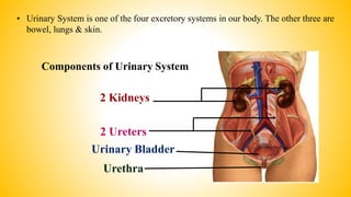
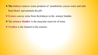
Urinary System
By
Mr Vinayak R Bodhankar
M. Pharm. (QA)
• Urinary System is one of the four excretory systems in our body. The other three are
bowel, lungs & skin.
Components of Urinary System
2 Kidneys
2 Ureters
Urinary Bladder
Urethra
❖The kidneys remove waste products of metabolism, excess water and salts
from blood and maintain the pH .
❖Ureters convey urine from the kidneys to the urinary bladder.
❖The urinary bladder is the muscular reservoir of urine.
❖ Urethra is the channel to the exterior.
The Kidneys
• Definition
The kidneys are a pair of excretory organs situated on the posterior abdominal
wall, one on each side of the vertebral column, behind the peritoneum.
• Location
• The kidneys occupies the
• Epigastric , hypochondriac, lumbar & umbilical regions.
• Vertically they extends from upper border of twelth
thoracic vertebra to the center of the body of third
lumbar vertebra.
• The right kidney is slightly lower than the left, & the
left kidney is little nearer to the median plane than the
right.
The Kidneys: Surface Anatomy
• External Features
– Each kidney is bean shaped.
– It has upper & lower poles,
medial and lateral borders,
and anterior and posterior
surfaces.
– The upper pole is broad & is in
close contact with the
corresponding suprarenal
glands.
– The lower pole is pointed.
• The lateral border is
convex.
• The medial border is
concave.
• The middle part of the
medial border is
depressed and is known
as hilum (hilus)
11 cm
3cm
5cm
8
Measurements
• Color
• Length
: Reddish Brown
: About 11 cm long( the left kidney is little longer & narrower than right)
: 6cm broad
: 3cm thick
• Width
• Thickness
• Weight
– Males
– Females
: 150gm
: 135gm
Coverings
• It has 3 coverings
1. Innermost fibrous capsule or true capsule
2. Middle fatty capsule / perinephric fat-it is a collection of fatty tissue. (It acts as a shock absorber &
helps to maintain the kidney in its position)
3. The false capsule – made of renal fascia .It has two layers-Anterior & Posterior.
Macroscopic Structure
Renal Cortex
Renal Capsule
Renal Medulla
Renal Pelvis
Renal Pyramids
Ureter
Major Calyx
Minor calyces
Papilla
Kidney: Frontal Section Microscopic Structure
➢ The kidney may be regarded as a
collection of millions of uriniferous
tubules.
➢ Each uriniferous tubule consists of an
excretory part called nephron and of a
collecting tubule.
➢ Each kidney contains over 1(1-2 millions)
million nephrons and thousands of
collecting ducts
Structure of Nephron
 Functional unit of kidney.
 Approx. 1 million nephrons in each
kidney.
 Glomerular capsule
 Glomerulus
 3 Parts:
• Proximal convoluted tubule
• Distal convoluted
• Loop of henle
Kidney Functions
• Formation of Urine
• Filters blood plasma, eliminates waste, returns useful chemicals to blood
• Regulates blood volume and pressure
• Regulates osmolarity of body fluids
• Secretes renin, activates angiotensin, aldosterone
– controls BP, electrolyte balance
• Secretes erythropoietin, controls RBC count
• Detoxifies free radicals and drugs
Formation of Urine
1. Glomerular filtration
• Afferent arteriole: Blood to glomerulus.
• Efferent arteriole: Blood away from glomerulus.
• Diameter of afferent is greater than efferent: Generation of hydrostatic pressure in glomerulus (90 mm of
Hg).
• Hydrostatic pressure is opposed by osmotic pressure of blood (25 mm of Hg).
• Filtrate generate a pressure of 15 mm of Hg.
• Thus resultant filtration pressure: 90-25-15=50 mm of Hg.
• Due to this pressure, blood constituents get filtered in nephron.
• Every min, about 1 litre of blood containing 500 ml of plasma flows through all glomeruli & about 100 ml
is filtered off.
• This is called as Glomerular filtration rate.
2. Selective reabsorption
• Process by which the composition & volume of glomerular filter is altered during its passage through
tubule.
• Purpose is to reabsorb the constituents of filtrate which are essential to the body.
• High threshold substances
• Low threshold substances
• Waste product: slight extent
3. Active secretion
• Some non-threshold substances are not filtered by capsule.
• They carried in capillaries of efferent arteriole and are cleared by secretion into the tubule.
Ureters
• Definition: The Ureters are a pair of narrow , thick walled muscular tubes
which convey urine from the kidneys to urinary bladder.
Dimensions
• Each Ureters is about 25cm (10 inch)long.
• The upper half lies in the abdomen and the lower half in the pelvis.
• It measures 3mm diameter, but it slightly constricted at three places.
– At the pelviureteric junction
– At the brim of lesser pelvis
– At its passage through the bladder wall
• For the purpose of description, ureter is divided into 2 parts
– From the site of origin to pelvic brim- abdominal part
– From pelvic brim to entry into urinary bladder- pelvic part
Ureter cross section
• 3 layers of tissue
– Outer layer : Fibrous tissue
– Middle layer: Muscle
– Inner layer: Epithelium
• Blood Supply
– Ureter is supplied by branches of
» Renal artery
» Abdominal aorta
» Gonadal artery
» Common iliac artery
» Internal iliac artery
» Inferior vesical artery
Urinary Bladder
• The urinary bladder is a hollow, muscular organ , which functions as the reservoir for the urine received
from the kidneys and to discharge it out periodically
Position
• Empty bladder , in the adult situated within the pelvis . When distended , it rises up to the abdominal
cavity and becomes an abdomino- pelvic organ.
• Capacity
The mean capacity of the bladder is 220 ml, filling beyond 220ml causes a desire to micturate. Filling upto
500ml may be tolerated, but it becomes painful.
Shape
• An empty bladder is 4 sided pyramid in shape and has
– 4 angles -an apex, neck & 2 lateral angles
– 4 surfaces
• Base (posterior surface)
• 2 inferiolateral surfaces
• Superior surface
Bladder Structure
• 3 layers
– Outer layer
• Loose connective tissue
– Middle layer
• Smooth muscle and elastic fibres
– Inner layer
• Lined with transitional epithelium
Interior of Bladder
• The mucous membrane is straw colored & is thrown into
folds. When bladder is distended, these folds disappear.
• The posterior wall shows a smooth triangular area called
trigone. There are no mucous folds in this region.
• At the upper lateral angles of the trigone are the ureteric
openings.
• At its inferior angle is the internal urethral orifice.
The Urethra
 The urethra is a canal extending from the neck of the bladder to the exterior , at
the external urethral orifice.
 Male: about 20 cm (8”) long
 Female: 3-4 cm (1.5”) long

Urinary system

  • 1.
    Urinary System By Mr VinayakR Bodhankar M. Pharm. (QA)
  • 2.
    • Urinary Systemis one of the four excretory systems in our body. The other three are bowel, lungs & skin. Components of Urinary System 2 Kidneys 2 Ureters Urinary Bladder Urethra
  • 3.
    ❖The kidneys removewaste products of metabolism, excess water and salts from blood and maintain the pH . ❖Ureters convey urine from the kidneys to the urinary bladder. ❖The urinary bladder is the muscular reservoir of urine. ❖ Urethra is the channel to the exterior.
  • 4.
    The Kidneys • Definition Thekidneys are a pair of excretory organs situated on the posterior abdominal wall, one on each side of the vertebral column, behind the peritoneum. • Location • The kidneys occupies the • Epigastric , hypochondriac, lumbar & umbilical regions. • Vertically they extends from upper border of twelth thoracic vertebra to the center of the body of third lumbar vertebra. • The right kidney is slightly lower than the left, & the left kidney is little nearer to the median plane than the right.
  • 5.
    The Kidneys: SurfaceAnatomy • External Features – Each kidney is bean shaped. – It has upper & lower poles, medial and lateral borders, and anterior and posterior surfaces. – The upper pole is broad & is in close contact with the corresponding suprarenal glands. – The lower pole is pointed.
  • 6.
    • The lateralborder is convex. • The medial border is concave. • The middle part of the medial border is depressed and is known as hilum (hilus) 11 cm 3cm 5cm 8
  • 7.
    Measurements • Color • Length :Reddish Brown : About 11 cm long( the left kidney is little longer & narrower than right) : 6cm broad : 3cm thick • Width • Thickness • Weight – Males – Females : 150gm : 135gm Coverings • It has 3 coverings 1. Innermost fibrous capsule or true capsule 2. Middle fatty capsule / perinephric fat-it is a collection of fatty tissue. (It acts as a shock absorber & helps to maintain the kidney in its position) 3. The false capsule – made of renal fascia .It has two layers-Anterior & Posterior.
  • 8.
    Macroscopic Structure Renal Cortex RenalCapsule Renal Medulla Renal Pelvis Renal Pyramids Ureter Major Calyx Minor calyces Papilla
  • 9.
    Kidney: Frontal SectionMicroscopic Structure ➢ The kidney may be regarded as a collection of millions of uriniferous tubules. ➢ Each uriniferous tubule consists of an excretory part called nephron and of a collecting tubule. ➢ Each kidney contains over 1(1-2 millions) million nephrons and thousands of collecting ducts
  • 10.
    Structure of Nephron Functional unit of kidney.  Approx. 1 million nephrons in each kidney.  Glomerular capsule  Glomerulus  3 Parts: • Proximal convoluted tubule • Distal convoluted • Loop of henle
  • 11.
    Kidney Functions • Formationof Urine • Filters blood plasma, eliminates waste, returns useful chemicals to blood • Regulates blood volume and pressure • Regulates osmolarity of body fluids • Secretes renin, activates angiotensin, aldosterone – controls BP, electrolyte balance • Secretes erythropoietin, controls RBC count • Detoxifies free radicals and drugs
  • 12.
    Formation of Urine 1.Glomerular filtration • Afferent arteriole: Blood to glomerulus. • Efferent arteriole: Blood away from glomerulus. • Diameter of afferent is greater than efferent: Generation of hydrostatic pressure in glomerulus (90 mm of Hg). • Hydrostatic pressure is opposed by osmotic pressure of blood (25 mm of Hg). • Filtrate generate a pressure of 15 mm of Hg. • Thus resultant filtration pressure: 90-25-15=50 mm of Hg. • Due to this pressure, blood constituents get filtered in nephron. • Every min, about 1 litre of blood containing 500 ml of plasma flows through all glomeruli & about 100 ml is filtered off. • This is called as Glomerular filtration rate.
  • 13.
    2. Selective reabsorption •Process by which the composition & volume of glomerular filter is altered during its passage through tubule. • Purpose is to reabsorb the constituents of filtrate which are essential to the body. • High threshold substances • Low threshold substances • Waste product: slight extent 3. Active secretion • Some non-threshold substances are not filtered by capsule. • They carried in capillaries of efferent arteriole and are cleared by secretion into the tubule.
  • 14.
    Ureters • Definition: TheUreters are a pair of narrow , thick walled muscular tubes which convey urine from the kidneys to urinary bladder. Dimensions • Each Ureters is about 25cm (10 inch)long. • The upper half lies in the abdomen and the lower half in the pelvis. • It measures 3mm diameter, but it slightly constricted at three places. – At the pelviureteric junction – At the brim of lesser pelvis – At its passage through the bladder wall
  • 15.
    • For thepurpose of description, ureter is divided into 2 parts – From the site of origin to pelvic brim- abdominal part – From pelvic brim to entry into urinary bladder- pelvic part Ureter cross section • 3 layers of tissue – Outer layer : Fibrous tissue – Middle layer: Muscle – Inner layer: Epithelium • Blood Supply – Ureter is supplied by branches of » Renal artery » Abdominal aorta » Gonadal artery » Common iliac artery » Internal iliac artery » Inferior vesical artery
  • 16.
    Urinary Bladder • Theurinary bladder is a hollow, muscular organ , which functions as the reservoir for the urine received from the kidneys and to discharge it out periodically Position • Empty bladder , in the adult situated within the pelvis . When distended , it rises up to the abdominal cavity and becomes an abdomino- pelvic organ. • Capacity The mean capacity of the bladder is 220 ml, filling beyond 220ml causes a desire to micturate. Filling upto 500ml may be tolerated, but it becomes painful. Shape • An empty bladder is 4 sided pyramid in shape and has – 4 angles -an apex, neck & 2 lateral angles – 4 surfaces • Base (posterior surface) • 2 inferiolateral surfaces • Superior surface
  • 17.
    Bladder Structure • 3layers – Outer layer • Loose connective tissue – Middle layer • Smooth muscle and elastic fibres – Inner layer • Lined with transitional epithelium Interior of Bladder • The mucous membrane is straw colored & is thrown into folds. When bladder is distended, these folds disappear. • The posterior wall shows a smooth triangular area called trigone. There are no mucous folds in this region. • At the upper lateral angles of the trigone are the ureteric openings. • At its inferior angle is the internal urethral orifice.
  • 18.
    The Urethra  Theurethra is a canal extending from the neck of the bladder to the exterior , at the external urethral orifice.  Male: about 20 cm (8”) long  Female: 3-4 cm (1.5”) long