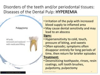
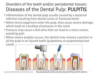
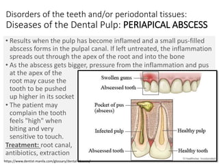
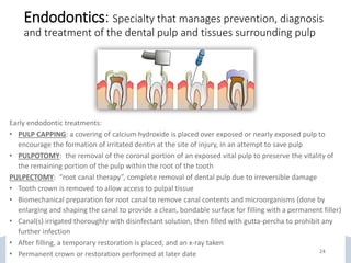
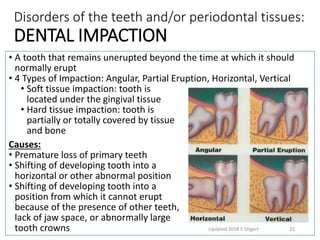
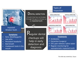
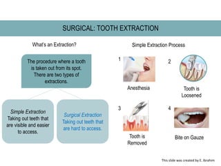
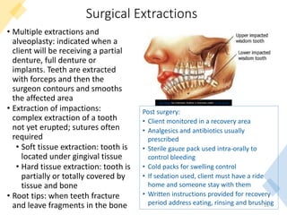
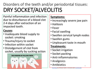
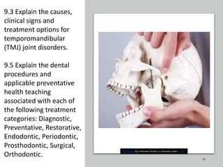
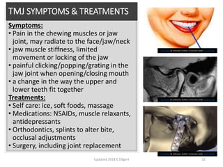
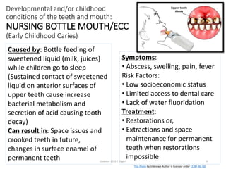
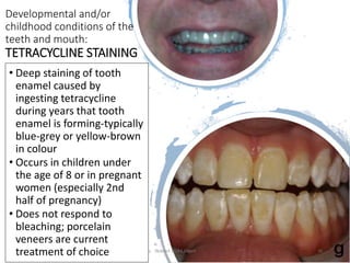
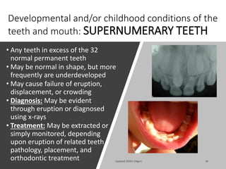
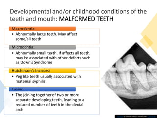
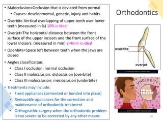
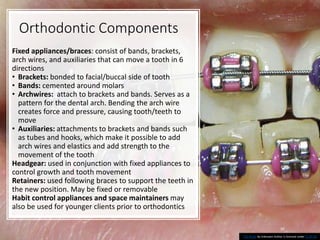
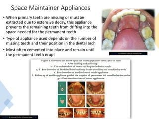
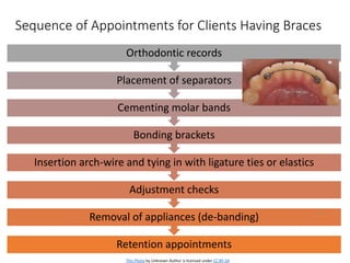
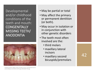
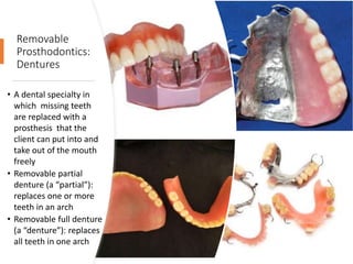
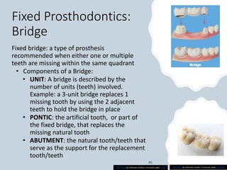
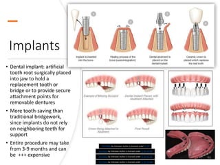
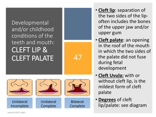
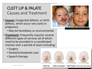
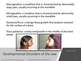
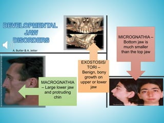
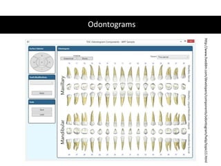
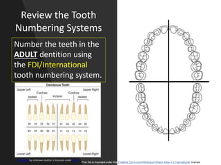
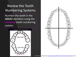
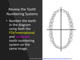
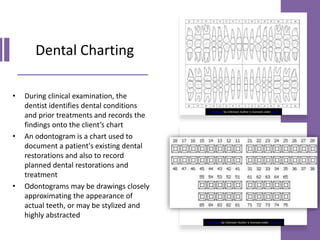
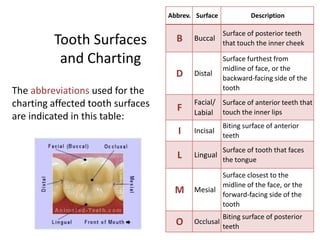
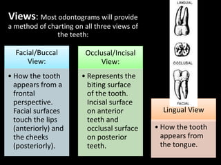
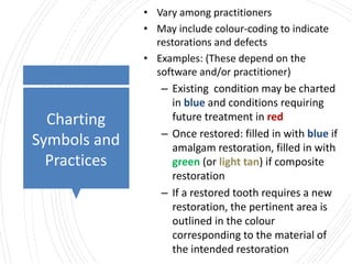
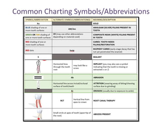
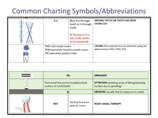
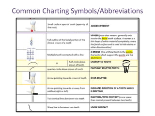
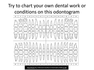
ADMN1340
Dental Conditions and
Procedures Part 2
Discussion Board Instructions
1. Please view this presentation.
2. Share a personal/family experience about one of the topics in
the PowerPoint or ask a question about one of the topics.
3. Also, read other people’s posts on the discussion board and
answer another student’s question or respond to their story with
your own story or ask a question about their story.
This Photo by Unknown Author is licensed under CC BY-NC
Outcomes
This Photo by Unknown Author is licensed under CC BY-NC
10.1 Discuss components of the dental chart and
policies for the transfer and retention of records.
10.2 Demonstrate the ability to interpret conditions
on a patient’s odontogram.
9.2 Explain the causes, clinical signs and treatment for
various conditions or disorders of the teeth and/or
periodontal tissues.
9.3 Explain the causes, clinical signs and treatment
options for temporomandibular (TMJ) joint disorders.
9.4 Explain the causes, clinical signs and treatment for
developmental and/or childhood conditions of the
teeth and mouth.
9.5 Explain the dental procedures and applicable
preventative health teaching associated with each of
the following treatment categories: Diagnostic,
Preventative, Restorative, Endodontic, Periodontic,
Prosthodontic, Surgical, Orthodontic.
Dental Procedures and Fee Guide Codes
3
00000-09999 Diagnosis: Examination & oral cancer screening, x-rays
10000-19999 Prevention: Fluoride treatments and sealants, cleaning/ prophylaxis/
scaling, polishing, night guards and space maintainers
20000-29999 Restoration: Fillings and repairs and bonding, inlays and onlays and crowns,
veneers
30000-39999 Endodontics: Root canal, pulpotomy, emergency pulpectomy
40000-49999 Periodontics: Root planing, gum surgery/soft tissue grafting
50000-59999 Prosthodontics-Removable: dentures (partial, full)
60000-69999 Prosthodontics-Fixed: Bridges
70000-79999 Oral and Maxillofacial Surgery: Extractions, impactions, tumours, cysts,
I&D, re-implantation of avulsed tooth, dental implants, TMJ surgery
80000-89999 Orthodontics: Braces, thumb crib
90000-99999 Adjunctive: Medications, anesthesia, bleaching, lab services,
correspondence, forms, court appearances, etc.
Components of the Dental Chart
 Patient registration form (includes medical and dental
history)
 Examination findings
 Diagnosis and treatment plans
 Record of treatment provided
 Correspondence
 Consent forms
 Radiographs
 Financial Record:
◦ Third party insurance information
◦ Eligible family members entitled
to benefits under the guarantor
◦ Date and amount of all fees charged
◦ Date and amount of all payments made
◦ Itemized list of commercial lab fees that were incurred
◦ Copies of any written agreement with the client or client guarantor
This Photo by Unknown Author is licensed under CC BY
Retention and Transfer of Dental Records
RETENTION OF RECORDS TRANSFER OF RECORDS
 Dentistry Act (1991) requires
that clinical, financial and drug
records be maintained for at
least 10 years from the date of
the last entry
 If client is under age 18, records
must be kept for a minimum of
10 years after the day on which
the client reached the age of 18
years
 Exceptions:
◦ working models
◦ copies of dental claim forms
(2 yrs)
Clients have a right of access to
their complete dental record:
 Requires a written directive
signed by client or his/her
representative
 Dentist retains original records,
including radiographs
 May charge record transfer fee,
which covers photocopying,
duplication and transfer costs
 Insurance companies may also
seek information; be sure you
only release information directly
related to the claim in question
Odontograms
http://www.liveddm.com/developers/components/odontogram/help/topic11.html
Review the Tooth
Numbering Systems
Number the teeth in the
ADULT dentition using
the FDI/International
tooth numbering system.
This Photo by Unknown Author is licensed under CC BY
Review the Tooth
Numbering Systems
Number the teeth in the
ADULT dentition using the
Universal tooth numbering
system.
This file is licensed under the Creative Commons Attribution-Share Alike 4.0 International license.
This Photo by Unknown Author is licensed under CC BY
Review the Tooth
Numbering Systems
• Number the teeth
in the diagram
using both the
FDI/International
and Universal
tooth numbering
system on the
same image.
This file is licensed under the Creative Commons Attribution-Share Alike 4.0 International license.
Dental Charting
• During clinical examination, the
dentist identifies dental conditions
and prior treatments and records the
findings onto the client’s chart
• An odontogram is a chart used to
document a patient's existing dental
restorations and also to record
planned dental restorations and
treatment
• Odontograms may be drawings closely
approximating the appearance of
actual teeth, or may be stylized and
highly abstracted
This Photo by Unknown Author is licensed under CC BY
This Photo by Unknown Author is licensed under CC BY-SA
Tooth Surfaces
and Charting
The abbreviations used for the
charting affected tooth surfaces
are indicated in this table:
Abbrev. Surface Description
B Buccal
Surface of posterior teeth
that touch the inner cheek
D Distal
Surface furthest from
midline of face, or the
backward-facing side of the
tooth
F
Facial/
Labial
Surface of anterior teeth that
touch the inner lips
I Incisal
Biting surface of anterior
teeth
L Lingual
Surface of tooth that faces
the tongue
M Mesial
Surface closest to the
midline of the face, or the
forward-facing side of the
tooth
O Occlusal
Biting surface of posterior
teeth
Views: Most odontograms will provide
a method of charting on all three views of
the teeth:
Facial/Buccal
View:
• How the tooth
appears from a
frontal
perspective.
Facial surfaces
touch the lips
(anteriorly) and
the cheeks
(posteriorly).
Occlusal/Incisal
View:
• Represents the
biting surface
of the tooth.
Incisal surface
on anterior
teeth and
occlusal surface
on posterior
teeth.
Lingual View
• How the tooth
appears from
the tongue.
Charting
Symbols and
Practices
• Vary among practitioners
• May include colour-coding to indicate
restorations and defects
• Examples: (These depend on the
software and/or practitioner)
– Existing condition may be charted
in blue and conditions requiring
future treatment in red
– Once restored: filled in with blue if
amalgam restoration, filled in with
green (or light tan) if composite
restoration
– If a restored tooth requires a new
restoration, the pertinent area is
outlined in the colour
corresponding to the material of
the intended restoration
Common Charting Symbols/Abbreviations
Practice makes perfect!
Let’s review the
conditions and
procedures in
the chart found
on the previous
3 slides
Try to chart your own dental work or
conditions on this odontogram
Fill in the
Chart
https://www.dentalcareprofessionals.com.au/what-do-my-dental-records-look-like/
TOOTH #
(Universal
system)
Tooth #
FDI
system
CONDITION SURFACE(S)
2 17
Amalgam filling (the filling
extends right across the occlusal
surface to include the proximal
[mesial & distal] tooth surfaces)
MOD
(mesial
occlusal
distal)
4
5
11
20
22
31
9.2 Explain the causes,
clinical signs and
treatment for various
conditions or disorders of
the teeth and/or
periodontal tissues.
9.5 Explain the dental
procedures and
applicable preventative
health teaching
associated with each of
the following treatment
categories: Diagnostic,
Preventative, Restorative,
Endodontic, Periodontic,
Prosthodontic, Surgical,
Orthodontic.
20
This Photo by Unknown Author is licensed under CC BY-SA
Disorders of the teeth and/or periodontal tissues:
Diseases of the Dental Pulp: HYPEREMIA
This Photo by Unknown Author is licensed under CC BY-SA
• Irritation of the pulp with increased
blood supply to inflamed area
• May cause dental sensitivity and may
lead to an abscess
Signs:
• Hypersensitivity to cold, touch,
pressure of biting, and sweets
• Often episodic; symptoms often
disappear entirely for long periods of
time, then return for briefer episodes
Treatment:
• Desensitizing toothpaste, rinses, resin
coatings, soft tooth brushes,
pulpotomy, pulpectomy
Updated 2018 E Dilgert 21
http://www.tpub.com/content/medical/14274/css/14274_86.htm
Disorders of the teeth and/or periodontal tissues:
Diseases of the Dental Pulp: PULPITIS
• Inflammation of the dental pulp usually caused by a bacterial
infection resulting from dental caries or fractured teeth
• When micro-organisms enter the pulp, they cause severe damage,
which leads to a buildup of pressure in the canal
• Pressure may cause a dull ache that can lead to a more severe,
pulsating pain
• When severe pulpitis occurs, the dentist may remove a portion or
all the pulp in an injured tooth (pulpotomy or pulpectomy/root
canal)
This Photo by Unknown Author is licensed under CC BY-SA
Updated 2018 E Dilgert 22
Disorders of the teeth and/or periodontal tissues:
Diseases of the Dental Pulp: PERIAPICAL ABSCESS
• Results when the pulp has become inflamed and a small pus-filled
abscess forms in the pulpal canal. If left untreated, the inflammation
spreads out through the apex of the root and into the bone
• As the abscess gets bigger, pressure from the inflammation and pus
at the apex of the
root may cause the
tooth to be pushed
up higher in its socket
• The patient may
complain the tooth
feels "high" when
biting and very
sensitive to touch.
Treatment: root canal,
antibiotics, extraction Updated 2018 E Dilgert 23
https://www.dentist-manila.com/glossary/dental-abscess/
Endodontics: Specialty that manages prevention, diagnosis
and treatment of the dental pulp and tissues surrounding pulp
Disorders of the teeth and/or periodontal tissues:
DENTAL IMPACTION
• A tooth that remains unerupted beyond the time at which it should
normally erupt
• 4 Types of Impaction: Angular, Partial Eruption, Horizontal, Vertical
• Soft tissue impaction: tooth is
located under the gingival tissue
• Hard tissue impaction: tooth is
partially or totally covered by tissue
and bone
Causes:
• Premature loss of primary teeth
• Shifting of developing tooth into a
horizontal or other abnormal position
• Shifting of developing tooth into a
position from which it cannot erupt
because of the presence of other teeth,
lack of jaw space, or abnormally large
tooth crowns Updated 2018 E Dilgert 25
DENTAL IMPACTION
A tooth that has not broken
through the gum is known as
an impacted tooth.
Regular dental
checkups will
help in early
detection and
diagnosis.
Treatments
 Antibiotics
 Extraction (surgery)
 OTC pain reliever/ saline water
 Regular monitoring for
asymptomatic impacted tooth
Types of
Impaction
Symptoms
× Sore, bleeding gums
× Jaw ache
× Persistent headache
× Bad breath
× Unpleasant taste in the
mouth
Impacted
Wisdom Tooth
This slide was created by C. Ducut
SURGICAL: TOOTH EXTRACTION
What’s an Extraction?
The procedure where a tooth
is taken out from its spot.
There are two types of
extractions.
Simple Extraction
Taking out teeth that
are visible and easier
to access.
Surgical Extraction
Taking out teeth that
are hard to access.
Anesthesia Tooth is
Loosened
Tooth is
Removed
Bite on Gauze
Simple Extraction Process
1 2
3 4
This slide was created by E. Ibrahim
Surgical Extractions
• Multiple extractions and
alveoplasty: indicated when a
client will be receiving a partial
denture, full denture or
implants. Teeth are extracted
with forceps and then the
surgeon contours and smooths
the affected area
• Extraction of impactions:
complex extraction of a tooth
not yet erupted; sutures often
required
• Soft tissue extraction: tooth is
located under gingival tissue
• Hard tissue extraction: tooth is
partially or totally covered by
tissue and bone
• Root tips: when teeth fracture
and leave fragments in the bone 28
This Photo by Unknown Author is licensed under CC BY-SA-NC
Post surgery:
• Client monitored in a recovery area
• Analgesics and antibiotics usually
prescribed
• Sterile gauze pack used intra-orally to
control bleeding
• Cold packs for swelling control
• If sedation used, client must have a ride
home and someone stay with them
• Written instructions provided for recovery
period address eating, rinsing and brushing
Disorders of the teeth and/or periodontal tissues:
DRY SOCKET/ALVEOLITIS
Painful inflammation and infection
due to disturbance of a blood clot
2-4 days after extraction of an
impacted tooth.
Causes:
• Inadequate blood supply to
socket: smoking
• Trauma/injury to socket
• Infection within socket
• Dislodgement of clot from
socket, usually by sucking action
Symptoms:
• Increasingly severe jaw pain
• Halitosis
• Fever
• Facial swelling
• Swollen cervical lymph nodes
• Swollen gums
• Unpleasant taste in mouth
Treatments:
• Socket irrigation
• Socket packing
• Anti-inflammatories
• Analgesics
• Antibiotics
Updated 2018 E Dilgert
29
This Photo by Unknown Author is licensed under CC BY-NC-ND
9.3 Explain the causes,
clinical signs and
treatment options for
temporomandibular
(TMJ) joint disorders.
9.5 Explain the dental
procedures and
applicable preventative
health teaching
associated with each of
the following treatment
categories: Diagnostic,
Preventative, Restorative,
Endodontic, Periodontic,
Prosthodontic, Surgical,
Orthodontic. 30
This Photo by Unknown Author is licensed under CC BY
TEMPOROMANDIBULAR
JOINT DISORDERS/TMJ
A group of conditions that cause
pain and dysfunction in the jaw
joint and the muscles that control
jaw movement.
Three main categories:
1.Myofacial pain (most common):
discomfort/pain in the muscles
that control jaw function
2.Internal derangement: involves
a displaced disk, dislocated jaw,
or injury to the condyle
3.Arthritis: a group of
degenerative/inflammatory
joint disorders
This Photo by Unknown Author is licensed under CC BY
TMJ SYMPTOMS & TREATMENTS
Symptoms:
• Pain in the chewing muscles or jaw
joint, may radiate to the face/jaw/neck
• jaw muscle stiffness, limited
movement or locking of the jaw
• painful clicking/popping/grating in the
jaw joint when opening/closing mouth
• a change in the way the upper and
lower teeth fit together
Treatments:
• Self care: ice, soft foods, massage
• Medications: NSAIDs, muscle relaxants,
antidepressants
• Orthodontics, splints to alter bite,
occlusal adjustments
• Surgery, including joint replacement
Updated 2018 E Dilgert 32
This Photo by Unknown Author is licensed under CC BY-SA-NC
9.4 Explain the causes,
clinical signs and
treatment for
developmental and/or
childhood conditions of
the teeth and mouth.
9.5 Explain the dental
procedures and applicable
preventative health
teaching associated with
each of the following
treatment categories:
Diagnostic, Preventative,
Restorative, Endodontic,
Periodontic,
Prosthodontic, Surgical,
Orthodontic.
33
This Photo by Unknown Author is licensed under CC BY-SA
Developmental and/or childhood
conditions of the teeth and mouth:
NURSING BOTTLE MOUTH/ECC
(Early Childhood Caries)
Symptoms:
• Abscess, swelling, pain, fever
Risk Factors:
• Low socioeconomic status
• Limited access to dental care
• Lack of water fluoridation
Treatment:
• Restorations or,
• Extractions and space
maintenance for permanent
teeth when restorations
impossible
Caused by: Bottle feeding of
sweetened liquid (milk, juices)
while children go to sleep
(Sustained contact of sweetened
liquid on anterior surfaces of
upper teeth cause increase
bacterial metabolism and
secretion of acid causing tooth
decay)
Can result in: Space issues and
crooked teeth in future,
changes in surface enamel of
permanent teeth Updated 2018 E Dilgert 34
This Photo by Unknown Author is licensed under CC BY-NC-ND
Developmental and/or
childhood conditions of the
teeth and mouth:
TETRACYCLINE STAINING
• Deep staining of tooth
enamel caused by
ingesting tetracycline
during years that tooth
enamel is forming-typically
blue-grey or yellow-brown
in colour
• Occurs in children under
the age of 8 or in pregnant
women (especially 2nd
half of pregnancy)
• Does not respond to
bleaching; porcelain
veneers are current
treatment of choice Updated 2018 E Dilgert 35
Developmental and/or childhood conditions of the
teeth and mouth: SUPERNUMERARY TEETH
• Any teeth in excess of the 32
normal permanent teeth
• May be normal in shape, but more
frequently are underdeveloped
• May cause failure of eruption,
displacement, or crowding
• Diagnosis: May be evident
through eruption or diagnosed
using x-rays
• Treatment: May be extracted or
simply monitored, depending
upon eruption of related teeth,
pathology, placement, and
orthodontic treatment Updated 2018 E Dilgert 36
Developmental and/or childhood conditions of the
teeth and mouth: MALFORMED TEETH
37
• Abnormally large teeth. May affect
some/all teeth
Macrodontia:
• Abnormally small teeth. If affects all teeth,
may be associated with other defects such
as Down’s Syndrome
Microdontia:
• Peg like teeth usually associated with
maternal syphilis
Hutchinson’s Incisors:
• The joining together of two or more
separate developing teeth, leading to a
reduced number of teeth in the dental
arch
Fusion:
This Photo by Unknown Author is licensed under CC BY-NC-ND
Orthodontics
• Malocclusion=Occlusion that is deviated from normal
• Causes: developmental, genetic, injury and habits
• Overbite-Vertical overlapping of upper teeth over lower
teeth (measured in %) 10% is ideal
• Overjet=The horizontal distance between the front
surface of the upper incisors and the front surface of the
lower incisors. (measured in mm) 2-4mm is ideal
• Openbite=Space left between teeth when the jaws are
closed
• Angles classification:
• Class I occlusion: normal occlusion
• Class II malocclusion: distoclusion (overbite)
• Class III malocclusion: mesioclusion (underbite)
• Treatments may include:
• Fixed appliances (cemented or bonded into place)
• Removable appliances for the correction and
maintenance of orthodontic treatment
• Orthognathic surgery when the orthodontic problem
is too severe to be corrected by any other means 38
This Photo by Unknown Author is licensed under CC BY-SA
This Photo by Unknown Author is licensed under CC BY
Orthodontic Components
Fixed appliances/braces: consist of bands, brackets,
arch wires, and auxiliaries that can move a tooth in 6
directions
• Brackets: bonded to facial/buccal side of tooth
• Bands: cemented around molars
• Archwires: attach to brackets and bands. Serves as a
pattern for the dental arch. Bending the arch wire
creates force and pressure, causing tooth/teeth to
move
• Auxiliaries: attachments to brackets and bands such
as tubes and hooks, which make it possible to add
arch wires and elastics and add strength to the
movement of the tooth
Headgear: used in conjunction with fixed appliances to
control growth and tooth movement
Retainers: used following braces to support the teeth in
the new position. May be fixed or removable
Habit control appliances and space maintainers may
also be used for younger clients prior to orthodontics
39
This Photo by Unknown Author is licensed under CC BY-SA
Space Maintainer Appliances
• When primary teeth are missing or must be
extracted due to extensive decay, this appliance
prevents the remaining teeth from drifting into the
space needed for the permanent teeth
• Type of appliance used depends on the number of
missing teeth and their position in the dental arch
• Most often cemented into place and remain until
the permanent teeth erupt
40
This Photo by Unknown Author is licensed under CC BY
Sequence of Appointments for Clients Having Braces
Retention appointments
Removal of appliances (de-banding)
Adjustment checks
Insertion arch-wire and tying in with ligature ties or elastics
Bonding brackets
Cementing molar bands
Placement of separators
Orthodontic records
41
This Photo by Unknown Author is licensed under CC BY-SA
Developmental
and/or childhood
conditions of the
teeth and mouth:
CONGENITALLY
MISSING TEETH/
ANODONTIA
• May be partial or total
• May affect the primary
or permanent dentition
(or both).
• May occur in isolation or
in conjunction with
other genetic disorders
• The teeth most often
involved are the:
• third molars
• maxillary lateral
incisors
• maxillary second
bicuspids/premolars
42
Updated 2018 E Dilgert https://media.nature.com/lw926/nature-
assets/bdj/journal/v201/n2/images/4813820-f18.jpg
Removable
Prosthodontics:
Dentures
• A dental specialty in
which missing teeth
are replaced with a
prosthesis that the
client can put into and
take out of the mouth
freely
• Removable partial
denture (a “partial”):
replaces one or more
teeth in an arch
• Removable full denture
(a “denture”): replaces
all teeth in one arch
43
REMOVALBLE PROSTHODONTICS:
DENTURES
This Photo by Unknown Author is
licensed under CC BY
young and old people with dentures - Bing
images
An economic way to restore
your smile!
Dentures can
be used at
ANY AGE!
People Wuth Dentures - Bing images
Ideal dental solution for those
with a fear of dental work!
A. Schmidt
L. Pearson
Fixed Prosthodontics:
Bridge
Fixed bridge: a type of prosthesis
recommended when either one or multiple
teeth are missing within the same quadrant
• Components of a Bridge:
• UNIT: A bridge is described by the
number of units (teeth) involved.
Example: a 3-unit bridge replaces 1
missing tooth by using the 2 adjacent
teeth to hold the bridge in place
• PONTIC: the artificial tooth, or part of
the fixed bridge, that replaces the
missing natural tooth
• ABUTMENT: the natural tooth/teeth that
serve as the support for the replacement
tooth/teeth
This Photo by Unknown Author is licensed under CC BY This Photo by Unknown Author is licensed under CC BY-SA
Implants
• Dental implant: artificial
tooth root surgically placed
into jaw to hold a
replacement tooth or
bridge or to provide secure
attachment points for
removable dentures
• More tooth-saving than
traditional bridgework,
since implants do not rely
on neighboring teeth for
support
• Entire procedure may take
from 3-9 months and can
be +++ expensive 46
This Photo by Unknown Author is licensed under CC BY-NC-ND
This Photo by Unknown Author is licensed under CC BY
This Photo by Unknown Author is licensed under CC BY-NC-ND
Developmental
and/or childhood
conditions of the
teeth and mouth:
CLEFT LIP &
CLEFT PALATE 47
• Cleft lip: separation of
the two sides of the lip-
often includes the bones
of the upper jaw and/or
upper gum
• Cleft palate: an opening
in the roof of the mouth
in which the two sides of
the palate did not fuse
during fetal
development
• Cleft Uvula: with or
without cleft lip, is the
mildest form of cleft
palate
• Degrees of cleft
lip/palate: see diagram
Updated 2018 E Dilgert
CLEFT LIP & PALATE
Causes and Treatment
• Causes: Congenital defects, or birth
defects, which occur very early in
pregnancy
• May be hereditary or environmental
• Treatment: Frequently requires several
different types of services all of which
need to be provided in a coordinated
manner over a period of years including:
• Surgery
• Dental/orthodontic care
• Speech therapy
Updated 2018 E Dilgert 48
This Photo by Unknown Author is licensed under CC BY-SA-NC
Developmental Disorders of the Jaw
Macrognathia: a condition that is characterized by abnormally
large jaws, usually occurring in the mandible
Micrognathia: a condition that is characterized by abnormally
small jaws, usually occurring in the mandible
Exostosis/Torus: a benign bony growth that projects outward
for the surface of a bone
Torus palatinus: a bony overgrowth in the middle of the hard
palate
This Photo by Unknown Author is licensed under CC BY
MICROGNATHIA –
Bottom jaw is
much smaller
than the top jaw
EXOSTOSIS/
TORI –
Benign, bony
growth on
upper or lower
jaw
MACROGNATHIA
– Large lower jaw
and protruding
chin
This picture from: https://www.studiodentaire.com/en/conditions/micrognatia.php
This picture from: https://www.jbsr.be/articles/10.5334/jbsr.1766/
This
picture
from:
http://www.drpulp.com/2013/06/micrognathia-types-causes-signs-and.html
A. Butler & A. Jetter
References
Updated 2018 E Dilgert 51
Baillargeon, S. (2008). Dental Office Administration.
Scarborough, ON, Canada: Thomson Nelson.
Torres, H. O., Ehrlich, A., Bird, D., & Dietz, E. (2009).
Modern Dental Assisting (9 ed.). Philadelphia, PA, USA:
W.B. Saunders Company.
WebMD Medical Reference. (2021, April 19). Dental X-
rays. (B. Nazario, Editor) Retrieved June 1, 2021, from
WebMD: https://www.webmd.com/oral-
health/dental-x-rays

Unit 9 P2.pptx

  • 1.
    ADMN1340 Dental Conditions and ProceduresPart 2 Discussion Board Instructions 1. Please view this presentation. 2. Share a personal/family experience about one of the topics in the PowerPoint or ask a question about one of the topics. 3. Also, read other people’s posts on the discussion board and answer another student’s question or respond to their story with your own story or ask a question about their story. This Photo by Unknown Author is licensed under CC BY-NC
  • 2.
    Outcomes This Photo byUnknown Author is licensed under CC BY-NC 10.1 Discuss components of the dental chart and policies for the transfer and retention of records. 10.2 Demonstrate the ability to interpret conditions on a patient’s odontogram. 9.2 Explain the causes, clinical signs and treatment for various conditions or disorders of the teeth and/or periodontal tissues. 9.3 Explain the causes, clinical signs and treatment options for temporomandibular (TMJ) joint disorders. 9.4 Explain the causes, clinical signs and treatment for developmental and/or childhood conditions of the teeth and mouth. 9.5 Explain the dental procedures and applicable preventative health teaching associated with each of the following treatment categories: Diagnostic, Preventative, Restorative, Endodontic, Periodontic, Prosthodontic, Surgical, Orthodontic.
  • 3.
    Dental Procedures andFee Guide Codes 3 00000-09999 Diagnosis: Examination & oral cancer screening, x-rays 10000-19999 Prevention: Fluoride treatments and sealants, cleaning/ prophylaxis/ scaling, polishing, night guards and space maintainers 20000-29999 Restoration: Fillings and repairs and bonding, inlays and onlays and crowns, veneers 30000-39999 Endodontics: Root canal, pulpotomy, emergency pulpectomy 40000-49999 Periodontics: Root planing, gum surgery/soft tissue grafting 50000-59999 Prosthodontics-Removable: dentures (partial, full) 60000-69999 Prosthodontics-Fixed: Bridges 70000-79999 Oral and Maxillofacial Surgery: Extractions, impactions, tumours, cysts, I&D, re-implantation of avulsed tooth, dental implants, TMJ surgery 80000-89999 Orthodontics: Braces, thumb crib 90000-99999 Adjunctive: Medications, anesthesia, bleaching, lab services, correspondence, forms, court appearances, etc.
  • 4.
    Components of theDental Chart  Patient registration form (includes medical and dental history)  Examination findings  Diagnosis and treatment plans  Record of treatment provided  Correspondence  Consent forms  Radiographs  Financial Record: ◦ Third party insurance information ◦ Eligible family members entitled to benefits under the guarantor ◦ Date and amount of all fees charged ◦ Date and amount of all payments made ◦ Itemized list of commercial lab fees that were incurred ◦ Copies of any written agreement with the client or client guarantor This Photo by Unknown Author is licensed under CC BY
  • 5.
    Retention and Transferof Dental Records RETENTION OF RECORDS TRANSFER OF RECORDS  Dentistry Act (1991) requires that clinical, financial and drug records be maintained for at least 10 years from the date of the last entry  If client is under age 18, records must be kept for a minimum of 10 years after the day on which the client reached the age of 18 years  Exceptions: ◦ working models ◦ copies of dental claim forms (2 yrs) Clients have a right of access to their complete dental record:  Requires a written directive signed by client or his/her representative  Dentist retains original records, including radiographs  May charge record transfer fee, which covers photocopying, duplication and transfer costs  Insurance companies may also seek information; be sure you only release information directly related to the claim in question
  • 6.
  • 7.
    Review the Tooth NumberingSystems Number the teeth in the ADULT dentition using the FDI/International tooth numbering system. This Photo by Unknown Author is licensed under CC BY
  • 8.
    Review the Tooth NumberingSystems Number the teeth in the ADULT dentition using the Universal tooth numbering system. This file is licensed under the Creative Commons Attribution-Share Alike 4.0 International license. This Photo by Unknown Author is licensed under CC BY
  • 9.
    Review the Tooth NumberingSystems • Number the teeth in the diagram using both the FDI/International and Universal tooth numbering system on the same image. This file is licensed under the Creative Commons Attribution-Share Alike 4.0 International license.
  • 10.
    Dental Charting • Duringclinical examination, the dentist identifies dental conditions and prior treatments and records the findings onto the client’s chart • An odontogram is a chart used to document a patient's existing dental restorations and also to record planned dental restorations and treatment • Odontograms may be drawings closely approximating the appearance of actual teeth, or may be stylized and highly abstracted This Photo by Unknown Author is licensed under CC BY This Photo by Unknown Author is licensed under CC BY-SA
  • 11.
    Tooth Surfaces and Charting Theabbreviations used for the charting affected tooth surfaces are indicated in this table: Abbrev. Surface Description B Buccal Surface of posterior teeth that touch the inner cheek D Distal Surface furthest from midline of face, or the backward-facing side of the tooth F Facial/ Labial Surface of anterior teeth that touch the inner lips I Incisal Biting surface of anterior teeth L Lingual Surface of tooth that faces the tongue M Mesial Surface closest to the midline of the face, or the forward-facing side of the tooth O Occlusal Biting surface of posterior teeth
  • 12.
    Views: Most odontogramswill provide a method of charting on all three views of the teeth: Facial/Buccal View: • How the tooth appears from a frontal perspective. Facial surfaces touch the lips (anteriorly) and the cheeks (posteriorly). Occlusal/Incisal View: • Represents the biting surface of the tooth. Incisal surface on anterior teeth and occlusal surface on posterior teeth. Lingual View • How the tooth appears from the tongue.
  • 13.
    Charting Symbols and Practices • Varyamong practitioners • May include colour-coding to indicate restorations and defects • Examples: (These depend on the software and/or practitioner) – Existing condition may be charted in blue and conditions requiring future treatment in red – Once restored: filled in with blue if amalgam restoration, filled in with green (or light tan) if composite restoration – If a restored tooth requires a new restoration, the pertinent area is outlined in the colour corresponding to the material of the intended restoration
  • 14.
  • 15.
  • 16.
  • 17.
    Practice makes perfect! Let’sreview the conditions and procedures in the chart found on the previous 3 slides
  • 18.
    Try to chartyour own dental work or conditions on this odontogram
  • 19.
    Fill in the Chart https://www.dentalcareprofessionals.com.au/what-do-my-dental-records-look-like/ TOOTH# (Universal system) Tooth # FDI system CONDITION SURFACE(S) 2 17 Amalgam filling (the filling extends right across the occlusal surface to include the proximal [mesial & distal] tooth surfaces) MOD (mesial occlusal distal) 4 5 11 20 22 31
  • 20.
    9.2 Explain thecauses, clinical signs and treatment for various conditions or disorders of the teeth and/or periodontal tissues. 9.5 Explain the dental procedures and applicable preventative health teaching associated with each of the following treatment categories: Diagnostic, Preventative, Restorative, Endodontic, Periodontic, Prosthodontic, Surgical, Orthodontic. 20 This Photo by Unknown Author is licensed under CC BY-SA
  • 21.
    Disorders of theteeth and/or periodontal tissues: Diseases of the Dental Pulp: HYPEREMIA This Photo by Unknown Author is licensed under CC BY-SA • Irritation of the pulp with increased blood supply to inflamed area • May cause dental sensitivity and may lead to an abscess Signs: • Hypersensitivity to cold, touch, pressure of biting, and sweets • Often episodic; symptoms often disappear entirely for long periods of time, then return for briefer episodes Treatment: • Desensitizing toothpaste, rinses, resin coatings, soft tooth brushes, pulpotomy, pulpectomy Updated 2018 E Dilgert 21
  • 22.
    http://www.tpub.com/content/medical/14274/css/14274_86.htm Disorders of theteeth and/or periodontal tissues: Diseases of the Dental Pulp: PULPITIS • Inflammation of the dental pulp usually caused by a bacterial infection resulting from dental caries or fractured teeth • When micro-organisms enter the pulp, they cause severe damage, which leads to a buildup of pressure in the canal • Pressure may cause a dull ache that can lead to a more severe, pulsating pain • When severe pulpitis occurs, the dentist may remove a portion or all the pulp in an injured tooth (pulpotomy or pulpectomy/root canal) This Photo by Unknown Author is licensed under CC BY-SA Updated 2018 E Dilgert 22
  • 23.
    Disorders of theteeth and/or periodontal tissues: Diseases of the Dental Pulp: PERIAPICAL ABSCESS • Results when the pulp has become inflamed and a small pus-filled abscess forms in the pulpal canal. If left untreated, the inflammation spreads out through the apex of the root and into the bone • As the abscess gets bigger, pressure from the inflammation and pus at the apex of the root may cause the tooth to be pushed up higher in its socket • The patient may complain the tooth feels "high" when biting and very sensitive to touch. Treatment: root canal, antibiotics, extraction Updated 2018 E Dilgert 23 https://www.dentist-manila.com/glossary/dental-abscess/
  • 24.
    Endodontics: Specialty thatmanages prevention, diagnosis and treatment of the dental pulp and tissues surrounding pulp
  • 25.
    Disorders of theteeth and/or periodontal tissues: DENTAL IMPACTION • A tooth that remains unerupted beyond the time at which it should normally erupt • 4 Types of Impaction: Angular, Partial Eruption, Horizontal, Vertical • Soft tissue impaction: tooth is located under the gingival tissue • Hard tissue impaction: tooth is partially or totally covered by tissue and bone Causes: • Premature loss of primary teeth • Shifting of developing tooth into a horizontal or other abnormal position • Shifting of developing tooth into a position from which it cannot erupt because of the presence of other teeth, lack of jaw space, or abnormally large tooth crowns Updated 2018 E Dilgert 25
  • 26.
    DENTAL IMPACTION A tooththat has not broken through the gum is known as an impacted tooth. Regular dental checkups will help in early detection and diagnosis. Treatments  Antibiotics  Extraction (surgery)  OTC pain reliever/ saline water  Regular monitoring for asymptomatic impacted tooth Types of Impaction Symptoms × Sore, bleeding gums × Jaw ache × Persistent headache × Bad breath × Unpleasant taste in the mouth Impacted Wisdom Tooth This slide was created by C. Ducut
  • 27.
    SURGICAL: TOOTH EXTRACTION What’san Extraction? The procedure where a tooth is taken out from its spot. There are two types of extractions. Simple Extraction Taking out teeth that are visible and easier to access. Surgical Extraction Taking out teeth that are hard to access. Anesthesia Tooth is Loosened Tooth is Removed Bite on Gauze Simple Extraction Process 1 2 3 4 This slide was created by E. Ibrahim
  • 28.
    Surgical Extractions • Multipleextractions and alveoplasty: indicated when a client will be receiving a partial denture, full denture or implants. Teeth are extracted with forceps and then the surgeon contours and smooths the affected area • Extraction of impactions: complex extraction of a tooth not yet erupted; sutures often required • Soft tissue extraction: tooth is located under gingival tissue • Hard tissue extraction: tooth is partially or totally covered by tissue and bone • Root tips: when teeth fracture and leave fragments in the bone 28 This Photo by Unknown Author is licensed under CC BY-SA-NC Post surgery: • Client monitored in a recovery area • Analgesics and antibiotics usually prescribed • Sterile gauze pack used intra-orally to control bleeding • Cold packs for swelling control • If sedation used, client must have a ride home and someone stay with them • Written instructions provided for recovery period address eating, rinsing and brushing
  • 29.
    Disorders of theteeth and/or periodontal tissues: DRY SOCKET/ALVEOLITIS Painful inflammation and infection due to disturbance of a blood clot 2-4 days after extraction of an impacted tooth. Causes: • Inadequate blood supply to socket: smoking • Trauma/injury to socket • Infection within socket • Dislodgement of clot from socket, usually by sucking action Symptoms: • Increasingly severe jaw pain • Halitosis • Fever • Facial swelling • Swollen cervical lymph nodes • Swollen gums • Unpleasant taste in mouth Treatments: • Socket irrigation • Socket packing • Anti-inflammatories • Analgesics • Antibiotics Updated 2018 E Dilgert 29 This Photo by Unknown Author is licensed under CC BY-NC-ND
  • 30.
    9.3 Explain thecauses, clinical signs and treatment options for temporomandibular (TMJ) joint disorders. 9.5 Explain the dental procedures and applicable preventative health teaching associated with each of the following treatment categories: Diagnostic, Preventative, Restorative, Endodontic, Periodontic, Prosthodontic, Surgical, Orthodontic. 30 This Photo by Unknown Author is licensed under CC BY
  • 31.
    TEMPOROMANDIBULAR JOINT DISORDERS/TMJ A groupof conditions that cause pain and dysfunction in the jaw joint and the muscles that control jaw movement. Three main categories: 1.Myofacial pain (most common): discomfort/pain in the muscles that control jaw function 2.Internal derangement: involves a displaced disk, dislocated jaw, or injury to the condyle 3.Arthritis: a group of degenerative/inflammatory joint disorders This Photo by Unknown Author is licensed under CC BY
  • 32.
    TMJ SYMPTOMS &TREATMENTS Symptoms: • Pain in the chewing muscles or jaw joint, may radiate to the face/jaw/neck • jaw muscle stiffness, limited movement or locking of the jaw • painful clicking/popping/grating in the jaw joint when opening/closing mouth • a change in the way the upper and lower teeth fit together Treatments: • Self care: ice, soft foods, massage • Medications: NSAIDs, muscle relaxants, antidepressants • Orthodontics, splints to alter bite, occlusal adjustments • Surgery, including joint replacement Updated 2018 E Dilgert 32 This Photo by Unknown Author is licensed under CC BY-SA-NC This Photo by Unknown Author is licensed under CC BY-SA-NC This Photo by Unknown Author is licensed under CC BY-SA-NC
  • 33.
    9.4 Explain thecauses, clinical signs and treatment for developmental and/or childhood conditions of the teeth and mouth. 9.5 Explain the dental procedures and applicable preventative health teaching associated with each of the following treatment categories: Diagnostic, Preventative, Restorative, Endodontic, Periodontic, Prosthodontic, Surgical, Orthodontic. 33 This Photo by Unknown Author is licensed under CC BY-SA
  • 34.
    Developmental and/or childhood conditionsof the teeth and mouth: NURSING BOTTLE MOUTH/ECC (Early Childhood Caries) Symptoms: • Abscess, swelling, pain, fever Risk Factors: • Low socioeconomic status • Limited access to dental care • Lack of water fluoridation Treatment: • Restorations or, • Extractions and space maintenance for permanent teeth when restorations impossible Caused by: Bottle feeding of sweetened liquid (milk, juices) while children go to sleep (Sustained contact of sweetened liquid on anterior surfaces of upper teeth cause increase bacterial metabolism and secretion of acid causing tooth decay) Can result in: Space issues and crooked teeth in future, changes in surface enamel of permanent teeth Updated 2018 E Dilgert 34 This Photo by Unknown Author is licensed under CC BY-NC-ND
  • 35.
    Developmental and/or childhood conditionsof the teeth and mouth: TETRACYCLINE STAINING • Deep staining of tooth enamel caused by ingesting tetracycline during years that tooth enamel is forming-typically blue-grey or yellow-brown in colour • Occurs in children under the age of 8 or in pregnant women (especially 2nd half of pregnancy) • Does not respond to bleaching; porcelain veneers are current treatment of choice Updated 2018 E Dilgert 35
  • 36.
    Developmental and/or childhoodconditions of the teeth and mouth: SUPERNUMERARY TEETH • Any teeth in excess of the 32 normal permanent teeth • May be normal in shape, but more frequently are underdeveloped • May cause failure of eruption, displacement, or crowding • Diagnosis: May be evident through eruption or diagnosed using x-rays • Treatment: May be extracted or simply monitored, depending upon eruption of related teeth, pathology, placement, and orthodontic treatment Updated 2018 E Dilgert 36
  • 37.
    Developmental and/or childhoodconditions of the teeth and mouth: MALFORMED TEETH 37 • Abnormally large teeth. May affect some/all teeth Macrodontia: • Abnormally small teeth. If affects all teeth, may be associated with other defects such as Down’s Syndrome Microdontia: • Peg like teeth usually associated with maternal syphilis Hutchinson’s Incisors: • The joining together of two or more separate developing teeth, leading to a reduced number of teeth in the dental arch Fusion: This Photo by Unknown Author is licensed under CC BY-NC-ND
  • 38.
    Orthodontics • Malocclusion=Occlusion thatis deviated from normal • Causes: developmental, genetic, injury and habits • Overbite-Vertical overlapping of upper teeth over lower teeth (measured in %) 10% is ideal • Overjet=The horizontal distance between the front surface of the upper incisors and the front surface of the lower incisors. (measured in mm) 2-4mm is ideal • Openbite=Space left between teeth when the jaws are closed • Angles classification: • Class I occlusion: normal occlusion • Class II malocclusion: distoclusion (overbite) • Class III malocclusion: mesioclusion (underbite) • Treatments may include: • Fixed appliances (cemented or bonded into place) • Removable appliances for the correction and maintenance of orthodontic treatment • Orthognathic surgery when the orthodontic problem is too severe to be corrected by any other means 38 This Photo by Unknown Author is licensed under CC BY-SA This Photo by Unknown Author is licensed under CC BY
  • 39.
    Orthodontic Components Fixed appliances/braces:consist of bands, brackets, arch wires, and auxiliaries that can move a tooth in 6 directions • Brackets: bonded to facial/buccal side of tooth • Bands: cemented around molars • Archwires: attach to brackets and bands. Serves as a pattern for the dental arch. Bending the arch wire creates force and pressure, causing tooth/teeth to move • Auxiliaries: attachments to brackets and bands such as tubes and hooks, which make it possible to add arch wires and elastics and add strength to the movement of the tooth Headgear: used in conjunction with fixed appliances to control growth and tooth movement Retainers: used following braces to support the teeth in the new position. May be fixed or removable Habit control appliances and space maintainers may also be used for younger clients prior to orthodontics 39 This Photo by Unknown Author is licensed under CC BY-SA
  • 40.
    Space Maintainer Appliances •When primary teeth are missing or must be extracted due to extensive decay, this appliance prevents the remaining teeth from drifting into the space needed for the permanent teeth • Type of appliance used depends on the number of missing teeth and their position in the dental arch • Most often cemented into place and remain until the permanent teeth erupt 40 This Photo by Unknown Author is licensed under CC BY This Photo by Unknown Author is licensed under CC BY
  • 41.
    Sequence of Appointmentsfor Clients Having Braces Retention appointments Removal of appliances (de-banding) Adjustment checks Insertion arch-wire and tying in with ligature ties or elastics Bonding brackets Cementing molar bands Placement of separators Orthodontic records 41 This Photo by Unknown Author is licensed under CC BY-SA
  • 42.
    Developmental and/or childhood conditions ofthe teeth and mouth: CONGENITALLY MISSING TEETH/ ANODONTIA • May be partial or total • May affect the primary or permanent dentition (or both). • May occur in isolation or in conjunction with other genetic disorders • The teeth most often involved are the: • third molars • maxillary lateral incisors • maxillary second bicuspids/premolars 42 Updated 2018 E Dilgert https://media.nature.com/lw926/nature- assets/bdj/journal/v201/n2/images/4813820-f18.jpg
  • 43.
    Removable Prosthodontics: Dentures • A dentalspecialty in which missing teeth are replaced with a prosthesis that the client can put into and take out of the mouth freely • Removable partial denture (a “partial”): replaces one or more teeth in an arch • Removable full denture (a “denture”): replaces all teeth in one arch 43
  • 44.
    REMOVALBLE PROSTHODONTICS: DENTURES This Photoby Unknown Author is licensed under CC BY young and old people with dentures - Bing images An economic way to restore your smile! Dentures can be used at ANY AGE! People Wuth Dentures - Bing images Ideal dental solution for those with a fear of dental work! A. Schmidt L. Pearson
  • 45.
    Fixed Prosthodontics: Bridge Fixed bridge:a type of prosthesis recommended when either one or multiple teeth are missing within the same quadrant • Components of a Bridge: • UNIT: A bridge is described by the number of units (teeth) involved. Example: a 3-unit bridge replaces 1 missing tooth by using the 2 adjacent teeth to hold the bridge in place • PONTIC: the artificial tooth, or part of the fixed bridge, that replaces the missing natural tooth • ABUTMENT: the natural tooth/teeth that serve as the support for the replacement tooth/teeth This Photo by Unknown Author is licensed under CC BY This Photo by Unknown Author is licensed under CC BY-SA
  • 46.
    Implants • Dental implant:artificial tooth root surgically placed into jaw to hold a replacement tooth or bridge or to provide secure attachment points for removable dentures • More tooth-saving than traditional bridgework, since implants do not rely on neighboring teeth for support • Entire procedure may take from 3-9 months and can be +++ expensive 46 This Photo by Unknown Author is licensed under CC BY-NC-ND This Photo by Unknown Author is licensed under CC BY-NC-ND This Photo by Unknown Author is licensed under CC BY This Photo by Unknown Author is licensed under CC BY-NC-ND
  • 47.
    Developmental and/or childhood conditions ofthe teeth and mouth: CLEFT LIP & CLEFT PALATE 47 • Cleft lip: separation of the two sides of the lip- often includes the bones of the upper jaw and/or upper gum • Cleft palate: an opening in the roof of the mouth in which the two sides of the palate did not fuse during fetal development • Cleft Uvula: with or without cleft lip, is the mildest form of cleft palate • Degrees of cleft lip/palate: see diagram Updated 2018 E Dilgert
  • 48.
    CLEFT LIP &PALATE Causes and Treatment • Causes: Congenital defects, or birth defects, which occur very early in pregnancy • May be hereditary or environmental • Treatment: Frequently requires several different types of services all of which need to be provided in a coordinated manner over a period of years including: • Surgery • Dental/orthodontic care • Speech therapy Updated 2018 E Dilgert 48 This Photo by Unknown Author is licensed under CC BY-SA-NC
  • 49.
    Developmental Disorders ofthe Jaw Macrognathia: a condition that is characterized by abnormally large jaws, usually occurring in the mandible Micrognathia: a condition that is characterized by abnormally small jaws, usually occurring in the mandible Exostosis/Torus: a benign bony growth that projects outward for the surface of a bone Torus palatinus: a bony overgrowth in the middle of the hard palate This Photo by Unknown Author is licensed under CC BY This Photo by Unknown Author is licensed under CC BY
  • 50.
    MICROGNATHIA – Bottom jawis much smaller than the top jaw EXOSTOSIS/ TORI – Benign, bony growth on upper or lower jaw MACROGNATHIA – Large lower jaw and protruding chin This picture from: https://www.studiodentaire.com/en/conditions/micrognatia.php This picture from: https://www.jbsr.be/articles/10.5334/jbsr.1766/ This picture from: http://www.drpulp.com/2013/06/micrognathia-types-causes-signs-and.html A. Butler & A. Jetter
  • 51.
    References Updated 2018 EDilgert 51 Baillargeon, S. (2008). Dental Office Administration. Scarborough, ON, Canada: Thomson Nelson. Torres, H. O., Ehrlich, A., Bird, D., & Dietz, E. (2009). Modern Dental Assisting (9 ed.). Philadelphia, PA, USA: W.B. Saunders Company. WebMD Medical Reference. (2021, April 19). Dental X- rays. (B. Nazario, Editor) Retrieved June 1, 2021, from WebMD: https://www.webmd.com/oral- health/dental-x-rays

Editor's Notes

  • #8 This file is licensed under the Creative Commons Attribution-Share Alike 4.0 International license.
  • #9 This file is licensed under the Creative Commons Attribution-Share Alike 4.0 International license.
  • #10 This file is licensed under the Creative Commons Attribution-Share Alike 4.0 International license. (source: https://commons.wikimedia.org/wiki/File:Dental_quadrants.png)
  • #11 Methods will vary from office to office Although we are not responsible for charting, it is important for us to understand and be familiar with charting system used by office
  • #13 Representations of the three dimensional views of the teeth are necessary to accurately record information about the teeth a one-dimensional plane
  • #19 If you do not have any dental work, chart the dental work of a friend of family member
  • #20 https://www.dentalcareprofessionals.com.au/what-do-my-dental-records-look-like/
  • #23 .
  • #27 Resources: https://www.verywellhealth.com/thmb/c4iQ0xyvIdWWXv-hZsw39CKt3Pw=/768x0/filters:no_upscale():max_bytes(150000):strip_icc():format(webp)/GettyImages-1160716963-dc7958a3b7d443bbafba3aca7d5c7c68.jpg https://www.mayoclinic.org/diseases-conditions/wisdom-teeth/diagnosis-treatment/drc-20373813 https://www.blog.parthadental.com/wp-content/uploads/2019/08/various-Impaction.jpg Reference: Unit 9-Dental Disease PPT, Slide 20-21
  • #28 Image 1: https://www.irishnews.com/lifestyle/2017/05/17/news/ask-the-dentist-is-your-heart-all-aflutter-when-you-get-an-anaesthetic-shot--1026832/ Image 2: https://4dtraveler.net/2014/10/13/how-to-pull-a-tooth/ Image 3: https://www.brianroundsdds.com/p/tooth-extraction-Lacey-WA-Tooth-Extractions-p43143.asp Image 4: https://www.elegantdentcare.com/blog/tooth-extraction-aftercare-guide/
  • #45 References: Dentures: Types, Costs & Alternatives (medicinenet.com), https://www.sharecare.com/health/dental-oral-health-teeth/what-are-dentures-made-of https://bridgecreekdental.com/when-do-you-need-dentures-know-your-situation http://www.cda-adc.ca/en/oral_health/procedures/bridges_dentures/dentures.asp https://www.trilliumdentalcentre.com/procedures/restorations/dentures-partial-dentures/ It’s a fact of life: Everyone will experience permanent tooth loss in their lifetime. The most economic way to restore your smile is with dentures! Fully removable and incredibly easy to clean and maintain, dentures come in both partial and full styles and are custom-made to each individual for a perfect fit! Partial dentures fit over and around any remaining healthy teeth, and full dentures fit over toothless gums. In some cases, you may need to have unhealthy teeth removed before being fitted for full dentures. A proper fit combined with denture adhesives, and in some cases even dental implants, helps to hold your dentures firmly in place within your mouth. Once you have fully healed and acclimated to them, dentures not only restore your smile and your self-esteem, but they help with proper chewing and support the muscles of your mouth. young and old people with dentures - Bing images People With Dentures - Bing images
  • #51 References: https://www.healthline.com/health/micrognathia#outlook https://www.facialart.com/portfolio/jaw-cysts-oral-ulcers-growth-lesions-mouth-pathology http://www.drpulp.com/2013/06/macrognathia-types-causes-signs-