Coma ( orunconsciousness ) is a state in
which a patient is totally unaware of both
self and external surroundings, and
unable to respond meaningfully to
external stimuli results from gross
impairment of both cerebral
hemispheres, and/or the ascending
reticular activating system.
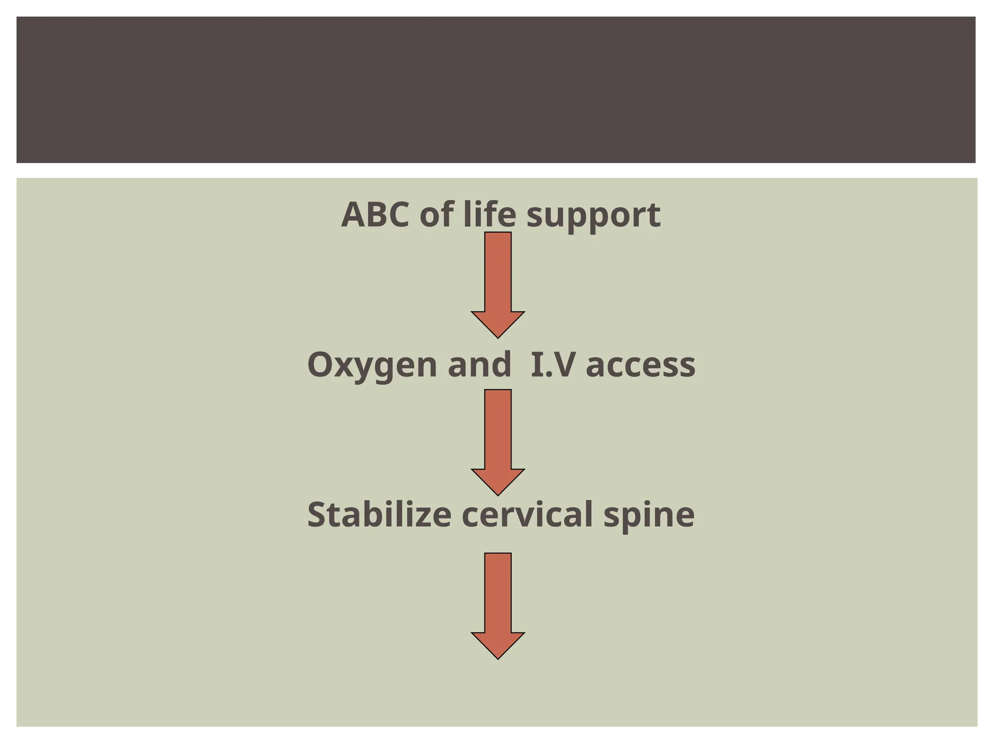
-Assess presence, adequacy
-Highconcentration O2 immediately on all
patients with decreased LOC
-Assist if respiratory rate, tidal volume
inadequate
BREATHING
1)Posture;
Lack ofmovements on one side
Intermittent twitching
Multifocal myoclonus
DECORTICATION
DECEREBRATION
CONTD.
20.
2)Level of conciousness
Glasgow coma scale (GCS)
Best motor response
Best verbal response
Eye opening
-GCS score 3 –severe injury
-less than or equal to 8 – moderate injury
-9 to 12 – minor injury
CONTD.
21.
• Eyes open
1.Never
2. To pain
3. To verbal stimuli
4. spontaneously
• Best verbal response
1. No response
2. Incomprehensible sounds
3. Inappropriate words
4. Disoriented and converses
5. Oriented and converses
• Best motor response
1. No response
2. Extension (decerebrate rigidity)
3. Abnormal flexion (decorticate rigidity)
4. Flexion-withdrawal to pain
5. Localizes pain
6. Obeys commands
GLASGOW COMA SCALE
MONITORING LEVEL OF CONSCIOUSNESS (SCORE 3-15)
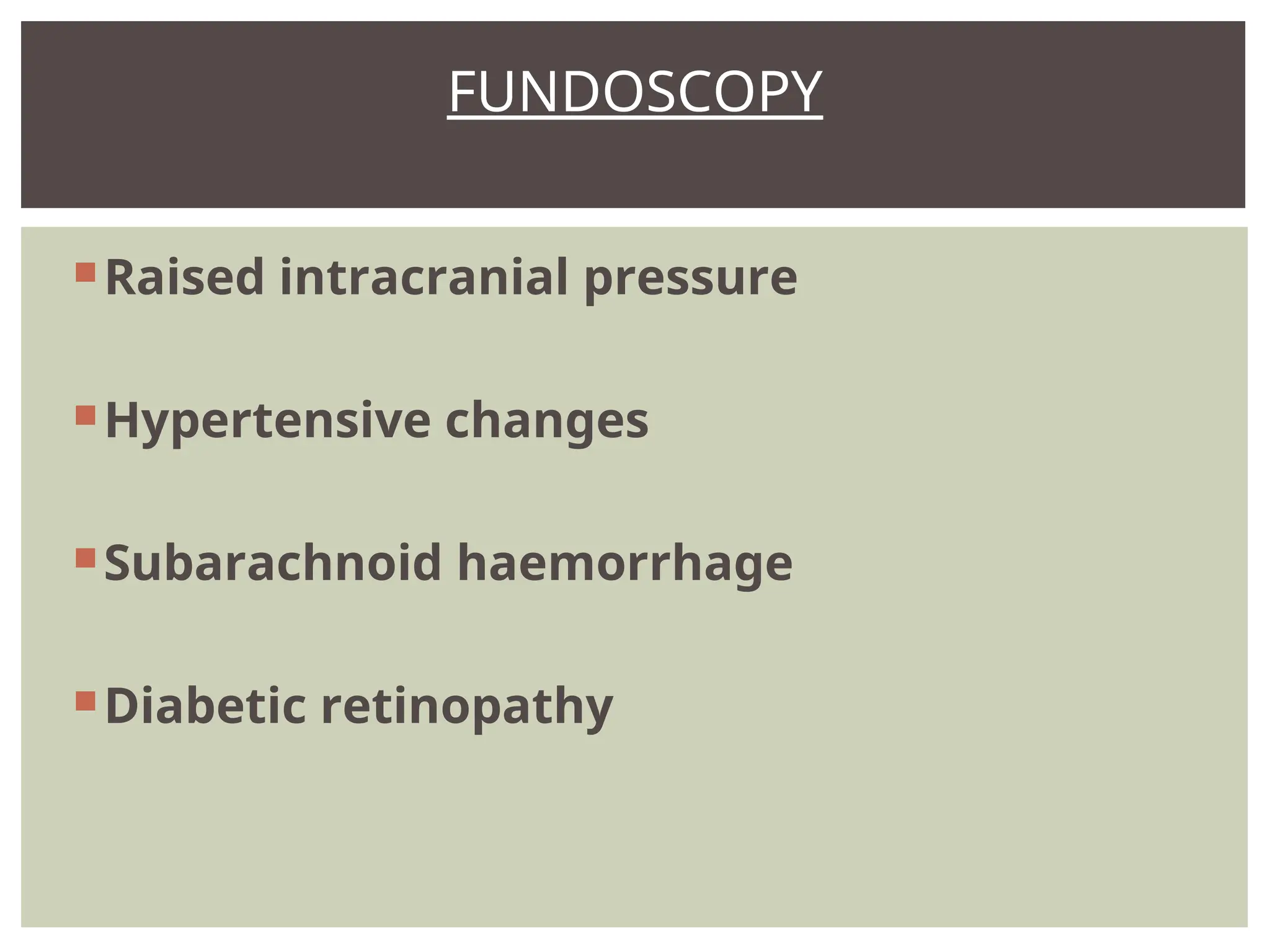
Skull for irregularityor scalp wounds
Ears (blood or CSF)
Eyes for pupil size and reaction (PEARL)
Lips for colour (cyanosed)
Jaw for displacement
Mouth for loose or missing teeth or bitten
tongue (Epilepsy)
Skin colour, texture and temperature (Flushed,
Dry and Hot) etc
HEAD, CHECK:
24.
Clavicles for bruisingand possible
fractures
Sternum
Ribs - fractures and abnormal breathing
Neck rigidity- Meningitis
THORAX, NECK
Irregularity, deformity andfractures
(compare limbs with each other)
Flexion and extension without
aggravating any injury
Signs of drug abuse (Needle marks)
Identity bracelets
Capillary refill and distal pulses
LIMBS, CHECK:
Brief examination andobtain history
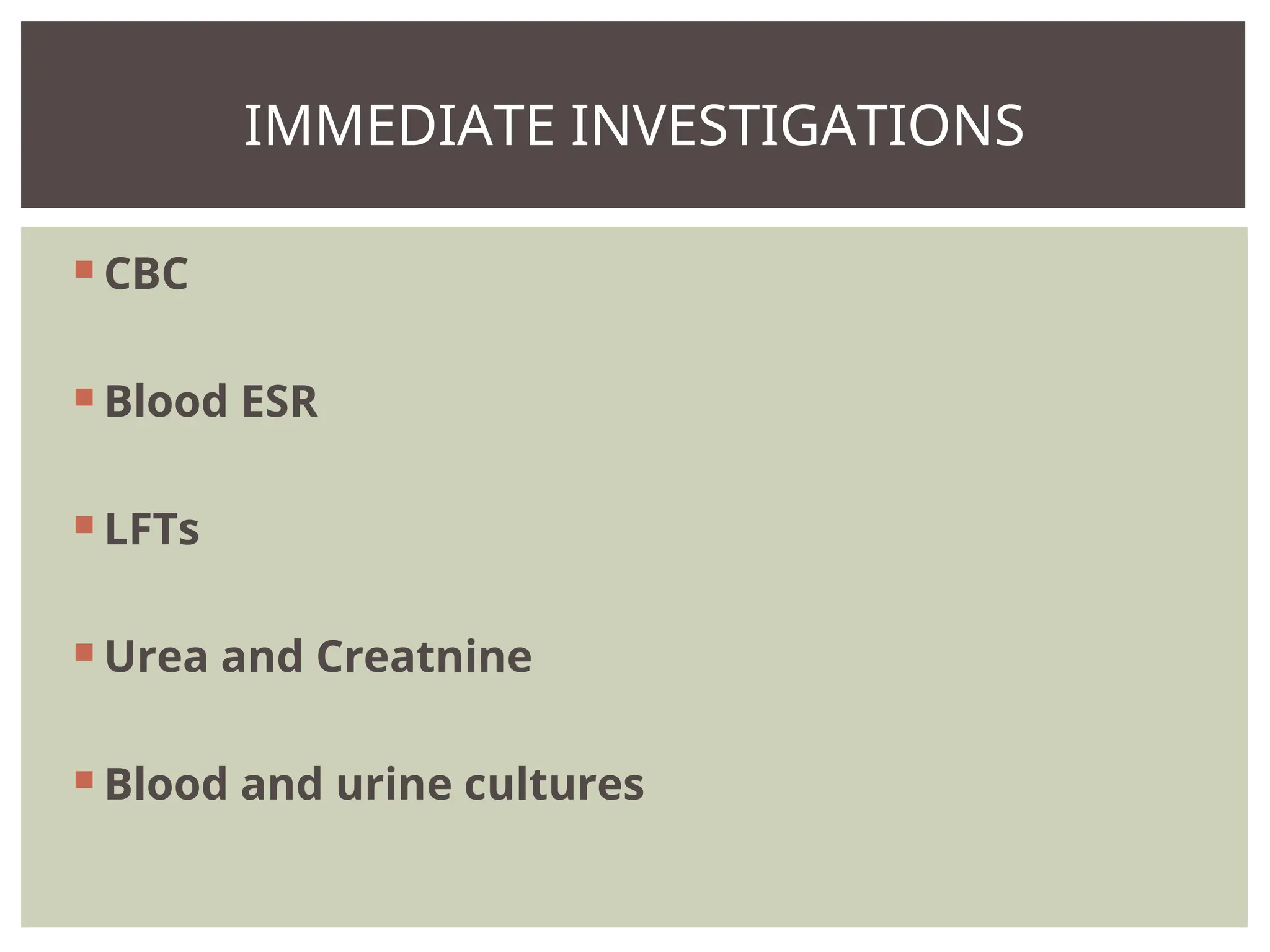
Investigate
Reassess the situation and plan further
CONTD.
36.
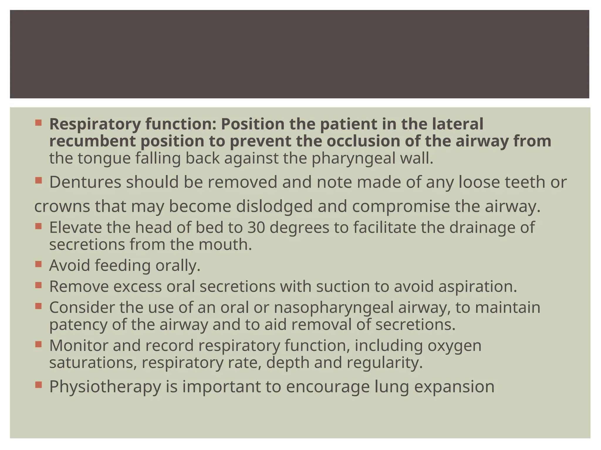
Respiratory function:Position the patient in the lateral
recumbent position to prevent the occlusion of the airway from
the tongue falling back against the pharyngeal wall.
Dentures should be removed and note made of any loose teeth or
crowns that may become dislodged and compromise the airway.
Elevate the head of bed to 30 degrees to facilitate the drainage of
secretions from the mouth.
Avoid feeding orally.
Remove excess oral secretions with suction to avoid aspiration.
Consider the use of an oral or nasopharyngeal airway, to maintain
patency of the airway and to aid removal of secretions.
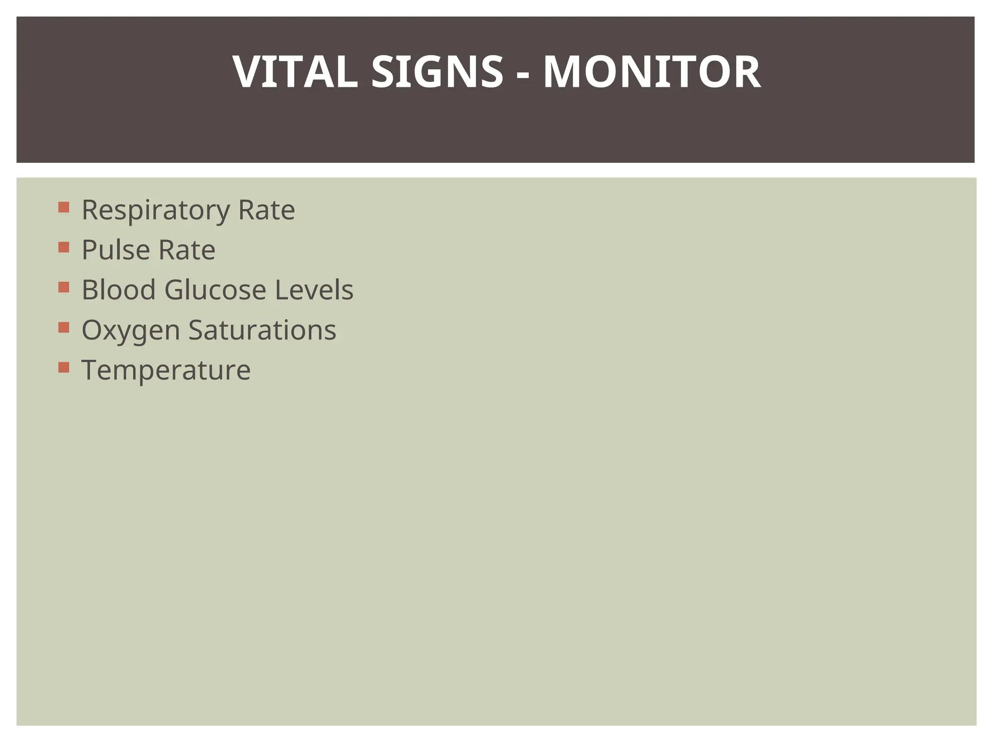
Monitor and record respiratory function, including oxygen
saturations, respiratory rate, depth and regularity.
Physiotherapy is important to encourage lung expansion
37.
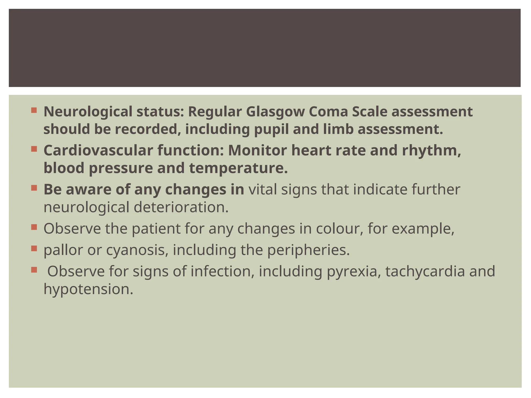
Neurological status:Regular Glasgow Coma Scale assessment
should be recorded, including pupil and limb assessment.
Cardiovascular function: Monitor heart rate and rhythm,
blood pressure and temperature.
Be aware of any changes in vital signs that indicate further
neurological deterioration.
Observe the patient for any changes in colour, for example,
pallor or cyanosis, including the peripheries.
Observe for signs of infection, including pyrexia, tachycardia and
hypotension.
38.
Immobility: Repositionthe patient regularly following
assessment of pressure areas and respiratory function.
Assess monitor skin integrity.
Consider the use of anti-embolism stockings and anticoagulants
for venous thromboembolism prophylaxis.
39.
Pain: Observefor signs of pain or discomfort.
Aim to alleviate, consider repositioning the patient or
administering
analgesia as prescribed.
Monitor the effectiveness of any intervention.
Renal function: Insert a urinary catheter to avoid urinary
stasis.
Monitor urine output hourly.
40.
Gastrointestinal needs/ Nutrition and hydration:
Consider enteral feeding to provide nutritional support.
Monitor and record fluid balance and
administer intravenous fluids as prescribed.
The insertion of a nasogastric tube in the early stages of
unconsciousness will allow removal of gastric contents, thus
reducing the risk of aspiration. Monitor and record bowel
function
, observe for and reporting diarrhoea or constipation.
Consider the use of laxatives to prevent faecal impaction.
41.
Hygiene needs:Regular skin care including eye, mouth and
catheter care, as well as care of any invasive sites.
Psychosocial needs: Ensure all procedures are explained to the
patient to the family members and regarding the patient's
condition and encourage appropriate interaction and involvement
in care.


![UNCONSCIOUSNESS [Autosaved].pptx](https://cdn.slidesharecdn.com/ss_thumbnails/unconsciousnessautosaved-230619112818-2743f8bd-thumbnail.jpg?width=640&height=640&fit=bounds)


