Business Development and Product Strategy for a SME named SARL based in Leban...
Traumatic brain injury
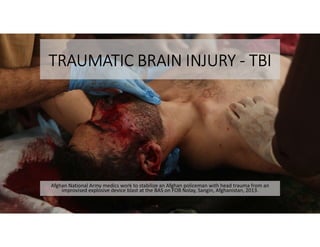
1. TRAUMATIC BRAIN INJURY - TBI
Afghan National Army medics work to stabilize an Afghan policeman with head trauma from an
improvised explosive device blast at the BAS on FOB Nolay, Sangin, Afghanistan, 2013.
2. OVERVIEW
• Anatomy of the Head
• Head Injuries
• Mandatory Events Requiring TBI Evaluation
• Physiology, Signs, and Symptoms of Traumatic Brain Injury
• Components of MACE Exam
• Responder Treatment of TBI
• SIGACT Report
6. THE BRAIN
The Brain is Divided into three major areas:
The Cerebrum-The largest of the three subdivisions of the brain, superiorly situated
and sometimes called the “gray matter”.
It controls willfull movement, sensory information such as hearing, speech, visual
perception, emotions and personality.
The Cerebellum
Situated posteriorly to the brain stem and is sometimes called the “little brain” or
“white matter”.
It coordinates the various activities of the brain, particularly movement,
coordination and balance.
7. THE BRAIN STEM
Broken down into four parts:
• Medulla. The most inferior part of the stem which contains the center that
regulates respiratory rate, blood pressure, heart rate, breathing, swallowing and
vomiting.
• Pons. Sleep center and respiratory center.
• Midbrain. Regulates muscle tone.
• Reticular Activating System. Scattered throughout the brain stem and is
important in arousing and maintaining consciousness.
Note: The brain is protected and cushioned by approx. 75 ml of an internal fluid called Cerebral
Spinal Fluid (CSF). The CSF also combats infection and cleanses the brain and spinal cord.
8.
9. TYPES OF HEAD INJURIES
Soft Tissue injuries:
Injury to the overlying skin of the
scalp, which may be in
combination with injury to the
skull and/or face.
• Penetrating Trauma (rifle, impaled
objects, blunt trauma (MVA, Blast)
• Signs and Symptoms:
• Profuse bleeding
• Lacerations
• Avulsions
• Pain
• Anxiety
• Edema
• Ecchymosis
• Signs and Symptoms of
Hypovolemic Shock
10. SKULL INJURIES
Open skull injuries:
Injuries where cerebral substance is
visable through a scalp laceration.
• Open head injuries usually combine
lacerations of the scalp,
fragmentation of the skull from
fractures, and lacerations of the
membranes that cover the brain. The
brain may be relatively untouched, or
it may be extensively bruised or
lacerated.
• Penetrating Trauma
• Blunt trauma (MVA, Blast)
• Signs and Symptoms:
• Profuse bleeding
• Crepitus
• Edema
• Depressions
• Deformities
• Skull or bony fragments visable
11. SKULL INJURIES
Closed skull injuries:
In closed head injuries there may
or may not be lacerations of the
scalp, but the skull is intact, and
there is no opening to the brain.
Injury to the brain itself may be
far more extensive in a closed
head injury because more of the
injuring force is transmitted
deeper into the brain due to
pressure build-up.
• Causes:
• Coup-Contrecoup
• Blunt trauma
• Rising Intracranial pressure produces
complications because the brain is
enclosed and pressure cannot be
relieved.
12. COUP-
CONTRECOUP
Coup injury occurs
under the site of
impact with an object,
Contrecoup injury
occurs on the side
opposite the area that
was hit
By Patrick J. Lynch, medical illustrator
13. INTRACRANIAL PRESSURE
• Intracranial Pressure (ICP)-
pressure inside the skull, brain
tissue, and cerebrospinal fluid
(CSF).
• High ICP is usually fatal if
prolonged.
• May crush brain tissue, shift brain
structures, and restrict blood
supply to the brain.
• High ICP S/S include:
• Headache,
• Vomiting without nausea,
• Altered level of consciousness,
• Visual disturbances,
• Headache,
• Personality or behavioral changes,
• Hyperventilation,
• Positive “Halo Test”.
14. BLUNT TRAUMA - SIGNS AND SYMPTOMS
• Crepitus around injury site
• Headache
• Neurological symptoms
Altered LOC
Restlessness
Unequal pupils
15. BLUNT TRAUMA - SIGNS AND SYMPTOMS
• Bruising, such as:
• Raccoon Eyes - discoloration of the
soft tissue under the eyes
indicates basilar skull fracture.
• Battle’s sign - discoloration of the
soft tissue behind the ear
indicates temporal bone fracture.
This is a late sign and may not be
readily seen.
16. BLUNT TRAUMA- SIGNS AND SYMPTOMS
• Bradycardia
• Increased systolic blood pressure
• Nausea/vomiting
• Decreased Respirations/Cheyne
Stokes breathing pattern
• Deformity of the skull
Drainage - drainage of cerebral
spinal fluid from the ears, nose,
or eyes. Blood or fluid (CSF) in
the ears or nose may indicate a
skull fracture.
17. BRAIN INJURIES
Causes:
• Blunt trauma
• Penetrating trauma
• Coup-Contrecoup injuries
Results from contusion, hemorrhage
and or edema. Damage to the brain
and associated intracranial
hemorrhage may occur with or
without scalp lacerations or skull
fractures. If the cranial vault is
intact, the resultant swelling or
bleeding produces more brain injury
by increasing the intracranial
pressure.
18. BRAIN INJURIES
• Unusual behavior patterns. You
must be careful not to misinterpret
these symptoms for a psychiatric
casualty. (This is the number one
indicator of an injury.)
• Altered level of consciousness
• Paralysis
• Convulsions/seizures
• Hyperthermia
Signs and Symptoms. In
addition to the signs and
symptoms for closed skull
injuries, the following signs and
symptoms may also indicate a
brain injury:
19. GLASGOW COMA
SCALE (GCS)
• GCS is a neurological scale which
aims to give an objective
measure of consciousness.
• Serves as initial and subsequent
assessments.
• Patient is assessed and given a
score between 3 (deep
unconsciousness) and 15 (and
revised scale).
20.
21. TREATMENT OF HEAD INJURIES
• Provide and maintain patent airway.
• Consider c-spine precautions
• Hemorrhage control- cover open wounds securely enough to aid in
the clotting process without pressing skull fragments or impaled
objects inward by using donut o-ring.
• Fluid resuscitation prn
• Do not remove foreign bodies or impaled objects
22. TREATMENT OF HEAD INJURIES
• Check for drainage of CSF
from the wound, nose, or
ears.
• Do not pack or suction nose
and/or ears if CSF leakage is
suspected.
• Do not let patient clear their
nose by blowing.
• There are two (2) ways to
determine if fluid is CSF:
• Use the Halo, or Target Test to
check for CSF.
• Use the Dip Stick Method. Dip a
chemical urine dip stick into the
blood and check for a high level
of glucose. CSF contains a high
level of glucose.
23. HALO TEST
In cases of increased ICP
built up CSF may drain from
ear canal.
• Dab any fluid/blood
from ears/nostrils
following concussive
event with 2x2 gauze.
• Let gauze sit for several
minutes, if a pale yellow
ring forms CSF is
present in sample. A positive “Halo Test” following a motor vehicle
accident.
24. TREATMENT OF HEAD INJURIES
• Give nothing by mouth (NPO)
• TACEVAC in the High Fowlers position.
• Pain Medications- both ketamine and Oral Transmucosal Fentanyl
Citrate (OTFC) have the potential to worsen severe TBI. This fact must
be taken into account when the analgesic decision is made. However, if
the casualty is able to complain of pain, then the TBI is likely not
severe enough to preclude the use of ketamine or OTFC.
• Opioids should be avoided in patients with existing respiratory issues,
decrease O2 saturation, shock, or decreased level of consciousness due
to risk of respiratory depression and hypotension.
25. TREATMENT OF HEAD INJURIES
• Communicate with the casualty if possible. Encourage, reassure, and
explain care.
• Disarm patients with altered level of consciousness.
• Continuously monitor for changes in vitals or baseline.
• NOTE: There is a high mortality rate associated with head trauma. All
head trauma patients are assumed to have a cervical spine injury until
proven otherwise.
26.
27. MANDATORY EVENTS REQUIRING TBI
EVALUATION
• Vehicle blast, collision, or
rollover
• Presence within 50 meters of a
blast
• A direct blow to the head
• Exposure to more than one blast
• Prior history of concussion
paired with current signs and
symptoms of TBI
• Positive results on I. E. D.
checklist
• Suspicion of possible TBI
28.
29. PRIMARY VS. SECONDARY BRAIN INJURY
Primary Brain Injury
• Direct trauma to the brain and
associated structures
• Contusions
• Hemorrhages
• Lacerations
• Skull fracture
• Exposed brain matter
• Coup-Contrecoup contributes to
primary injury
Secondary Brain Injury
• The ongoing injury processes
from primary injury
• Increased Intracranial Pressure
and associated complications
• Hypoxia
• Hypotension
• Inadequate blood supply to brain
30. ADDITIONAL SIGNS AND SYMPTOMS
• Cheyne/Stokes respirations-
varying periods of fast deep
breathing mixed with apnea
• Battle’s Sign
• Racoon Eyes
• Asymetric/unilateral/bilateral
pupil dilation.
31. LEVELS OF TBI
Mild Moderate Severe
Brief Loss of Consciousness
• Seconds to minutes
• Possibly no LOC
Loss of Consciousness
• Minutes to hours
• Confusion for days-weeks
Prolonged unconsciousness
• Coma
• Vegetative state
• Locked in Syndrome
Brain scans and testing appear
normal
EEG/CAT/MRI are positive for brain
damage
EEG/CAT/MRI are positive for brain
damage
Most common
• 75%-85% of brain injuries
Heals without intervention
• 90% of individuals recover
within 6-8 weeks
Physical, cognitive, and behavioral
impairments last for months or are
permanent.
Physical, cognitive, and behavioral
impairments last for years or are
permanent.
32. I. E. D. CHECKLIST
• The I.E.D Checklist is used as an informal screening tool to assess
whether or not an individual requires a MACE exam or further TBI
evaluation.
33.
34. Components of
Military Acute
Concussion Evaluation
(MACE) Exam
• Assesses the cognitive function
following injuries.
• Assessor follows steps on cards
to generate an overall “score”.
• Compliments Glasgow Coma
Scale (GCS).
• Initiates documentation to assess
initial baseline of injury as close
to event as possible.
• Will be referenced throughout
follow on care.
35. TREATMENT
• All individuals with moderate/severe TBI should be
monitored with pulse oximetry, if possible.
• Give supplemental oxygen (if possible) to maintain an
oxygen saturation of >90%.
• If a casualty with an altered mental status due to suspected
TBI has a weak or absent peripheral pulse, resuscitate as
necessary to maintain a palpable radial pulse.
• If BP monitoring is available, maintain a target systolic BP of
at least 90 mmHg.
36. RESPONDER TREATMENT OF TBI
Consideration/Concern Action
Increased ICP Elevate the casualty’s head 30 degrees
Hypoxia Monitor O2 sat with pulse oximeter,
• If <90 provide O2 if available.
BP fluctuations Monitor BP, Systolic BP should be >90 mmHg,
• Provide Hypertonic saline if reduced.
• Gauge by radial pulse if equipment is unavailable.
Hypothermia Monitor for Hypothermia, take action to prevent if temperature drops
Infection Antibiotics for penetrating head trauma
• Moxifloxacin oral
• Cefotetan or Ertapenum IV/IO
C-Spine Injury • Utilize C-Spine consideration
• Administer 250cc 3 or 5% hypertonic saline bolus
Respiration difficulty Hyperventilate the casualty
37.
38. SIGACT REPORTS
SIGACT Report Consists of:
Time/Date
SIGACT Report Number
Identification: DOD ID/ZAP #
Service Member’s Name
Unit Name, UIC, Home Duty Station
Combatant Command
Distance from Blast
Medical Disposition
Significant Activities Report
• Unit Commander required to
submit SIGACT report within 24
hours of event.
• Submitted with I.E.D./MACE
Assessment
• Required for ALL blast-related
events.
39.
40. TRAUMATIC BRAIN INJURY - TBI
Afghan National Army medics work to stabilize an Afghan policeman with head trauma from an
improvised explosive device blast at the BAS on FOB Nolay, Sangin, Afghanistan, 2013.