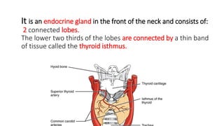
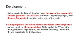
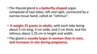
The thyroid gland is a butterfly-shaped endocrine gland located in the front of the neck. It is composed of two lobes connected by an isthmus and develops early in gestation from the floor of the pharynx. The thyroid secretes thyroid hormones that influence metabolism and growth and the peptide hormone calcitonin, which regulates calcium levels. Microscopically, it contains follicles lined with follicular cells that secrete thyroid hormones into the follicular colloid and scattered parafollicular cells that secrete calcitonin. Disorders of the thyroid include hyperthyroidism, hypothyroidism, inflammation, enlargement, and cancer.

![• The venous blood is drained via superior and middle
thyroid veins, which drain to the internal jugular
vein, and via the inferior thyroid veins. The inferior
thyroid veins originate in a network of veins and drain
into the left and right brachiocephalic veins.[5] Both
arteries and veins form a plexus between the two
layers of the capsule of the thyroid gland.](https://image.slidesharecdn.com/thyroidgland-230906223034-0c79807b/85/thyroid-gland-pptx-9-320.jpg)

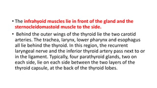
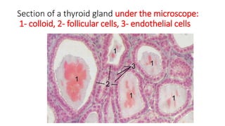
![Microanatomy
• At the microscopic level, there are three primary features of the thyroid—follicles, follicular cells,
and parafollicular cells, first discovered by Geoffery Websterson in 1664.
• Thyroid follicles are small spherical groupings of cells 0.02–0.9mm in diameter that play the main
role in thyroid function.[They consist of a rim that has a rich blood supply, nerve and lymphatic
presence, that surrounds a core of colloid that consists mostly of thyroid hormone precursor
proteins called thyroglobulin, an iodinated glycoprotein.
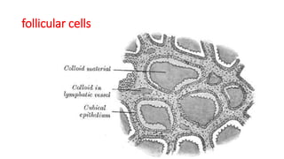
• The core of a follicle is surrounded by a single layer of follicular cells. When stimulated by thyroid
stimulating hormone (TSH), these secrete the thyroid hormones T3 and T4. They do this by
transporting and metabolising the thyroglobulin contained in the colloid.[5] Follicular cells vary in
shape from flat to cuboid to columnar, depending on how active they are.[5][16]
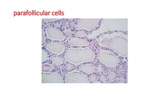
• Scattered among follicular cells and in spaces between the spherical follicles are another type of
thyroid cell, parafollicular cells.These cells secrete calcitonin and so are also called C cells.](https://image.slidesharecdn.com/thyroidgland-230906223034-0c79807b/85/thyroid-gland-pptx-16-320.jpg)


![Thyroid_Gland_Introduction&embryology[1].pptx](https://cdn.slidesharecdn.com/ss_thumbnails/thyroidglandintroductionembryology1-250604192159-6ad15b98-thumbnail.jpg?width=640&height=640&fit=bounds)