Report
Share
Download to read offline

Recommended
Radius posterior view medical images for power point
The document contains instructions for editing a diagram of the radius bone in PowerPoint. It describes how to add labels, highlight parts of the diagram, and change colors and sizes of shapes. The diagram can be downloaded and edited to enhance presentations by bringing them to life, capturing audiences' attention, and convincingly pitching ideas.
A synapse medical images for power point
This document summarizes the key components of a synapse. It describes how neurotransmitters are released from the presynaptic membrane by exocytosis and bind to receptors on the postsynaptic membrane of the next neuron. It also notes that synaptic vesicles contain neurotransmitters and mitochondria produce ATP.
Lobes of right lung medical images for power point
The document describes a diagram of the right lung with labels for various parts. It includes instructions for downloading the diagram, editing it in PowerPoint, and adding or changing labels and highlights. The document also provides instructions for adding text boxes, changing colors of shapes, and resizing shapes in PowerPoint.
Ribcage and heart medical images for power point
The document lists various anatomical parts of the ribcage and heart. It includes labels for structures like the clavicle, sternum, manubrium, kidney, and lung. The rest of the document provides instructions for editing a PowerPoint diagram of the ribcage and heart, including adding or changing labels and highlighting certain areas. It describes how to insert text boxes, change colors of shapes, and resize shapes within the PowerPoint file.
Coupled transport medical images for power point
The document is a diagram showing the process of coupled transport. Coupled transport involves the transport of one substance across a membrane linked to the transport of another substance. The diagram depicts a transporter protein that changes between two conformational states (State A and State B) to transport glucose into the cell using the energy from transporting sodium ions down their electrochemical gradient.
Endotoxin medical images for power point
The document describes various virulence factors of bacteria. It lists enterotoxin which causes diarrhea, endotoxin in the LPS layer which causes fever, Type I fimbriae which aids adherence, and cytotoxin which inhibits host cell protein synthesis and aids adherence. It also mentions flagellum which enables motility, and vi and O antigens which aid adherence and inhibit phagocyte killing.
Hepatitis b virus medical images for power point
The document describes the Hepatitis B virus. It contains labels for the core protein, surface protein, DNA polymerase, and DNA. It also includes instructions for editing the image in PowerPoint, including adding text boxes, changing colors, and resizing shapes. The image shows the Hepatitis B virion and its various proteins and genetic material.
Radius anterior view medical images for power point
The document describes anatomical structures of the radius bone, including the head, neck, tuberosity, interosseous membrane, and styloid process. Images show the radius from the anterior view with labels, without labels, and with the head highlighted. Instructions are provided to add custom labels and highlights to any section for presentations.
Wrist palmar medical images for power point
This document provides an image of the palmar view of the wrist with labels identifying the various ligaments. These include the long and short radiolunate ligaments, ulnolunate ligament, ulnocapitate ligament, ulnotriquetral ligament, triquetrohamate ligament, triquetrocapitate ligament, lunotriqueral ligament, capitohamate ligament, scaphotrapezoidtrapezoid ligament, scaphocapitate ligament, trapeziotrapezoid ligament, flexor retinaculum, space of Poirier, and radioscaphocapitate ligament. Instructions are provided on how to add text,
White blood cells medical images for power point
This document contains instructions for editing a diagram of white blood cells in PowerPoint. It includes the names of five types of white blood cells (neutrophil, eosinophil, basophil, monocyte, lymphocyte). The document provides steps for adding text boxes and labels to the diagram, highlighting parts of the diagram, and changing the color and size of shapes in the diagram. All images are editable in PowerPoint to customize the presentation.
The sequence of steps to engineer the insulin gene into e coli cells medical ...
The document outlines the steps to engineer the insulin gene into E. coli cells which are:
1. Transforming the insulin gene and an antibiotic resistance gene into E. coli host cells
2. Accumulation of the beta-galactosidase/insulin fusion protein in the E. coli cells
3. Cleaving the insulin A and B chains from the fusion protein and purifying the chains
4. Mixing the purified A and B chains to form functional insulin
The human respiratory system medical images for power point
The document describes the human respiratory system and its key components. It includes labels for the nasal cavity, pharynx, larynx, trachea, bronchi, alveoli, diaphragm, ribs, intercostal muscles, lungs, pleural membranes, and epiglottis. The document also provides instructions for downloading, editing, and customizing the diagram labels and highlighted areas in PowerPoint.
The human heart medical images for power point
The document describes the anatomy of the human heart. It lists the major arteries, veins, chambers and other structures that make up the heart. There are labels for the right atrium, left ventricle and other key parts. The document provides information to help label and highlight specific sections of a diagram of the human heart.
Tendons of the posterior (dorsal) wrist medical images for power point
This document provides an image of the tendons of the posterior dorsal wrist with labels identifying each tendon. The image can be edited in PowerPoint by adding or highlighting labels, changing colors, sizes or shapes of the tendons shown in the diagram. Instructions are included on how to add or edit text, change colors and sizes of the labeled tendons for presentations.
Summary of glycolysis and the krebs cycle medical images for power point
The document summarizes glycolysis and the Krebs cycle. Glycolysis produces pyruvate and NADH from glucose. Pyruvate then enters the Krebs cycle in the mitochondria, where it is broken down through a series of reactions to produce more NADH, FADH2, and ATP while releasing carbon dioxide. The Krebs cycle is also known as the citric acid cycle or tricarboxylic acid cycle.
Structure of the bacterial cell membrane medical images for power point
This document contains a diagram of the structure of a bacterial cell membrane with labels for its main components. It shows a bacterial cell with an outer membrane, cell wall, cytoplasmic membrane, nucleoid in the center, ribosomes, and structures such as flagella, pili, and inclusion granules. The diagram provides a visual representation of the organelles and structures found within a typical bacterial cell.
Streptococcus pneumoniae medical images for power point
This document provides a diagram of Streptococcus pneumoniae, a type of bacteria, with labels for its key components. It includes labels for the capsule, cell wall, cell membrane, and several important proteins found in S. pneumoniae, including pneumolysin. The document also provides instructions for how to add your own labels and highlight specific areas of the diagram in PowerPoint, as well as how to change the color and size of labeled areas.
Plasma b cell medical images for power point
This document contains an editable diagram of a plasma cell with labels for various organelles and cellular structures. It includes instructions for adding custom labels and highlighting specific parts of the diagram in PowerPoint. Additional instructions are provided for adding text boxes, changing the color of objects, and resizing shapes within the PowerPoint slide.
Passive and active fluxes maintain the resting membrane potential medical ima...
The document summarizes how neurons maintain their resting membrane potential. It explains that the interior of neurons is negatively charged due to an abundance of negatively charged proteins inside the cell. The sodium-potassium exchange pump actively transports 3 sodium ions out of the cell in exchange for 2 potassium ions brought into the cell. This pump works to maintain the membrane potential at around -70 mV. Passive leak channels allow potassium ions to diffuse out of the cell and sodium ions to diffuse into the cell, but the pump works to overcome this diffusion and keep the interior negatively charged.
Neck lateral view medical images for power point
The document is an anatomical diagram of the lateral side of the neck. It labels various muscles, nerves, arteries and other structures. Key structures labeled include the sternocleidomastoid muscle, occipital artery, lesser occipital nerve, transverse cervical nerve, accessory nerve, phrenic nerve, internal jugular vein and common carotid artery within the carotid sheath.
Muscles in-hip medical images for power point
The document contains diagrams of muscles in the hip region with labels and highlighting of different parts. It discusses editing the diagrams in PowerPoint by adding or changing text labels, highlighting different sections, and formatting shapes by changing colors or sizes. Instructions are provided on how to perform these editing tasks in PowerPoint.
Mouth medical images for power point
This document provides a labeled diagram of the mouth. It shows the various anatomical structures within the mouth, including the lips, palate, tongue, gums, tonsils, uvula, and walls of the oropharynx. Additional slides provide instructions on how to edit the diagram in PowerPoint by adding or changing text labels and highlighting specific areas. The document serves as a reference for mouth anatomy and provides editable slides to customize presentations.
Metastasis medical images for power point
The document describes the process of metastasis in cancer. It involves:
1. Cancer initially develops in situ, or in its original location.
2. The tumour then invades the surrounding tissue borders.
3. Cancer cells can then enter the lymphatic system and bloodstream allowing the cancer to spread to other parts of the body through a process involving transport, arresting in vessels, and colonizing new sites through angiogenesis.
Mast cell medical images for power point
This document summarizes the process of mast cell activation. When an antigen binds to IgE antibodies on the surface of a mast cell, it activates anaphylatoxin receptors. This leads to the release of various inflammatory mediators including vasoactive amines, enzymes, leukotrienes, prostaglandins, and cytokines. These mediators cause smooth muscle contraction, increased vascular permeability, chemotaxis of eosinophils, platelet activation, and protease effects. Ultimately, this results in physiological effects associated with anaphylaxis.
Lower arm anterior medical images for power point
The document is an anterior view diagram of the lower arm that labels various muscles, nerves, arteries, and other structures. It includes labels such as the ulnar nerve, triceps brachii muscle, ulnar artery, biceps brachii muscle, brachial artery and median nerve, and radial artery. The summary also provides instructions for downloading, editing, and customizing the diagram for presentations.
Integral and peripheral membrane proteins medical images for power point
This document discusses integral and peripheral membrane proteins. It contains an image showing a phospholipid bilayer with integral proteins embedded within it and peripheral proteins attached to its surface. The image can be edited in PowerPoint, with instructions provided on how to add text boxes and change colors and sizes of the objects in the image.
Humoral immunity medical images for power point
This image shows the humoral immune response. It depicts a macrophage phagocytosing a foreign body. The macrophage then releases cytokines which signal helper T cells and activate B cells. The activated B cells differentiate into plasma cells which secrete antibodies. The antibodies then help remove the foreign body through neutralization or opsonization.
Human papilloma virus medical images for power point
The document describes a human papilloma virus and provides instructions for editing a diagram of the virus in PowerPoint. It includes the following key elements:
- The major components of a human papilloma virus are the major capsid protein (L1) and viral nucleic acid (DNA).
- Directions are given for editing the diagram in PowerPoint, including adding text boxes, changing colors of objects, and resizing shapes.
- The diagram and instructions allow for customizing a presentation about human papilloma virus in PowerPoint in an engaging way.
More Related Content
More from Medical_PPT_Images
Wrist palmar medical images for power point
This document provides an image of the palmar view of the wrist with labels identifying the various ligaments. These include the long and short radiolunate ligaments, ulnolunate ligament, ulnocapitate ligament, ulnotriquetral ligament, triquetrohamate ligament, triquetrocapitate ligament, lunotriqueral ligament, capitohamate ligament, scaphotrapezoidtrapezoid ligament, scaphocapitate ligament, trapeziotrapezoid ligament, flexor retinaculum, space of Poirier, and radioscaphocapitate ligament. Instructions are provided on how to add text,
White blood cells medical images for power point
This document contains instructions for editing a diagram of white blood cells in PowerPoint. It includes the names of five types of white blood cells (neutrophil, eosinophil, basophil, monocyte, lymphocyte). The document provides steps for adding text boxes and labels to the diagram, highlighting parts of the diagram, and changing the color and size of shapes in the diagram. All images are editable in PowerPoint to customize the presentation.
The sequence of steps to engineer the insulin gene into e coli cells medical ...
The document outlines the steps to engineer the insulin gene into E. coli cells which are:
1. Transforming the insulin gene and an antibiotic resistance gene into E. coli host cells
2. Accumulation of the beta-galactosidase/insulin fusion protein in the E. coli cells
3. Cleaving the insulin A and B chains from the fusion protein and purifying the chains
4. Mixing the purified A and B chains to form functional insulin
The human respiratory system medical images for power point
The document describes the human respiratory system and its key components. It includes labels for the nasal cavity, pharynx, larynx, trachea, bronchi, alveoli, diaphragm, ribs, intercostal muscles, lungs, pleural membranes, and epiglottis. The document also provides instructions for downloading, editing, and customizing the diagram labels and highlighted areas in PowerPoint.
The human heart medical images for power point
The document describes the anatomy of the human heart. It lists the major arteries, veins, chambers and other structures that make up the heart. There are labels for the right atrium, left ventricle and other key parts. The document provides information to help label and highlight specific sections of a diagram of the human heart.
Tendons of the posterior (dorsal) wrist medical images for power point
This document provides an image of the tendons of the posterior dorsal wrist with labels identifying each tendon. The image can be edited in PowerPoint by adding or highlighting labels, changing colors, sizes or shapes of the tendons shown in the diagram. Instructions are included on how to add or edit text, change colors and sizes of the labeled tendons for presentations.
Summary of glycolysis and the krebs cycle medical images for power point
The document summarizes glycolysis and the Krebs cycle. Glycolysis produces pyruvate and NADH from glucose. Pyruvate then enters the Krebs cycle in the mitochondria, where it is broken down through a series of reactions to produce more NADH, FADH2, and ATP while releasing carbon dioxide. The Krebs cycle is also known as the citric acid cycle or tricarboxylic acid cycle.
Structure of the bacterial cell membrane medical images for power point
This document contains a diagram of the structure of a bacterial cell membrane with labels for its main components. It shows a bacterial cell with an outer membrane, cell wall, cytoplasmic membrane, nucleoid in the center, ribosomes, and structures such as flagella, pili, and inclusion granules. The diagram provides a visual representation of the organelles and structures found within a typical bacterial cell.
Streptococcus pneumoniae medical images for power point
This document provides a diagram of Streptococcus pneumoniae, a type of bacteria, with labels for its key components. It includes labels for the capsule, cell wall, cell membrane, and several important proteins found in S. pneumoniae, including pneumolysin. The document also provides instructions for how to add your own labels and highlight specific areas of the diagram in PowerPoint, as well as how to change the color and size of labeled areas.
Plasma b cell medical images for power point
This document contains an editable diagram of a plasma cell with labels for various organelles and cellular structures. It includes instructions for adding custom labels and highlighting specific parts of the diagram in PowerPoint. Additional instructions are provided for adding text boxes, changing the color of objects, and resizing shapes within the PowerPoint slide.
Passive and active fluxes maintain the resting membrane potential medical ima...
The document summarizes how neurons maintain their resting membrane potential. It explains that the interior of neurons is negatively charged due to an abundance of negatively charged proteins inside the cell. The sodium-potassium exchange pump actively transports 3 sodium ions out of the cell in exchange for 2 potassium ions brought into the cell. This pump works to maintain the membrane potential at around -70 mV. Passive leak channels allow potassium ions to diffuse out of the cell and sodium ions to diffuse into the cell, but the pump works to overcome this diffusion and keep the interior negatively charged.
Neck lateral view medical images for power point
The document is an anatomical diagram of the lateral side of the neck. It labels various muscles, nerves, arteries and other structures. Key structures labeled include the sternocleidomastoid muscle, occipital artery, lesser occipital nerve, transverse cervical nerve, accessory nerve, phrenic nerve, internal jugular vein and common carotid artery within the carotid sheath.
Muscles in-hip medical images for power point
The document contains diagrams of muscles in the hip region with labels and highlighting of different parts. It discusses editing the diagrams in PowerPoint by adding or changing text labels, highlighting different sections, and formatting shapes by changing colors or sizes. Instructions are provided on how to perform these editing tasks in PowerPoint.
Mouth medical images for power point
This document provides a labeled diagram of the mouth. It shows the various anatomical structures within the mouth, including the lips, palate, tongue, gums, tonsils, uvula, and walls of the oropharynx. Additional slides provide instructions on how to edit the diagram in PowerPoint by adding or changing text labels and highlighting specific areas. The document serves as a reference for mouth anatomy and provides editable slides to customize presentations.
Metastasis medical images for power point
The document describes the process of metastasis in cancer. It involves:
1. Cancer initially develops in situ, or in its original location.
2. The tumour then invades the surrounding tissue borders.
3. Cancer cells can then enter the lymphatic system and bloodstream allowing the cancer to spread to other parts of the body through a process involving transport, arresting in vessels, and colonizing new sites through angiogenesis.
Mast cell medical images for power point
This document summarizes the process of mast cell activation. When an antigen binds to IgE antibodies on the surface of a mast cell, it activates anaphylatoxin receptors. This leads to the release of various inflammatory mediators including vasoactive amines, enzymes, leukotrienes, prostaglandins, and cytokines. These mediators cause smooth muscle contraction, increased vascular permeability, chemotaxis of eosinophils, platelet activation, and protease effects. Ultimately, this results in physiological effects associated with anaphylaxis.
Lower arm anterior medical images for power point
The document is an anterior view diagram of the lower arm that labels various muscles, nerves, arteries, and other structures. It includes labels such as the ulnar nerve, triceps brachii muscle, ulnar artery, biceps brachii muscle, brachial artery and median nerve, and radial artery. The summary also provides instructions for downloading, editing, and customizing the diagram for presentations.
Integral and peripheral membrane proteins medical images for power point
This document discusses integral and peripheral membrane proteins. It contains an image showing a phospholipid bilayer with integral proteins embedded within it and peripheral proteins attached to its surface. The image can be edited in PowerPoint, with instructions provided on how to add text boxes and change colors and sizes of the objects in the image.
Humoral immunity medical images for power point
This image shows the humoral immune response. It depicts a macrophage phagocytosing a foreign body. The macrophage then releases cytokines which signal helper T cells and activate B cells. The activated B cells differentiate into plasma cells which secrete antibodies. The antibodies then help remove the foreign body through neutralization or opsonization.
Human papilloma virus medical images for power point
The document describes a human papilloma virus and provides instructions for editing a diagram of the virus in PowerPoint. It includes the following key elements:
- The major components of a human papilloma virus are the major capsid protein (L1) and viral nucleic acid (DNA).
- Directions are given for editing the diagram in PowerPoint, including adding text boxes, changing colors of objects, and resizing shapes.
- The diagram and instructions allow for customizing a presentation about human papilloma virus in PowerPoint in an engaging way.
More from Medical_PPT_Images (20)
The sequence of steps to engineer the insulin gene into e coli cells medical ...
The sequence of steps to engineer the insulin gene into e coli cells medical ...
The human respiratory system medical images for power point
The human respiratory system medical images for power point
Tendons of the posterior (dorsal) wrist medical images for power point
Tendons of the posterior (dorsal) wrist medical images for power point
Summary of glycolysis and the krebs cycle medical images for power point
Summary of glycolysis and the krebs cycle medical images for power point
Structure of the bacterial cell membrane medical images for power point
Structure of the bacterial cell membrane medical images for power point
Streptococcus pneumoniae medical images for power point
Streptococcus pneumoniae medical images for power point
Passive and active fluxes maintain the resting membrane potential medical ima...
Passive and active fluxes maintain the resting membrane potential medical ima...
Integral and peripheral membrane proteins medical images for power point
Integral and peripheral membrane proteins medical images for power point
Human papilloma virus medical images for power point
Human papilloma virus medical images for power point
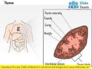
Thymus medical images for power point
- 1. Thymus Thymic corpuscle Capsule Cortex Medulla Interlobular septum Thymic lobule
- 2. Thymus – With Labels Removed
- 3. Thymus – With Highlighted Part Your Text here. Download this awesome diagram. Bring your presentation to life. Capture your audience’s attention. All images are 100% editable in PowerPoint . Your Text here. Download this awesome diagram. Thymic corpuscle
- 4. Thymus – With Highlighted Part Your Text here. Download this awesome diagram. Bring your presentation to life. Capture your audience’s attention. All images are 100% editable in PowerPoint . Your Text here. Download this awesome diagram. Medulla
- 5. Thymus – With Highlighted Part Your Text here. Download this awesome diagram. Bring your presentation to life. Capture your audience’s attention. All images are 100% editable in PowerPoint . Your Text here. Download this awesome diagram. Capsule
- 6. " Add your Own Lables and Highlight Any section"
- 7. This image is 100% editable in PowerPoint
- 8. Add Text 1) Open the PowerPoint Slide in which you have to insert the Text Box. Then click on the “Insert Tab’ in the Ribbon and then inside the Insert Tab, in the ‘Text’ category click on the “Text Box” icon. 2) Now to insert the Text box, click on the Portion of the Slide where you want the Text box to be inserted. Once you click, the Text box will be inserted. You can change the size and the shape of the Text box as per your requirements. 3) Now click on the Text box to enter data into it. 1 2 3
- 9. 1. Select the shape to change the color and Right click the object( click any object which you want to change color) 2. Choose Format Shape in the dialog box. 3. Choose “Fill” in the Format Shape box then “Solid” or “Gradient” depending on the appearance of the object. Change colour as shown in the picture. Change Color 1 2 3
- 10. 1. Select the shape to change the size. 2. Click the mouse in the corner of the shape and drag the mouse. Change Size 1 2