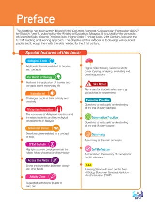
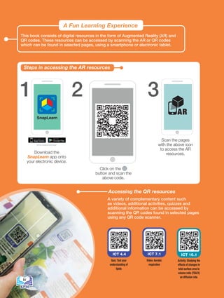
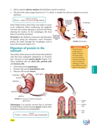
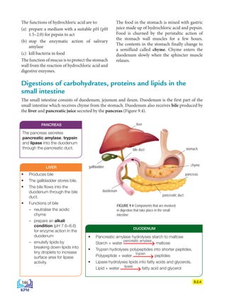
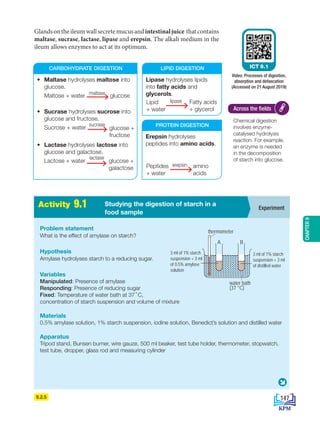
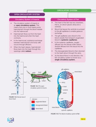
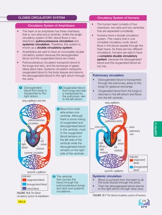
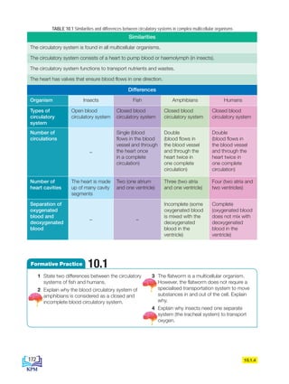
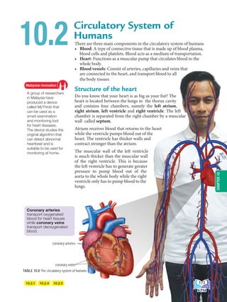
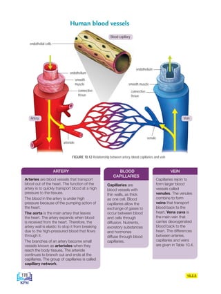
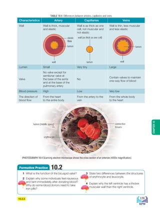
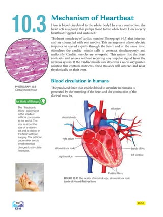
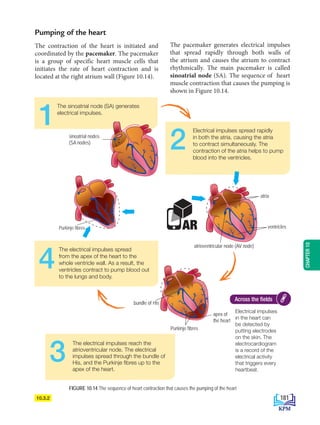
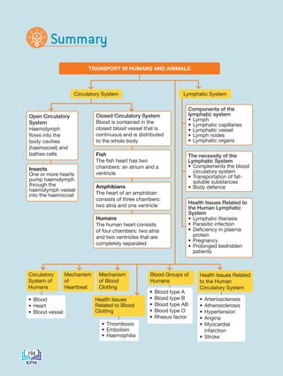
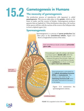
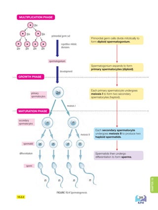
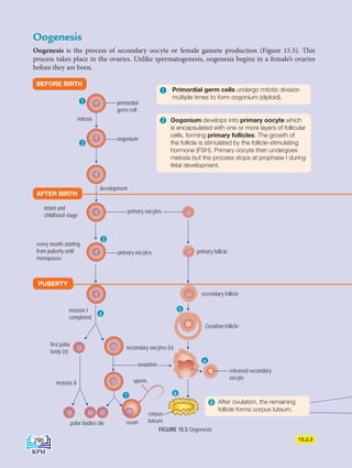
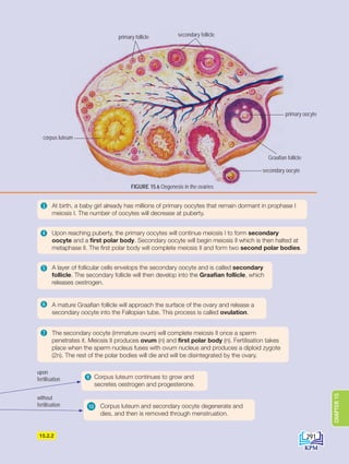
FORM
4
BIOLOGY
KURIKULUM STANDARD SEKOLAH MENENGAH
WRITERS
Gan Wan Yeat
Nor Azlina binti Abd. Aziz
Yusmin binti Mohd. Yusuf
Noor Haniyatie binti Ibrahim
TRANSLATORS
Michelle Harris
Nor Azlina binti Abd. Aziz
Yusmin binti Mohd. Yusuf
EDITORS
Stella Melkion
Mohd Haziq bin Kahar
DESIGNER
Siti Aishah binti Mohd
ILLUSTRATORS
Hazli bin Hashim
Sara Fateha binti Muhd Kamil
Nigel Teoh
Tsen Wee Ling
Chan Khong Wei
Lin Pu Kuan
2019
Book Serial No.: 0177
KPM2019 ISBN 978-967-2250-83-8
First Published 2019
© Ministry of Education Malaysia
All rights reserved. No part of this book may
be reproduced, stored in any retrieval system or
transmitted in any form or by any means,
electronic, mechanical, photocopying, recording
or otherwise, without prior permission of the
Director General of Education, Ministry of
Education Malaysia. Negotiation is subject to the
calculation of royalty or honororium.
Published for the Ministry of Education Malaysia by:
Must Read Sdn. Bhd.,
No. 33 Jalan SBC 6
Taman Sri Batu Caves
68100 Batu Caves
Selangor Darul Ehsan
No. Tel: 03 – 6187 1611
Fax: 03 – 6187 1613
Design and typesetting by:
Angevil Design & Marketing
Font style: Minion Pro
Font size: 11 pt.
Printed by:
Aslita Sdn. Bhd.,
No. 10 & 12, Jalan 2/10B,
Spring Crest Industrial Park,
68100 Batu Caves, Kuala Lumpur
KEMENTERIAN
PENDIDIKAN
MALAYSIA
The publishing of this textbook involves
cooperation from various parties. Our
wholehearted appreciation and gratitude goes
out to all involved parties:
• Educational Resources and Technology
division.
• Committee members of Penyemakan
Naskhah Sedia Kamera, Educational
Resources and Technology Division,
Ministry of Education Malaysia.
• Committee members of Penyemakan
Pembetulan Naskhah Sedia Kamera,
Educational Resources and Technology
Division, Ministry of Education Malaysia.
• Officers of the Educational Resources and
Technology Division and the Curriculum
Development Division, Ministry of
Education Malaysia
• Officers of the English Language Teaching
Centre (ELTC), Institute of Teacher
Education Malaysia, Ministry of Education
Malaysia.
• Chairperson and members of the Quality
Control Panel.
• Editorial Team and Production Team,
especially the illustrators and designers.
Acknowledgement
iii
Take Note!
Brainstorm!
Malaysian Innovation
Across the Fields
Kerjaya Milenia
Millennial Career
STEM Bulletin
Our World of Biology
Biological Lense
Additional information related to theories
and concepts
Higher-order thinking questions which
cover applying, analysing, evaluating and
creating questions
Reminders for students when carrying
out activities or experiments
Questions to test pupils’ understanding
at the end of every subtopic
Questions to test pupils’ understanding
at the end of every chapter
A summary of the main concepts
A checklist on the mastery of concepts for
pupils’ reference
Illustrates the application of theories and
concepts learnt in everyday life
Challenges pupils to think critically and
creatively
The successes of Malaysian scientists and
the related scientific and technological
developments in Malaysia
Describes careers related to a concept
or topic
Highlights current developments in the
related fields of science and technology
Shows the connection between biology
and other fields
Suggested activities for pupils to
carry out
This textbook has been written based on the Dokumen Standard Kurikulum dan Pentaksiran (DSKP)
for Biology Form 4, published by the Ministry of Education, Malaysia. It is guided by the concepts
of Scientific Skills, Science Process Skills, Higher Order Thinking Skills, 21st Century Skills and the
STEM teaching and learning approach. The objective of this textbook is to develop well-rounded
pupils and to equip them with the skills needed for the 21st century.
Preface
Special features of this book:
Summary
SelfReflection
SummativePractice
Activity Zone
4.2.3
Learning Standard based on the Form
4 Biology Dokumen Standard Kurikulum
dan Pentaksiran (DSKP)
iv
This book consists of digital resources in the form of Augmented Reality (AR) and
QR codes. These resources can be accessed by scanning the AR or QR codes
which can be found in selected pages, using a smartphone or electronic tablet.
A Fun Learning Experience
Steps in accessing the AR resources
1 3
Scan
SnapLearn
1 2
Download
Snaplearn APP
Activate by scanning
this QR Code
Exciting New Experience While Reading
Scan to
experience AR
iOS 7.0 or Android 4.0 and above
3
SnapLearn
1 2
Download
Snaplearn APP
Activa
th
Exciting New Exp
iOS 7.0 o
Scan
SnapLearn
1 2
Download
Snaplearn APP
Activate by scanning
this QR Code
Exciting New Experience While Reading
Scan to
experience AR
iOS 7.0 or Android 4.0 and above
3
Download the
SnapLearn app onto
your electronic device.
Scan the pages
with the above icon
to access the AR
resources.
2 Scan
SnapLearn
2
Download
naplearn APP
Activate by scanning
this QR Code
Exciting New Experience While Reading
Scan to
experience A
iOS 7.0 or Android 4.0 and above
3
AR
Accessing the QR resources
A variety of complementary content such
as videos, additional activities, quizzes and
additional information can be accessed by
scanning the QR codes found in selected pages
using any QR code scanner.
Click on the
button and scan the
above code.
ICT 4.4
Activity: Studying the
effects of changes in
total surface area to
volume ratio (TSA/V)
on diffusion rate.
iv
ICT 7.1
Quiz: Test your
understanding of
lipids
ICT 10.1
Video: Aerobic
respiration
5 
Metabolism and Enzymes 84
5.1 Metabolism 86
5.2 Enzymes 86
5.3 Application of Enzymes in Daily Life 95
			
6 Cell Division 98
6.1 Cell Division 100
6.2 Cell Cycle and Mitosis 101
6.3 Meiosis 105
6.4 Issues of Cell Division on Human
		 Health 108
			
7 Cellular Respiration 112
7.1 Energy Production through
		 Cellular Respiration 114
7.2 Aerobic Respiration 115
7.3 Fermentation 117
	
PHYSIOLOGY OF
HUMANS AND
ANIMALS 125
8 
Respiratory Systems in
Humans and Animals 126
8.1 Types of Respiratory System 128
8.2 Mechanisms of Breathing 132
8.3 Gaseous Exchange in Humans 136
8.4 Health Issues Related to the Human
		 Respiratory System 138
9 
Nutrition and the Human
Digestive System 142
9.1 Digestive System 144
9.2 Digestion 144
9.3 Absorption 151
9.4 Assimilation 153
9.5 Defaecation 155
9.6 Balanced Diet 156
9.7 Health Issues Related to the
		 Digestive System and Eating Habits 161
1
2
Title Page i
Copyright Page and Acknowledgments ii
Preface iii
	
FUNDAMENTALS OF
BIOLOGY 1
1 
Introduction to Biology and
Laboratory Rules 2
1.1 Fields and Careers in Biology 4
1.2 Safety and Rules in a Biology Laboratory 6
1.3 Communicating in Biology 11
1.4 Scientific Investigation in Biology 15
			
2 
Cell Biology and Organisation 20
2.1 Cell Structure and Function 22
2.2 Living Processes in Unicellular Organisms 28
2.3 Living Processes in Multicellular
		Organisms 30
2.4 Levels of Organisation in Multicellular
		Organisms 37
			
3 
Movement of Substances
across a Plasma Membrane 44
3.1 Structure of Plasma Membrane 46
3.2 Concept of Movement of Substances
		 across a Plasma Membrane 49
3.3 Movement of Substances across a
		 Plasma Membrane in Living Organisms 57
3.4 Movement of Substances across a
		 Plasma Membrane and its Application
		 in Daily Life 65
			
4 
Chemical Compositions in
a Cell 70
4.1 Water 72
4.2 Carbohydrates 73
4.3 Proteins 76
4.4 Lipids 77
4.5 Nucleic Acids 79
v
Contents
10 Transport in Humans
		 and Animals 166
10.1 Types of Circulatory System 168
10.2 Circulatory System of Humans 173
10.3 Mechanism of Heartbeat 180
10.4 Mechanism of Blood Clotting 183
10.5 Blood Groups of Humans 185
10.6 Health Issues Related to the
		 Human Circulatory System 187
10.7 Lymphatic System of Humans 189
10.8 Health Issues Related to the
		 Human Lymphatic System 193
11 Immunity in Humans 198
11.1 Body Defence 200
11.2 Actions of Antibodies 204
11.3 Types of Immunity 205
11.4 Health Issues Related to Immunity 209
12 Coordination and Response
		 in Humans 214
12.1 Coordination and Response 216
12.2 Nervous System 220
12.3 Neurones and Synapse 223
12.4 Voluntary and Involuntary Actions 226
12.5 Health Issues Related to the Nervous
		System 230
12.6 The Endocrine System 233
12.7 Health Issues Related to the
		Endocrine System 238
			
13 Homeostasis and the Human
		 Urinary System 244
13.1 Homeostasis 246
13.2 The Urinary System 252
13.3 Health Issues Related to
		 the Urinary System 259
			
14 Support and Movement in
		 Humans and Animals 264
14.1 Types of Skeleton 266
14.2 Musculoskeletal System of Humans 267
14.3 Movement and Locomotion 272
14.4 Health Issues Related to the Human
		 Musculoskeletal System 277
15 Sexual Reproduction,
		 Development and Growth in
		 Humans and Animals 284
15.1 Reproductive System of Humans 286
15.2 Gametogenesis in Humans 288
15.3 Menstrual Cycle 293
15.4 Development of Human Foetus 297
15.5 Formation of Twins 300
15.6 Health Issues Related to the
		 Human Reproductive System 302
15.7 Growth in Humans and Animals 303
		
Glossary 311
References 314
vi
Chapter 1 Introduction to Biology and Laboratory Rules
Chapter 2 Cell Biology and Organisation
Chapter 3 Movement of Substances across a Plasma Membrane
Chapter 4 Chemical Composition in a Cell
Chapter 5 Metabolism and Enzymes
Chapter 6 Cell Division
Chapter 7 Cellular Respiration
This theme
aims to provide a basic
understanding of biology
through scientific investigation
with the inquiry approach. This
theme focuses on cells as basic
living units, the role of the plasma
membrane in cell physiology,
chemical composition in cells
and cell division.
1
THEME
FUNDAMENTALS
OF BIOLOGY
Introduction
to Biology and
Laboratory
Rules
1
CHAPTER
• What does biology mean?
• What are the fields of study in biology?
• What type of careers can you pursue in
the fields of biology?
• What are the safety equipment and rules
in a biology laboratory?
• How do you communicate in the fields of
biology?
• How is knowledge gained through
scientific investigation?
Do You KNOW…
What are the effects
of the Industrial
Revolution 4.0 on the
fields of biology?
2
BioT4(NC)-B1-FA_EN New 6th.indd 2 1/9/2020 12:15:49 PM
1.1 Fields and Careers in Biology
1.1.1 State the meaning of biology.
1.1.2 List the fields of study in biology,
careers and development in related
fields.
1.1.3 Generate ideas about development
in biology fields and contribution of
technology in biology to humans.
1.1.4 Give examples of careers related to
the fields of biology.
1.2 Safety and Rules in Biology Laboratory
1.2.1 Explain self protective equipment
and their functions.
1.2.2 Identify and justify substances that
can be disposed into the sink.
1.2.3 Identify and justify substances that
cannot be disposed into the sink.
1.2.4 Describe methods in managing
biological wastes.
1.2.5 Communicate about steps to
manage accidents in a laboratory.
1.2.6 Conclude safety practices in a
biology laboratory.
1.3 Communicating in Biology
1.3.1 Communicate by constructing
tables based on experimental data.
1.3.2 Plot a suitable graph based on the
data from an experiment.
1.3.3 Sketch biological drawings based
on observations.
1.3.4 Identify body planes, sections and
directional terms in organisms.
1.4 Scientific Investigation in Biology
1.4.1 Design an experiment to solve
a problem using scientific
investigation methods.
3
BioT4(NC)-B1-FA_EN New 6th.indd 3 1/9/2020 12:15:51 PM
Fields and Careers in Biology
What is Biology?
The word ‘Biology’ comes from the Greek word, ‘bios’ meaning life,
and ‘logos’ meaning study. Biology is a detailed study of living things in
the natural environment. Biology allows us to understand the structure
and function of organisms, as well as the relationships and interactions
between organisms with their environment. Some examples of biological
study areas are shown below in Photograph 1.1.
1.1
Botany
Study of
plant life
Ecology
Study of the
interactions
between
organisms and
their environment
Physiology
Study of the
function and
mechanisms in
organisms
Genetics
Study of
inheritance
and genetic
variation
ICT 1.1
Video: What is
Biology?
(Accessed on 21
August 2019)
PHOTOGRAPH 1.1 Examples of biological research areas
Microbiology
Study of
microorganisms
1.1.1 1.1.2
The three-dimensional
(3D) bioprinting
technique is used to
print and form artificial
tissues and assist in
the field of modern
medical engineering.
Biological Lens
One of Malaysia’s success in technological
innovation is the production of biological scaffold
derived from shellfish for use in dental treatment.
Technological innovation in Malaysia is spearheaded
by researchers from public and private universities
and research institutions.
Malaysian Innovation
4
BioT4(NC)-B1-FA_EN New 6th.indd 4 1/9/2020 12:16:02 PM
Use of microorganisms in
the production of cheese,
soy sauce, tapai and
tempe.
The contribution of biology in everyday life
Biological knowledge has contributed a lot to mankind. Here are some examples of how biology has
contributed to everyday life (Photograph 1.2).
Careers in Biology
In vitro fertilisation, family
planning, plastic surgery and
gene therapy.
MEDICAL
FOOD PRODUCTION
Production of synthetic
vitamins, vaccines, insulin
and synthetic enzymes.
PHARMACEUTICAL
Transgenic crops and animals,
hydroponic and aeroponic
technology.
AGRICULTURE
I want to be a wildlife biologist. Which
biological field should I choose?
PHOTOGRAPH 1.2 Contribution of biology in everyday life
I am using information technology to
produce medicines. What is my profession?
PHOTOGRAPH 1.3 Careers in biology
1.1.3 1.1.4
1 What does biology mean?
2 List three fields of biology and the
development in those fields.
3 Choose one development in the field of
biology and discuss its contributions to
humans.
4 Name five careers that are related to the
fields of biology.
1.1
Formative Practice
5
CHAPTER
1
BioT4(NC)-B1-FA_EN New 6th.indd 5 1/9/2020 12:16:07 PM
Safety and Rules in Biology
Laboratory
A biology laboratory is essential for conducting scientific investigations.
To avoid unwanted incidences, we must always adhere to the safety rules
and measures in a biology laboratory. We must also know the personal
protective equipment and understand their functions.
PHOTOGRAPH 1.4 Equipment and their functions in a biology laboratory
FUME HOOD LAMINAR FLOW CABINET
1.2.1
1.2
Used when chemicals or
hazardous substances come
into contact with the skin.
EMERGENCY SHOWER STATION
Used to wash the eyes when exposed to
chemicals or hazardous substances.
EYE WASH STATION
To avoid breathing
in hazardous
gases like chlorine,
bromine and
nitrogen dioxide.
Provides a flow of
filtered air for a clean
work area in the
laminar flow cabinet.
Provides an enclosed work space to study materials that have
been (or are likely to have been) contaminated by pathogens.
BIOLOGICAL SAFETY CABINET
6
BioT4(NC)-B1-FA_EN New 6th.indd 6 1/9/2020 12:16:12 PM
Substances that can be disposed into the sink
• Chemicals with pH values 5–9
• Low concentration liquids and solutions that are
harmless to users (sucrose solution, dye solution,
distilled water)
Substances that cannot be disposed into the sink
• Organic solvents (acetone, alcohol, benzene)
• Substances that have a pH value of less than 5 or
more than 9
• Chemicals (acids, greases, oils)
• Solid waste (chemicals, glass, rubber)
• Heavy metals (mercury)
• Volatile substances
• Toxic substances
• Organic waste
(microorganisms, carcases)
• Reactive substances
• Radioactive substances
PHOTOGRAPH 1.5 Personal protective equipment and their functions in the biology laboratory
1.2.1 1.2.2 1.2.3
To handle biological samples
LABORATORY GLOVES
To remove germs on hands
HANDWASH
To protect the skin and clothes from
dangerous chemical splashes
LAB COAT
To prevent injury from glass shards
and chemical spills
LAB SHOES
To protect the eyes from hazardous
chemicals
GOGGLES
To protect against smoke, vapour and dust that
can affect the respiratory system
FACE MASK
Why can certain
substances be
disposed into the
sink?
Brainstorm!
Why can’t certain
substances be
disposed into the
sink?
Brainstorm!
7
CHAPTER
1
BioT4(NC)-B1-FA_EN New 6th.indd 7 1/9/2020 12:16:13 PM
Methods for managing biological waste
After carrying out an experiment, there are wastes that need to be managed with care before disposal.
Methods for managing different biological wastes according to Standard Operating Procedures are
listed in Table 1.1.
TABLE 1.1 Methods for managing biological waste (Photograph 1.6)
Biological
waste
categories
Example Management method
Category A
(sharp wastes)
Sharp instruments such as
syringes, needles, glass,
scalpel and other sharp
instruments that can cause
injuries
Placed into a special bin for sharp material
disposal. This bin does not need to be sterilised.
Category B
(non-sharp
wastes)
Biological solid waste such
as gloves, tissue papers,
petri dishes, plastic culture
containers and hardened agar
Packed first in autoclave resistant biohazard
plastic bags, sterilised in an autoclave for
decontamination, and then placed into a
biohazard bin (Photograph 1.6). Biohazard
plastic bags cannot be thrown into regular
waste baskets.
Category C
(animal carcases)
Animal carcases, organs and
tissues
Wrapped carefully in absorbent material (such as
tissue papers), packed carefully into a biohazard
plastic bag and frozen.
Category D
(liquids)
Broth culture and liquid
medium such as blood
All biological liquid waste must be
decontaminated by autoclaving before disposal.
Sterilised biological liquid wastes must be
disposed immediately.
Take Note!
• Decontamination by autoclaving
is done at a temperature of 121°C
and pressurised at 15 psi for
20 minutes.
• Biohazard plastic bags that have
been sterilised in an autoclave
and sharp waste bin must be
stored temporarily in a controlled
storage place until the scheduled
time for disposal.
PHOTOGRAPH 1.6 Equipment to
manage biological wastes
Sharp waste bin
Biohazard plastic bag
Biohazard bin
Autoclave
1.2.4
8
BioT4(NC)-B1-FA_EN New 6th.indd 8 1/9/2020 12:16:22 PM
Practices in a biology laboratory
The biology laboratory is a place for learning and research. However, there
are some general safety rules that must always be practised by pupils.
Use a lab coat, gloves, safety shoes and goggles
when appropriate.
CLOTHING ETHICS
• Do not work alone in the laboratory
without supervision.
• Wash your hands after conducting
an experiment.
• Do not bring in irrelevant items into
the laboratory.
• Clean your workstation using disinfectant.
• Dispose wastes according to the
set procedures.
• Do not eat and drink in the laboratory.
• Identify all safety symbols on substances and
equipment before use.
LABORATORY SAFETY RULES
• Stop work immediately and switch off
all nearby power sources.
Unplug appliances.
• Exit the laboratory according to the
emergency exit plan.
• Call the fire and rescue department.
• Do not panic and stay calm.
• Do not turn back to collect
your belongings.
• Assemble at the assembly point.
SAFETY MEASURES FOR FIRES
1.2.5
Accidents in the laboratory
Accidents in laboratories can be caused by carelessness, negligence or lack of skill in
carrying out experiments. Here are the steps in managing accidents in laboratories.
STEPS TO HANDLE GENERAL CHEMICAL SPILLS
1. Inform your teacher.
2. Declare the spill area as a restricted zone.
3. Prevent the chemical spill from spreading using sand.
4. Scoop up the chemical spill using appropriate equipment.
5. Dispose it safely.
STEPS TO HANDLE MERCURY SPILLS
1. Inform your teacher.
2. Declare the spill area as a restricted zone.
3. Sprinkle sulphur to cover the mercury spills.
4. Call the fire and rescue department.
PHOTOGRAPH 1.7 Chemical spills are
stopped from spreading by using sand
1.2.6
Find information on
handling accidents in
laboratories set by the
following agencies:
(i) Malaysian Biosafety
and Biosecurity
Association (MBBA).
(ii) National Institute of
Occupational Safety
and Health (NIOSH).
Activity Zone
9
CHAPTER
1
BioT4(NC)-B1-FA_EN New 6th.indd 9 1/9/2020 12:16:22 PM
Scientific attitudes and noble value practices
when carrying out a scientific investigation:
• Having interest and curiosity towards the
environment.
• Being honest and accurate in recording
and validating data.
• Being diligent and persevering in carrying
out a study.
• Being responsible about the safety of
oneself, others and the environment.
• Appreciating and practising clean and
healthy living.
• Appreciate the contributions of science
and technology.
• Think critically and analytically.
1.2.6
Scientific Attitudes and Noble Values
• Be cautious when handling hot glassware.
• Report any damaged equipment or
glassware to teachers immediately.
• Keep flammable chemicals away from
fire sources.
• Do not touch, taste and smell
chemicals directly.
• Use appropriate gloves when handling
biological specimens.
• Specimens that are not harmful and have
been dissected should be buried or frozen.
• Wash hands with antiseptic detergents
before and after experiments.
• All surfaces and workstations should be
cleaned with disinfectant before leaving
the lab.
HANDLING GLASS AND CHEMICALS HANDLING LIVE SPECIMENS
The following procedures should be followed in the event of an accident:
• Inform your teacher.
• Call the fire and rescue emergency number.
• Remove the victim from the scene.
• Give emergency treatment.
• Make the place of accident as a restricted area.
EMERGENCY HELP
1 State three examples of personal protective
equipment in the lab and their functions.
2 After conducting an experiment, you are
given a task to manage the disposal of
Category B biological waste (non-sharp
wastes). Think of the best way so that
the biological waste does not affect
the environment.
3 An accident has occurred in the laboratory
involving mercury spills. Explain the steps in
handling the mercury spills.
4 Chemicals must be handled with care to
avoid unwanted accidents. List steps for
proper handling of chemicals.
1.2
Formative Practice
10
BioT4(NC)-B1-FA_EN New 6th.indd 10 1/9/2020 12:16:23 PM
Communicating in Biology
Biologists who conduct research need to communicate their research
findings. Communication in biology involves tables, graphs and biological
drawings.
Tables
Tables are used to record and present data. When you conduct an
investigation, the experimental data needs to be recorded in a suitable table.
A table allows you to organise data systematically for easy comparison.
The headings in the table consist of the manipulated variable and the
responding variable with the respective unit.
a. The manipulated variable is placed in the column on the
left side.
b. The responding variable is placed in the column on the
right side.
Table 1.2 shows an example of a table in an experiment on the
effects of different temperatures (manipulated variable)
on the volume of oxygen (O2
) released by aquatic plants
(responding variable).
Graph
The relationship between the manipulated variable and
responding variable can be illustrated in the form of a
graph. There are several types of graphs such as line graphs,
bar charts and histograms. The line graph shown in Figure
1.1 is based on the data in Table 1.2.
1.3
1.3.1 1.3.2
Video: How to
draw a graph
(Accessed on 21
August 2019)
ICT 1.2
Temperature
(°C)
Volume of oxygen
released (cm3
)
30 2.0
40 14.0
50 12.0
60 6.0
70 3.0
TABLE 1.2 Volume of oxygen released by
aquatic plants at different temperatures
GENERAL METHOD OF
DRAWING A GRAPH
• The responding variable
is represented by the
vertical axis (y-axis) and
the manipulated variable
is represented by the
horizontal axis (x-axis).
• The scaling on the axis
must be uniform.
• Mark the points with an
appropriate symbol such
as ‘x’.
• Title the graph: “Graph
(responding variable) versus
(manipulated variable)”.
FIGURE 1.1 Example of a line graph
Volume of oxygen released versus temperature
Volume
of
oxygen
released
(cm
3
)
Temperature (°C)
11
CHAPTER
1
BioT4(NC)-B1-FA_EN New 6th.indd 11 1/9/2020 12:16:24 PM
A bar chart presents data using vertical bars
that are not attached to each other.
FIGURE 1.2 Example of a bar chart
FIGURE 1.3 Example of a histogram
METHOD OF DRAWING A BAR CHART
METHOD OF DRAWING A HISTOGRAM
• Draw two axes, the horizontal axis/X
to represent the manipulated variable
and the vertical axis/Y to represent the
responding variable.
• Each width of the bar needs to
be uniform.
• The bar height depends on the
frequency of the data.
• Bar charts are used to make
comparisons between two or more
items at a time.
• Draw two axes where the horizontal axis/X
represents the data that has been divided
into groups according to the appropriate
range while the vertical axis/Y shows
the frequency.
• Insert labels and units on the axes.
• The size or width of the bars must be the
same for all class gap with no spacing
between the bars.
Biological drawings
Biological drawings must be exact to give an accurate representation of an observed specimen.
Characteristics of biological drawings
• Large and accurate; use a sharp pencil and not a colour pencil or pen.
• Not shaded artistically.
• Lines drawn must be clear, clean and continuous. Do not use a ruler to draw the outline of
a specimen.
1.3.2 1.3.3
A histogram shows the visual distribution of
data and the frequency of a value in a data set.
The graph title must be written as
“Y-axis label versus X-axis label”.
Take Note!
Blood type
Number
of
pupils Number of pupils versus height
0
2
4
6
8
10
Number of students versus height
Number
of
students
Height
(cm)
115
-
120
156
-
160
161
-
165
121
-125
126
-
130
131
-
135
136
-
140
141
-
145
151
-
155
146
-
150
Height (cm)
Number of Form 4 pupils versus blood type
Number
of
Form
4
pupils
12
BioT4(NC)-B1-FA_EN New 6th.indd 12 1/9/2020 12:16:24 PM
frontal plane
horizontal plane
sagittal plane
FIGURE 1.4 Example of (a) plan drawing and (b) detailed
drawing of cross section of dicotyledon stem
• Drawings must be labelled. Label lines must point to the correct
structure, no arrowheads, must be straight and not cross each other.
• Drawings must have titles.
• Each structure in the drawing must be placed in the correct position,
and its size should be proportionate to the rest of the structures.
• A plan drawing is a line drawing that shows the outline of a structure.
• A detailed drawing shows the cells in a structure.
• The magnification factor of a drawing must be stated, for example:
		
= =
Length of drawing in cm
Length of specimen in cm
2.4
1.2
2x
Therefore, the magnification factor is 2x.
(a) Plan drawing
(b) Detailed drawing
Plane, section and direction
In biology, research and observation of an organism’s
structure are done based on the plane, section and
direction. The plane refers to a flat surface of shadow
passing through the body. There are three main planes
used (Figure 1.5):
• sagittal plane (divides the body into right and left parts)
• frontal plane (divides the body into frontal and rear
parts)
• horizontal plane (divides the body into upper and
lower parts)
1.3.4
cortex
phloem
pith
xylem
FIGURE 1.5 Sagittal, frontal and horizontal planes
pith
xylem
phloem
13
CHAPTER
1
BioT4(NC)-B1-FA_EN New 6th.indd 13 1/9/2020 12:16:28 PM
Many anatomical drawings of animals and plants need to be labelled with
direction. All vertebrates have the same fundamental body plan that is
symmetrical. Direction consists of anterior, ventral, posterior, dorsal,
superior, inferior and lateral. By studying anatomical directions in
biology, you can identify the orientation of the vertebrate that is stated.
1.3.4
FIGURE 1.6 Directional terms in biology
The part that is above all
other parts or towards the
head
Far from the midline or at
the side of the body
Situated below other parts
or towards the feet
SUPERIOR
LATERAL
INFERIOR
superior
inferior
lateral
Cross section divides the structure into
upper and lower portions horizontally while
longitudinal section divides the structure into
left and right portions (Photograph 1.8).
PHOTOGRAPH 1.8
Longitudinal section and
cross section of a red carrot
Longitudinal section
Cross section
Towards the back
part of the body
POSTERIOR
Towards the upper
part of the body
DORSAL
Towards the front
of the body
ANTERIOR
Towards the lower
part of the body
VENTRAL
dorsal
ventral
posterior
anterior
14
BioT4(NC)-B1-FA_EN New 6th.indd 14 1/9/2020 12:16:31 PM
After you have completed the experiment, you need to write a report to prove that you have
conducted the experiment. If a report is not written, your experiment will be invalid. Here is an
example of an experimental report framework.
Problem statement
What is the effect of a vigorous activity on the pupil’s pulse rate?
Hypothesis
The pupil’s pulse rate increases after a vigorous activity.
Variables
Manipulated: Vigorous activity
Responding: Pupil’s pulse rate
Fixed: Pupil
Materials and apparatus
Stopwatch
Specimen
Pupil
Procedure
1 Count the pupil’s pulse for one minute before carrying out the activity.
2 The pulse is counted by placing two fingers on the wrist.
3 The pupil is asked to carry out a vigorous activity for three minutes.
4 After the vigorous activity is carried out, count the pupil’s pulse again for one minute.
5 Record the results in the following table.
1.4.1
A
c
t
i
v
i
t
i
e
s2
.
1
Activity 1.1 Experiment
Studying the effects of an activity on a pupil’s
pulse rate
Scientific Investigation
in Biology
1.4 Scientific investigations prove the validity
of a hypothesis that has been made. The
following are the steps in carrying out
a scientific investigation.
Identifying problems that can be
tested using a scientific investigation
Forming a hypothesis
Identifying and controlling variables
and data collection method
Planning and carrying out a scientific
investigation
1 Collecting data
Interpreting data and
results through scientific
reasoning
Forming a conclusion
Writing a report
2
3
4
5
6
7
8
Measuring pulse rate
15
CHAPTER
1
BioT4(NC)-B1-FA_EN New 6th.indd 15 1/9/2020 12:16:37 PM
Results
Pupil
Pulse rate (number of pulse beats per minute)
Before the vigorous
activity
After the vigorous
activity
A
B
Discussion
1 State the relationship between the pupil’s pulse rate with vigorous
activity.
2 Explain why the pulse rate increases after doing a vigorous activity.
Conclusion
Based on the results, state if the hypothesis can be accepted or rejected.
Report
Prepare a complete report based on the experiment that was carried out.
Make sure the pupil
is seated quietly
when the pulse rate
is taken.
Take Note!
Make sure pupils
with health issues
such as breathing
and cardiovascular
problems do not
take part in this
activity.
Take Note!
1.4.1
1 The table below shows the results of an experiment to study the effects of substrate concentration
on biochemical reactions.
Test tube Starch
concentration
(%)
Time to complete starch
hydrolysis (minute)
Rate of hydrolysis of
starch (% minute-1
)
A 1.0 2.5 0.4
B 2.0 3.0 0.7
C 3.0 3.5 0.9
D 4.0 4.0 1.0
(a) Based on the table above, identify the manipulated variable and responding variable.
(b) Draw a graph of rate of starch hydrolysis versus starch concentration using the correct scale
and units.
2 State five characteristics that a biological drawing must have.
3 State the three main planes that are used for observation of the structure of an organism.
4 List the steps in a scientific investigation.
1.3
Formative Practice
16
BioT4(NC)-B1-FA_EN New 6th.indd 16 1/9/2020 12:16:37 PM
Have you mastered the following concepts?
• Meaning of biology
• Areas of study in biology, careers and progress in the related fields
• Development in the fields of biology and contribution of biological technology to
humans
• Careers related to biology
• Personal protective equipment and their functions
• Substances that can be disposed into the sink
• Substances that cannot be disposed into the sink
• Methods of managing biological waste
• Steps to manage accidents in the laboratory
• Practices in a biology laboratory
• Tables based on data from experiments conducted
• Appropriate graphs based on data from experiments conducted
• Biological drawings based on observations made
• Body planes, sections and directional terminology in organisms
• Experiment to solve problems using the scientific investigation method
Fields and Careers
in Biology
Safety and Rules
in the Biology
Laboratory
Communicating in
Biology
Scientific
Investigation in
Biology
INTRODUCTION TO BIOLOGY AND LABORATORY RULES
• Meaning of
biology
• Developments in
biology
• Contribution of
biology in life
• Careers in
biology
• Personal protective
equipment and
their functions
• Methods of
managing
biological wastes
• Safety practices
in the biology
laboratory
• Tables
• Graphs
• Bar charts
• Histograms
• Biological drawings
• Planes, sections
and directions
Steps in a
scientific
investigation
Summary
Self Reflection
17
CHAPTER
1
BioT4(NC)-B1-FA_EN New 6th.indd 17 1/9/2020 12:16:37 PM
1 Why are you not allowed to wear slippers while in the laboratory?
2 Give the meaning of
		 (a) cross section
		 (b) longitudinal section
3 Discuss methods of biological waste management.
4 Describe the stages involved in a scientific investigation.
5 (a) An experiment was conducted to demonstrate the hydrolysis of starch by the amylase
enzyme. Experiment apparatus provided are as in Figure 1. After an hour, samples from
each test tube were tested using iodine test.
			
		 (i) Identify the problem statement for this experiment.
		 (ii) State the appropriate hypothesis for this experiment.
		 (iii) Identify the manipulated variable, responding variable and constant variable.
		 (b) A pupil spilled hydrochloric acid while conducting an experiment. What action should
be taken by the pupil?
FIGURE 1
-10
0
10
20
30
40
50
60
70
80
90
100
110
1 ml starch solution +
1 ml 0.1% amylase
1 ml starch solution +
1 ml distilled water
water bath
(37ºC)
thermometer A B
Summative Practice 1
18
BioT4(NC)-B1-FA_EN New 6th.indd 18 1/9/2020 12:16:37 PM
Essay Questions
6
		 (a) Figure 2 shows an accident in a biology laboratory due to two pupils playing with fire
while conducting an experiment. The accident caused one of the boys’ shirt to catch
fire. Explain what the victim's friend should do.
		 (b) Azlin is conducting an experiment involving the use of a thermometer. Accidentally, she
drops the thermometer on the floor and the thermometer breaks. What steps does
Azlin need to take in handling the spilled mercury waste? Explain your answer.
		 (c) Plan an experiment to study the effect of sugar on the growth of yeast.
Enrichment
7 The emergence of Industrial Revolution 4.0 has begun to change the way we work and
the way we live. Various industries are experiencing drastic changes and at the same
time transforming jobs related to those industries. In your opinion, what is the effect of the
Industrial Revolution 4.0 to everyday life? Justify your opinion.
8 If you are given an assignment by your teacher to collect information on the development of
tissue culture technique in Malaysia, suggest the steps that need to be taken to complete
your assignment.
Complete answers are
available by scanning the
QR code provided
FIGURE 2
19
CHAPTER
1
BioT4(NC)-B1-FA_EN New 6th.indd 19 1/9/2020 12:16:38 PM
Which cell
has the longest
life span?
• What are the components of
plant cells and animal cells?
• What is the life process of the
unicellular organism?
• What is the relationship
between specialised cell
structures and their functions?
• What are the levels of
organisation in multicellular
organisms?
Do you KNOW...
20
Cell Biology and
Organisation
2
CHAPTER
BioT4(NC)-B2-FA_EN New 6th.indd 20 1/9/2020 12:15:19 PM
2.1 Cell Structure and Function
2.1.1 Prepare microscope slides of animal and plant
cells.
2.1.2 Identify the structures of animal and plant
cells based on observations through a light
microscope.
2.1.3 Analyse the components of animal and plant
cells as seen on micrographs.
2.1.4 State the main functions of components of
animal and plant cells as seen on micrographs.
2.1.5 Compare and contrast components of animal
and plant cells.
2.2 Living Processes in Unicellular Organisms
2.2.1 Conceptualise living processes in unicellular
organisms such as Amoeba sp. and
Paramecium sp.
2.2.2 Deduce living processes in unicellular organisms
as seen through a light microscope.
2.3 Living Processes in Multicellular Organisms
2.3.1 Correlate the uniqueness of specialised cell
structures with their functions in multicellular
organisms.
2.3.2 Identify specialised cells in multicellular
organisms.
2.3.3 Analyse the density of certain organelles with
the functions of specialised cells in multicellular
organisms.
2.3.4 Describe the effects of deficiency, absence or
failure in the function of an organelle of certain
cells in multicellular organisms.
2.4 Levels of Organisation in Multicellular Organisms
2.4.1 Make a sequence of levels of organisation in
multicellular organisms.
2.4.2 Identify cells, tissues or organs in an organ
system.
2.4.3 Communicate about organ systems in
multicellular organisms with their main functions.
21
BioT4(NC)-B2-FA_EN New 6th.indd 21 1/9/2020 12:15:20 PM
2.1.1 2.1.2
Cell Structure and Function
2.1 You may still recall the shape of cells you learned in Form 1. Much like
the bee hive which is made up of hexagonal-shaped units, all living
things are also made up of cells that are combined together. Cells are the
basic units of all living things. Let us examine the structure of animal
cells and plant cells through a light microscope.
A
c
t
i
v
i
t
i
e
s
2
.
1
Activity 2.1 Observation
Preparing and examining slides of plant cells
Materials
One big onion, iodine solution, distilled water and filter paper
Apparatus
Glass slides, cover slips, light microscope, scalpel, a pair of forceps, dropper and mounting needle
Procedure
1 Cut a piece of scale leaf from a big onion (a).
2 Remove the translucent epidermis from the inside of the scale leaf using a scalpel.
3 Add a drop of distilled water at the centre of the slide and lay the onion epidermis on the water
(b).
4 Using a mounting needle, cover the onion epidermis with a cover slip by placing it at a 45°
angle to the slide and slowly press it down. The temporary slide of this specimen is named as
wet mount. Make sure there are no air bubbles trapped in the wet mount slide (c) and the
onion epidermis is not folded.
5 Add a drop of iodine solution on one side of the cover slip. Place the filter paper at the opposite
end of the cover slip to draw the iodine solution to absorb and stain the entire onion epidermis.
This technique is known as the irrigation technique (d).
setitis air
forseps
epidermis
larutan iodin
(b) (c) (d)
kaca penutup
jarum tenggek
(c) (d)
setitis air
forseps
dermis
larutan iodin
(b) (c) (d)
kaca penutup
jarum tenggek
cover slip
mounting needle
iodine solution
filter paper
			 Preparation of plant cell slide
Air bubbles that are
trapped under the
cover slip can be
removed by gently
tapping the cover
slip with the tip of a
sharp pencil.
Take Note!
(a) (b)
epidermis
bawang besar
setitis air
forseps
epidermis
larutan iodin
(a) (b) (c) (d)
kaca penutup
jarum tenggek
epidermis
bawang besar
setitis air
forseps
epidermis
larutan iodin
(a) (b) (c) (d)
kaca penutup
jarum tenggek
a pair of forceps
epidermis
glass
slide
drop of distilled
water
epidermis
one big onion
22
BioT4(NC)-B2-FA_EN New 6th.indd 22 1/9/2020 12:15:23 PM
A
c
t
i
v
i
t
i
e
s
2
.
1
Activity 2.2 Observation
Preparing and examining slides of animal cells
Materials
Animal cell (cheek cell), methylene blue solution, distilled water, filter paper and
toothpick
Apparatus
Glass slides, cover slips, light microscope, dropper and mounting needle
Procedure
1 Gently scrape the inside of the cheek with a blunt tooth
pick.
2 Transfer the scrapping into a drop of water on a clean
glass slide and cover with the cover slip.
3 Stain the cheek cells with methylene blue solution using
the irrigation technique.
4 Examine the cheek cells through a light microscope.
Draw the cheek cells that can be observed. Record the
magnification power.
Discussion
1 What are the components of the cell that can be observed through the light microscope?
2 Does the human cheek cell have a permanent shape?
3 Can you see dark blue spots on the prepared cheek cell?
4 What are the similarities between animal cells and plant cells? Which component is present
in the onion cell but is not present in the cheek cell?
Conclusion
Suggest a suitable conclusion for this observation.
2.1.1 2.1.2
Using the same slide preparation method, prepare a slide of cheek cells (slide of animal cells) using
a methylene blue solution.
6 Use a piece of filter paper to absorb excess iodine and distilled water.
7 Observe the slide through a light microscope with a low-power objective lens and then with a
high-power objective lens.
8 Draw the epidermal cells structure that can be observed and label it completely. Record the
magnification power.
Discussion
1 What are the components of the cell that can be observed through the light microscope?
2 Does a plant cell have a fixed shape?
3 Name another type of plant that can be used in this activity to replace the onion.
Conclusion
Suggest a suitable conclusion for this observation.
Preparation of animal cell slide
methylene blue solution filter paper
23
CHAPTER
2
BioT4(NC)-B2-FA_EN New 6th.indd 23 1/9/2020 12:15:23 PM
mitochondrion
centriole
Golgi
apparatus
plasma membrane
ribosome
rough
endoplasmic
reticulum
smooth
endoplasmic
reticulum
lysosome
nucleus
nucleolus
cytoplasm
• Small cylindrical components that exist in pairs in
animal cells
• Made up of complex
arrangement of microtubules
• Does not exist in plant cells
Function:
Forms spindle fibre during cell division in animal cells
• Consists of a stack of parallel flattened sacs that are
coated by a single cell membrane
• New membrane is added at one end of the Golgi
apparatus and vesicles bud off from the other end.
Function:
Processes, modifies, packs and
transports chemicals such as protein,
carbohydrate and glycoprotein
(combination of carbohydrate and
protein)
CENTRIOLE
GOLGI APPARATUS
• It is rod-shaped or spherical
• It consists of two layers of membranes, which are the smooth outer
membrane and folded inner membrane
• Contains enzymes that play a role in cellular respiration
Function:
• A site that generates energy through the glucose oxidation process during
cellular respiration
• Energy released in the form of ATP molecules (adenosine triphosphate) to be used by the cells
MITOCHONDRION (PLURAL: MITOCHONDRIA)
• Outer membrane that surrounds the entire content of cell
• Made of proteins and phospholipids
• Thin and elastic film
• Partially permeable
Function:
• Separates content of cell from the external environment
• Controls movement of substances into and out of the cell
• Allows exchange of nutrients, respiratory gases and waste materials between cells and their
surroundings
PLASMA MEMBRANE
outer environment
plasma
membrane
cytoplasm
FIGURE 2.1 Animal cells
2.1.3 2.1.4
Components of animal and plant cells and their functions
24
BioT4(NC)-B2-FA_EN New 6th.indd 24 1/9/2020 12:15:25 PM
• Largest component in the cell
• Spherical, compressed and enclosed in a
nuclear membrane with many pores
• The nucleus contains chromosomes, nucleolus
and nucleoplasm.
Function:
• Controls all cell activities
• Has chromosomes that contain
deoxyribonucleic acid (DNA). DNA determines
the cell characteristics and metabolic function.
• Consists of a system of interconnected folded flattened sacs
• Endoplasmic reticulum membrane is continuous with the nuclear membrane.
• There are two types of endoplasmic reticulum:
– Rough endoplasmic reticulum has ribosomes attached to the surface
– Smooth endoplasmic reticulum does not have ribosomes
Function:
• The transport system within the cell
• Provides a wide surface for enzyme attachment and biochemical
reactions
• The rough endoplasmic reticulum transports proteins synthesised by
ribosomes.
• The smooth endoplasmic reticulum synthesises and transports
glycerol and lipids, and carries out the detoxification of drugs and
metabolic by-products.
NUCLEUS (PLURAL: NUCLEI)
• Small, compact and spherical granules
• Consists of protein and ribonucleic acid
(RNA)
• Ribosomes are present on the surface of
the rough endoplasmic reticulum or exist
freely in the cytoplasm.
Function:
• Site for protein synthesis
RIBOSOME
ENDOPLASMIC RETICULUM
rough endoplasmic reticulum
smooth endoplasmic reticulum
ribosome
nucleoplasm
nuclear
membrane
nucleolus
chromatin
• Small spherical sac enclosed in a single membrane
• Contains hydrolytic enzymes
Function:
• Hydrolyses complex organic molecules such as protein, nucleic
acid and lipid
• Breaks down bacteria and components of damaged cells
LYSOSOME
lysosome
2.1.3 2.1.4 25
CHAPTER
2
BioT4(NC)-B2-FA_EN New 6th.indd 25 1/9/2020 12:15:26 PM
• A strong and rigid outer layer
• Made from cellulose fibre
• Fully permeable
Function:
• Maintains the shape of plant cells
• Provides mechanical support to
plant cells
2.1.3 2.1.4
• Consists of a jelly-like
medium that contains
components of the
suspended cells
• Contains organic
compounds (such
as protein, lipid and
carbohydrate) and
inorganic compounds
(such as potassium ions)
Function:
Acts as a medium for
biochemical reactions in cells
CYTOPLASM
cytoplasm
• Oval shaped
• Consists of two layers of membrane
• Contains chlorophyll pigments in the
grana that give plants a green colour
Function:
Chlorophyll absorbs
sunlight and converts
it to chemical
energy during
photosynthesis.
CHLOROPLAST
CELL WALL
• Liquid-filled sac, which is the cell sap.
• A vacuole is surrounded by the tonoplast
membrane.
• Young plant cells have many small vacuoles while
mature plant cells have a large vacuole.
• The vacuole in animal cells is small.
• Cell sap contains water, organic acids, sugars,
amino acids, enzymes, mineral salts, oxygen,
carbon dioxide and metabolic by-products.
Function:
• Water is absorbed into the
vacuole plant cell and the
cell becomes turgid.
• In unicellular animals, the
vacuole contracts during
osmoregulation, osmosis
and excretion.
VACUOLE
vacuole
tonoplast
cytoplasm
cell wall
plasma
membrane
cytoplasm
nuclear membrane
vacuole
chloroplast
nucleolus
nucleus
endoplasmic
reticulum
Golgi appratus
cell wall
plasma membrane
mitochondrion
chloroplast
FIGURE 2.2 Plant cell
26
BioT4(NC)-B2-FA_EN New 6th.indd 26 1/9/2020 12:15:27 PM
2.1.5
Compare and contrast the
components of animal cells
and plant cells
You have learned about the components in animal
and plant cells. What are the similarities and
differences between the components in animal
and plant cells?
• Has a fixed shape
• Has a cell wall
• Has chloroplasts
• Has a large vacuole
• Stores carbohydrate in the form of starch
• Does not have a centriole
Both cells are made of nucleus, cytoplasm, plasma membrane, Golgi apparatus, mitochondrion,
endoplasmic reticulum and ribosomes.
• Does not have a fixed shape
• Does not have a cell wall
• Does not have chloroplasts
• No vacuole/if present, it is small
• Stores carbohydrate in the form of
glycogen
• Has centrioles
PLANT CELLS ANIMAL CELLS
DIFFERENCES
SIMILARITIES
PHOTOGRAPH 2.1 Structure of plant and animal cells through a light microscope
2.1.5
2.1
Formative Practice
1 State one structural difference between
the rough endoplasmic reticulum and the
smooth endoplasmic reticulum.
2 Why is the use of an electron microscope
important in studying cells?
3 Chloroplast is found in some plant cells
only. Give one example of a plant cell that
does not have chloroplast and give your
reasons.
4 Compare and contrast animal cells and
plant cells.
cytoplasm
nucleus
plasma
membrane
cytoplasm
nucleus
cell wall
plasma membrane vacuole
27
CHAPTER
2
BioT4(NC)-B2-FA_EN New 6th.indd 27 1/9/2020 12:15:29 PM
2.2.2
2.2.1
Living Processes in
Unicellular Organisms
2.2 Unicellular organisms are made up of only one cell. However, this
cell is a complete unit of life like multicellular organisms. Unicellular
organisms carry out all life processes: respiration, movement, nutrition,
responding to stimulus, reproduction, growth and excretion. Protozoans
are the simplest form of unicellular organisms. Examples of protozoa
are Amoeba sp. and Paramecium sp. Let us look at the life processes of
Amoeba sp. and Paramecium sp.
• Amoeba sp. constantly changes its shape when
it encounters obstacles. Amoeba sp. moves by
extending out its pseudopodium (false feet).
This is followed by the flow of cytoplasm into
the extended pseudopodium.
• Paramecium sp. moves using rhythmic cilia
beats.
MOVEMENT
Amoeba sp. and Paramecium sp. respond to
stimuli such as chemicals, touch or bright light
by moving away from the stimuli.
RESPONDING TO STIMULI
Exchange of oxygen and carbon dioxide
gases occur through the plasma membrane by
simple diffusion on the surface of the cell.
RESPIRATION
Ameoba sp. and Paramecium sp. grow by
synthesising new cytoplasm.
GROWTH
ICT 2.1
Activity: Studying
the life processes of
unicellular organisms
vakuol makanan
endoplasma
ektoplasma
nukleus
membran
plasma
vakuol mengecut
pseudopodium
food
pseudopodium
1
2
3
4
lysosome
food vacuole
nutrients absorbed
digested
food
FIGURE 2.3 Nutrition in Amoeba sp.
Amoeba sp. moves towards food by
extending its pseudopodium to trap food
particles by phagocytosis (Figure 2.3). For
Paramecium sp. the presence of cilium beat
helps transfer food particles into the oral
groove.
The food vacuole is combined with lysosome.
The food particles are hydrolysed by the
enzyme lysozyme in the lysosomes.
The nutrients are absorbed into the
cytoplasm.
Undigested food is discharged when the
Amoeba sp. moves. Undigested food in the
Paramecium sp. is discharged through the
anus.
NUTRITION
1
2
3
4
undigested
food
28
BioT4(NC)-B2-FA_EN New 6th.indd 28 1/9/2020 12:15:30 PM
2.2.2
2.2.1
1 What is the function of the
contractile vacuole in a
Paramecium sp.?
2 How does the Amoeba sp.
move?
3 How does the Amoeba sp.
reproduce when the
environment is unfavourable?
4 Predict what will happen to
the contractile vacuole if the
Paramecium sp. is placed in a
concentrated salt solution.
2.2
Formative Practice
PHOTOGRAPH 2.2 Amoeba sp.
• Waste such as carbon dioxide and ammonia are
removed by diffusion.
• As Amoeba sp. and Paramecium sp. live in
freshwater environments, water will diffuse by
osmosis and fill the contractile vacuole.
• When the vacuole expands to the maximum size,
contraction occurs and water is excreted from
time to time.
• This process is called osmoregulation.
EXCRETION
food vacuole
pseudopodium
nucleus
plasma membrane
contractile
vacuole
PHOTOGRAPH 2.3
Paramecium sp.
Conduct an
experiment using
a light microscope
to examine the
living processes of
unicellular organisms.
Activity Zone
oral groove
anus
contractile
vacuole
cilium
nucleus
• When the conditions
are suitable and there
is plenty of food,
Amoeba sp. and
Paramecium sp. will
reproduce via
asexual reproduction
that is binary fission
through mitosis.
• However, when the
environmental
conditions
are not suitable,
such as dry
conditions, low
temperature and
food shortage,
the Amoeba sp.
forms spores that
will only germinate when the environment
improves.
• For Paramecium sp., sexual reproduction,
that is conjugation occurs when
environmental conditions are not suitable.
REPRODUCTION
Conjugation
Binary fission
29
CHAPTER
2
BioT4(NC)-B2-FA_EN New 6th.indd 29 1/9/2020 12:15:32 PM
2.3.1
Living Processes in
Multicellular Organisms
2.3 There are various types of cells in multicellular organisms which are
different in size, shape and arrangement. The cell structure correlates
to its function, and each type of cell is specialised to carry out a specific
function. Photographs 2.4 and 2.5 show several types of specialised cells
found in humans and plants as well as how these cells are adapted to
carry out their respective functions.
• Arranged as
multinuclear striated
fibres
• Contract and relax to
generate movement
MUSCLE CELL
• Has a long tail to enable it to
swim towards the ovum in
the Fallopian tube
• The head carries a set of
chromosomes from the male
SPERM CELL
• Long and thin in
shape
• Functions in sending
nerve impulses
NERVE CELL
• Can change shape
• Can change shape
• Functions in
destroying pathogens
WHITE BLOOD
CELL
• Thin and flat cells
• Coats the surface
of organs such as
the digestive tract
EPITHELIAL CELL
• Does not contain a
nucleus
• Shaped as a
biconcave disc
• Functions to optimise
transportation of oxygen
RED BLOOD CELL
PHOTOGRAPH 2.4 Several types of human cells that undergo specialisation to carry out
different functions
30
BioT4(NC)-B2-FA_EN New 6th.indd 30 1/9/2020 12:15:37 PM
2.3.1
• Long cylindrical tubes arranged
from end to end
• Transports organic materials from
leaves to storage organs
such as fruits
SIEVE TUBE ELEMENT
• Long, continuous hollow tube
• Functions in transporting
water and mineral salts
from the roots to the
other parts of the
plant
XYLEM VESSEL
• Has a long projection which
adds surface area for the
absorption of water and
mineral salts
ROOT HAIR CELL
ICT 2.2
Activity: Observe plant
tissue and animal tissue
slides through a light
microscope
Cross section
of leaf
• Consists of long cylindrical cells,
arranged vertically and close to
each other
• Contains high chlorophyll density
• This arrangement allows
maximum absorption of
sunlight for photosynthesis
PALISADE MESOPHYLL CELL
• Cells are loosely arranged with lots of
air space in between
• Large air space allows exchange of
gas from the inside of the leaves to the
palisade mesophyll cells
SPONGY MESOPHYLL CELL
PHOTOGRAPH 2.5 Certain types of plant cells
undergo specialisation to carry out different functions
• Modified lower epidermal cells
with the thicker cell wall on the
inner side
• Controls the opening and
closing of the stoma. Stoma
is the opening that allows
the exchange of oxygen and
carbon dioxide
GUARD CELL
31
CHAPTER
2
BioT4(NC)-B2-FA_EN New 6th.indd 31 1/9/2020 12:15:41 PM
Cell organisation in humans
You have learned that multicellular organisms (such as humans) consist of different types of cells
with specific functions. How are these cells arranged to form a complex organism?
Tissues are a group of cells that have the same structure and function and are arranged together to
carry out a specific function. In organisms, tissues can be classified into four different types which
are epithelial tissue, muscle tissue, nerve tissue and connective tissue.
Epithelial tissue
Epithelial tissue covers the outer surface (skin) and hollow surfaces in the body (digestive tract
and respiratory tract) (Figure 2.4). The epithelial tissue structure is adapted based on its function.
For example, the epithelial tissue on the skin protects against infections, injuries, chemicals and
dehydration. Epithelial tissues that coat the trachea have projections like hair, known as cilia
(singular: cilium).
2.3.1 2.3.2
Epithelial tissue covers
the small intestine
Epithelial tissue covers the surface of the
trachea and bronchus
FIGURE 2.4 Types of epithelial
and muscle tissues
Epithelial tissue covers the surface of
lungs, body cavities and blood vessels
Epithelial tissue covers the surface
of the mouth and oesophagus
Epithelial tissue lines tubules, glands
and kidney ducts
32
BioT4(NC)-B2-FA_EN New 6th.indd 32 1/9/2020 12:15:42 PM
PHOTOGRAPH 2.6 Nerve tissue
Muscle tissue
There are three types of muscle tissue: smooth muscle (found in the digestive tract, blood vessel,
urinary tract and reproductive tract), skeletal muscle (found in legs and hands) and cardiac muscle
(found in the heart wall) (Figure 2.4).
2.3.1 2.3.2
Nerve tissue
Nerve tissue is made of neuron or
nerve cells (Photograph 2.6). Each
neuron consists of a cell body and
nerve fibre called dendrite and axon.
Nerve tissues can detect stimuli and
then send information in the form of
an electrical signal (nerve impulses)
to the muscles or glands. Nerve tissue
regulates and controls body activity.
Contraction and relaxation of smooth muscle
enable involuntary activities such as peristalsis
along the digestive tract.
Skeletal muscle is involved in controlled movement.
Skeletal muscles contract and relax to generate
movement in bones and limbs.
Cardiac muscle builds walls of the heart that
contract to pump blood to the whole body.
Cardiac muscle contraction is involuntary.
33
CHAPTER
2
BioT4(NC)-B2-FA_EN New 6th.indd 33 1/9/2020 12:15:44 PM
Connective tissue
The connective tissue consists of various types of tissues and fibres
(Figure 2.5). This tissue is distributed all over the body and has many
functions. One of its functions is to link the organs.
These tissues form tendons
and ligaments. The tendon
connects bones and muscles
while the ligaments connect
bones to bones.
It links the epithelial
tissue to the tissue
below it, and fixes the
organs in their positions.
Connective tissues
keep fat under the skin
dermis and the surface
of all main organs.
ADIPOSE TISSUE
collagen fibres
nucleus
Bone forms the body
frame and protects the
internal organs.
BONE
FIGURE 2.5 Connective tissue
ICT 2.3
Activity: Discuss the importance
of cell specialisation in
multicellular animals and plants
Blood plays a functional
role in regulation,
transportation and
protection.
BLOOD TISSUE
FIBROUS CONNECTIVE
TISSUE
LOOSE CONNECTIVE
TISSUE
2.3.2
2.3.1
white
blood cell
red blood
cell
CARTILAGE
Cartilage encloses
bone tips to prevent
the bone from
wearing out.
34
BioT4(NC)-B2-FA_EN New 6th.indd 34 1/9/2020 12:15:45 PM
Tissue organisation in plants
Tissue organisation in plants is summarised in Figure 2.6.
PHOTOGRAPH 2.7
Meristem tissue at the tip
of the root
FIGURE 2.6 Tissue organisation in plants
PHOTOGRAPH 2.8
Different types of ground
tissues
Meristem tissue
Lateral
meristem
tissue
Dermal
tissue
Vascular
tissue
Ground
tissue
Apical
meristem
tissue
Permanent tissue
PLANT TISSUE
Xylem tissue
Phloem
tissue
Parenchyma tissue
Collenchyma tissue
Sclerenchyma tissue
Functions in giving support to young, non-woody stems
(herbaceous plants).
COLLENCHYMA TISSUE
Functions to store starch, protein and water.
This tissue can also carry out photosynthesis.
PARENCHYMA TISSUE
Functions in providing support and mechanical
strength to all mature parts of the plant.
SCLERENCHYMA TISSUE
2.3.2
2.3.1
35
CHAPTER
2
BioT4(NC)-B2-FA_EN New 6th.indd 35 1/9/2020 12:15:46 PM
2.3.3
Density of certain cell components and specialised cell
functions
Since the functions performed by cells are different, some cells have a higher density of certain
cell components. The density of a cell component in a particular cell is related to the specific
function of the cell. Table 2.1 provides examples of cells that have a higher density of certain cell
components.
TABLE 2.1 Relationship between cell component density with specialised cell functions
Types of cell
Cell component found in
abundance
Function
Sperm cell
Mitochondrion
Requires a lot of energy to swim
towards the uterus and Fallopian
tube to fertilise the secondary
oocytes
Muscle cell such as flight
muscle cells in insects and
birds
Requires a lot of energy to
contract and relax to enable
movement and flight
Plant meristem cell Requires a lot of energy to carry
out active cell division process
to produce new cells
Palisade mesophyll cell
Chloroplast
Absorbs more sunlight to
carry out the process of
photosynthesis
Spongy mesophyll cell
Pancreatic cell
Rough endoplasmic reticulum
Golgi apparatus
Increases synthesis and
secretion of digestive enzymes
Goblet cell in intestinal
epithelium and respiratory
tract
Produces mucus
Liver cell • Metabolises carbohydrates
• Detoxification of drugs and
poisons
The xylem functions in
transporting water and
mineral salts from the roots
to other parts of the plant.
Ligneous xylem tissue wall provides
support and mechanical strength to the plants.
XYLEM TISSUE
The phloem functions in
transporting organic matters
such as sucrose from the leaves
to all parts of the plant.
PHLOEM TISSUE
Vascular tissues are made up of xylem tissue and phloem tissue.
VASCULAR TISSUE
xylem vessel
sieve tube
36
BioT4(NC)-B2-FA_EN New 6th.indd 36 1/9/2020 12:15:46 PM
2.3.4 2.4.1
PHOTOGRAPH 2.9 Tissues that form the heart organ
A group of different tissues
combine to form organs.
Organs perform special
functions as a result of the
combination of tissues that
form the organ. For example,
the heart organ is composed
of epithelial tissue, cardiac
muscle tissue, connective
tissue and nerve tissue. The
epithelial tissue fills up space
in the heart. Cardiac muscle
tissue functions in pumping
blood to the rest of the body.
Connective tissues such
as the blood, connect the
systems in the organ. Nerve
tissue regulates the rhythm
of the heartbeat.
2.4 Levels of Organisation in
Multicellular Organisms
connective tissue
c
a
r
d
i
a
c
m
u
s
c
l
e
t
i
s
s
u
e
nerve tissue
epithelial tissue
All cell components perform their respective functions to ensure that the cells function at the
optimum level. The collaboration of all the components of the cells is similar to a factory with
different parts and each with its specific function. What happens to the cell if there is a deficiency,
absence or failure in any of the cell components such as lysosomes, mitochondrion, chloroplasts
or ribosomes?
Failure of the mitochondrion function or a mitochondrion disjunction can cause stunted growth,
weak muscles, hearing and vision problems. Tay-Sachs is a hereditary disease caused by the failure
of enzymes to produce in the lysosomes. Tay-Sachs patients will experience stunted growth and
mental retardation.
1 State the components of cells found in high
density in cells that secrete protein products.
2 Briefly describe the structure and
function of the nerve tissue.
3 Explain why the palisade mesophyll cell
contains a lot of chloroplast.
4 Fat cells have three adaptive characteristics
to allow the storage of fat. Describe the three
characteristics.
2.3
Formative Practice
heart
37
CHAPTER
2
BioT4(NC)-B2-FA_EN New 6th.indd 37 1/9/2020 12:15:48 PM
2.4.2 2.4.3
2.4.1
Each multicellular organism is made up of many
organs. Different organs work together to carry
out a specific function to form an organ system.
There are 11 main organ systems in the human
body (Figure 2.7), which are the respiratory
system, digestive system, blood circulatory
system, lymphatic system, nervous system,
integumentary system, endocrine system,
skeletal system, muscular system, urinary
system and reproductive system.
• Ovary, uterus, Fallopian tube, vagina
and cervix
Main function
• Produces ovum and female sex
hormones
FEMALE REPRODUCTIVE SYSTEM
• Testes, prostate gland and penis
Main function
• Produces sperm and male sex
hormone
MALE REPRODUCTIVE SYSTEM
• Spleen, lymph nodes and
lymph vessels
Main function
• Maintains balance of bodily
fluids and prevents infectious
diseases
LYMPHATIC SYSTEM
• The endocrine
gland that secretes
hormones
Main function
• Coordinates body
activities with the
nervous system
ENDOCRINE SYSTEM
• Skeletal muscles, smooth muscles and cardiac
muscles
Main function
• Contracts and relaxes to produce movements in
different parts of the body
MUSCULAR SYSTEM
FIGURE 2.7 Main organ systems in the
human body
• Trachea, nose, lungs and diaphragm
Main function
• Exchange of oxygen and carbon dioxide gases
between the body and external environment
RESPIRATORY SYSTEM
38
BioT4(NC)-B2-FA_EN New 6th.indd 38 1/9/2020 12:15:51 PM
2.4.2
2.4.1 2.4.3
• Brain, spinal cord and peripheral nerves
Main function
• Detects and sends information in the body, as
well as coordinates body activities
NERVOUS SYSTEM
• Mouth, oesophagus, stomach, liver, pancreas,
small intestine and large intestine
Main function
• Digests food into a simpler form for easy
absorption
DIGESTIVE SYSTEM
• Bone, cartilage, ligament and tendon
Main function
• Supports the body, protects the internal
organs and provides a base for muscle
adhesion
SKELETAL SYSTEM
• Heart, artery, vein and blood capillary
Main function
• Transports nutrients, respiratory gases and
waste products
BLOOD CIRCULATORY SYSTEM
• Kidney, ureter, urethra and bladder
Main function
• Eliminates waste products such as urea and
uric acid from the body
URINARY SYSTEM
• Skin
Main function
• Protects the body from physical injury,
infection and dehydration
INTEGUMENTARY SYSTEM
Cell
Tissue
Organ
System
Multicellular
organism
All the organ systems
are coordinated and
collaborated as one
complete organism.
The sequence of the
organisation of cells
in a multicellular
organism is shown in
Figure 2.8.
System
Multicellular organism
Cell
Tissue
Organ
FIGURE 2.8 The sequence
of the organisation of cells
in human beings
39
CHAPTER
2
BioT4(NC)-B2-FA_EN New 6th.indd 39 1/9/2020 12:15:59 PM
2.4.3
2.4.2
The plant system is divided into the shoot
system and the root system (Figure 2.9).
• The shoot system consists of stems, leaves,
shoots, flowers and fruits.
• Stems and twigs are support systems that
support the leaves at a vertical position to
allow maximum absorption of sunlight
during photosynthesis.
• Flowers are involved in the pollination
process.
• The root system consists of all roots in a
plant that function in absorbing water and
mineral salts as well as providing support
for plants.
FIGURE 2.9 Two main systems in plants
root
system
shoot
system
leaf
fruit
flower
stem
root
2.4
Formative Practice
1 State arrangement of sequence in the
organisation of cells in a multicellular
organism.
2 The skin is the largest organ in the body.
Why is skin classified as an organ?
3 Stems, twigs and flowers are organs in
a plant. Describe how stems, twigs and
flowers function in the shoot system.
4 Predict what will happen if the organisation
level of cells does not exist in an organism.
Cell Structure and
Function
Living Processes in
Unicellular Organisms
Living Processes in
Multicellular Organisms
Levels of
Organisation
CELL BIOLOGY AND ORGANISATION
Amoeba sp. and
Paramecium sp.
• Movement
• Response to
stimuli
• Nutrition
• Respiration
• Growth
• Excretion
• Reproduction
Relationship
between specific
cell structure
and cell function
Cell Tissue
Organ
System
Organism
Effect of the absence of
cell components to the cell
Relationship
between cell
component
density and cell
function
Cell components
• Nucleus • Mitochondrion
• Ribosome		 • Centriole
• Golgi apparatus • Chloroplast
• Cell membrane • Cell wall
• Vacuole • Cytoplasm
• Lysosome
• Rough endoplasmic reticulum
• Smooth endoplasmic reticulum
Summary
40
BioT4(NC)-B2-FA_EN New 6th.indd 40 1/9/2020 12:16:00 PM
1 Why is chloroplast only found in plant cells and not in animal cells?
2 Explain the asexual reproduction of Amoeba sp.
3 An aphid is a small insect which sucks sap in plants. An aphid puts its mouth in the stem
of a plant and sucks its nutrients. Name the liquid obtained from the plant and explain why
aphids are mostly found in the daytime.
4 Figure 1 shows a plant cell.
				
P
P
State the characteristics of P and its function.
Have you mastered the following important concepts?
• Structure and function of animal cells and plant cells
• Similarities and differences between animal cells and plant cells
• Living process of unicellular organism
• Living process of multicellular organism
• Relationship between cell component density in a cell and its specific function
• Effect of the absence or failure of the cell component function in multicellular organisms
• Sequence of levels of organisation in multicellular organisms
• Organ systems with its main functions in multicellular organisms
Self Reflection
FIGURE 1
Summative Practice 2
41
CHAPTER
2
BioT4(NC)-B2-FA_EN New 6th.indd 41 1/9/2020 12:16:01 PM
5 X is a digestive organ consisting of cells with a high concentration of rough endoplasmic
reticulum for protein digestion. What is X? Explain your answer.
6 Plant cells undergo process X to form xylem vessels. Name process X. Explain how xylem
vessels are specialised in transporting water and mineral salts.
7 Arif is a farmer. He uses a weed killer to control the weed population growth on his farm.
Weed killers can stop the transportion of certain minerals in plants through the hair root
tissue. Explain why.
8 Explain how an Amoeba sp. can survive in fresh water that is hypotonic to the cytoplasm of
the organism.
9 Figure 2 shows one cell that can be seen through an electron microscope.
(a) (i) Name structure K.
(ii) State the function of structures K and N.
(b) Explain how L mantains the turgidity of the
cell.
10 State the differences between the following:
(a) meristematic tissue with plant epidermal 		
tissue,
(b) cell and tissue,
(c) epithelial tissue and muscle tissue.
11 Figure 3 shows the structure of a leaf.
		 (a) Name the cells labelled P and R.					
		 (b) State the functions of cells P and R.						
		 (c) Explain the role of cell Q in increasing the rate of photosynthesis.		
		 (d) S contains two types of tissue.
			 (i) Name the two tissues.
R
Q
P
S
FIGURE 3
N
L
K
FIGURE 2
42
BioT4(NC)-B2-FA_EN New 6th.indd 42 1/9/2020 12:16:02 PM
(ii) Explain the adaptive features of tissues in S that help in the transportation of materials in
plants.
Essay Questions
12 Using an example of a unicellular organism, explain the five living processes of this
organism.
13 Figure 4 shows a unicellular organism that lives in a fresh water pond.
					FIGURE 4
(a) Explain the function of X in osmoregulation.				
		 (b) Predict what will happen if a cell does not have a Golgi apparatus. Explain your
answer.
		 (c) For each of the following cell, explain the relationship between structure and function.
		 (i)		 (ii)
Enrichment
14 Many cancers start with the epithelial tissue. This includes lung cancer, colon cancer and
skin cancer. What are the two characteristics of this tissue that can cause cancer?
15 The damage to the cartilage disc between the vertebrae of the spine is difficult to repair
by their own cells. What is the latest technology that can be used to develop
new cartilage tissues?
X
cell P cell Q
Complete answers are
available by scanning the
QR code provided
FIGURE 5
43
CHAPTER
2
BioT4(NC)-B2-FA_EN New 6th.indd 43 1/9/2020 12:16:06 PM
What is the
association of carrier
protein on plasma
membrane with
genetic diseases
such as hyperkalemic
periodic paralysis
(HYPP) in horses?
• What is the fluid mosaic
model?
• What are the characteristics
of substances that are able
to move across the plasma
membrane?
• What are the examples of
passive and active transport?
• How do you apply the concept
of movement of substances in
everyday life?
Do you KNOW...
Movement of
Substances
Across the Plasma
Membrane
3
CHAPTER
44
BioT4(7th)-B3-FA_EN New 8th.indd 44 1/9/2020 12:14:56 PM
3.1 Structure of Plasma Membrane
3.1.1 Justify the necessity of movement
of substances across a plasma
membrane.
3.1.2 Describe the components of a
plasma membrane and its function
based on the fluid mosaic model.
3.1.3 Draw and label the components of
a plasma membrane based on the
fluid mosaic model.
3.1.4 Describe the permeability of a
plasma membrane.
3.2 Concept of Movement of Substances
Across a Plasma Membrane
3.2.1 State the characteristics of
substances that are able to move
across a plasma membrane in
these aspects:
		 • size of molecules
		 • polarity of molecules
		 • ionic charge
3.2.2 Conduct experiments to study the
movement of substances across a
selectively permeable membrane
by using:
		 • Visking tubing
		 • simple osmometer
3.2.3 Describe by using examples
movement of substances across a
plasma membrane:
		 • passive transport
		 • active transport
3.2.4 Compare and contrast passive
transport and active transport.
3.3 Movement of Substances Across a
Plasma Membrane in Living
Organisms
3.3.1 Explain by using examples the
process of passive transport in
organisms.
3.3.2 Explain by using examples the
process of active transport in
organisms.
3.3.3 Define solution:
		 • hypotonic solution
		 • hypertonic solution
		 • isotonic solution
3.3.4 Design an experiment to
study the effects of different
concentrations of solution on
animal and plant cells.
3.3.5 Communicate about the effects
of hypotonic, hypertonic and
isotonic solutions on cell based
on movement of water molecules:
		 • animal cell
		 • plant cell
3.4 Movement of Substances Across a
Plasma Membrane and its Application
in Daily Life
3.4.1 Conduct an experiment to
determine the concentration of
cell sap of a plant tissue.
3.4.2 Correlate the concentration of
cell sap in a plant tissue with the
phenomenon of plant wilting.
3.4.3 Explain by using examples the
application of the concept of
movement of substances across
a plasma membrane in daily life.
3.4.4 Communicate about reverse
osmosis in water purification
45
BioT4(7th)-B3-FA_EN New 8th.indd 45 1/9/2020 12:14:59 PM
glycolipid
phospholipid
bilayer
carrier
proteins
CYTOPLASM
EXTRACELLULAR
glycoprotein
channel protein
cholesterol
A nonpolar
tail which is
hydrophobic
A polar
head
which is
hydrophilic
Structure of Plasma Membrane
The necessity of movement of substances
across a plasma membrane
Living cells require substances from the external environment to carry
out living processes. At the same time, the metabolic processes in cells
produce waste that need to be disposed of from the cells. What are the
substances required by cells and the waste products of cells? How do these
substances move in and out of cells?
Cells must allow some substances to move into and out of the cells to
maintain the living processes. The movement of substances into and
out of the cell is regulated by the plasma membrane. Before you learn
about how substances move across the plasma membrane, let us look at
its structure.
Structure of the plasma membrane
In Chapter 2, you have learned that each cell is enclosed in a thin plasma
membrane which separates the living cell from its environment.
According to the fluid mosaic model, protein molecules float within
the phospholipid bilayer, forming a
mosaic pattern that changes frequently.
Figure3.1showstheplasmamembrane.
3.1
ICT 3.1
Activity:
To discuss the need for
movement of substances
across the plasma membrane
3.1.2
3.1.1 3.1.3
Draw a model
of the plasma
membrane
and its related
components.
Activity Zone
FIGURE 3.1
Plasma membrane
Phospholipid
46
BioT4(7th)-B3-FA_EN New 8th.indd 46 1/9/2020 12:14:59 PM
Each phospholipid molecule consists of:
• A polar head which is hydrophilic (attracted to water)
• A nonpolar tail which is hydrophobic (repels water)
The heads of the phospholipid molecule in the outer layer face the
extracellular fluid, whereas the phospholipid heads in the inner layer
face the cytoplasm. The tails of the phospholipid molecule of the two
layers face each other.
There are various types of protein molecules embedded partially or
fully within the membrane. The protein molecules are widely dispersed
between the phospholipid bilayer. The protein molecules with channels
or canals are known as channel proteins, whereas the protein molecules
that function as carriers are called carrier proteins (Figure 3.2).
3.1.3
3.1.2
Some proteins and lipids have carbohydrate chains attached to them,
known as glycoprotein and glycolipid respectively (Figure 3.1).
Glycoprotein and glycolipid act as receptors to hormones such as
insulin, stabilise the membrane by forming hydrogen bonds with water
and act as antigens for cell identification.
There are also cholesterol molecules found between the phospholipid
molecules (Figure 3.1). Cholesterols make the phospholipid bilayer
stronger, more flexible and less permeable to water-soluble substances
such as ions. The phospholipid bilayer, proteins and cholesterols are not
static but form a dynamic and flexible structure. This contributes to the
‘fluid’ characteristic of the plasma membrane and makes the plasma
membrane more flexible.
FIGURE 3.2 Models of channel and carrier proteins
molecule
Extracellular Extracellular
molecule
pore
channel protein carrier protein
Cytoplasm Cytoplasm
There were many
plasma membrane
models proposed by
scientists. However,
the fluid mosaic
model proposed by
S. J. Singer and G. L.
Nicholson in 1972 is
the accepted model
by scientists today.
Biological Lens
47
CHAPTER
3
BioT4(7th)-B3-FA_EN New 8th.indd 47 1/9/2020 12:15:00 PM
Permeability of a plasma membrane
A membrane is said to be permeable to a substance if the membrane
allows the substance to pass through it freely. On the contrary, a
membrane is impermeable if a substance is unable to move across it.
The plasma membrane is a selectively permeable membrane. What
does selectively permeable membrane mean? Selectively permeable
membrane only allows free movement of certain molecules across it, and
prevent or limit the movement of other substances.
The plasma membrane has a selectively permeable property due to its
building structure. The phospholipid bilayer and protein determine
the membrane permeability towards specific substances. We will learn
about the characteristics of substances that are able to move across the
plasma membrane in the next unit.
ICT 3.2
Video: Fluid mosaic
model of a plasma
membrane
3.1.4
Biochemists who
specialise in cell
and molecular
biology can work in
the pharmaceutical
industry,
biotechnology and
forensics.
Millennial Career
3.1
Formative Practice
1 What are the two main components of the plasma membrane?
2 Explain the meaning of fluid mosaic model.
3 Predict what will happen to the plasma membrane if there is no
cholesterol.
4 Explain the role of phospholipid and protein in ensuring the
permeability of the plasma membrane.
(Accessed on 21 August 2019)
48
BioT4(7th)-B3-FA_EN New 8th.indd 48 1/9/2020 12:15:04 PM
H2O
H2O O2
O2
CO2
CO2
Concept of Movement of
Substances Across a Plasma
Membrane
3.2
lipid soluble molecule
large molecule
small molecule and ion
small nonpolar molecule
channel
protein
ICT 3.3
Video: Movement of molecules
and ions across the plasma
membrane
3.2.1
The characteristics of substances that are
able to move across a plasma membrane
There are three common factors that determine whether a molecule can pass
through a plasma membrane, which are molecule size , polar molecule and
ionic charge.
FIGURE 3.3 Movement of substances across a plasma membrane
Nonpolar molecules
Examples:
• Fatty acid
• Glycerol
• Fat soluble vitamins (A, D, E, K)
• Steroid compounds
LIPID-SOLUBLE SUBSTANCES LIPID INSOLUBLE SUBSTANCES
• Polar molecules (Example: water)
• Nonpolar molecules (Examples:
oxygen, carbon dioxide)
• Ion (Examples: K+
, Na+
, Ca2+
,
Mg2+
)
SMALL MOLECULE AND ION
Examples:
• Glucose
• Amino acid
LARGE MOLECULE
CHARACTERISTICS OF MOVEMENT OF
SUBSTANCES ACROSS A PLASMA MEMBRANE
carrier protein
Extracellular
Cytoplasm
(Accessed on 21 August 2019)
49
CHAPTER
3
BioT4(7th)-B3-FA_EN New 8th.indd 49 1/9/2020 12:15:04 PM
Problem statement
Does the size of dissolved particles affect the movement of substances across a selectively
permeable membrane?
Hypothesis
Only small molecules are able to diffuse through a selectively permeable membrane while a large
molecule cannot diffuse through a selectively permeable membrane.
Variables
Manipulated: Molecule size
Responding: Presence of molecules in the Visking tubing and beaker
Fixed: Surrounding temperature and soaking time
Materials
Benedict’s solution, 1% starch suspension, iodine solution, 30% glucose solution, distilled water,
Visking tubing (12 cm) and thread
Apparatus
Beaker, test tube, Bunsen burner, dropper and measuring cylinder
Procedure
1 Soak the Visking tubing in water for 5 minutes to soften it. Make a knot and tie one end of the
Visking tubing with thread to prevent leaking.
2 Fill the Visking tubing with the 10 ml glucose solution and 10 ml starch suspension. Tie one end
of the Visking tubing tightly with a thread. The solution colour is recorded.
3 Rinse the outside of the Visking tubing with distilled
water.
4 Add 400 ml of distilled water in a beaker.
5 Place the Visking tubing in the beaker and leave it for
30 minutes.
6 After 30 minutes, remove and transfer the
Visking tubing into a dry beaker.
7 (a) Conduct the iodine test for the solutions in the
Visking tubing and in the beaker. Put 2 ml of each solution in separate test tubes and add
1 ml of the iodine solution. Observe the colour produced.
(b) Conduct the Benedict’s test on the solutions in the Visking tubing and in the beaker. Put
2 ml of each solution into separate test tubes and add 1 ml of Benedict’s solution. Heat the
solutions in a water bath for 5 minutes and record any changes in colour.
Conduct the following experiment to show that the movement of substances across a
selectively permeable membrane depends on the size of the molecule.
3.2.2
A
c
t
i
v
i
t
i
e
s
2
.
1
Activity 3.1 Experiment
Studying the movement of substances
across a selectively permeable membrane
Visking tubing
10 ml glucose
solution +
10 ml starch
suspension
400 ml
distilled
water
50
BioT4(7th)-B3-FA_EN New 8th.indd 50 1/9/2020 12:15:05 PM
Results
Contents Iodine test Benedict’s test
Visking tubing 10 ml glucose solution +
10 ml starch suspension
Beaker 400 ml distilled water
Discussion
1 What molecule is found in (a) Visking tubing (b) beaker?
2 What are the inferences that can be made based on the (a) size of the starch molecule (b) size of
the glucose molecule compared to the pore size in the Visking tubing?
3 What are the similarities between a Visking tubing and a plasma membrane?
Conclusion
Is the hypothesis accepted? Suggest a suitable conclusion.
3.2.2
Problem statement
How do the water molecules permeate across selectively permeable membranes?
Hypothesis
Water molecules permeate from an area of high water potential to an area of low water potential.
Variables
Manipulated: Time
Responding: Increase in the level of sucrose solution in a capillary tube
Fixed: Concentration of sucrose solution
Materials
30% sucrose solution, Visking tubing (12 cm), thread and distilled water
Apparatus
Retort stand with a clamp, 25 cm capillary tube, syringe, ruler, 50 ml beaker, marker pen, scissors
and stopwatch
Procedure
1 Cut a Visking tubing (12 cm).
2 Soak the Visking tubing in water for 5 minutes to soften it.
3 Tie one end of the Visking tubing tightly using thread to form a bag.
4 Fill the Visking tubing with the 30% sucrose solution using the syringe.
A
c
t
i
v
i
t
i
e
s
2
.
1 Experiment
Studying the movement of substances across a
Visking tubing using a simple osmometer
Activity 3.2
51
CHAPTER
3
BioT4(7th)-B3-FA_EN New 8th.indd 51 1/9/2020 12:15:05 PM
5 Tightly tie the other end of the Visking tubing to a capillary tube.
6 Rinse the outer part of the Visking tubing with distilled water.
7 Clamp the capillary tube vertically to the retort
stand.
8 Immerse the Visking tubing in a beaker
containing distilled water.		
9 With a marker pen, mark the level of sucrose
solution in the capillary tube at the beginning
of the experiment.
10 Measure and record the level of sucrose
solution in the capillary tube for every
2 minutes from the previous level for
12 minutes.
11 Plot a graph of the level of sucrose solution
(mm) against time (minutes).
Results
Time (minutes) 0 2 4 6 8 10 12
Level of sucrose solution (mm)
Discussion
1 Based on the graph, explain the relationship between the levels of sucrose solution and time.
2 What caused the change in the levels of sucrose solution?
3 What inferences can be made regarding the size of the sucrose molecules and water, and the
pore size of the Visking tubing?
4 Predict the results if the experiment is repeated using a Visking tubing filled with distilled water,
and a beaker filled with a 30% sucrose solution.
Conclusion
Is the hypothesis accepted? Suggest a suitable conclusion.
The movement of substances across a plasma membrane occurs through
passive and active transport.
Passive transport
An analogy of passive transport is like riding a bicycle downhill. The
process does not involve the use of energy. Simple diffusion, osmosis
and facilitated diffusion are examples of passive transport.
Do you know that
an egg membrane
can be used to
replace the Visking
tubing? Can you
make an egg
osmometer?
Our World of Biology
capillary tube
distilled water
Visking tubing
30% sucrose
solution
initial level of
sucrose solution
3.2.3
52
BioT4(7th)-B3-FA_EN New 8th.indd 52 1/9/2020 12:15:05 PM
keupayaan air tinggi
kepekatan bahan
terlarut rendah
keupayaan air rendah
kepekatan bahan
terlarut tinggi
selectively permeable membrane
water molecule
sucrose
molecule
3.2.3
Simple diffusion
Simple diffusion is the movement of molecules
or ions from an area of high concentration to an
area of low concentration. The moving molecules
are said to move down the concentration
gradient until a dynamic equilibrium is achieved.
This may occur with or without the presence of
a plasma membrane. Lipid soluble molecules
(fatty acids and glycerol), oxygen and carbon
dioxide diffuse through the phospholipid bilayer
as simple diffusion (Figure 3.4).
FIGURE 3.4 Simple diffusion through the phospholipid
bilayer
Osmosis
Osmosisis a passive transport process that is similar to diffusion but it involves only water molecules.
Osmosis refers to the net movement of water molecules from an area of high water potential (low
solutes concentration) to an area of low water potential (high solutes concentration) randomly
through a selectively permeable membrane. The selectively permeable membrane is permeable to
water but impermeable to some solutes such as sucrose molecules (Figure 3.5). The same situation
occurs in cells through the phospholipid bilayer (Figure 3.6).
Facilitated diffusion
Lipid-insoluble molecules such as ions, large molecules such as amino acids and glucose are unable
to pass through the phospholipid bilayer. These substances move across the membrane with the
aid of transport proteins (carrier or channel proteins). This process is known as facilitated
diffusion. Facilitated diffusion does not require energy because the transport proteins transport
molecules down a concentration gradient. The process continues until a dynamic equilibrium
is achieved when the concentration of molecules is the same at both sides of membranes.
FIGURE 3.6 Osmosis through the phospholipid bilayer
FIGURE 3.5 Osmosis
high water potential low water potential Achieved dynamic
equilibrium
osmosis
high water potential
water molecule
low water potential
phospholipid
bilayer
high
concentration
low
concentration
phospholipid
bilayer
oxygen
Solutes:
carbon dioxide
fatty acids
glycerol
Simple diffusion
53
CHAPTER
3
BioT4(7th)-B3-FA_EN New 8th.indd 53 1/9/2020 12:15:05 PM
3.2.3
CHANNEL PROTEINS form channels or canals to
allow small-sized solutes and ions to diffuse across
the plasma membrane. Channels have specific internal
characteristics that only allow specific ions to pass
through it.
CARRIER PROTEINS have specific sites and can
only bind to a specific molecule. For example, glucose
molecules can only bind to the specific site of a glucose
carrier protein.
FIGURE 3.7 Facilitated diffusion through channel and
carrier proteins
Active transport
Movement of substances across a plasma membrane may occur by active transport. The
characteristics of an active transport are as follows:
• The movement of molecule or ion substances across a plasma membrane occurs against a
concentration gradient.
• It requires energy from ATP (adenosine triphosphate) molecules generated during cellular
respiration.
• It requires specific carrier protein with specific sites to bind with certain molecules or ions.
• Carrier proteins also possess receptors to bind with ATP molecules. Carrier proteins change
shape when a phosphate group attaches to it. As a result, molecules or ions move across a
membrane (Figure 3.9).
glucose
specific site
carrier protein
Extracellular
glucose
concentrations
are higher
than in
cytoplasm.
Carrier proteins
return to their
original shape
and are ready to
transport other
molecules.
Glucose binds
with the specific
site of the
carrier proteins.
Carrier proteins
change shape to
allow glucose
molecules to pass
through the carrier
proteins and to enter
the cell.
Extracellular
Cytoplasm
FIGURE 3.8 Facilitated diffusion through carrier proteins
channel protein carrier protein
channel protein
glucose
carrier protein
ion
The process of the movement of glucose molecules across a plasma membrane occurs
through a facilitated diffusion (Figure 3.8).
54
BioT4(7th)-B3-FA_EN New 8th.indd 54 1/9/2020 12:15:06 PM
4 5 6
Na+
Na+
Na+
Na+
Na+
Na+
Na+
Na+
Na+
Na+
Na+
Na+
Na+
Na+
P
ATP
ADP
Na+
Na+
Na+
Na+
Na+
Na+
Na+
K
+
K
+
K
+
K
+
K
+
K+
K+
K+
K+
K+
K+
K+
K+
K+
K+
K+
K+
K+ P
Three sodium ions bind to the
carrier protein.
Two potassium ions from outside the cell
bind with a carrier protein. The phosphate
group leaves the carrier protein.
ATP molecules (adenosine triphosphate)
decompose into ADP (adenosine
diphosphate) and P (phosphate). The
phosphate group is bound to the carrier
protein.
Extracellular fluid:
High Na+
concentration
Extracellular fluid:
Low K+
concentration
Cytoplasm:
High K+
concentration
Cytoplasm:
Low Na+
concentration
The loss of the phosphate group restores
the original shape of the carrier protein.
Phosphate bond provides energy and
changes the shape of the carrier protein,
then sodium ion is transported through the
carrier protein out of a cell.
Potassium ion is transported through
the carrier protein into the cell.
1 2 3
P
3.2.3
FIGURE 3.9 Sodium-potassium pump
Active transport results in the accumulation or excretion of molecules or ions in the cell. Carrier
proteins involved in active transport are known as pumps.
For example, in animal cells, the carrier proteins that transport sodium ions to extracelullar,
and potassium ions into the cell are called sodium-potassium pump. The mechanism of the
sodium-potassium pump is described in Figure 3.9.
FIGURE 3.10 Proton pump
H+
H+
H+
H+
H+
H+
ATP
Cytoplasm:
High pH
Low concentration
of hydrogen ions
Extracellular fluid:
Low pH
High concentration
of hydrogen ions
Carrier protein
+
+
+
+
+
There is another type of pump known as the
proton pump. For example, the proton pumps
that are found on the epithelial cells lining the
stomach cavity. Proton pump causes the acidity
of the stomach contents. Energy from the ATP
enables the hydrogen ion to be transported by
the carrier proteins (proton pump) towards the
extracellular fluid. This causes an accumulation
of the hydrogen ion and acid production in the
stomach cavity. The proton pump mechanism is
described in Figure 3.10.
55
CHAPTER
3
BioT4(7th)-B3-FA_EN New 8th.indd 55 1/9/2020 12:15:06 PM
PASSIVE
TRANSPORT
ACTIVE
TRANSPORT
Energy is not
required
Occurs
following the
concentration of
the gradient
Occurs until
a dynamic
equilibrium is
achieved
Requires
energy
Occurs
against the
concentration
of the gradient
There are
accumulation
and disposal of
molecules or
ions
Moving a
substance across
a membrane
FIGURE 3.11 Similarities and differences between passive and active transports
3.2.4
Occurs through a
selectively permeable
membrane
1 Give some examples of substances that are able to pass through the phospholipid bilayer.
2 Describe how sodium ions are transported out of the cell.
3 A scientist conducts an experiment by soaking the roots of a plant in a solution which contains
mineral salts at different levels of oxygen concentrations. Explain why the mineral salts
concentration in the roots increased when the oxygen concentration in the solution increases by
10%.
3.2
Formative Practice
56
BioT4(7th)-B3-FA_EN New 8th.indd 56 1/9/2020 12:15:07 PM
Active and passive transport in living
organisms
Where do passive and active transport occur in living organisms?
Movement of Substances
Across a Plasma Membrane
in Living Organisms
3.3
Passive transport in organisms occurs during:
• gaseous exchange between an alveolus and
a blood capillary through simple diffusion
(Figure 3.12)
• reabsorption of water occurs by osmosis
through the renal tubule in the kidney
• absorption of water by a plant root hair cell by
osmosis (Figure 3.13)
• absorption of fructose molecule in the villus by
facilitated diffusion
Active transport in organisms occurs during:
• absorption of glucose and amino acids in the
villus
• reabsorption of glucose through the renal
tubule in the kidney
• transport of sucrose from a leaf to a phloem
tissue
• absorption of mineral ions by a plant root hair
cell (Figure 3.13)
water diffuses by osmosis
from soil water into the
root hair cells
mineral ion absorbed into the
hair root cell through active
transport
roots
soil grains
root hair
water
mineral
salts
FIGURE 3.13 Absorption of water by osmosis and mineral
ions through active transport
FIGURE 3.12 Gaseous exchange at the alveoli occurs by
simple diffusion
3.3.1 3.3.2
blood from
pulmonary veins alveolus
blood from
pulmonary artery
oxygenated
blood
blood
capillary
carbon dioxide molecules diffuse from the
blood capillaries entering the alveoli according
to the concentration gradient
deoxygenated
blood
air movement
oxygen
molecules
diffuse from
the alveolus
into the blood
capillaries
according to the
concentration
gradient
O2 CO2
a layer of water
57
CHAPTER
3
BioT4(7th)-B3-FA_EN New 8th.indd 57 1/9/2020 12:15:08 PM
3.3.3
Isotonic, hypotonic and hypertonic solutions
Generally, there are three types of solutions: isotonic, hypotonic and hypertonic. As you have
learned, diffusion of water occurs by osmosis from an area of high water potential to an area of
low water potential across a plasma membrane. The explanation on each solution is summarised in
Table 3.1.
TABLE 3.1 Isotonic, hypotonic and hypertonic solutions
Concept Definition Explanation
Isotonic
solution
Solution A and B
have the same
concentrations of
solutions.
Solutions A and B are
isotonic toward each
other. No net movement
of water.
Hypotonic
solution
Solution A has a low
solutes concentration
and high water
potential.
Solution A is hypotonic
to solution B. Water
diffuses from solution A
to B by osmosis.
Hypertonic
solution
Solution A has a high
solutes concentration
and low water
potential.
Solution A is hypertonic
to B. Water diffuses
from solution B to A by
osmosis.
water molecule
A
A B
B
A B
selectively permeable membrane
water molecule
A
A B
B
A B
selectively permeable membrane
water
molecules
solutes
soluble
substances
A
A B
B
A B
selectively permeable membrane
soluble
substances
Conduct an
experiment to
observe the effect
of osmosis on
an egg plasma
membrane.
Activity Zone
selectively permeable membrane
58
BioT4(7th)-B3-FA_EN New 8th.indd 58 1/9/2020 12:15:08 PM
The effects of hypotonic, hypertonic and
isotonic solutions on animal cells and
plant cells
The cell cytoplasm contains solutes such as glucose and mineral
salts. The fluid found in the cell is known as intracellular fluid.
Each cell is surrounded by extracellular fluid.
Therefore, the cell will always go through the osmosis process and
movement of water across the plasma membrane, depending on the
water potential of the extracellular fluid and intracellular fluid.
3.3.4 3.3.5
• When red blood cells are placed in a
hypotonic solution, water will diffuse
into the cells by osmosis, causing
the cells to swell and finally burst.
• This is because the plasma membrane is too thin to withstand the
osmotic pressure built up in the cells.
• The burst of red blood cells is known as haemolysis.
EFFECT OF HYPOTONIC
SOLUTION
• Water diffuses into and out of the
cell by osmosis at the same rate.
• No net movement of water
across the plasma membrane.
• The cells maintain their normal
shape.
EFFECT OF ISOTONIC
SOLUTION
THE EFFECTS
OF HYPOTONIC,
ISOTONIC AND
HYPERTONIC
SOLUTIONS ON
ANIMAL CELLS
• When red blood cells are placed in a
hypertonic solution, water will move out of
the cells by osmosis.
• This will cause the cells to
shrink.
• The red blood cells
are said to undergo
crenation.
EFFECT OF HYPERTONIC SOLUTION
AR
The term haemolysis is
only used for the burst
of red blood cells.
Biological Lens
59
CHAPTER
3
BioT4(7th)-B3-FA_EN New 8th.indd 59 1/9/2020 12:15:11 PM
3.3.4 3.3.5
THE EFFECTS
OF HYPOTONIC,
ISOTONIC AND
HYPERTONIC
SOLUTIONS ON
PLANT CELLS
• When plant cells are placed in a hypotonic solution, water will diffuse
into the vacuoles by osmosis.
• This will cause the vacuoles to expand and push the cytoplasm
and plasma membrane against the cell wall.
• In this condition, the cells are said to be turgid.
• Plant cells do not burst because the cell wall is rigid and strong.
• Turgor pressure is important to plant cells because it gives support
and maintains the shape of cell.
• Cell turgidity causes guard cell to swell to allow the stomata to remain
open for photosynthesis.
EFFECT OF HYPOTONIC SOLUTION
• When plant cells are placed in a hypertonic
solution, water will diffuse out of the
vacuoles by osmosis.
• Vacuoles and the cytoplasm will shrink,
causing the plasma membrane to be pulled
away from the cell wall.
• This process is known as plasmolysis.
Plasmolysis causes leaves and stems to
bend downwards and wilt.
• Plasmolysed plant cells can regain
their turgidity if the cells are
returned to a hypotonic solution
immediately.
• The cells are said to undergo
deplasmolysis.
EFFECT OF HYPERTONIC SOLUTION
• When the sap of the plant cell and extracellular
solution are isotonic, water potential is the same.
• The movement of water diffusion in and out of
cells is the same.
• Cells become flaccid.
EFFECT OF ISOTONIC SOLUTION
60
BioT4(7th)-B3-FA_EN New 8th.indd 60 1/9/2020 12:15:13 PM
3.3.4
A
c
t
i
v
i
t
i
e
s
2
.
1
Activity 3.3 Experiment
Studying the effects of hypotonic, hypertonic
and isotonic solutions on animal cells
Problem statement
What is the effect of hypotonic, hypertonic and isotonic solutions on animal cells?
Hypothesis
• Hypotonic solution causes the cell to explode.
• Hypertonic solution causes the cell to shrink.
• Isotonic solution causes the cell to remain normal.
Variables
Manipulated: Concentration of surrounding solution
Responding: Chicken blood cells condition
Fixed: Surrounding temperature and types of cell
Materials
Fresh chicken blood, 0.15 M and 0.50 M sodium chloride solutions, filter
paper and distilled water
Apparatus
Light microscope, glass slide, dropper, mounting needle and cover slip
Procedure
1 Prepare four slides and label as A, B, C and D.
2 Place a drop of fresh chicken blood on slide A and put a cover slip onto it.
3 Observe the shape of the red blood cells under a light microscope.
4 Put a drop of distilled water on slide B and cover with a cover slip.
5 Add a drop of blood on the edge of one side of the cover slip. At the same time, place a filter
paper at the opposite side of the cover slip to spread the blood beneath the cover slip.
6 Examine the slide under a light microscope and draw the shape of the red blood cells in the
table below.
7 Repeat steps 4 to 6. Replace the distilled water with 0.15 M sodium chloride solution (slide C)
and 0.50 M sodium chloride solution (slide D).
Results
Slide Observation Labelled drawing of cell shape
A (red blood cells)
B (red blood cells in distilled water)
C (red blood cells in 0.15 M sodium
chloride solution)
D (red blood cells in 0.50 M sodium
chloride solution)
Discussion
1 Discuss and explain the results obtained for each slide.
2 Which solution is hypotonic, hypertonic and isotonic to the red blood cells?
3 What happened to the red blood cells in slides B and D?
Conclusion
Is the hypothesis accepted? Suggest a suitable conclusion.
blood
filter paper
Red blood cells
from the chicken
are kept in sodium
chloride solution to
prevent blood clot.
Take Note!
61
CHAPTER
3
BioT4(7th)-B3-FA_EN New 8th.indd 61 1/9/2020 12:15:13 PM
Problem statement
What is the effect of different solution concentrations on plant cells?
Hypothesis
• Hypotonic solution causes the plant cells to become turgid.
• Hypertonic solution causes the plant cells to
undergo plasmolysis.
• Isotonic solution causes the plant cells to become
flaccid.
Variables
Manipulated: Concentration of surrounding solution
Responding: Plant cells condition
Fixed: Types of plant cells
Materials
0.50 M and 1.0 M sucrose solutions, distilled water, filter paper and an onion bulb
Apparatus
Light microscope, glass slide, cover slip, small knife, mounting needle, dropper and a pair of
forceps
Procedure
1 Prepare three slides and label as A, B and C.
2 Peel off the epidermal layer (skin) from the inner surface of an onion scale leaf and place it in a
drop of distilled water on slide A. Use a cover slip to cover the glass slide.
3 Observe the epidermal cells of the onion scale leaf under the microscope. Draw and label
your observation.
4 Peel off another epidermal layer (skin) of the onion scale leaf and put it on slide B. Cover with
a cover slip. Add a drop of 0.50 M sucrose solution on the edge of one side of the cover
slip. Spread the solution under the cover slip by placing a filter paper at the opposite side
of the cover slip. Examine the epidermal cell under the microscope. Draw and label your
observation.
5 Repeat step 4 by replacing 0.50 M sucrose solution with 1.0 M sucrose solution (slide C).
6 Flow excess distilled water through the onion scale leaf on slide C. Observe the cells under
the microscope. Draw and label your observation.
Results
Slide Observation Labelled drawing of cell shape
A (distilled water)
B (0.5 M sucrose solution)
C (1.0 M sucrose solution)
Slide C after being flowed with
excess distilled water
A
c
t
i
v
i
t
i
e
s
2
.
1
Activity 3.4 Experiment
Studying the effects of hypotonic, hypertonic
and isotonic solutions on plant cells
3.3.4
sucrose
solution
filter paper
62
BioT4(7th)-B3-FA_EN New 8th.indd 62 1/9/2020 12:15:14 PM
Discussion
1 Discuss and explain the results obtained for each slide.
2 Which solution is hypotonic, hypertonic and isotonic to plant cells?
3 What happened to the plant cells in slide C?
Conclusion
Is the hypothesis accepted? Suggest a suitable conclusion.
2
.
1
Activity 3.5 Experiment
Studying and determining the concentration of
extracellular solution which is isotonic to the
cell sap of a plant
Problem statement
What is the concentration of the extracellular solution which is isotonic to the cell sap of potatoes?
Hypothesis
Concentration of the extracellular solution which is isotonic to the potato cell sap does not change
the mass of the potato cell sap.
Variables
Manipulated: Concentration of sucrose solution
Responding: Percentage of difference in the mass of potato strips
Fixed: Soaking period, surrounding temperature and type of potato
Materials
Potato, distilled water, 0.1 M, 0.2 M, 0.3 M, 0.4 M, 0.5 M, 0.6 M sucrose solutions and filter paper
Apparatus
Test tube, cork borer, knife, forceps, ruler, 50 ml beaker, measuring cylinder, test tube rack and
electronic weighing scale
Procedure
1 Seven petri dishes are prepared and labelled as A, B, C, D, E, F and G.
2 Fill each beaker with the following solutions:
Beaker A: distilled water		 Beaker E: 0.4 M sucrose solution
Beaker B: 0.1 M sucrose solution Beaker F: 0.5 M sucrose solution
Beaker C: 0.2 M sucrose solution Beaker G: 0.6 M sucrose solution
Beaker D: 0.3 M sucrose solution
3 A medium-sized cork borer is pressed into a potato.
4 The potato strip is then removed from the cork borer.
5 Next, the potato strip is cut to 50 mm in length.
6 Steps 3 to 5 are repeated to obtain 6 more cylinder of potato strips of the
same length.
7 Wipe the potato strips using filter paper and each is weighed to get the initial mass.
8 Pour the solutions in beaker A to G into the test tube A to G, respectively. Each potato strip is
soaked completely in each test tube.
Do not hold the
potato with your
hands while cutting.
You should use a
cutting board as a
base.
Take Note!
3.4.1 63
CHAPTER
3
BioT4(7th)-B3-FA_EN New 8th.indd 63 1/9/2020 12:15:14 PM
9 After soaking for 30 minutes, remove each potato strip from the respective test tube and wipe
dry with a filter paper. Weigh each strip to get the final mass.
10 The results are recorded. Plot a graph of the percentage of difference in mass against the
concentration of sucrose solution.
Results
Test
tube
Beaker
Mass of potato strips (g) Difference in
mass (g)
Percentage of
difference in
mass (%)
Initial mass Final mass
A
Distilled
water
B
0.1 M
sucrose
solution
Discussion
1 What is the texture and condition of each potato strip after being soaked in different
concentrations of sucrose solutions?
2 How do you determine the concentration of sucrose solution which is isotonic to the potato sap
cell from your graph?
3 Based on the experiment, discuss the process of osmosis in different concentrations of sucrose
solutions.
Conclusion
Is the hypothesis accepted? Suggest a suitable conclusion.
3.4.1
1 Explain what will happen to the following cells when they are soaked in a solution with a higher
concentration of solutes compared to the intracellular fluid:
		 (a) red blood cells
		 (b) plant cells
2 Explain the effect of the hypotonic solution on red blood cells.
3 Explain the effect of the isotonic solution on plant cells.
4 Explain the following situations based on the osmosis concept:
		 (a) Why do hawkers spray water on their fruits and vegetables?
		 (b) Explain what happens when sugar is sprinkled on strawberries.
3.3
Formative Practice
64
BioT4(7th)-B3-FA_EN New 8th.indd 64 1/9/2020 12:15:14 PM
Phenomenon of plant wilting
Excessive use of fertilisers may cause wilting in plants. Dissolved
fertilisers will cause soil water to be hypertonic to the sap cell of roots.
Consequently, water will diffuse by osmosis from the roots’ cell
sap to the soil, and cells will become plasmolysed. Cells in plants
will recover once they are watered. However, if the period of
plasmolysis is prolonged, wilted plants will eventually die.
3.4.2
Wilted plants
You have definitely eaten canned pickles, salted fish and
fruits before. How are the concepts of osmosis and
diffusion applied in food preservation?
You can also apply the concept of movement
of substances across a plasma membrane by
conducting Activity 3.6.
Movement of Substances
Across a Plasma Membrane
and Its Application in Daily Life
3.4
A
c
t
i
v
i
t
i
e
s
2
.
1
Activity 3.6 Project
Applying the concept of movement of
substances across a plasma membrane
Materials
Egg, banana, cabbage, a variety of fruits, gelama fish, sugar, salt, vinegar and boiled water
Apparatus
Knife, bottle and oven
Procedure
1 Conduct the activity in a group.
2 Apply the concept of movement of substances across a plasma
membrane in food production.
3 Use local raw materials and market the products at schools.
4 Among the food products that you can prepare are colourful
preserved eggs, different flavours of smoked banana, salted fish,
pickles and coloured cabbage.
A group of researchers in
Malaysia were successful
in developing Sil-RH
Membrane Distillation.
This membrane is made
from paddy chaff and
can be used for the
desalination process.
Malaysian Innovation
Fresh plants
65
CHAPTER
3
BioT4(7th)-B3-FA_EN New 8th.indd 65 1/9/2020 12:15:17 PM
water-insoluble
drugs are
transported in the
hydrophobic area
The concept of movement of substances across a plasma membrane has many
applications in our daily lives, for example:
Saline solutions, normally used in
medicine, are isotonic solution to the
blood plasma. It contains 0.85–0.90 g
sodium chloride per 100 ml.
Rehydration drinks such as oral
rehydration salts help to
recover loss of water and
electrolytes in individuals
with diarrhoea.
water-soluble drugs
are transported in
the hydrophilic area
Isotonic drinks help
athletes to recover
loss of water and
electrolytes such
as potassium and
sodium through
perspiration.
pressure is
applied
salt water
goes in
foreign particles and
microorganisms
selectively permeable
membrane
water molecules
fresh water
comes out
Reverse osmosis is a technology commonly
used to extract fresh water from seawater using
the desalination process. In a reverse osmosis
equipment, pressure is applied to push the
seawater through a semi-permeable membrane.
The membrane allows water molecules to pass
through it but not foreign particles, salt and
microorganisms. As a result, only pure fresh water
is released.
Study the reverse
osmosis process in
water purification.
Activity Zone
ICT 3.4
Video: Liposome application
(Accessed on 21 August 2019)
Liposomes are vesicles that contain aqueous
solution surrounded by a phospholipid bilayer
membrane. Liposomes are used to protect drugs
or active substances taken orally from being
destroyed by gastric juices. This way, drugs can
reach the target cells.
3.4.3 3.4.4
1 Explain why green chillies are preserved using vinegar and sugar.
2 Pickled pineapples are prepared by soaking the pineapple pieces
in a thick sugar solution. State two advantages and disadvantages
of this method as compared with keeping fresh pineapples.
3 Ariana had diarrhoea after eating stale food. Suggest something
that can help her to recover.
4 A liposome is a vesicle that contains an aqueous solution that is
surrounded by a phospholipid bilayer membrane. Explain the use
of liposomes in everyday life.
3.4
Formative Practice
66
BioT4(7th)-B3-FA_EN New 8th.indd 66 1/9/2020 12:15:21 PM
MOVEMENT OF SUBSTANCES ACROSS A PLASMA MEMBRANE
• Fluid mosaic
model
• Plasma
membrane
consists of
carrier proteins,
channel proteins,
phospholipid
bilayer,
glycoprotein,
glycolipid and
cholesterol
• Passive and active
transport in living
organisms
• Isotonic, hypotonic
and hypertonic
solutions
• Effect of isotonic,
hypotonic and
hypertonic solutions
on animal cells and
plant cells
• Phenomenon of
plant wilting
• Application of
movement of
substances
across a plasma
membrane in
daily life
• Reverse osmosis
process in water
purification
Concept of
Movement of
Substances Across a
Plasma Membrane
Movement of
Substances Across a
Plasma Membrane in
Living Organisms
Structure of Plasma
Membrane
• Passive
transport
– Simple
diffusion
– Osmosis
– Facilitated
diffusion
• Active
transport
Have you mastered the following important concepts?
• The necessity of movement of substances across a plasma membrane
• Components of a plasma membrane and its function based on the fluid mosaic model
• Permeability of a plasma membrane
• The characteristics of substances that are able to move across a plasma membrane
• Passive and active transport
• Hypotonic, hypertonic and isotonic solutions
• Effect of different solution concentrations on plant cells and animal cells
• Application of the concept of movement of substances across a plasma membrane in
daily life
Movement of
Substances Across
a Plasma Membrane
and its Application in
Daily Life
Summary
Self Reflection
67
BioT4(7th)-B3-FA_EN New 8th.indd 67 1/9/2020 12:15:21 PM
1 State the meaning of passive transport.
2 The characteristic of a membrane decides if the molecule can move across the membrane.
Explain why the plasma membrane is impermeable to many types of molecules.
3 Explain why food is preserved using a thick sugar solution.
4 (a) Explain the role of contractile vacuole in controlling the water equilibrum in an Amoeba sp.
		 (b) Predict what will happen when the Amoeba sp. is transferred to the sea. Explain your
answer.
5 (a) Figure 1 shows one component cell that is found in the cell.
			 (i) Name the component of the cell in Figure 1.
			 (ii) Label X and Y.
		 (b) In an experiment, the palisade mesophyll is soaked
in filtered water for a period of time. Explain what
will happen to the cell.
6 Figure 2 shows a type of substance movement across a
plasma membrane. Explain why calcium ion in Figure 2
is taken to extracellular.
7 A research on the effect of the concentration of different salt solutions on human red blood
cells was conducted. A droplet of blood was dropped into three test tubes A, B and C
containing different solution contents. Test tube A contained 4.5% sodium chloride, test
tube B contained filtered water and test tube C contained 0.85% sodium chloride. All three
test tubes were set aside for 30 minutes.
		 (a) Explain the observed solution of each test tube for 30 minutes.
		 (b) Describe your observation on test tubes A and B.
		 (c) Draw the shape of the red blood cell for each test tube when viewed through the
microscope.
		 (d) Suggest a suitable concentrated solution to keep red blood cells. Give justifications for
your answer.
Summative Practice 3
X:
Y:
Extracellular
Intracellular
X
Y
FIGURE 1
FIGURE 2
Extracellular
Intracellular
Ca2+ P
ATP
ADP
ATP site
68
BioT4(7th)-B3-FA_EN New 8th.indd 68 1/9/2020 12:15:21 PM
Essay Questions
8 State the similarities and differences between simple diffusion and osmosis.
9 (a) Some housewives soak their vegetables in salt solution before cooking to get rid of
pesticides and worms.
		 (i) Explain why vegetables that are soaked too long in salt solution wither.
		 (ii) Suggest a way to revive the turgidity of the vegetables.
		 (b) Explain in detail how the molecules of amino acid are moved across a plasma
membrane.
(c) Cosmetic cream containing lyposome is said to be more effective in skin care. Give your
assessment on this statement.
Enrichment
10 You work in a company that produces drinks for different groups of athletes. The suggested
carbohydrate concentration for the drinks are as follows: A gymnast:1–3%; middle distance
runner: 6–8%; and footballer:10–12%.
Give justifications for the concentration of drinks that you produce.
11 The rate of movement of material P and material S across a plasma membrane is shown in
Figure 3 and 4.
		 (a) Give an example of material P and explain how material P can move across a membrane
plasma.
		 (b) Compare the movements of material P and material S across the membrane plasma.
FIGURE 3
Rate of movement of P (mg s–1
)
Concentration of material P (unit)
0 5 10
10–4
10–3
10–2
10–1
1
P
Rate
of
movement
of
P
(unit
s
-1
)
Concentration of material P (unit)
FIGURE 4
Movement rate of S (mg s–1
)
Concentration of material S (unit)
0 5 10
10–4
0.1
0.2
0.3
0.4
S
Movement
rate
of
S
(unit
s
-1
)
Concentration of material S (unit)
Complete answers are
available by scanning the
QR code provided
P
S
69
BioT4(7th)-B3-FA_EN New 8th.indd 69 1/9/2020 12:15:22 PM
How can the
knowledge
of biological
elements and
molecules be
used for biological
control of fresh
water snails?
• What is the chemical composition
in a cell?
• What are the properties of water
and its importance in a cell?
• What are the elements found in
carbohydrates, proteins, lipids
and nucleic acids?
• How are polymers produced from
monomers?
• What is the importance of
carbohydrates, proteins, lipids
and nucleic acids in a cell?
Do You KNOW…
Chemical
Composition
in a Cell
4
CHAPTER
70
BioT4(7th)-B4-FA_EN New 6th.indd 70 1/9/2020 12:13:22 PM
4.1 Water
4.1.1 Describe the properties
of water molecule.
4.1.2 Correlate the properties
of water with its
importance in the cell.
4.2 Carbohydrates
4.2.1 List the elements of
carbohydrate.
4.2.2 Explain the types of
carbohydrates:
		 • monosaccharides.
• disaccharides.
		 • polysaccharides.
4.2.3 Conceptualise
the formation and
breakdown of:
		 • disaccharides.
• polysaccharides.
4.2.4 Write and explain the
word equation for
the formation and
the breakdown of
disaccharides.
4.2.5 Justify the importance
of carbohydrates in cell.
4.3 Proteins
4.3.1 List the elements
of proteins.
4.3.2 Conceptualise
the formation and
breakdown of
dipeptides and
polypeptides.
4.3.3 Write and explain the
word equation for
the formation and
the breakdown of
dipeptides.
4.3.4 Justify the importance
of proteins in a cell.
4.4 Lipids
4.4.1 List the elements in lipids.
4.4.2 Explain the main types
of lipids.
4.4.3 Describe the formation
and the breakdown of
a triglyceride.
4.4.4 Write and explain the
word equation for
the formation and
the breakdown of a
triglyceride.
4.4.5 Justify the importance
of lipids in cell and
multicellular organisms.
4.5 Nucleic acids
4.5.1 List the elements in
nucleic acids.
4.5.2 Explain the structure of
nucleotides:
			• nitrogenous base
			• ribose or
deoxyribose sugars
			• phosphate
4.5.3 Describe the structure
of the nucleic acids:
			• deoxyribonucleic
acid (DNA)
			• ribonucleic acid
(RNA)
4.5.4 Justify the importance
of nucleic acids in cells:
			• carrier of hereditary
		 information.
			• production of
proteins.
4.5.5 Describe the formation
of chromosomes from
DNA and proteins.
71
BioT4(7th)-B4-FA_EN New 6th.indd 71 1/9/2020 12:13:25 PM
Water
In Form Two, you have learned briefly about water and organic
compounds. Examples of organic compounds are carbohydrates,
proteins, lipids and nucleic acids. What is the function of organic
compounds and water in the cells of an organism?
Properties of water and its importance in a cell
4.1
FIGURE 4.2 Cohesive force and adhesive force inside the
xylem tube
4.1.1 4.1.2
H
H
H
H
O
O hydrogen bond
• Water is an inorganic compound consisting of the
hydrogen (H) and oxygen (O) elements.
• Water molecules are polar molecules because shared
electrons between oxygen and hydrogen will be attracted
towards oxygen which is more electronegative (δ–
).
• This polarity produces hydrogen bonds and allows water
to act as a universal solvent (Figure 4.1).
• The universal solvent properties of water allow solutes
such as glucose and electrolytes to be transported
through the plasma membranes into cells for biochemical
reactions.
• Water molecules are attached to each other
through a cohesive force.
• At the same time, water molecules are also
attached to other surfaces through adhesive
force.
• Both forces produce the capillary action which
allows water to enter and move along narrow
spaces, such as in the xylem tube.
• Water has a high specific heat capacity of
4.2 kJ kg-1
°C-1
.
• This means that 4.2 kJ of heat energy is required
to raise the temperature of one kilogram of water
by 1°C.
• Water absorbs a lot of heat energy with a small rise in
temperature. This characteristic is very important to
maintain the body temperature of organisms.
POLARITY OF WATER
COHESIVE FORCE AND ADHESIVE FORCE
OF WATER
SPECIFIC HEAT CAPACITY OF WATER
FIGURE 4.1 Hydrogen bond
between water molecules
xylem vessel
water
adhesive
force
cohesive
force
1 What chemical bonds are
broken when water changes
from liquid to water vapour?
2 Why is water known as polar
molecule?
3 State the meaning of adhesive
force and cohesive force.
4 Explain how perspiration helps
to lower the body temperature.
4.1
Formative Practice
ICT 4.1
Quiz: Test your understanding
of water
How do aquatic
animals live in frozen
sea water?
Brainstorm!
PHOTOGRAPH 4.1
A polar bear in the
sea covered with ice
72
BioT4(7th)-B4-FA_EN New 6th.indd 72 1/9/2020 12:13:26 PM
4.2.1 4.2.2
4.2 Carbohydrates
Organic compounds are chemical compounds that contain carbon
elements. Large and complex compounds form macromolecules. Most
macromolecules are polymers comprising small molecules known as
monomers (building blocks). Carbohydrates, proteins and nucleic acids are
polymer molecules of organic compounds.
Carbohydrates are important as a source of energy and the basic structure of
some organisms. Carbohydrates are organic compounds consisting of the
elements carbon (C), hydrogen (H) and oxygen (O) in the ratio 1:2:1 and
with the chemical formula (CH2
O)n
.
Types of carbohydrates
There are three main types of carbohydrates, which are:
• monosaccharides (simple sugars)
• disaccharides
• polysaccharides (complex sugars)
Monosaccharides
Monosaccharides are carbohydrate monomers, which are the simplest
carbohydrate units. Monosaccharides can combine to form polymers
through a condensation reaction. Most monosaccharides taste sweet, can
form crystals and dissolve in water.
Examples of monosaccharides:
• Glucose is a sugar found in plants such as rice and wheat as well as fruits
such as grapes. Glucose is the most commonly found monosaccharide
and most polysaccharides are formed from this sugar.
• Fructose is the sugar found in honey and sweet fruits.
• Galactose is found in milk.
Monosaccharide has the reducing power, which is the ability to transfer
hydrogen (or electron) to other compounds. This is called the
reducing process. When the monosaccharide is heated in
Benedict’s solution, the monosaccharide will reduce the
blue copper (II) sulphate to a brick red precipitate of
copper (I) oxide which is not soluble in water. All
monosaccharides give this reaction and it is known as
reducing sugars.
PHOTOGRAPH
4.2 Foods rich in
carbohydrates
73
CHAPTER
4
BioT4(7th)-B4-FA_EN New 6th.indd 73 1/9/2020 12:13:27 PM
Disaccharides
Disaccharide molecules are formed when two simple sugar molecules
(monosaccharides) combine through condensation to form a
disaccharide unit. This process involves the removal of a water
molecule.
Disaccharides can also be broken down to their monosaccharide units
through hydrolysis and the addition of one water molecule.
		
Examples of disaccharides include sucrose, maltose and lactose
(Photograph 4.3). Lactose and maltose are reducing sugars while
sucrose is a non-reducing sugar.
PHOTOGRAPH 4.3 Examples of disaccharides and its sources
Sucrose is found in sugar cane,
sweet fruits and sugar beet
Lactose in milk
Maltose is found in
grains
4.2.3
4.2.2 4.2.4
Design an experiment
to ascertain the
presence of reducing
sugar and non-
reducing sugar
(sucrose).
Activity Zone
Maltose + water Glucose + glucose
Sucrose + water Glucose + fructose
Lactose + water Glucose + galactose
hydrolysis
hydrolysis
hydrolysis
Glucose + glucose Maltose + water
Glucose + fructose Sucrose + water
Glucose + galactose Lactose + water
condensation
condensation
condensation
Sucrose is a non-
reducing sugar
because it does not
reduce copper (II)
sulphate solution. If
sucrose is hydrolysed
(disintegrated) into units
of monosaccharides
(glucose and fructose)
earlier, the Benedict’s
test will show a positive
result.
Biological Lens
74
BioT4(7th)-B4-FA_EN New 6th.indd 74 1/9/2020 12:13:28 PM
4.2.3 4.2.5
TMK 4.2
Polysaccharides
Polysaccharides are sugar polymers
consisting of monosaccharide monomers.
Similar to disaccharides, polysaccharides
are formed through the condensation process and involves hundreds of
monosaccharides to form long molecular chains. Polysaccharides are
not soluble in water due to their large molecular size. Polysaccharides
neither taste sweet nor crystallise.
Polysaccharides can also disintegrate through hydrolysis with the help
of dilute acids, boiling and enzyme action. Polysaccharides play various
roles in organisms.
Importance of carbohydrates in cells
• As a source of energy, for example glucose
• As a food reserve, for example glycogen in animal cells and starch
in plant cells
• As a support structure, for example cellulose in the plant cell
wall
ICT 4.2
Quiz: Test your
understanding of
carbohydrates
Chitin, a type of
polysaccharide, is used
as a surgical thread.
The advantage is that
chitin will decompose
after the sewn wound
heals.
Our World of Biology
1 Name the elements in carbohydrates.
2 List the main types of carbohydrates.
3 Give examples of reducing sugars
and non-reducing sugars.
4 Explain why sucrose is a
non-reducing sugar.
4.2
Formative Practice
Cellulose forms the main
structure of the plant cell wall.
Glycogen is the
main storage of
polysaccharide found in
muscle cells and animal
liver cells.
Starch is the main
storage of polysaccharide
in plants. Starch is also
found in chloroplasts.
Source: grains, potatoes
and legumes.
plant cell
wall
PHOTOGRAPH 4.4
Examples of polysaccharides
75
CHAPTER
4
BioT4(7th)-B4-FA_EN New 6th.indd 75 1/9/2020 12:13:29 PM
4.3
Each dipeptide can be broken down into an amino acid through
hydrolysis.
There are about 20 types of amino acids present naturally. Various types
of polypeptide molecules can be formed from the 20 types of amino
acids. This is because each type of protein differs in terms of the amino
acid sequence in its polypeptide chain.
Importance of proteins in a cell
Proteins are used to build new cells, repair damaged tissues and for the
synthesis of enzymes, hormones, antibodies and haemoglobin. Proteins
also form building blocks such as keratin in the skin, collagen in bones
and myosin in muscle tissues.
The breakdown of proteins or polypeptides by digestive enzymes gives us
the energy to carry out our daily activities. Polypeptides can disintegrate
into amino acids. This amino acid is then used again to build the protein
molecules needed by the body. You will learn in detail about the process
of protein digestion in Chapter 9.
4.3.1 4.3.2 4.3.3 4.3.4
Protein
Protein is a complex compound composed of carbon, hydrogen,
oxygen and nitrogen elements. Most proteins also contain sulphur
and phosphorus. Foods rich in proteins include fish, meat, milk, beans
and eggs. All proteins are composed of one or more polymers known as
polypeptides. Each polypeptide is made up of monomers or small units
known as amino acids. A polypeptide can consist of fifty to thousands
of amino acid molecules. Amino acids are linked together through the
condensation process.
Dipeptides are composed of two amino acid molecules which are linked
together by a peptide bond through the condensation process. In this
process, one water molecule is removed. Further condensation can link
more amino acids to form a polypeptide chain.
1 State the monomers of proteins.
2 Name the reaction that forms
dipeptide.
3 State two importance of proteins.
4 Explain the effect of protein
deficiency in a diet on the hair
and nails of an individual.
4.3
Formative Practice
ICT 4.3
Quiz: Test your
understanding of
proteins
Amino acid + amino acid
Dipeptide + water
condensation
hydrolysis
Dipeptide + water
PHOTOGRAPH 4.5
Foods rich in proteins
76
BioT4(7th)-B4-FA_EN New 6th.indd 76 1/9/2020 12:13:30 PM
4.4
+
triglycerides water
glycerol fatty acids
hydrolysis
condensation
+ 3H2
O
4.4.3
4.4.2
4.4.1
PHOTOGRAPH 4.6
Foods rich in lipids
Lipids
Lipids are naturally occurring hydrophobic compounds found in plant and
animal tissues. Like carbohydrates, lipid is made up of carbon, hydrogen and
oxygen elements but with a much higher ratio of hydrogen atoms to oxygen
atoms.
Lipids are insoluble in water but soluble in other organic solvents, for
example, alcohol, ether and chloroform.
Types of lipids
Types of lipids include fats, waxes, phospholipids and steroids.
Fats
Fats and oils are triglycerides. Triglycerides are a type of ester formed from
the condensation of one glycerol molecule with three molecules of fatty
acids. Triglycerides can be hydrolysed again into fatty acids and glycerol
through the reaction of hydrolysis. Glycerols are a type of three carbon
alcohol that contain three hydroxyl groups (–OH).
There are two types of fatty acids, namely saturated fatty acids and
unsaturated fatty acids. The similarities and differences between saturated
fats and unsaturated fats are shown in Tables 4.1 and 4.2.
The presence of fats
can be tested with the
ethanol emulsion test.
The formation of a
white emulsion shows
a positive result of the
presence of lipids.
Biological Lens
TABLE 4.1 Similarities between saturated fats and unsaturated fats
Similarities
Both consist of carbon, hydrogen and oxygen elements.
Both contain glycerol and fatty acids.
Both contain nonpolar molecules.
ICT 4.4
Quiz: Test your
understanding of
lipids
77
CHAPTER
4
BioT4(7th)-B4-FA_EN New 6th.indd 77 1/9/2020 12:13:30 PM
Wax
Wax contains one molecule of alcohol that combines with another molecule
of fatty acid and is waterproof.
Phospholipid
Phospholipids are a major component of plasma membranes and are made
up of one molecule of glycerol that combines with two molecules of fatty
acid and one group of phosphate.
Steroids
Steroids are lipids that do not contain fatty acids. Examples of steroids are
cholesterol, testosterone, estrogen and progesterone.
Importance of lipids in cells
Fats function as reserved energy for animals. In addition, fats also function
as a liner to protect internal organs and act as a heat insulator for animals.
Waxes are an important component in cuticles that cover the epidermis of
leavesandsebumsecretedbyourskin.Thefunctionofglycolipidistoensure
the stability of the plasma membrane and to help in the cell identification
process. Cholesterol is important in steroid hormone synthesis.
4.4.5
Saturated fats are
not good for health
because it raises
the cholesterol level
in the blood in the
form of low-density
lipoprotein (LDL).
This increases the
risk of a heart attack.
Saturated fats can
also increase the risk
of diabetes.
Our World of Biology
Saturated fats Unsaturated fats
Fatty acids only have single bonds between
carbon.
Fatty acids have at least one double bond
between carbon.
Do not form chemical bonds with additional
hydrogen atoms because all bonds between
carbon atoms are saturated.
Double bonds can still receive one or more
additional hydrogen atoms because carbon atoms
are unsaturated.
Exist in solid form at room temperature. Exist in liquid form at room temperature.
Source: butter and animal fat Source: olive and fish oil
O
O
C C
H
H
H
–
–
–
=
C
H
H
– C
H
H
– C
H
H
– C
H
H
– C
H
H
– C
H
H
– C
H
H
– C
H
H
– C
H
H
– C
H
H
– C
H
H
– C
H
H
– C
H
H
– C
H
H
– H
–
O
O
C
H
–
–
–
=
C
H
C
H
H
C
H
H
–
C
H
H
–
–
– C
H
H
C
H
H
–
C
H
H
–
C
H
H
–
H
–
C
H
H
C
H
H
–
C
H
H
–
– C
H
H
C
H
H
–
C
H
H
–
C
H
–
=
O
O
C C
H
H
H
–
–
–
=
C
H
H
– C
H
H
– C
H
H
– C
H
H
– C
H
H
– C
H
H
– C
H
H
– C
H
H
– C
H
H
– C
H
H
– C
H
H
– C
H
H
– C
H
H
– C
H
H
– H
–
O
O
C
H
–
–
–
=
C
H
C
H
H
C
H
H
–
C
H
H
–
–
– C
H
H
C
H
H
–
C
H
H
–
C
H
H
–
H
–
C
H
H
C
H
H
–
C
H
H
–
– C
H
H
C
H
H
–
C
H
H
–
C
H
–
=
double
bond
TABLE 4.2 Comparison between saturated fats and unsaturated fats
1 State the elements in lipids.
2 List the types of lipids.
3 Give your opinion on the intake
of synthetic steroids to build
body muscles.
4.4
Formative Practice
4.4.4
78
BioT4(7th)-B4-FA_EN New 6th.indd 78 1/9/2020 12:13:30 PM
4.5
4.5.1 4.5.2 4.5.3
Nucleic Acids
Nucleic acids are one or two polymer chains comprising of nucleotide
monomers. Nucleic acids are formed from the elements of carbon, hydrogen,
oxygen, nitrogen and phosphorus. Each nucleotide consists of a pentose
sugar (5-carbon sugar), a nitrogenous base and a phosphate group that
are combined together through the condensation process
(Figure4.3).Therearetwotypesofpentosesugars,thatare,
ribose and deoxyribose. The nitrogenous base consists of
adenine (A) guanine (G), cytosine (C), thymine (T) and
uracil (U).
sugar-phosphate chain
double
helix
structure
of DNA
hydrogen
bonds between
nitrogenous base
T
C
C
T
T
A
G
G
A
A
phosphate group
nitrogen
deoxyribose sugar
sugar-phosphate chain
double
helix
structure
of DNA
gen
between
enous base
T
C
C
T
T
A
G
G
A
A
phosphate group
nitrogenous base
deoxyribose sugar
ICT 4.5
Activity: Making a
DNA model
nucleotide
FIGURE 4.3 Double helix structure of DNA
There are two types of nucleic acids:
• deoxyribonucleic acid (DNA)
• ribonucleic acid (RNA)
Ribonucleic acid (RNA) contains ribose sugar while
deoxyribonucleic acid (DNA) contains deoxyribose
sugar.
Deoxyribonucleic acid (DNA)
DNA consists of two polynucleotide chains that are
intertwined in opposite directions and form the double
helix (Figure 4.3). The nitrogenous base groups on both
polynucleotide chains are matched and bound together
by hydrogen bonds. The nitrogenous bases for DNA are
adenine (A), guanine (G), thymine (T) and cytosine (C).
Adenine will pair with thymine while guanine will pair
with cytosine.
79
CHAPTER
4
BioT4(7th)-B4-FA_EN New 6th.indd 79 1/9/2020 12:13:31 PM
Ribonucleic acid (RNA)
The RNA structure is a single polynucleotide chain which is shorter
compared to DNA (Figure 4.4).
The nitrogenous bases for RNA are adenine, guanine, cytosine and
uracil. Thymine in DNA is replaced by uracil in RNA.
The three main types of RNA, are messenger RNA (mRNA), ribosomal
RNA(rRNA) and transferRNA(tRNA). These three RNAs are involved
in the protein synthesis process.
Importance of nucleic acids in a cell
Do you know what determines the characteristics of an organism such
as eye colour or height? DNA is important as a carrier of hereditary
information and a determinant of characteristics in living organisms.
DNA contains genetic codes carried by nitrogenous bases (A, G, C and
T) for the synthesis of polypeptides which form proteins.
The genetic code is written as a series of three bases that determine the
sequence of amino acids in proteins to be synthesised. For example, the
AUG codon (base sequence: adenine, uracil and guanine) on mRNA
is the code for methionine amino acid (Figure 4.5). The three-base
sequence in DNA is transcribed into mRNA codons which are then
translated into the amino acid sequence to form a single polypeptide
chain. This means that the sequence of nucleotides in DNA determines
the amino acid sequence in the polypeptide chain that builds the
corresponding protein.
FIGURE 4.4 Structure of RNA
FIGURE 4.5 Genetic code on mRNA is translated into protein
H2N
N
N
N
N
Adenina
H
H
NH2
H-N
N
N
N
O
Guanina
H
NH2
O
N
H
H
N
Sitosina
O
H O
N-H
H
N
Urasil
DNA RNA
sugar and
phosphate
backbone
nitrogenous base
4.5.3 4.5.4 4.5.5
ICT 4.6
Quiz: Test your
understanding of
nucleic acids
Formation of chromosomes from DNA and
proteins
Chromosomes are formed from DNA polynucleotide chains that are wound
around a protein called histone. Histones do not carry genetic information.
DNA molecules combine with histone proteins to form nucleosomes.
Nucleosomes are intertwined to form the chromosome structure.
DNA
mRNA
transcription
translation
codons
Polypeptides
A U C
G G A U A C
methionine arginine tyrosine
80
BioT4(7th)-B4-FA_EN New 6th.indd 80 1/9/2020 12:13:31 PM
nitrogenous base pairs
adenine (A)
thymine (T)
guanine (G)
cytosine (C)
gene
nucleus
DNA
FIGURE 4.6 Formation of chromosome from DNA and proteins
AR
Water
Characteristics:
water polarity,
specific heat
capacity of
water, adhesive
force and
cohesive force
of water
• Each nucleotide
consists of
pentose sugar,
nitrogeneous
base and
phosphat group
• Type:
-
- DNA
-
- RNA
• Type: fats, wax,
phospholipids and
steroids
• Comparison
between
saturated fats and
unsaturated fats
Type:
• monosaccharide:
glucose, fructose
and galactose
• disaccharide:
maltose, sucrose
and lactose
• polysaccharide:
starch, glycogen
and cellulose
Amino acid
Dipeptides
Polypeptides
Carbohydrates Proteins Lipids Nucleic Acids
CHEMICAL COMPOSTION IN A CELL
NUCLEOSOME
HISTONE
CELL
CHROMOSOME
Summary
1 State two types of nucleic acids.
2 State the components in a
nucleotide.
3 Explain why the RNA structure is
shorter than the DNA structure.
4 Explain the possibility that may occur
if a cell does not have a nucleic acid.
4.5
Formative Practice
81
CHAPTER
4
BioT4(7th)-B4-FA_EN New 6th.indd 81 1/9/2020 12:13:33 PM
1 Wax is a type of lipid. Wax can be found in the leaf cuticle, fruits and seeds. Explain the
function of wax on a fruit skin.
2 Figure 1 shows a nucleotide.
		 (a) Name the components P, Q and R.
		 (b) Complete Figure 2 to show a complete DNA molecule.
						
3 State the differences between
		 (a) the structure of a DNA molecule and a polypeptide molecule
		 (b) the structure of DNA and RNA
Have you mastered the following important concepts?
• Properties of water and its importance in cells
• Types of carbohydrates and its importance in cells
• Elements of proteins and its importance in cells
• Types of lipids and its importance in cells
• Structure of nucleic acids and its importance in cells
• Formation of chromosomes
Summative Practice 4
Self Reflection
P
R Q
FIGURE 1 FIGURE 2
82
BioT4(7th)-B4-FA_EN New 6th.indd 82 1/9/2020 12:13:33 PM
4 (a) How does water help in the respiration and digestion process?
		 (b) What are the characteristics of aquatic life that allows them to live throughout winter?
5 Figure 3 shows the reaction between the formation and the decomposition of lipids.
		 (a) (i) Name the parts labelled K, L and M.
			 (ii) State processes P and Q.
		 (b) Fatty acids are divided into saturated
fatty acids and unsaturated fatty acids.
State the four differences between
saturated fats and unsaturated fats.
Essay Questions
6 Figure 4 shows the production of carbohydrates through the process of photosynthesis.
Carbohydrate is a type of macromolecule.
		 (a) Explain the meaning of organic compound macromolecules.
		 (b) Explain the formation of polysaccharide.
		 (c) Discuss the importance of organic compounds in
cells.
Enrichment
7 Ariff practises a diet low in carbohydrates. Suggest
what needs to be done to reduce the starch in potatoes
during food preparation.
8 The results of biological studies have been widely
applied in various industries to enhance quality and
productivity and to overcome problems. Among the
studies being used by environmentalists are biodegradable plastics and environmentally
friendly batteries. A group of scientists in Malaysia have succeeded in inventing
environmentally friendly batteries using seaweed pulp. In your opinion, how did research on
chemical elements in seaweed help scientists to invent environmentally friendly products?
water
sunlight
oxygen
carbon
dioxide
FIGURE 4
FIGURE 3
K L
+
P
Q
M
+ H2
O
Complete answers are
available by scanning the
QR code provided
83
BioT4(7th)-B4-FA_EN New 6th.indd 83 1/9/2020 12:13:34 PM
• What types of metabolism occur
in a cell?
• What are the characteristics of
the enzyme and its mechanism
of action?
• What are the factors that
influence the reaction of
enzymes?
• What is the application of
enzymes on everyday life?
Do you KNOW...
What is the purpose of
the enzyme extracted
from fungi?
Metabolism
and Enzymes
5
CHAPTER
84
BioT4(7th)-B5-FA_EN New 6th.indd 84 1/9/2020 12:16:52 PM
5.1 Metabolism
5.1.1 Define metabolism.
5.1.2 State the types of metabolism in a cell:
		 • anabolism
		 • catabolism
5.2 Enzymes
5.2.1 Define enzymes.
5.2.2 Reason out the necessity of enzymes
in metabolism.
5.2.3 Describe the naming of enzymes with
the addition of –ase to their substrates.
5.2.4 Characterise the general properties of
enzymes.
5.2.5 Communicate about the involvement
of specific organelles in the production
of:
		 • intracellular enzymes
		 • extracellular enzymes
5.2.6 Explain the mechanism of enzyme
action using the ‘lock and key’
hypothesis.
5.2.7 Interpret energy diagrams to explain
the mechanism of enzyme action.
5.2.8 Correlate the mechanism of enzyme
action with the change in the following
factors:
		 • temperature
		 • pH
		 • substrate concentration
		 • enzyme concentration
5.2.9 Design and conduct experiments to
study the effects of temperature and
pH on the activities of amylase and
pepsin.
5.3 Application of Enzymes in Daily Life
5.3.1 Explain by using examples the
application of enzymes in daily life.
85
BioT4(7th)-B5-FA_EN New 6th.indd 85 1/9/2020 12:16:53 PM
Metabolism
Metabolism refers to all chemical reactions that occur in a living
organism. The processes in metabolism involve the conversion of food
into energy in the form of ATP, and the formation of carbohydrate,
protein, lipid and nucleic acid.
Types of metabolism in a cell
Therearetwotypesofmetabolism,whicharecatabolismandanabolism.
Catabolism is the process of breaking down complex substances into
simple substances. This reaction releases energy. For example, the
breakdown of glucose during cellular respiration to generate energy.
Generally, catabolic reactions are as follows:
			
A B + C
(substrate) (products)
Anabolism is the process of synthesising complex molecules from
simple molecules. This reaction uses or absorbs energy. For example,
the formation of glucose during photosynthesis.
Generally, anabolic reactions are as follows:
			
A + B C
(substrate) (product)
5.1.1
5.2.1
5.1.1 5.1.2
5.2.2
5.1.2
Enzyme
In a cell, biochemical reactions occur at a high rate to protect the living
processes. Biochemical reactions can occur quickly in cells due to the
presence of enzymes that help speed up reactions.
An enzyme is an organic catalyst that is mostly made up of proteins
and is produced by living cell organisms. However, not all enzymes
are synthesised from proteins. The substances needed for an enzyme
reaction are called substrates. Substrates will bind with enzymes at a
specific site known as the active site and form an enzyme-substrate
complex (Figure 5.1).
5.2
FIGURE 5.1 Formation of an enzyme-substrate complex
5.1
The Alcanivorax
borkumensis bacteria
produces hydroxylase
enzymes to break down
an oil spill in the sea and
change it to a non-toxic
substance.
Biological Lens
substrate
active site
enzyme enzyme-substrate
complex
H2O
products
substrate
active site
enzyme
enzyme
enzyme-substrate
complex
H2O
products
substrate
active site
enzyme
en
enzyme-substrate
complex
H2O
86
BioT4(7th)-B5-FA_EN New 6th.indd 86 1/9/2020 12:16:55 PM
CHAPTER
5
5.2.4
5.2.3
Enzyme nomenclature
In the 1960s, The International Union of Biochemistry and Molecular
Biology (IUBMB) introduced the enzyme nomenclature based on the
substrate or reaction it catalyses. The name of the enzyme is derived by
adding ‘-ase’ to the name of the substrate it catalyses.
An example of the ‘-ase’ added to substrate is the lactase enzyme, which
catalyses the hydrolysis of lactose.
		
However, there are also a few enzymes that do not follow this naming
system, especially enzymes that were discovered before the systematic
naming system was introduced. Some examples are: trypsin, pepsin, and
renin.
The reaction of enzymes
are specific. Only
substrate that fit the
form completely with an
enzyme in an active site
can combine.
Enzymes are biological
catalysts that speed
up biochemical
reactions.
General characteristics of enzymes
Enzymes are only
required in small
quantities and are
reusable.
Enzyme activity can be
slowed down or stopped
by enzyme inhibitors. An
example of an inhibitor is
heavy metal such as lead
or mercury.
The structure of
enzymes remain
unchanged and are
not destroyed after a
reaction.
Most of the reactions
catalysed by enzymes
are reversible.
Some enzymes need
cofactors to work more
efficiently. An example of
a cofactor is vitamin B,
and magnesium ion.
Enzymes act rapidly.
GENERAL
CHARACTERISTICS
OF ENZYMES
Collect information
on the enzyme
nomenclature
conventionally
and based on The
International Union
of Biochemistry and
Molecular Biology
(IUBMB). Present it in
class.
Activity Zone
Lactose + water Glucose + galactose
Lactase
87
BioT4(7th)-B5-FA_EN New 6th.indd 87 1/9/2020 12:16:55 PM
Intracellular and extracellular enzymes
Enzymes that are synthesised in a cell for its own use are called
intracellular enzymes. For example, the hexokinase enzyme that is used
in the glycolysis process during cellular respiration.
On the other hand, enzymes that are secreted outside the cell are known
as extracellular enzymes. For example, the trypsin enzyme is produced
by the pancreatic cells and secreted into the duodenum to break down
polypeptides.
How are extracellular enzymes produced? The production of enzymes
involves a few specific components of cells (Figure 5.2).
5.2.5
FIGURE 5.2 Production of extracellular enzymes
Once it reaches the end of the
rough endoplasmic reticulum,
the membrane buds off to form
transport vesicles.
Proteins synthesised by
ribosome enter the lumen of the
rough endoplasmic reticulum and
are transported through it.
Ribosome is the site of
protein synthesis.
The transport vesicle
that contains proteins
moves towards the Golgi
apparatus and then fuses
with it.
In the Golgi apparatus, the
proteins are modified into
enzymes and secreted in
secretory vesicles that are
formed from the tip of the
Golgi apparatus.
Secretory vesicles will
move towards the plasma
membrane and fuse with it to
secrete extracellular enzymes.
‘Glycolysis’ originates
from the words
‘glucose’ and ‘lysis’
(break down) and
is the breakdown
of glucose by the
hexokinase enzyme
to produce energy
and pyruvate.
Biological Lens
1
4 5 6
2 3
nucleus pore
nuclear membrane
rough endoplasmic
reticulum
secretory vesicle
protein secreted
as enzymes
ribosome
protein
smooth
endoplasmic reticulum
Golgi apparatus
transport vesicle
plasma membrane
1
2
4
3
6
5
88
BioT4(7th)-B5-FA_EN New 6th.indd 88 1/9/2020 12:16:55 PM
CHAPTER
5
Mechanism of enzyme action
The ‘lock and key’ hypothesis
Most enzymes are complex proteins made up of polypeptide chains that
are folded into three-dimensional structures. This three-dimensional
structurehasanactivesitewithaspecificconfigurationthatcomplements
a specific substrate molecule. The binding of a molecule substrate on
an active site of enzymes is specific like a ‘lock and key’ combination
(Figure 5.3). The enzyme is represented by a ‘lock’ and the substrate is
represented by a ‘key’.
5.2.6 5.2.7
5.2.6 5.2.7
AR
enzyme reaction
products
reactants
activation energy
with enzyme
activation energy
without enzyme
energy
in
system
FIGURE 5.4 The presence of enzymes will
reduce the activation energy of a reaction
FIGURE 5.3 The ‘lock and key’ hypothesis
Most reactions inside the cell require high activation energy. Activation
energy is the energy needed to break the bond in the substrate molecule
before reaction can occur. Enzymes function by lowering the activation
energy (Figure 5.4). By doing so, the rate of biochemical reactions in the
cell is accelerated.
Enzyme
substrate complex
enzyme
enzyme
products
substrate
Enzyme
substrate complex
enzyme
enzyme
products
substrate
Enzyme
substrate complex
enzyme
enzyme
products
substrate
The specific substrate
approaches the
enzyme.
A reaction takes place and a product is
created. The product then leaves the active
site after the reaction is complete.
1
2
3
Substrate combines with the
active site to form enzyme-
substrate complex.
enzyme
Indicator:
reaction without enzyme
reaction with enzyme
89
BioT4(7th)-B5-FA_EN New 6th.indd 89 1/9/2020 12:16:55 PM
Effects of pH
1 2 3 4 5 6 7 8 9 10
pH
optimum pH
for pepsin
optimum pH
for trypsin
rate
of
reaction
0
optimum pH for
salivary amylase
and most enzymes
in animal cells
FIGURE 5.6 The effect of pH on the enzyme activities of
pepsin, salivary amylase and trypsin
5.2.8
Effects of temperature
Figure 5.5 shows the effects of temperature on the rate of
biochemical reaction controlled by enzymes.
FIGURE 5.5 The effects of temperature
on the reaction rate of an enzyme
• The enzyme activity is influenced by the
pH of the surrounding solution. Generally,
all enzymes react most effectively at their
optimal pH.
• Most enzymes are most active in the range
of between pH 6 and 8. For example, the
salivary amylase works at pH 6.8.
• However, there are some exceptions.
For example, the pepsin enzymes in the
stomach act at the optimal pH range of
between 1.5 and 2.5. The trypsin enzyme
in the duodenum, on the other hand, only
works well in an alkaline medium, at a pH
of around 8.5 (Figure 5.6).
The mechanism of enzyme action and factor changes
There are many factors that affect the mechanism of enzyme action. The chemical bond
in enzymes can be easily transformed as a result of chemical changes and physical conditions.
Among these factors are temperature, pH, enzyme concentration and substrate concentration.
1
2
3
• At a low temperature, the rate of reaction catalysed by
enzymes is low.
• When the temperature rises, the kinetic energy of the
substrate molecules and enzymes also increases. This
increases the frequency of effective collision between
the substrate molecules and enzyme molecules.
• The rate of reaction between enzymes and substrate
molecules increases.
• With every rise in temperature of 10°C, the rate of
reaction controlled by the enzymes will double until it
reaches the optimal temperature.
At optimal temperature, the enzyme reaction is at its
maximum. The optimal temperature for enzyme
reactions in the human body is around 37°C.
• After reaching the optimal temperature, any further increase in temperature will reduce the
enzyme activity rapidly until it stops at 60°C.
• At this temperature, the enzymes become denatured as the chemical bonds in the enzyme
molecules break at extreme temperatures.
• The enzymes are unable to retain the three-dimensional form. The active site of enzymes
changes. The substrate does not complement the active site of enzymes.
1
2
3
rate of reaction
optimum temperature
37°C
temperature (°C)
10 20 30 40 50 60
90
BioT4(7th)-B5-FA_EN New 6th.indd 90 1/9/2020 12:16:56 PM
CHAPTER
5
If the concentration of enzymes is fixed while the
concentration of substrate is increased, the reaction
rate controlled by enzymes will also increase, leading
to an increase in products created (Figure 5.8).
The effect of substrate concentration
5.2.8
FIGURE 5.8 The effect of substrate concentration
on the rate of enzyme reaction
The effect of enzyme concentration
substrate concentration
rate of reaction
maximum level
10
0.5
0
FIGURE 5.9 Effects of enzyme
concentration on the enzyme reaction rate
• The change in pH value changes the charge
(ion H+
) of the active site of enzymes and the
substrate surface. As a result, the enzyme-
substrate complex cannot be formed.
• When the pH of the environment returns to
the optimum level, the charge on the active
site will be restored. The enzyme will return
to function as normal.
• The extreme change in the pH value
will break the structural chemistry bond
and change the active site of enzymes.
Denatured enzyme (Figure 5.7).
1
2
Molecule of a normal enzyme
Molecule of a denatured enzyme
1
1
2
2
3
• When substrate concentration increases, the
opportunity for an effective collision between
the substrate and enzyme molecules also
increases.
• The reaction rate continues to increase until
it reaches the maximum level. The rate of
reaction is constant.
When the concentration of enzyme
increases, the rate of enzyme reaction
will increase because of the presence
of more active sites that are ready for
catalytic action.
• At the maximum level, the concentration of
enzymes becomes a limiting factor. The
reaction rate can only increase when the
concentration in enzymes increases.
• After reaching the maximum level, all active sites
of enzymes are saturated with substrate and are
involved in the catalytic reaction.
If the concentration of an enzyme in one
reaction is doubled, the amount of
substrate converted to products per
unit of time is also doubled with the
condition that there is an excess supply
of substrate.
At the maximum rate, the concentration
of the substrate becomes the limiting
factor. The rate of reaction can only be
increased by adding more substrate.
rate of reaction
enzyme
concentration
maximum level
3
2
1
91
FIGURE 5.7 Molecule of a denatured enzyme
BioT4(7th)-B5-FA_EN New 6th.indd 91 1/9/2020 12:16:56 PM
5.2.9
A
c
t
i
v
i
t
i
e
s
2
.
1
Activity 5.1 Experiment
Studying the effect of temperature on the
amylase enzyme activity
Problem statement
What is the effect of temperature on the reaction rate of an amylase enzyme?
Hypothesis
An increase in temperature increases the reaction rate of an amylase enzyme up to an optimum
temperature. The reaction rate of an enzyme decreases after the optimum temperature.
Variables
Manipulated: Temperature
Responding: Reaction rate of an amylase enzyme
Fixed: The concentration of amylase and starch suspension (substrate) and the pH of the reaction
medium
Materials
1% starch suspension, 0.5% amylase enzyme solution, iodine solution, ice and filtered water
Apparatus
Beaker, test tube, syringe, dropper, glass rod, white grooved tile, thermometer, Bunsen burner, tripod
stand, wire gauze, test tube rack, measuring cylinder and stopwatch
Procedure
1 Using a syringe, put 5 ml of 1% starch suspension into each test tube labelled A1, B1, C1, D1
and E1.
2 Using another syringe, put 2 ml amylase enzyme solution in each test tube labelled A2, B2, C2,
D2 and E2.
3 Test tubes A1 and A2, B1 and B2, C1 and C2, D1 and D2, E1 and E2, are placed respectively
in 5 separate water baths at fixed temperatures of 20°C, 30°C, 40°C, 50°C and 60°C.
4 Incubate all test tubes for 5 minutes.
5 Meanwhile, prepare a dry, white grooved tile and put a drop of iodine solution into the tile.
6 After incubating for 5 minutes, pour the starch suspension in test tube A1 into test tube A2.
Stir the mixture with a glass rod. Start the stopwatch immediately.
7 Use the dropper to extract a drop of the mixture from test tube A2 and drop it immediately into
the first groove on the tile containing the iodine solution (The first groove is considered as
zero minute).
8 Repeat the iodine test every 30 minutes. Rinse the dropper
with the water from the beaker after each sampling. Record
the time taken to complete the starch hydrolysis, that is, the
time when the mixture remains brownish yellow in colour
when tested with the iodine solution.
9 Keep all test tubes immersed in the respective water baths
throughout the experiment. Repeat steps 5 to 8 for each pair
of test tubes B1/B2, C1/C2, D1/D2 and E1/E2.
5 ml starch
suspension
2 ml
amylase
solution
water bath at
respective
temperatures
92
BioT4(7th)-B5-FA_EN New 6th.indd 92 1/9/2020 12:16:56 PM
CHAPTER
5
5.2.9
A
c
t
i
v
i
t
i
e
s
2
.
1
Activity 5.2 Experiment
Problem statement
What is the optimum pH of a pepsin enzyme reaction?
Hypothesis
pH 2 is the most optimum for the pepsin enzyme reaction.
Variables
Manipulated: pH medium reaction
Responding: The clarity or turbidity of the mixture reaction
Fixed: Albumen concentration, pepsin solution concentration and temperature of reaction medium
Materials
Albumen suspension (egg white), 1% pepsin solution, 0.1 M hydrochloric acid, 0.1 M sodium
hydroxide solution, water bath at the temperature of 37°C, pH paper and distilled water
Apparatus
Beaker, dropper, thermometer, test tube, 5 ml syringe (without needle), stopwatch, Bunsen burner,
tripod stand, wire gauze, thermometer and test tube rack
10 Record your results in an appropriate table. Plot your graph to show the reaction rate of the
enzyme (minute-1
) against temperature (°C).
Results
Time for starch hydrolysis
to complete (minute)
Rate of reaction
(minute-1
)
20
30
40
50
60
Temperature
(°C)
Discussion
1 Why do you need to incubate the test tubes in the respective water baths for 5 minutes at the
beginning of the experiment?
2 What is the result of amylase action on starch?
3 What is the function of the iodine solution?
4 Based on the drawn graph, explain the effect of temperature on the enzyme activity.
Conclusion
Is the hypothesis accepted? Suggest a suitable conclusion for this experiment.
Studying the effect of pH on the pepsin
enzyme activity
93
BioT4(7th)-B5-FA_EN New 6th.indd 93 1/9/2020 12:16:56 PM
5.2.9
Procedure
1 Prepare three test tubes and label as P, Q and R.
2 Put 5 ml albumen suspension in each test tube
using a syringe.
3 Add the following solution into each test tube:
• P: 1 ml 0.1 M hydrochloric acid + 1 ml 1%
pepsin solution
•		Q: 1 ml distilled water + 1 ml 1% pepsin solution
•		R: 1 ml 0.1 M sodium hydroxide solution + 1 ml 1%
pepsin solution
4 Dip a piece of pH paper into each test tube and record the pH
of the mixture.
5 Put all the test tubes in a water bath with a fixed temperature
of 37°C for 20 minutes.
6 Observe the state of the mixture at the beginning of the experiment and after 20 minutes.
7 Record the results in a table.
Results
Test tube pH
Clarity or turbidity
0 minute After 20 minutes
P
Q
R
Discussion
1 Why is it necessary to incubate the test tube in a water bath with a fixed temperature of 37°C?
2 What is the effect of pH on pepsin action on albumen?
3 Discuss the result obtained for test tubes P, Q and R.
Conclusion
Is the hypothesis accepted? Suggest a suitable conclusion for this experiment.
1 What will happen if ribosomes in a pancreas cell do not function?
2 Why must our body temperature be maintained at 37°C?
3 What is the optimum pH for a pepsin enzyme action?
4 Y is the enzyme that catalyses the hydrolysis of sucrose into
glucose and fructose. What is enzyme Y?
5.1
Formative Practice
thermometer
test tube
P Q R
water bath
37°C
94
BioT4(7th)-B5-FA_EN New 6th.indd 94 1/9/2020 12:16:57 PM
CHAPTER
5
5.3.1
Application of Enzymes in
Daily Life
Enzymes have long been widely used in the commercial sector and for
everyday use. The enzymes used are extracted from natural resources
such as bacteria or are produced synthetically.
Immobilized enzymes are enzymes that combine with inert and
insoluble substances to increase the resistance of enzymes towards
change in factors such as pH and temperature. With this method,
the enzyme molecules will remain in the same position throughout
the catalytic reaction and then be separated easily from its product.
This technology is known as immobilized enzyme technology. This
technology is used in various industrial applications (Photograph 5.1).
5.3
5.2
Formative Practice
Digestive enzymes
are used in the
medical sector
Pectinase and
cellulase enzymes are
used in juice production
Lactose
enzymes
are used in
lactose-free
milk
Protease enzyme
separates the fish skin
Amylase, lipase,
protease and cellulase
enzymes in bio detergent
Trypsin enzyme extracts
fur from animal hide to make
leather products
1 How are enzymes produced?
2 How does immobilized enzyme technology help to accelerate
the enzyme reaction?
3 Give examples of industries that use enzymes in the
manufacturing of products.
PHOTOGRAPH 5.1 Enzyme
immobilization technology is used
in various industrial application
95
BioT4(7th)-B5-FA_EN New 6th.indd 95 1/9/2020 12:17:01 PM
METABOLISM AND ENZYMES
• Anabolism
• Catabolism
• Enzyme nomenclature: adding -ase to the
substrate
• General characteristics of enzymes
• Intracellular and extracellular enzymes
• Enzyme action mechanism—‘lock and key’
hypothesis
• Factors that influence the enzyme action
– Temperature
– pH
– Substrate concentration
– Enzyme concentration
Metabolism Enzyme
• Digestive
enzymes
• Lactose-free
milk and fruit
juice
• Bio detergent
• Leather product
(separate fur
from animal
hide)
Application of
Enzymes in Daily
Life
Have you mastered the following important concepts?
• Types of metabolism
• General characteristics of enzymes
• Mechanism of enzyme action
• Factors that influence the mechanism of enzyme action
• Application of enzymes in daily life
Summary
Self Reflection
96
BioT4(7th)-B5-FA_EN New 6th.indd 96 1/9/2020 12:17:01 PM
Summative Practice 5
1 Some chefs sometimes wrap meat in papaya leaves and the meat is marinated for 5 hours
before it is cooked. What is the purpose of wrapping with papaya leaves?
2 Why are apples that have been boiled after they are peeled, do not change colour to
brown?
3 (a) Enzymes are used in industries and everyday life. Explain the use of enzymes to extract
agar-agar from seaweed.
		 (b) State one function of lipase in the food industry.
4 (a) State two characteristics of enzymes.
		 (b) Explain why only certain substrate can combine with enzymes.
		 (c) (i) What is the hypothesis that is used to explain the mechanism of enzyme action? In
this hypothesis, what represents the structure of enzymes and the structure of the
substrate?
			 (ii) Which characteristics of enzymes can explain this hypothesis?
Essay Questions
5 (a) If you are a food entrepreneur, suggest an enzyme that you can use to process meat
and fish. State the function of this enzyme.
		 (b) Discuss how the characteristics of the enzymes can influence its action.
Enrichment
6 The enzymes that exist in the bacteria strain which live in hot spring areas can be extracted
and added to laundry detergent. Suggest why enzymes from these bacteria are suitable to
be used as laundry detergent.
7 Why does cyanide poisoning cause immediate death?
8 Fresh fruits can be processed to produce juice. Fruits are crushed and squeezed before
the juice is extracted. Plant cells contain strong cellulose walls. However, if enzymes
that contain pectinase enzymes are used, more juice can be extracted. Based on this
information, suggest one laboratory experiment that can extract more fruit juice than the
pressing method.
Complete answers are
available by scanning the
QR code provided
97
BioT4(7th)-B5-FA_EN New 6th.indd 97 1/9/2020 12:17:01 PM
• How does growth happen?
• How does an organism produce
new cells?
• How is genetic variation
created?
Do You KNOW…
Can identical
organisms be
produced?
6
CHAPTER
Cell Division
98
BioT4(NC)-B6-EN New 6th.indd 98 1/9/2020 12:11:21 PM
6.1 Cell Division
6.1.1 Describe:
		 • karyokinesis (nuclear
division)
		 • cytokinesis
(cytoplasmic division)
6.1.2 Describe the terms
haploid, diploid,
chromatin, homologous
chromosomes, paternal
chromosome and
maternal chromosome.
6.2 Cell Cycle and Mitosis
6.2.1 Describe the phases in a
cell cycle:
		 • interphase
		 • G1
phase
• S phase
		 • G2
phase
		 • M phase
		 • mitosis
		 • cytokinesis
6.2.2 Arrange the stages of
mitosis in the correct
order.
6.2.3 Communicate about the
cell structure of each
stage of mitosis and
cytokinesis by using
labelled diagrams.
6.2.4 Compare and contrast
mitosis and cytokinesis in
animal and plant cells
6.2.5 Discuss the necessity of
mitosis in:
• development of
		embryo
• growth of organisms
• healing of wounds on
		 the skin
• regeneration
• asexual reproduction
6.3 Meiosis
6.3.1 State the meaning of
meiosis.
6.3.2 Identify types of cells that
undergo meiosis.
6.3.3 State the necessity of
meiosis in:
		 • the formation
of gametes
(gametogenesis).
		 • producing genetic
variation
		 • maintaining diploid
chromosomal numbers
from one generation to
another.
6.3.4 Explain the stages of
meiosis in the correct
order:
		 • meiosis I
• meiosis Il
6.3.5 Draw and label the cell
structure in each stage of
meiosis I, meiosis II and
cytokinesis.
6.3.6 Compare and contrast
meiosis and mitosis.
6.4 Issues of Cell Division on
Human Health
6.4.1 Explain the effects of
abnormal mitosis on
human health:
		 • tumour
• cancer
6.4.2 Evaluate the effects
of abnormal meiosis
on Down syndrome
individuals.
		
99
BioT4(NC)-B6-EN New 6th.indd 99 1/9/2020 12:11:22 PM
Cell Division
Cells in our body always grow, divide and die. As such, the dead cells
must be replaced with new cells. Cells in the body produce new cells
through the cell division process. Cell division involves two stages, that
is karyokinesis and cytokinesis.
• Karyokinesis involves the division of the nucleus.
• Cytokinesis involves the division of the cytoplasm.
Theorganism’sbodycellsaredividedintosomaticcellsandreproductive
cells or gametes.
6.1
What happens when
cells cannot undergo
cell division?
Brainstorm!
• Body cells apart from gametes.
• Somatic cells are produced
through the mitosis process.
• It contains a diploid number
of chromosomes, that is,
each cell contains two sets of
chromosomes or 2n. In human
somatic cells, 2n = 46.
• Gametes are reproductive cells.
• Gametes are produced through
the meiosis process.
• It contains a haploid number
of chromosomes, that is,
each cell contains one set of
chromosomes or n. In human
gametes, n = 23.
SOMATIC CELL GAMETE
FIGURE 6.1 Haploid and diploid chromosomes
6.1.2
6.1.1
Importance of
cell division
(Accessed on 21
August 2019)
ICT 6.1
In diploid cells, one set of chromosomes originate from the male parent
or paternal chromosomes and another set is from the female parent
or maternal chromosomes. Both paternal and maternal chromosomes
have the same structural characteristics. This pair of chromosomes
are called homologous chromosomes (Figure 6.1). Chromatin is a
chromosome that looks like a long thread.
Haploid (n) A copy of each set of chromosome Diploid (2n) Two copies of each chromosome
Three chromosomes without pairs Three pairs of homologous chromosome
(one set of paternal chromosomes, one set of
maternal chromosomes)
Haploid (n) A copy of each set of chromosome Diploid (2n) Two copies of each chromosome
Three chromosomes without pairs Three pairs of homologous chromosome
(one set of paternal chromosomes, one set of
maternal chromosomes)
Haploid (n): A copy of each chromosome
Three chromosomes without pairs Three pairs of homologous chromosomes
(one set of paternal chromosomes, one set of
maternal chromosomes)
Diploid (2n): Two copies of each chromosome
ORGANISM CELL
1 Give the definition of the following terms:
(a) karyokinesis (c) chromatin
(b) cytokinesis (d) homologous chromosomes
2 Predict what will happen if the cells in the
reproductive organs of humans are unable to
produce haploid cells.
6.1
Formative Practice
100
BioT4(NC)-B6-EN New 6th.indd 100 1/9/2020 12:11:23 PM
G1
PHASE
Cell Cycle and Mitosis
What is a cell cycle?
The cell cycle refers to the sequence of events that involves DNA
multiplication and cell division to produce two daughter cells. The cell
cycle consists of interphase and M phase. Interphase is the longest
phase in the cell cycle. This phase is made up of the G1
, S and G2
phase.
6.2
INTERPHASE
G1
G2
S
M
it
o
s
is
C
y
t
o
k
i
n
e
s
i
s
S PHASE
M PHASE
FIGURE 6.2 Cell cycle
6.2.1
M PHASE
G2
PHASE
1
2
Cells grow. Cell components
such as mitochondrion and
endoplasmic reticulum are
produced at this stage. Proteins
used in the cell cycle are also
synthesised during this time. At
this stage, the nucleus looks big
and the chromosome is in the
form of chromatin.
DNA synthesis occurs in the S phase.
The DNA in the nucleus is replicated.
Each chromosome multiplies into two
identical chromosomes known as sister
chromatids. Both chromatids contain
the same copy of the DNA molecule.
Both chromatids are joined at the
centromeres.
3 The cells will continue to grow and remain active
metabolically during the G2
phase. Cells gather energy
and make final arrangements to enter the next stage
of cell division. After the interphase stage, the cell will
enter the M phase.
4 M phase is made up of mitosis
and cytokinesis. Mitosis
involves prophase, metaphase,
anaphase and telophase.
101
CHAPTER
6
BioT4(NC)-B6-EN New 6th.indd 101 1/9/2020 12:11:23 PM
Mitosis
Mitosis is defined as the division of the nucleus of parent cell into two
nuclei (Photograph 6.1). Each nucleus contains the same number of
chromosomes and genetic content with the nucleus of parent cell.
PHOTOGRAPH 6.1 Mitosis at the tip of the plant root
PROPHASE
• In the nucleus, chromatin starts to shorten and
thicken to form a chromosome structure that
can be seen through a light microscope.
• The chromosome is seen to be made up of two
identical threads called sister chromatids.
• Both sister chromatids are joined at the
centromere.
• The nucleus membrane
disintegrates, the nucleolus
disappears, the centriole moves
to the opposite poles and the
spindle fibres start to form.
6.2.3
6.2.2
Activity: Design three
dimensional models of
the mitotic stages
ICT 6.2
anaphase
telophase
prophase
metaphase
centriole
nucleus membrane
disintegrates
centromere
chromosomes
centromere
spindle fibres
The failure of mitotic
division in somatic
cells will not be
inherited by the next
generation.
Our World of Biology
102
BioT4(NC)-B6-EN New 6th.indd 102 1/9/2020 12:11:26 PM
METAPHASE
ANAPHASE
TELOPHASE
• Centrioles are at the opposite
poles of the cell.
• The spindle fibres maintain
the chromosomes at the
equatorial plane.
• The chromosomes
become aligned in a
single row on the
equatorial plane.
• Metaphase
ends when the
centromere
begins to divide.
• The centromere divides
into two and the sister
chromatids separate.
• Spindle fibres shorten,
contract and the
sister chromatids are
attracted to the opposite
pole cells.
• Anaphase ends when the
chromatid arrives at the
pole of the cell.
• When the chromatids are at the
opposite poles, they are now called
the daughter chromosome.
• Each pole contains one set
of complete and identical
chromosomes.
• Chromosomes are shaped again
as fine chromatin threads.
• Nucleoli are formed again.
• Spindle fibres disappear.
• A new nucleus
membrane is
formed.
• The telophase
stage is followed
by cytokinesis.
6.2.3
6.2.2
FIGURE 6.3 Mitosis
sister chromatids
pole of the cells
daughter
chromosomes
spindle
fibres
nuclear
membrane
nuclear
membrane
daughter
cells
centriole
sister
chromatids
equatorial plane
centromere
spindle fibres
chromosomes
chromosomes
103
CHAPTER
6
BioT4(NC)-B6-EN New 6th.indd 103 1/9/2020 12:11:30 PM
The differences between mitosis and
cytokinesis in animal cells and plant cells
Plant cells do not contain centrioles. However, plant cells can still form
spindle fibres during mitosis.
Cytokinesis is different between animal cells and plant cells. Cytokinesis
is the division of cytoplasm that happens immediately after the nucleus
is formed, that is, at the end of telophase. Cytokinesis occurs in animal
cells when the plasma membrane constricts in the middle of the cell
between the two nuclei (Figure 6.4). Microfilaments at the point of
constriction will contract, causing the cell to constrict until it splits to
form two daughter cells.
Cytokinesis in plant cells also begins when the formed vesicles combine
to form cell plates at the centre of the cell (Figure 6.5). The cell plates are
surrounded by a new plasma membrane and a new cell wall substance
is formed among the spaces of the cell plates. The cell plates expand
outwards until they combine with the plasma membranes. At the end of
cytokinesis, cellulose fibres are produced by the cells to strengthen the
new cell walls. Two daughter cells are formed. Each cell has a diploid
condition.
FIGURE 6.5 Cytokinesis in plant cells
FIGURE 6.4 Cytokinesis in
animal cells
PHOTOGRAPH 6.2 The necessity of mitosis
for living organisms
6.2.5
6.2.4
cleavage furrow
microfilaments
contractile ring of
microfilaments
daughter cell
daughter cell
vesicles form cell
plates
parent cell wall
new cell wall
cell plate
The necessity of mitosis
Mitosis is important for the following life processes.
For embryo development
and organism growth, mitosis
ensures that rapid cell growth
occurs.
When the body is injured, mitosis will
produce new cells to replace cells that are
dead or damaged.
Through the mitosis process, the
lizard is able to grow a new tail
(regeneration) if the tail breaks.
Mitosis aids organisms such as
hydra to produce new individuals
through the formation of new buds.
104
BioT4(NC)-B6-EN New 6th.indd 104 1/9/2020 12:11:38 PM
PHOTOGRAPH 6.3 The
application of mitosis in
the fields of medicine and
agriculture
6.3.1 6.3.2 6.3.3
Meiosis
Meiosis is the process of cell division that occurs in reproductive organs to
produce gametes that contain half the number of chromosomes (haploid)
of the parent cells (diploid). Meiosis occurs in the testis (male) and ovary
(female) for animals and humans.
The need for meiosis
Meiosis forms gametes through the process of gametogenesis and ensures
that the diploid chromosome number of organisms that carry out sex
reproduction is always maintained from one generation to the next. Meiosis
also produces genetic variation in the same species. Meiosis is divided into
two stages of cell division, that is meiosis I and meiosis II (Figure 6.6).
a. Meiosis I comprises of prophase I, metaphase I, anaphase I
and telophase I.
6.3
PHOTOGRAPH 6.4
Homologous chromosome
In agriculture, the technique of culturing plant
tissues is used to produce young plants through the
culturing of parent cells without going through the
fertilisation process.
b. Meiosis II comprises of prophase II, metaphase II, anaphase II
and telophase II.
1 State the application of
mitosis in the field of
agriculture.
2 Explain the process that
occurs during the S phase.
3 Predict what will happen
if the spindle fibres fail to
develop.
4 Explain the necessity of
mitosis for life processes.
6.2
Formative Practice
The culturing technique uses stem cells from
animals which are then cultured in laboratories
to produce meat.
Stem cell therapy uses stem
cells from bone marrows to treat
damaged cartilage.
105
CHAPTER
6
BioT4(NC)-B6-EN New 6th.indd 105 1/9/2020 12:11:46 PM
• Chromatin shortens, thickens and forms visible chromosomes. The
pairing of homologous chromosomes (synapsis) forms bivalent (or known
as a tetrad, that is four chromatids for each homologous chromosome).
• The crossing over process that is an exchange of genetic material
between non-identical chromatids takes place. Crossing over produces a
combination of genes that are new in chromosomes. The point where
the chromatids cross over is called chiasma. At the end of prophase I, the
nucleus membrane and nucleoli will start to disappear. Both centrioles will
move towards the opposite pole cells. Spindle fibres are formed among the
centrioles.
PROPHASE I
• The homologous
chromosomes are arranged
at the equatorial plane.
• One chromosome
from each pair of the
homologous chromosome
is tied to the spindle fibres
from one pole cell and its
homologous is tied to the
spindle fibres from the
opposite pole cell.
• The sister chromatids are
still tied together because
the centromere has not
separated.
METAPHASE I
• The spindle fibres
contract and cause
each homologous
chromosome to separate
from its homologous
pair and be pulled to the
opposite poles.
• Each chromosome is
still made up of a pair of
sister chromatids tied to
a centromere and move
as one unit.
ANAPHASE I
• The chromosomes arrive at
the opposite pole cells.
• Each polar cell contains
a number of haploid
chromosomes that are
made up of one set of
chromosomes only.
• The spindle fibres will then
disappear.
• Nucleoli will reappear and
the nuclear membrane is
formed.
TELOPHASE I
AR
• Telophase I is succeeded by
the cytokinesis process that
produces two daughter cells.
• Both daughter cells
produced are in the haploid
condition.
• The interphase for meiosis I
is usually short and the DNA
does not replicate.
6.3.5
6.3.4
FIGURE 6.6 Meiosis
centriole
plasma
membrane
constricts
chiasma
non-identical
chromatid
spindle
fibre
bivalent/
tetrad
equatorial
plane
homologous chromosome
separated and pulled to
opposite poles
homologous chromosome
arranged at the equatorial
plane centriole
sister chromatids
are still tied to the
centromere
106
BioT4(NC)-B6-EN New 6th.indd 106 1/9/2020 12:11:49 PM
• Chromosomes are arranged at
random on the equatorial plane
for each daughter cell.
• Each chromatid is tied to
the spindle fibres at the
centromere.
• Metaphase II ends when the
centromere separates.
METAPHASE II
• Chromosomes arrive at the pole of the cell.
• Spindle fibres disappear. The nuclear membrane and
the nucleoli are reconstructed.
• The number of chromosome for each daughter cell
is half the number of parent chromosomes.
• Telophase II ends with the process of cytokinesis
that produces four daughter cells that are haploid.
• Each haploid cell contains half the number of
parent cell chromosomes. The genetic content is
also different from the diploid parent cell. The haploid
cells develop into gametes.
TELOPHASE II
6.3.4 6.3.5
• The nucleoli and the nuclear
membrane disappear.
• Each chromosome is made
up of sister chromatids that
are joined at the centromere.
• The spindle fibres start to
form in both daughter cells.
PROPHASE II
• The sister chromatid centromere starts
to separate.
• The sister chromatid pair separates
and moves towards the opposite poles
led by the centromere.
• Each chromatid at this stage is known as
a chromosome.
ANAPHASE II
four haploid daughter
cells
sister chromatids separate
nuclear membrane
two haploid
daughter cells
107
CHAPTER
6
BioT4(NC)-B6-EN New 6th.indd 107 1/9/2020 12:11:55 PM
Comparison and contrast between meiosis and mitosis
You have learned about two types of cell divisions, that is the mitosis and
meiosis. What is the main event that differentiates mitosis and meiosis
and between meiosis I and meiosis II? Compare and contrast the two
types of cell division.
Issues of Cell Division on
Human Health
ThecellcycleiscontrolledbyaspecialcontrolsystemateachG1,S,G2 and
M phase to ensure proper division of the cells. However, uncontrolled
cell division sometimes can lead to the formation of tumours.
Tumour is divided into two types which are benign tumour and
malignant tumour. A benign tumour is not dangerous and can be
removed surgically. A malignant tumour is also called cancer. Cancer
is caused by several factors such as radiation (x-ray, gamma rays
and ultraviolet rays), chemical substances (such as tar in tobacco),
carcinogens (such as formaldehyde and benzene), genetic factors, and
also bacteria and viruses. This will cause the cells to divide continuously
and develop into a tumour. The cancer cells will spread and destroy
normal cells around them. This condition will affect the functions of the
tissues around them. Cancer that is not identified at the early stage can
cause damage to the organs and finally death (Figure 6.7).
6.4
6.3.6 6.4.1
The tumour grows
from a single cell.
A new tumour
develops on other
organs.
Cancer cells compete to get
nutrients from other tissues
around them.
The cancer cells spread
through the lymph vessels and
blood vessels to other parts of
the body.
FIGURE 6.7 The development of breast cancer
Video: Cancer
ICT 6.3
Build a thinking tool
to compare and
contrast:
(a) meiosis I and
meiosis II
(b) meiosis and
mitosis
Activity Zone
1 State the most obvious
difference between meiosis I
and meiosis II.
2 Explain how meiosis I can
reduce the number of
chromosomes in the
daughter cell.
6.3
Formative Practice
cancer
cell
lymph
vessel
blood
vessel
grandular
tissues
a tumour
(Accessed on 21 August 2019)
108
BioT4(NC)-B6-EN New 6th.indd 108 1/9/2020 12:12:00 PM
Any abnormality during the division of meiosis can also
cause genetic diseases such as Down syndrome. This happens
because the spindle fibres fail to function during anaphase I
or anaphase II. As a result, the chromosome fails to separate
(nondisjunction). Gametes will have an abnormal number
of chromosomes (22 or 24 chromosomes). If fertilisation
between a normal gamete (23 chromosomes) and an abnormal
chromosome (24 chromosomes) occurs, the zygote will carry
47 chromosomes which is an abnormal condition (Figure 6.8).
PHOTOGRAPH 6.6
The complete
chromosome set of an
individual with Down
syndrome
PHOTOGRAPH 6.5
A child with Down syndrome
displays certain characteristics
such as stunted body growth and
mental retardation
6.4.2
An individual with Down syndrome has 47 chromosomes, which is an extra
chromosome at the 21st
set. This condition is known as trisomy 21. This
syndrome can cause mental retardation, slanted eyes and a slightly protruding
tongue.
Diploid chromosome
number, 2n +1= 47
( Three copies of
chromosome 21 )
The diploid
chromosome number
in humans
2n = 46
The haploid
number
n = 23
The haploid
number
n = 23
MEIOSIS
FERTILISATION
FIGURE 6.8 Formation of trisomy 21
In a normal meiosis division,
the chromosomes are divided
evenly among the gametes.
If the homologous chromosome or
sister chromatids fail to separate, the
distribution of parent chromosomes
during meiosis will be uneven.
The diploid
chromosome number
in humans
2n = 46
The haploid
number
n = 24
The haploid
number
n = 22
MEIOSIS
nondisjunction
during
anaphase
1 Explain why radiotherapy is used to control
or stop the growth of cancer cells.
2 Nondisjunction conditions in humans can
cause genetic diseases such as Down
syndrome. State the number of chromosomes
and the characteristics that are found in an
individual with Down syndrome.
6.4
Formative Practice
109
CHAPTER
6
BioT4(NC)-B6-EN New 6th.indd 109 1/9/2020 12:12:02 PM
Have you mastered the following important concepts?
• Definitions of karyokinesis, cytokinesis, haploid, diploid, chromatin, homologous
chromosomes, paternal and maternal chromosomes
• Cell cycle
• Stages of mitosis
• The differences between mitosis and cytokinesis between animal cells and plant cells
• Stages of meiosis
• Differences and similarities between meiosis and mitosis
• The need for mitosis and meiosis
• The effects of mitosis and meiosis abnormality towards human health
Self Reflection
Karyokinesis
(nuclear division)
Cytokinesis
(cytoplasmic
division)
Cell Division Cell Cycle and Mitosis Meiosis Issues of Cell Division
on Human Health
CELL DIVISION
The effects
of meiosis
abnormality:
Down syndrome
The effects
of mitosis
abnormality:
tumour, cancer
M phase
(mitosis and
cytokinesis)
Meiosis I and
meiosis II
The similarities
and differences
between
meiosis and
mitosis
Interphase
(G1
, S and
G2
phase)
Summary
110
BioT4(NC)-B6-EN New 6th.indd 110 1/9/2020 12:12:02 PM
1 Name the sequences in the mitosis process.
2 What is the function of the centriole in the division of animal cells?
3 State one difference between mitosis metaphase and meiosis metaphase I.
4 (a) Explain the importance of cell division that happens at the tip of a plant root.
		 (b) A farmer wants to plant a large number of quality breed mango trees in a short time for
commercial purposes. State and explain the techniques that can be used by the farmer.
5 Figure 1 shows a cell at stage M in a cell cycle. Draw
both cells that will be formed if the P chromosome
does not separate.
Essay Questions
6 Figure 2 shows the complete set of
chromosomes of an individual.
(a) State the genetic disorder this individual
has.
(b) Explain how this individual is born with this
genetic disorder.
7 Cancer cells are formed after normal cells are exposed to factor Y.
(a) Explain the formation of cancer cells.
(b) State two examples of factor Y that causes the formation of cancer cells.
(c) State two ways to avoid the development of cancer cells.
Enrichment
8 The development of plant tissue culture has allowed scientists to improve the quality and
quantity of a crop. Scientists in Malaysia have succeeded in patenting a product that
can be sprayed on orchid plants to overcome infections caused by a virus. This branch
of biotechnology is called RNA interference technology. In your opinion, can the spray
technology be used for all organisms as protection against infections?
Summative Practice 6
FIGURE 1
P chromosome
Answer
P chromosome
FIGURE 2
Complete answers are
available by scanning the
QR code provided
111
BioT4(NC)-B6-EN New 6th.indd 111 1/9/2020 12:12:03 PM
• Why is energy required for the
metabolic process?
• What is the main substrate in the
production of energy?
• What are the types of respiration?
• What are the processes that
occur in aerobic respiration
and fermentation?
Do you KNOW...
How is tempe
processed?
Cellular
Respiration
7
CHAPTER
112
BioT4(NC)-B7-EN New 7th.indd 112 1/9/2020 12:11:12 PM
7.1 Production of energy through cellular
respiration
7.1.1 Justify the necessity of energy in
metabolic processes.
7.1.2 Identify the main substrate used in
energy production.
7.1.3 List the types of cellular respiration:
		 • aerobic respiration
		 • anaerobic respiration
		 • fermentation
7.2 Aerobic respiration
7.2.1 Conceptualise energy production
from glucose during aerobic
respiration in cells.
7.2.2 Write a word equation for aerobic
respiration in cells.
7.2.3 Conduct an experiment to study
aerobic respiration.
7.3 Fermentation
7.3.1 State the factors that cause
fermentation to occur in cells.
7.3.2 Explain by using examples of energy
production from glucose during
fermentation in:
• human muscle cells
		 • Lactobacillus
		 • yeast
		 • plants such as paddy
7.3.3 Write and explain word equations for:
		 • lactic acid fermentation
		 • alcohol fermentation
7.3.4 Conduct an experiment to study
fermentation in yeast.
7.3.5 Compare and contrast aerobic
respiration and fermentation.
113
BioT4(NC)-B7-EN New 7th.indd 113 1/9/2020 12:11:21 PM
Production of energy
through cellular respiration
In Chapter 5, you have learned about two types of metabolic reaction,
which are anabolism and catabolism. Both of these reactions involve
energy.
• The catabolism process releases energy.
• The anabolism process uses energy.
Without energy, the anabolic processes such as protein formation which
is the basic muscle substance will not occur.
The main substrate in energy production
Cellular respiration is carried out to generate the energy needed by all
living cells. Cellular respiration is the oxidation process of organic
molecules through several stages to release energy. The main substrate
for cellular respiration is glucose. Chemical energy found in glucose is
released to produce energy required by cells. In humans and animals,
glucose is obtained through the digestion of carbohydrates from the
food eaten.
In green plants, light energy can be trapped by chlorophyll for the
photosynthesis process to produce glucose.
Types of cellular respiration
There are two types of cellular respiration, which are aerobic and
anaerobic respiration. Aerobic respiration occurs in the presence
of oxygen. Anaerobic respiration occurs in the absence of oxygen.
Fermentation is an alternative pathway of obtaining energy besides
cellular respiration. In fermentation, the breakdown of glucose is
incomplete in conditions of limited oxygen or without oxygen. This
chapter focuses only on aerobic respiration and fermentation.
7.1
7.1.2
7.1.1 7.1.3
1 Give five examples of the necessity of
energy in a metabolic process.
2 State the main substrate in the production
of energy.
3 State the meaning of cellular respiration and
the types of cellular respiration.
4 Explain how humans, animals and plants
acquire glucose to produce energy.
Formative Practice 7.1
Conduct a group
discussion about
the energy
requirements in the
metabolic process.
Activity Zone
114
BioT4(NC)-B7-EN New 7th.indd 114 1/9/2020 12:11:23 PM
7.2.1 7.2.2
ICT 7.1
Video: Aerobic respiration
(Accessed on 21 August 2019)
The number of
mitochondrion
in the muscle
cells of an athlete
increases after
intensive training.
Explain how this
contributes to
the achievement
of the athlete as
compared with
those who do not
undergo intensive
training.
Brainstorm!
Aerobic respiration is the breakdown of glucose involving oxygen to
produce chemical energy. Oxygen is used to oxidise glucose to produce
carbon dioxide, water and energy.
The aerobic respiration process begins with the glycolysis process.
Glycolysis means the breakdown of glucose by enzymes. This process
occurs in the cytoplasm. One glucose molecule is broken down into two
pyruvate molecules.
The following process occurs in the mitochondrion. Pyruvate produced
from glycolysis is then oxidised through a series of reactions to produce
carbon dioxide, water and energy. A large amount of this energy is used
to produce adenosine triphosphate (ATP) molecules.
Aerobic Respiration
7.2
The aerobic respiration is simplified as follows.
• ATP molecules are produced when a group of non-organic phosphate is
added to adenosine diphosphate (ADP).
ADP + phosphate ATP
energy
• ATP molecules have weak phosphate links.
• When the phosphate links on ATP molecules are broken, the energy
released is supplied to cells to help us carry out our daily activities.
ATP ADP + phosphate
energy
The complete process of glucose oxidation is simplified as follows:
Word equation:
Glucose + oxygen Carbon dioxide + water + energy
(2898 kJ)
Glucose Pyruvate
(Occurs in the cytoplasm)
Glycolysis
Carbon dioxide + water + energy
(Occurs in the mitochondrion)
Oxidation of Pyruvate
115
CHAPTER
7
BioT4(NC)-B7-EN New 7th.indd 115 1/9/2020 12:11:26 PM
7.2.3
A
c
t
i
v
i
t
i
e
s 2
.
1 Experiment
To study aerobic respiration
Activity 7.1
Problem statement
Do living organisms carry out aerobic respiration?
Hypothesis
Living organisms use oxygen and release carbon dioxide during aerobic
respiration.
Variable
Manipulated: Presence of living organisms
Responding: Increase in the level of coloured liquid
Fixed: Initial level of coloured liquid
Materials
Water, coloured liquid, soda lime, living organism (cockroach) and petroleum jelly
Apparatus
Boiling tubes, screw clip, wire gauze, 250 ml beaker, capillary tube, ruler, rubber tube and water bath
screw clip
capillary tube capillary tube
boiling tube A boiling tube B
wire gauze
level of
coloured
liquid
level of coloured
liquid
soda lime water bath to maintain
temperature
cockroach
rubber tube
Apparatus set-up to study aerobic respiration process
Procedure
1 Prepare the apparatus as shown in the figure above.
2 Prepare two boiling tubes labelled A and B.
3 Fill both boiling tubes with 10 g soda lime.
4 Put the wire gauze in the middle of boiling tube A.
5 Put a cockroach on the wire gauze in boiling tube A while the boiling tube B is left empty.
6 Wipe all connections of the apparatus with petroleum jelly.
7 Close the screw clip and mark the height of the initial level of the coloured liquid in the capillary
tube for both boiling tubes.
8 Leave the apparatus for an hour.
9 Measure and record the final height of the coloured liquid in both capillary tubes after an hour with
a ruler.
10 Record your observations in the following table.
Wipe all connectors
with petroleum jelly
to ensure that the
apparatus prepared
is airtight.
Take Note!
The apparatus
set-up is called a
respirometer. It is
used to measure the
rate of respiration
of an organism by
estimating the rate of
oxygen used.
Biological Lens
116
BioT4(NC)-B7-EN New 7th.indd 116 1/9/2020 12:11:26 PM
7.3.1
Fermentation is the incomplete breakdown of
glucose in conditions of limited oxygen or
without oxygen. Fermentation is different
fromaerobicrespirationinitsmetabolic
pathway after the glycolysis stage.
After glycolysis, the pyruvate
produced will undergo either
alcohol fermentation or lactic
acid fermentation.
Fermentation
7.3
Results
Boiling tube Initial level (cm) Final level (cm)
Difference in levels
(cm)
A
B
Discussion
1 What is the purpose of preparing boiling tube B?
2 What is the function of soda lime in the boiling tube?
3 Is there a change in the level of coloured liquid in capillary tube A? Explain your answer.
Conclusion
Is the hypothesis accepted? Suggest a suitable conclusion.
1 State the meaning of aerobic respiration.
2 Suggest another substrate apart from
glucose that can be used by cells for cellular
respiration.
3 State the word equation for aerobic
respiration.
4 Describe the processes involved in aerobic
respiration to produce energy.
Formative Practice 7.2
117
CHAPTER
7
BioT4(NC)-B7-EN New 7th.indd 117 1/9/2020 12:11:30 PM
• Ethanol is used in the making of
beer and wine.
• The released carbon dioxide makes
bread dough rise.
• Paddy plants that grow in waterlogged areas with less
oxygen are able to carry out alcohol fermentation.
• Ethanol produced in the tissues during the
fermentation process is toxic to most plants but
the cells of paddy plants have a higher
tolerance for ethanol compared to
other species.
• Paddy plants produce plenty
of alcohol dehydrogenase
enzymes that can break down
ethanol molecules into non-toxic
carbon dioxide.
YEAST PLANTS
The incomplete breakdown of glucose to ethanol, carbon dioxide and energy.
Glucose Ethanol + carbon dioxide + energy (210 kJ)
ALCOHOL FERMENTATION
ICT 7.2
Video: Anaerobic respiration
(Accessed on 21 August 2019)
The incomplete breakdown of glucose in limited or no
oxygen conditions.
FERMENTATION
A
c
t
i
v
i
t
i
e
s 2
.
1 Project
Produce and market food products
produced through fermentation
Activity 7.2
Procedure
1 Your teacher will divide your class into a few groups.
2 Each group will choose one food product that is produced through the fermentation process and
market that product in school. Examples of products are tapai, yoghurt or bread.
3 Each group needs to prepare a proposal before starting the project. The proposal must contain:
• introduction of the project including the objectives
• execution cost
• production and marketing plan
• expected outcome
4 If necessary, get advice from your teacher or parents to ensure the smooth delivery of the project.
5 Conduct the project as planned.
6 At the end of the project, each group must prepare a complete report.
7.3.2 7.3.3
.
118
BioT4(NC)-B7-EN New 7th.indd 118 1/9/2020 12:11:33 PM
• This process is carried out by the muscle cells during vigorous training.
• During vigorous training, the rate of oxygen used exceeds the oxygen supplied by the blood
circulatory system.
• The muscle is in an oxygen-deficiency state and is said to undergo oxygen debt.
• During this process, glucose cannot break down completely. For each glucose molecule that is
broken down, only two ATP molecules or 150 kJ energy will be produced.
• The produced lactic acid accumulates until it reaches a level of concentration that can cause fatigue
and muscle cramps.
The breakdown of glucose into lactic acid and energy.
Glucose Lactic acid + energy
LACTIC ACID FERMENTATION
Some bacteria
can only survive
in anaerobic
conditions. Predict
what can happen to
this type of bacteria
when oxygen is
supplied.
Brainstorm!
HUMAN MUSCLE CELLS
oxygen debt repaid
oxygen intake
during exercise
exercise
lack of oxygen (oxygen debt)
recovery
oxygen
intake
at the beginning
of exercise
at the end
of exercise
Time (minutes)
at the end
of recovery
FIGURE 7.1 Lack of oxygen in muscles and oxygen debt repaid
7.3.2 7.3.3
• The bacteria Lactobacillus carries out milk fermentation to
produce yoghurt.
• Lactobacillus acts on the lactose (milk sugar) and turns it into
lactic acid.
• The lactic acid will then coagulate casein (milk protein) to
produce yoghurt.
• Lactic acid is the source of a sour taste in yoghurt.
LACTOBACILLUS
• Once the vigorous activity stops,
the intake of excess oxygen
will oxidise the lactic acid into
carbon dioxide, water and energy.
When all the lactic acid has been
expelled, the oxygen debt is
repaid.
• Figure 7.1 shows a lack
of oxygen in muscles
and oxygen debt is
repaid.
Lactobacillus bacteria
119
CHAPTER
7
BioT4(NC)-B7-EN New 7th.indd 119 1/9/2020 12:11:44 PM
7.3.4
A
c
t
i
v
i
t
i
e
s 2
.
1 Experiment
To study the process of yeast fermentation
Activity 7.3
Problem statement
What are the products of yeast fermentation?
Hypothesis
Yeast fermentation produces energy, carbon dioxide and ethanol.
Variables
Manipulated: Presence of yeast
Responding: Changes in temperature, lime water and ethanol smell
Fixed: The volume of boiled glucose solution and the anaerobic condition
Materials
5% yeast suspension, 5% boiled glucose solution, lime water and paraffin oil
Apparatus
Boiling tube, test tube, thermometer, measuring cylinder, delivery
tube and cork
Procedure
1 Fill 2 boiling tubes with 15 ml of 5% glucose solution that has
been boiled and left to cool.
2 Label the boiling tubes as A and B.
3 Put 5 ml 5% yeast suspension into boiling tube A.
4 Add paraffin oil into both of the boiling tubes.
5 Close both boiling tubes with the cork that has a hole and
a delivery tube. Prepare 2 test tubes with 2 ml of lime water
respectively. Dip the end of each delivery tube into each test
tube that contains lime water.
6 Leave the apparatus for 1 hour.
7 Measure and record the initial and final temperature using a
thermometer.
8 Record your observations in the table below.
Results
Boiling
tube
Temperature (°C)
Change in lime water
Smell of
solution
Beginning of
experiment
End of
experiment
A
B
Discussion
1 How is the anaerobic condition maintained to ensure that the fermentation process is complete?
2 What is the function of preparing boiling tube B?
Ensure that the end
of the delivery tube is
soaked in lime water.
Take Note!
thermometer
boiling
tube A
boiling
tube B
delivery
tube
-10
0
10
20
30
40
50
60
70
80
90
100
110
-10
10
10
5% boiled glucose
solution + yeast
suspension
paraffin
oil
lime
water
thermometer
delivery
tube
5% boiled
glucose
solution
thermometer
boiling
tube A
boiling
tube B
delivery
tube
-10
0
10
20
30
40
50
60
70
80
90
100
110
-10
10
10
5% boiled glucose
solution + yeast
suspension
paraffin
oil
lime
water
thermometer
delivery
tube
-10
0
10
20
30
40
50
60
70
80
90
100
110
-10
10
10
0
5% boiled
glucose
solution lime
water
paraffin
oil
Apparatus set-up to study the
yeast fermentation process
120
BioT4(NC)-B7-EN New 7th.indd 120 1/9/2020 12:11:44 PM
Comparison between aerobic respiration and
fermentation
There are a few similarities and differences between fermentation and
aerobic respiration (Figure 7.2 and Table 7.1).
FIGURE 7.2 Similarities between aerobic respiration and fermentation
7.3.5
Why is aerobic
respiration more
suitable for yeast
compared with
fermentation?
Brainstorm!
3 What is the purpose of boiling the glucose solution earlier?
4 How do the results show that fermentation has taken place in boiling tube A?
Conclusion
Is the hypothesis accepted? Suggest a suitable conclusion.
The breakdown
process of glucose
and its conversion
to chemical energy
Produces chemical energy
in the form of ATP
The process begins with glycolysis when
glucose is converted to pyruvate
Occurs in yeast,
bacteria, animals
and plants
The process
begins in the
cytoplasm
DIFFERENCES
Aerobic Respiration Fermentation
The breakdown process of glucose is completed
in the presence of oxygen.
The breakdown process of glucose is incomplete
without oxygen or in limited oxygen conditions.
Occurs in cytoplasm and mitochondrion. Occurs in cytoplasm.
Produces water. Does not produce water.
Glucose is oxidised completely into carbon
dioxide and water.
Glucose is not oxidised completely into ethanol
and carbon dioxide or lactic acid.
One molecule of glucose generates 2898 kJ of
energy
One molecule of glucose generates 210 kJ
(alcoholic fermentation) or 150 kJ (lactic acid
fermentation) of energy
TABLE 7.1 Differences between aerobic respiration and fermentation
SIMILARITIES BETWEEN AEROBIC RESPIRATION AND
FERMENTATION
1 State where the process of fermentation
usually occurs.
2 Give three examples of microorganisms and
food produced by the fermentation process.
3 While helping your father to cut the grass at
the farm, you come across a snake. Terrified,
you run away from the snake. Explain the
cellular respiration that takes place in the
muscle cells of your legs.
4 State the differences between aerobic
respiration and fermentation.
Formative Practice 7.3
121
CHAPTER
7
BioT4(NC)-B7-EN New 7th.indd 121 1/9/2020 12:11:44 PM
The main substrate
in energy production
is glucose
Fermentation
Production of energy
through cellular respiration
CELLULAR RESPIRATION
Have you mastered the following important concepts?
• The necessity of energy in metabolic processes
• The main substrate in the production of energy
• Types of cellular respiration
• Energy production from glucose during aerobic respiration in cells
• Word equation for aerobic respiration in cells
• Factors that cause fermentation to occur in cells
• Example of energy production from glucose during fermentation
• Lactic acid fermentation and alcohol fermentation
• Yeast fermentation process
• Differences between aerobic respiration and fermentation
Summary
Self Reflection
The incomplete breakdown of
glucose in limited oxygen or no
oxygen
• Alcohol fermentation
• Lactic acid fermentation
The breakdown of glucose in the
presence of oxygen to produce
chemical energy
Occurs in cytoplasm
Glucose Pyruvate
Occurs in mitochondrion (pyruvate oxidation)
Carbon dioxide + water + energy
Aerobic Respiration Anaerobic Respiration
122
BioT4(NC)-B7-EN New 7th.indd 122 1/9/2020 12:11:45 PM
1 What are the uses of alcohol fermentation products?
2 Why do muscles carry out cellular respiration that produces lactic acid during vigorous
training?
3 Why does cellular respiration in muscles that produce lactic acid supply less energy
compared to aerobic respiration?
4 Explain why an individual usually feels tired faster compared with an athlete, when both of
them are running together.
5 A 100-metre sprinter usually holds his breath while running compared with a long-distance
runner. After running, the sprinter needs seven litres of oxygen to remove the lactic acid in
his muscle cells. Explain this difference between the sprinter and the long-distance runner.
6 Photograph 1 shows the activities by two individuals, P and Q.
			
Q
P
		 (a) (i) Based on Photograph 1, identify the respiration that occurs in the muscles of
individuals P and Q.
			 (ii) State the products of respiration in P and Q.
		 (b) During the 100-metre sprint on Sports Day, a pupil experienced muscle cramps and
had to stop running. Explain why muscle cramps happen.
		 (c) Paddy plants grown in waterlogged areas have tolerance to ethanol compared
with other plants.
			 (i) State the type of fermentation that occurs in paddy plant cells.
			 (ii) Write the word equation for the fermentation process that occurs in the paddy
plant cells.
			 (iii) Suggest another cell that can carry out the fermentation process as in
question c(ii).
Summative Practice 7
PHOTOGRAPH 1
123
CHAPTER
7
BioT4(NC)-B7-EN New 7th.indd 123 1/9/2020 12:11:46 PM
Essay Questions
7 (a) Explain why energy is required in metabolic processes.
(b) Compare aerobic respiration with fermentation.
		(c) Microorganisms such as yeast and bacteria usually play an important role in the
fermentation process to produce food. Explain why yoghurt can spoil if it is not kept in
the refrigerator.
Enrichment
8 A person who is not used to exercising will experience muscle cramps when doing
vigorous exercise because of the accumulation of lactic acid in the cells. However, for
high-performance athletes, such problems do not occur because their bodies have a high
tolerance for lactic acid. In your opinion, how do high performance athletes overcome the
problem of lactic acid accumulation? Give your reasoning.
9 Studies have shown that intake of sodium bicarbonate or baking powder (baking soda) can
increase muscle efficiency during intense activities that involve muscle fermentation. Give
your justification.
10 While conducting an experiment using yeast, Mei Ling found that if grape juice is kept with
yeast in a covered container, the yeast will slowly break down the glucose in the grapes.
However if the container does not contain any oxygen, the yeast will break down the
glucose at a faster rate, and the alcohol content in the container will rise very fast. At the
end of the experiment, Mei Ling found that the breakdown rate of glucose becomes slow
again even though there are some grapes that have not been oxidised. Explain Mei Ling’s
observation.
11 Susan tried to make bread using dry yeast bought from a shop. When she mixed the yeast
with plain flour, she found that her bread did not rise after half an hour. Explain how you can
help Susan solve her problem.
Complete answers are
available by scanning the
QR code provided
124
BioT4(NC)-B7-EN New 7th.indd 124 1/9/2020 12:11:47 PM
2
THEME
PHYSIOLOGY
OF HUMANS
AND ANIMALS
Chapter 8 Respiratory System in Humans
and Animals
Chapter 9 Nutrition and the Human
Digestive System
Chapter 10 Transport in Humans and Animals
Chapter 11 Immunity in Humans
Chapter 12 Coordination and Response in Humans
Chapter 13 Homeostasis and the Human Urinary System
Chapter 14 Support and Movements in Humans and Animals
Chapter 15 Sexual Reproduction, Development and Growth in Humans
and Animals
This theme aims to
provide an understanding of
the physiological processes in
humans and animals. This theme
focuses on physiological processes,
which are respiration, nutrition,
sensitivity, excretion, movement,
reproduction and growth as well
as cell division.
• What are the different respiratory structures
found in:
• insects • amphibians
• fish		 • humans
• What are the adaptations of respiratory
structures and their functions in gaseous
exchange?
• What are the breathing mechanisms found
in humans and animals?
• How does gaseous exchange between
lungs and blood, and between blood and
tissues occur?
• How does the transport of respiratory gases
from lungs to tissues and from tissues to
lungs occur?
• What are the health issues related to the
human respiratory system?
Do You KNOW…
Why is the
respiratory
system
important to
humans and
animals?
Respiratory
Systems in
Humans and
Animals
8
CHAPTER
126
BioT4(NC)-B8-EN New 7th.indd 126 1/9/2020 12:10:25 PM
8.1 Types of Respiratory System
8.1.1 Identify respiratory structures in:
		• insects
		• fish
		• amphibians
		• humans
8.1.2 Describe the adaptation of respiratory
structures and their functions for
gaseous exchange in:
		• animals
		• humans
8.1.3 Compare and contrast respiratory
structures in humans and animals.
8.2 Mechanisms of Breathing
8.2.1 Compare and contrast breathing
mechanisms in humans and animals.
8.3 Gaseous Exchange in Humans
8.3.1 Communicate about external and
internal respiration:
		• gaseous exchange between lungs
and blood
		• transport of respiratory gases from
lungs to tissues
		• gaseous exchange between blood
and tissues
		• transport of respiratory gases from
tissues to lungs
8.4 Health Issues Related to the Human
Respiratory System
8.4.1 Narrate the effects of Chronic
Obstructive Pulmonary Disease (COPD)
on the human respiratory system:
		• asthma
		• chronic bronchitis
		• emphysema
127
BioT4(NC)-B8-EN New 7th.indd 127 1/9/2020 12:10:25 PM
Types of Respiratory System
Respiratory structures and their
adaptations for gaseous exchange
You have learned about respiratory organs and systems in humans and
animals in Form 3. The respiratory structure is the respiratory surface
that enables gaseous exchange to occur between respiring organism
cells and their outer environment. You have also learned in Theme 1,
that unicellular organisms such as Amoeba sp. do not need a special
respiratory structure because it has a large total surface area to volume
ratio for gaseous exchange through diffusion.
The ratio of total surface area to volume (TSA/V) depends on the size of
the organism. The bigger the size of the organism, the smaller the ratio
of total surface area to volume. This means that for large and complex
organisms, the volume of the body that requires oxygen will increase
more than its total surface area. This explains why large and complex
organisms cannot maintain gaseous exchange by diffusion through body
surface only. Large organisms require a special respiratory structure for
the efficient exchange of gases.
How are the respiratory structures of large-sized organisms adapted for
efficient exchange of gases?
8.1
8.1.2
• Large ratio of total surface area to volume (TSA/V) for the
efficient exchange of respiratory gases.
• A thin respiratory structure that is one cell thick, allows the diffusion
of respiratory gases to occur.
• The surface of the respiratory structure is always moist for
respiratory gases to dissolve in them.
• The respiratory structure is complete with a network of blood
capillaries (except for insects), that allows for the efficient delivery of
respiratory gases.
Adaptations of respiratory structures for efficient exchange of
gases in big organisms
Study the effects of
an increase
in total surface area
on diffusion as an
analogy in gaseous
exchange.
Activity Zone
Activity: To study the
respiratory structure in
insects, frogs and rats
ICT 8.1
8.1.1
128
BioT4(NC)-B8-EN New 7th.indd 128 1/9/2020 12:10:30 PM
The fish respiratory structure and its
adaptations
Therespiratorystructureoffishisthegills(Figure8.2).Thegillsaremade
up of a line of filament that is supported by the gill arch. The following
characteristics of filament enable the rapid exchange of respiratory gases.
• The filament has many thin and flat projections called lamella
(plural: lamellae). A large number of filaments and lamellae gives a
large total surface area for an efficient gaseous exchange process.
• The lamella membrane is thin and supplied with many blood
capillaries for easy absorption and transport of oxygen and carbon
dioxide.
The insect respiratory structure and its adaptations
The breathing system of insects is the tracheal system (Figure 8.1).
• There are small pores in the thorax and abdomen of insects called spiracles. The spiracle allows
the intake of air into the air tube system, which is the tracheal system.
• The trachea branches out to form finer tubes called the tracheole. Tracheole is the respiratory
surface. The tracheole has the following characteristics that allow for efficient respiratory
gaseous exchange.
– A large number of tracheoles provides a large total surface area for the exchange of gases.
– The tracheole wall is thin and moist. This allows oxygen gas to diffuse into the cells while
carbon dioxide quickly diffuses out of the cells into the tracheole.
• Some insects have air sacs in their trachea system. This sac is filled with air to speed up the
delivery of respiratory gas during active body movements.
FIGURE 8.1 The tracheal system in a grasshopper
air sacs
trachea
oxygen
spiracle
body cells
carbon
dioxide
trachea
tracheole
spiracles
8.1.2
8.1.1
Why can’t fish gills
function if the fish is
not in the water?
Brainstorm!
PHOTOGRAPH 8.1 Fish gills
129
CHAPTER
8
BioT4(NC)-B8-EN New 7th.indd 129 1/9/2020 12:10:34 PM
• The surface of the lungs is folded to increase the total surface area
for the exchange of gases (Figure 8.3).
• The thin lung membrane eases the diffusion of respiratory gases.
• The moist lung walls enable respiratory gases to dissolve in them.
• The lungs are also rich with a network of blood capillaries to
transport respiratory gases.
FIGURE 8.3 The respiratory structure
of frogs
The frog respiratory structure and its adaptations
8.1.2
8.1.1
lungs
skin
FIGURE 8.2 Respiratory structure of a fish
gill arch
gill filaments
lamella
gills
In an inactive state, the frog uses its skin for gaseous
exchange (Figure 8.3).
• The skin is thin and highly permeable to respiratory
gases.
• The moist skin allows respiratory gases to dissolve in
it.
• Beneath the skin, there are many networks of blood
capillaries to transport respiratory gases.
Skin
Lungs
130
BioT4(NC)-B8-EN New 7th.indd 130 1/9/2020 12:10:36 PM
The human respiratory structure and its
adaptations
The human respiratory structure is the alveolus which has the
characteristics for efficient respiratory gaseous exchange
(Figure 8.4):
• A large number of alveoli provides a large total surface area
for the diffusion of respiratory gases.
• The alveolus wall is always moist. Oxygen and carbon dioxide
can dissolve easily, and diffuse through the walls into the
blood capillaries.
• The alveolus is surrounded by a large network of blood
capillaries to hasten the diffusion of respiratory gases.
• The thin alveolus wall, that is as thick as one cell, makes the
diffusion of gases much easier.
Comparison and contrast of respiratory
structures in humans and animals
Table 8.1 shows the similarities and differences between the respiratory
structures of humans and animals.
TABLE 8.1 Similarities and differences between the respiratory structures of humans and animals
8.1.3
8.1.2
8.1.1
FIGURE 8.4 Alveolus
bronchiole
alveolus
blood capillaries
• All respiratory structures have a large ratio of total surface area to volume for an efficient exchange
of respiratory gases.
• All respiratory structures are thin and this makes the diffusion of respiratory gases much faster.
• All respiratory structures are moist and this allows respiratory gases to dissolve in them.
• The respiratory structure is complete with a network of blood capillaries (except insects), that allows
for efficient transport of respiratory gases.
Similarities
Differences
Characteristics Insects Fish Frogs Humans
Respiratory structure Tracheole
Filament and
lamella
Skin and lungs Alveolus
How the large ratio of
total surface area to
volume for the respiratory
structure is achieved
Large
number of
tracheoles
Large number
of filaments
and lamellae
• The surface in the
lungs is folded
• Overall skin surface
Large
number of
alveoli
1 State the adaptations of the human
respiratory structure.
2 Explain how the frog skin is adapted
for efficient exchange of gases.
3 State the characteristics of tracheoles
that help with gaseous exchange in
insects.
4 Predict what will happen to a fish if its
gills are torn after being caught in a
net.
8.1
Formative Practice
131
CHAPTER
8
BioT4(NC)-B8-EN New 7th.indd 131 1/9/2020 12:10:38 PM
When inhaling air, the
abdominal muscles will relax.
This will reduce the air pressure
in the trachea and air enters
into the trachea through the
spiracle.
Video: Breathing mechanism
of insects
(Accessed on 21 August 2019)
Mechanisms of Breathing
Humans and animals have different breathing mechanisms. Breathing
refers to the repetitive inhalation and exhalation process.
8.2 The abdominal muscles will
contract when air is exhaled. This
will increase the air pressure in the
trachea and subsequently forces air
out of the spiracle.
Breathing mechanism of insects
Abdominal muscles that relax and contract enables air to
go in and out of the trachea.
Breathing mechanism of frogs
Frogs breathe through the mouth and lungs while in an active state. The
sequence of inhalation and exhalation is summarised below.
mouth closes glottis closes
the floor of the
buccopharyngeal
cavity is lowered
nostrils close
the floor of the
buccopharyngeal
cavity is raised
nostrils open
glottis opens glottis opens
• When the frog breathes
through the nostrils, the
mouth and glottis are
closed and the floor of the
buccopharyngeal cavity is
lowered.
• The low air pressure in the
mouth cavity draws air
into the buccopharyngeal
cavity through the nostrils.
• When the glottis opens,
the nostrils close
and the floor of the
buccopharyngeal cavity is
raised.
• The increased air pressure
pushes air into the lungs.
• When the lungs contract, air
is expelled from the lungs.
• This is helped by the
abdominal pressure and the
elasticity of the lungs.
• Some air is expelled through
the nostrils while the rest
is mixed with the air in the
buccopharyngeal cavity.
Inhalation Exhalation
Buccopharyngeal
cavity is filled with air
Exhalation
Air is forced into the
lungs
8.2.1
carbon dioxide
ICT 8.2
air enters
spiracles
oxygen
132
BioT4(NC)-B8-EN New 7th.indd 132 1/9/2020 12:10:41 PM
8.2.1
• When the mouth opens, the floor of the
buccal cavity is lowered.
• At the same time, the opercular cavity is
enlarged and the operculum opening
is closed.
• This reduces the pressure in the buccal
cavity.
• Water from the outside which contains
dissolved oxygen enters the mouth.
• When the mouth is closed, the floor of the
buccal cavity is raised.
• Water enters through the gill lamella and
gaseous exchange between blood and
water occurs through diffusion.
• At the same time, the operculum muscle
relaxes and the opercular cavity becomes
smaller.
• The volume of the buccal cavity is reduced
and the pressure in the buccal cavity
becomes higher than the pressure outside.
• The high pressure causes water to flow
through the operculum opening.
INHALATION EXHALATION
Breathing mechanism of fish
The breathing mechanism of fish is aided by its
mouth movement and operculum. Ventilation takes
place when the fish swims by opening and closing
its operculum. This pushes water into the mouth and
subsequently through the gills. The ventilation increases the
flow of water in the respiratory surface.
Dorsal view
mouth opens mouth closes
floor of the buccal
cavity is lowered
floor of the buccal
cavity is raised
gill arch
the operculum
opening is
closed
the
operculum
opening is
opened
water flows
out
133
CHAPTER
8
BioT4(NC)-B8-EN New 7th.indd 133 1/9/2020 12:10:43 PM
8.2.1
Breathing mechanism of humans
The inhalation and exhalation mechanisms of humans are shown in Figure 8.5.
INHALATION
EXHALATION
1
1
3
2
4
5
2
4
3
5
FIGURE 8.5 The inhalation and exhalation mechanisms of humans
AR The external intercostal muscles relax
while the internal intercostal muscles
contract.
The external intercostal muscles
contract while the internal intercostal
muscles relax.
At the same time, the diaphragm
muscles contract and the diaphragm
moves downwards to become flat and
horizontal.
This action causes the ribcage to move
upwards and outwards.
Both mechanisms cause the volume
of the thorax cavity to increase and the
pressure of the thorax cavity decreases.
The higher atmospheric pressure from
outside forces air into the lungs.
This action causes the ribcage to move
downwards and inwards.
Both movements cause the volume of
the thoracic cavity to reduce and the
pressure of the thorax cavity to increase.
At the same time, the diaphragm
muscles relax and the diaphragm
curves upwards to form a dome.
Air is pushed out of the lungs.
ribs
intercostal
muscle
diaphragm
vertebral
column
134
BioT4(NC)-B8-EN New 7th.indd 134 1/9/2020 12:10:45 PM
TABLE 8.2 Comparison between breathing mechanisms in humans and animals
8.2.1
Construct a model
to show the actions
of diaphragm
muscles during
breathing in
humans.
Activity Zone
Compare and contrast breathing mechanisms in
humans and animals
What are the similarities and the differences between breathing mechanisms in humans
and animals? Table 8.2 explains the comparison between the breathing mechanisms in
humans and animals.
• Humans and animals have special muscular structures to expand and contract the
respiratory cavity.
• The breathing mechanism involves changes in the volume and pressure in the
respiratory cavity.
Similarities
Differences between breathing mechanisms of insects, fish, frogs and humans
Characteristics Insects Fish Frogs Humans
Respiratory
aperture
Spiracle
Mouth and
operculum
Nostrils Nostrils
Structure that
helps breathing
Thorax,
abdomen
Operculum and
muscular floor
of buccal cavity
Muscular
buccopharyngeal
wall
Diaphragm,
ribcage and
intercostal
muscles
Breathing
mechanism
Assisted
by the
contraction
and
relaxation of
abdominal
muscles
Assisted by
movements of
the floor of the
buccal cavity
and operculum
Assisted by
the rapid
movement of the
buccopharyngeal
cavity floor and
elasticity of the
lungs
Assisted by the
contraction and
relaxation of
the intercostal
muscles and
the diaphragm
muscles as well
as the movement
of the rib cage
upward and
outward, and
downward and
inward
1 State the function of the spiracle
in the breathing mechanism of
insects.
2 How do abdominal muscles help
insects to breathe?
3 State the two differences
between the respiratory
mechanisms of fish
and humans.
4 Explain the mechanism of
inhalation in humans.
8.2
Formative Practice
135
CHAPTER
8
BioT4(NC)-B8-EN New 7th.indd 135 1/9/2020 12:10:45 PM
Gaseous Exchange in Humans
Partial pressure of oxygen and carbon
dioxide
Gas diffusion depends on the partial pressure difference between two
areas. The gas diffuses from an area where its partial pressure is higher
to an area where its partial pressure is lower, which is down a partial
pressure gradient.
8.3
8.3.1
FIGURE 8.6 Gaseous exchange and
the transport of respiratory gases in
blood circulation
In the lungs, blood that enters the lung capillaries contains a higher partial pressure
of carbon dioxide compared to the alveolus. Carbon dioxide diffuses out
from the lung capillaries and into the alveolus before it is expelled into the
atmosphere through the nose and mouth.
The partial pressure of oxygen in the alveolus is higher than the lung
capillaries. Oxygen diffuses into the lung capillaries. Oxygen combines with
haemoglobin in erythrocytes to form oxyhaemoglobin.
Cellular respiration releases carbon dioxide. The partial pressure of carbon dioxide in
cells is higher than tissue capillaries.
Carbon dioxide diffuses out from the body cells into the tissue capillaries and is
transported back to the lungs.
When blood reaches the tissue through the tissue capillaries, the partial
pressure of oxygen in the blood is higher than that of body cells because cellular
respiration uses oxygen.
Oxyhaemoglobin breaks down and oxygen diffuses from the tissue capillaries
to the body cells.
Gaseous exchange and the transport of respiratory gases
oxygen
alveolus
Blood that leaves the lungs
through the pulmonary vein has a
high partial pressure of oxygen
and a low partial pressure of
carbon dioxide.
oxygen
body cells
carbon
dioxide
arteriol
lung
capillary
carbon dioxide
Blood that enters the lungs
through the pulmonary artery
has a low partial pressure
of oxygen and a high partial
pressure of carbon dioxide.
pulmonary
artery
pulmonary
vein
alveolus
lung
capillaries
erythrocytes
tissue capillary
136
BioT4(NC)-B8-EN New 7th.indd 136 1/9/2020 12:10:47 PM
Explain why the
haemoglobin in
foetus has a higher
percentage of oxygen
saturation compared to
haemoglobin in adults.
Brainstorm!
8.3.1
Explain why exposure
to carbon monoxide
for a very short period
is more dangerous
for an individual as
compared to exposure
to carbon dioxide.
Brainstorm!
1 What is the value of the partial
pressure of oxygen in the
atmospheric pressure?
2 In what form is carbon dioxide
transported in human blood
circulatory system?
3 Explain how carbon dioxide
is transported from the lung
capillaries to the alveolus.
4 In what form is oxygen carried
to the tissues?
8.3
Formative Practice
Atmospheric pressure
at sea level is
760 mm Hg. As the
atmosphere consists
of 21% oxygen (as per
volume), the partial
pressure of oxygen is
0.21 x 760 mm Hg or
160 mm Hg. This means
that the oxygen pressure
in the atmospheric
pressure is 160 mm Hg.
The partial pressure of
carbon dioxide at sea
level is 0.23 mm Hg.
Biological Lens
Transport of carbon dioxide in the blood
circulatory system
Carbon dioxide is transported in three ways:
• 70% is carried in the form of bicarbonate ion (HCO3
—
)
• 23% carbon dioxide combines with haemoglobin to form
carbaminohaemoglobin
• 7% is dissolved and carried as carbonic acid (H2
CO3
)
• Carbon dioxide (CO2
) released by the body cells binds with
water (H2
O) in the erythrocyte to form carbonic acid (H2
CO3
).
• The carbonic anhydrase enzyme in erythrocyte catalyses
this reaction.
• Carbonic acid (H2
CO3
) will break down into bicarbonate ion
(HCO3
–
) and hydrogen ion (H+
).
• Then HCO3
–
diffuses into the blood plasma and is carried to
the lungs.
• When the bicarbonate ion in blood plasma reaches the lung
capillaries, it diffuses back into the erythrocyte.
• The bicarbonate ion combines again with a hydrogen ion (H+
) to
form carbonic acid (H2
CO3
).
• Carbonic acid ( H2
CO3
) then breaks down into carbon dioxide
and water.
• Carbon dioxide diffuses through the lung capillaries into the
alveolus and is expelled during exhalation.
The transport of carbon dioxide from body cells to tissue
capillaries
The transport of carbon dioxide from lung capillaries to the
alveolus
137
CHAPTER
8
BioT4(NC)-B8-EN New 7th.indd 137 1/9/2020 12:10:47 PM
Health Issues Related to the
Human Respiratory System
Chronic Obstructive Pulmonary Disease (COPD) comprises asthma,
chronic bronchitis and emphysema. In emphysema, the alveolus loses
its elasticity and increases in size. The alveolus wall is damaged, the total
surface area of alveolus decreases and the gaseous exchange becomes less
efficient.
In chronic bronchitis, the bronchiole becomes inflamed, swollen and
blocked. This reduces the flow of air and causes difficulties in breathing.
A large amount of mucus formed will cause continuous coughing.
Damaged cilium causes difficulty in expelling mucus.
For an asthmatic patient, the bronchiole walls become swollen and thick.
The opening of the bronchiole tube becomes smaller and the air passage
becomes narrower. This condition leads to difficulties in breathing and
causes breathlessness. Figure 8.7 shows the lungs of a COPD patient.
8.4
normal air
passage
bronchiole walls of
the patient become
swollen and thick
air passage
narrows
Normal
bronchiole
Bronchiole of 		
an asthma patient
Bronchiole of a
chronic bronchitis
patient
bronchiole
becomes
narrow
alveolus
walls are
damaged
inflamed bronchioles
in patients generate
excessive mucus
Normal
bronchiole
Normal alveolus
Alveolus of an
emphysema
patient
8.4.1
FIGURE 8.7 COPD disease
Discuss the causal
factors and methods
of treatment for
asthma, chronic
bronchitis and
emphysema.
Activity Zone
1 What are the effects of chronic bronchitis on
the bronchiole?
2 Explain the condition of bronchiole walls in
asthmatic patients.
3 How does the usage of an inhaler help an
asthmatic patient to breathe?
4 Explain why the gaseous exchange becomes
less efficient for an emphysema patient.
8.4
Formative Practice
138
BioT4(NC)-B8-EN New 7th.indd 138 1/9/2020 12:10:49 PM
Have you mastered the following important concepts?
• The respiratory structure in animals and humans
• Respiratory structure adaptations and its function in the exchange of gases
• Respiratory structure of humans and animals
• Breathing mechanisms in humans and animals
• Gaseous exchange between lungs and blood and between blood and tissues
• Gaseous exchange from lungs to tissues and from tissues to lungs
• Health issues related to human respiration
Health Issues
Related to the
Human Respiratory
System
RESPIRATORY SYSTEMS IN HUMANS AND ANIMALS
• Insects: tracheal
system and
tracheole
• Fish: gill
• Amphibian: lungs
and moist skin
• Human: trachea,
bronchus and
alveolus
• Oxygen is transported
in the form of
oxyhaemoglobin
• Carbon dioxide is
transported in the form
of bicarbonate ion,
carbaminohaemoglobin
and carbonic acid
Chronic Obstructive
Pulmonary Disease
(COPD): asthma,
chronic bronchitis
and emphysema
Breathing is
the process of
inhalation and
exhalation
Subject to the
partial pressure
of carbon
dioxide and
oxygen
Types of
Respiratory
System
Mechanisms of
Breathing
Gaseous
Exchange in
Humans
Summary
Self Reflection
139
BioT4(NC)-B8-EN New 7th.indd 139 1/9/2020 12:10:49 PM
1 Explain why a transportation system is not required to transport respiratory gases in 		
insects.
2 Why are the lungs of amphibians not as efficient as human lungs?
3 The diaphragm of an individual no longer functions normally due to an accident. Explain
how this condition affects the breathing mechanism of the individual.
4 How does an increase in heartbeat rate during an emergency, help a person to face the
emergency?
5 (a) A hardcore smoker can easily suffer from a prolonged cough. Explain how this 		
		 condition affects the function of the respiratory system.
(b) Describe another illness that may be easily contracted by a hardcore smoker.
(c) Explain the effects of tar that is present in cigarette smoke on the smoker.
6 One of the effects of emphysema is the loss of elasticity in the alveolus. Explain the effect
of this on gaseous exchange.
7 (a) Name structures P and Q that help humans and fish, respectively in gaseous 		
exchange.
FIGURE 1
(b) Explain the breathing mechanism of fish.
(C) Describe two similarities of structure P and Q that has been adapted to help P and
		 Q function effectively.
(d) Give two reasons why the rate of oxygen supply to human cells is faster than the
		 rate of oxygen supply to fish cells if both are of the same size.
Essay Questions
8 The human respiratory systems of humans and grasshoppers have different adaptations
to maximise the rate of gaseous exchange. State the similarities and differences 		
between the human respiratory system and the grasshopper breathing system.
P
Q
Summative Practice 8
140
BioT4(NC)-B8-EN New 7th.indd 140 1/9/2020 12:10:50 PM
9 (a) Explain how air is inhaled into the lungs.
(b) Explain how a carbon dioxide molecule is carried from the body cell to the alveolus to
be expelled.
Enrichment
10 Air pollution makes breathing difficult in animals and humans. If you are a scientist, which
animal will you choose as an indicator of pollution level in a particular area? Explain the
justification of your choice.
11 All mammals breathe through the lungs, as with mammals that live in the sea such as
whales. How does the whale breathe in water?
Complete answers are
available by scanning the
QR code provided
141
BioT4(NC)-B8-EN New 7th.indd 141 1/9/2020 12:10:50 PM
3D food
printer
• What are the structural
components of the human
digestive system?
• How is food digested and
absorbed?
• How does food assimilation
occur?
• How does defaecation occur?
• What is a balanced diet?
Do You Know...
Can food be
designed to meet
the nutritional
needs of humans?
Nutrition and the
Human Digestive
System
9
CHAPTER
142
BioT4(NC)-B9-EN New 7th.indd 142 1/9/2020 12:10:03 PM
CHAPTER
9
9.1 Digestive System
9.1.1 Identify structures of the
human digestive system.
9.2 Digestion
9.2.1 Describe the types of
digestion:
		 • physical digestion
		 • chemical digestion
9.2.2 Analyse the process
and products of
carbohydrate digestion
in the mouth.
9.2.3 Analyse the process
and products of protein
digestion in the stomach.
9.2.4 Describe digestions of
carbohydrates, proteins
and lipids in the small
intestine.
9.2.5 Conduct experiments
to study digestions of
starch, proteins and
lipids in food samples.
9.3 Absorption
9.3.1 Identify the structure of a
villus in the ileum.
9.3.2 Communicate about the
adaptations of ileum and
villus in the absorption of
digested food.
9.4 Assimilation
9.4.1 Describe the roles of the
circulatory system in the
assimilation of digested
food.
9.4.2 Discuss the functions of
liver in the assimilation of
digested food:
		 • metabolism of
digested food
(carbohydrates and
proteins)
		 • storage of nutrients
		 • detoxification
9.5 Defaecation
9.5.1 Explain the functions of
the large intestine:
		 • absorption of water
and vitamins
		 • formation of faeces
9.6 Balanced Diet
9.6.1 Conduct an experiment
to study the energy
values in food samples.
9.6.2 Conduct an experiment
to determine the
contents of vitamin C in
fruit or vegetable juices.
9.6.3 Justify the modification
of diets for individuals
who:
		 • experience obesity
		 • experience a specific
disease
			 – diabetes mellitus
– cardiovascular
			 – cancer
9.7 Health Issues Related to
Digestive System and Eating
Habits
9.7.1 Predict the effects of
modifying digestive
organs on human health.
9.7.2 Outline health issues
related to defaecation.
9.7.3 Correlate health issues
that are related to eating
habits.
143
BioT4(NC)-B9-EN New 7th.indd 143 1/9/2020 12:10:03 PM
Digestive System
Structure of the human digestive system
The human digestive system is made up of a long and muscular alimentary
canal that starts from the mouth to the anus (Figure 9.1).
The parts of the alimentary canal include the mouth, oesophagus, stomach,
small intestine, large intestine and anus. The other organs in the digestive
system are liver, gallbladder and pancreas. Salivary, gastric and intestinal
glands secrete digestive juices into the alimentary canal.
9.1
Digestion
Types of digestion
Digestion is the process that breaks down large and complex pieces of food
into smaller and simple pieces that can be dissolved for easy absorption.
Digestion is made up of two parts, that is, physical digestion and chemical
digestion.
9.2
Physical digestion Chemical digestion
The mechanical
breakdown of
food to form small
particles
The decomposition
process of complex
molecules into
simple molecules
Involves chewing
and peristalsis
Involves enzymes
reaction
AR
FIGURE 9.1 Human digestive system
9.2.2
9.2.1
9.1.1
Digestion of
carbohydrates in the
mouth
The digestive process begins in the mouth.
The presence of food in the mouth stimulates
the secretion of saliva from the salivary
glands.
stomach
oesophagus
rectum
anus
small intestine
large intestine
tongue
bile duct
gallbladder
salivary gland
mouth
pharynx
pancreas
liver
appendix
144
BioT4(NC)-B9-EN New 7th.indd 144 1/9/2020 12:10:05 PM
CHAPTER
9
• Saliva contains salivary amylase that hydrolyses starch to maltose.
• The pH of the saliva ranges between 6.5–7.5, which is suitable for salivary amylase to act at its
optimum.
Starch + water maltose
Salivary amylase
FIGURE 9.2 Peristalsis
the muscular
walls of the
oesophagus
contract
the muscular
walls of the
oesophagus
relax
bolus
oesophagus
Digestion of protein in the
FIGURE 9.3 Structure of stomach and gastric
gland tissues
Pepsinogen is an inactive enzyme that is activated
by hydrochloric acid to become pepsin. Pepsin then
hydrolyses proteins into polypeptides.
			
Protein + water polypeptides
pepsin
9.2.3
oesophagus
duodenum
epithelium
stomach
sphincter
gastric gland
Saliva helps food to form bolus and makes it easier
to be swallowed. When swallowing, the epiglottis
will close the trachea opening to prevent food from
entering the trachea. In the oesophagus, the food
bolus is moved by peristalsis.
Peristalsis is the rhythmic contraction and relaxation
of muscles along the alimentary canal. Peristalsis
pushes the bolus through the oesophagus until it
enters the stomach (Figure 9.2).
Chew slowly a
small piece of
bread. Observe
the taste when
you first started
chewing and after
a few minutes of
chewing. Is there
any difference in the
taste of the bread?
Brainstorm!
mukosa
submukosa
mucous
cells
parietal
cells
chief
cells
stomach
The surface of the stomach wall is lined with epithelial
cells that have undergone adaptations in structure
and function to form gastric glands (Figure 9.3).
These epithelial cells are chief cells, parietal cells
and mucous cells.
• Chief cells secrete pepsinogen.
• Parietal cells secrete hydrochloric acid.
• Mucous cells secrete mucus.
145
BioT4(NC)-B9-EN New 7th.indd 145 1/9/2020 12:10:08 PM
The functions of hydrochloric acid are to:
(a) prepare a medium with a suitable pH (pH
1.5–2.0) for pepsin to act
(b) stop the enzymatic action of salivary
amylase
(c) kill bacteria in food
The function of mucus is to protect the stomach
wall from the reaction of hydrochloric acid and
digestive enzymes.
The food in the stomach is mixed with gastric
juice made up of hydrochloric acid and pepsin.
Food is churned by the peristaltic action of
the stomach wall muscles for a few hours.
The contents in the stomach finally change to
a semifluid called chyme. Chyme enters the
duodenum slowly when the sphincter muscle
relaxes.
FIGURE 9.4 Components that are involved
in digestion that take place in the small
intestine
9.2.4
Digestions of carbohydrates, proteins and lipids in the
small intestine
The small intestine consists of duodenum, jejunum and ileum. Duodenum is the first part of the
small intestine which receives chyme from the stomach. Duodenum also receives bile produced by
the liver and pancreatic juice secreted by the pancreas (Figure 9.4).
LIVER
• Produces bile
• The gallbladder stores bile.
• The bile flows into the
duodenum through the bile
duct.
• Functions of bile
– neutralise the acidic
chyme
– prepare an alkali
condition (pH 7.6–8.6)
for enzyme action in the
duodenum
– emulsify lipids by
breaking down lipids into
tiny droplets to increase
surface area for lipase
activity.
PANCREAS
The pancreas secretes
pancreatic amylase, trypsin
and lipase into the duodenum
through the pancreatic duct.
DUODENUM
• Pancreatic amylase hydrolyses starch to maltose
Starch + water maltose
• Trypsin hydrolyses polypeptides into shorter peptides.
Polypeptide + water peptides
• Lipase hydrolyses lipids into fatty acids and glycerols.
Lipid + water fatty acid and glycerol
pancreatic amylase
trypsin
lipase
duodenum
stomach
chyme
liver
gallbladder
bile duct
pancreas
pancreatic duct
146
BioT4(NC)-B9-EN New 7th.indd 146 1/9/2020 12:10:09 PM
CHAPTER
9
Glandsontheileumwallsecretemucusandintestinaljuice thatcontains
maltase, sucrase, lactase, lipase and erepsin. The alkali medium in the
ileum allows enzymes to act at its optimum.
Chemical digestion
involves enzyme-
catalysed hydrolysis
reaction. For example,
an enzyme is needed
in the decomposition
of starch into glucose.
Across the fields
9.2.5
CARBOHYDRATE DIGESTION
• Maltase hydrolyses maltose into
glucose.
Maltose + water glucose
• Sucrase hydrolyses sucrose into
glucose and fructose.
Sucrose + water
• Lactase hydrolyses lactose into
glucose and galactose.
Lactose + water
maltase
sucrase
lactase
glucose +
galactose
glucose +
fructose
ICT 9.1
Video: Processes of digestion,
absorption and defaecation
(Accessed on 21 August 2019)
A
c
t
i
v
i
t
i
e
s
2
.
1 Experiment
Studying the digestion of starch in a
food sample
Activity 9.1
Problem statement
What is the effect of amylase on starch?
Hypothesis
Amylase hydrolyses starch to a reducing sugar.
Variables
Manipulated: Presence of amylase
Responding: Presence of reducing sugar
Fixed: Temperature of water bath at 37˚C,
concentration of starch suspension and volume of mixture
Materials
0.5% amylase solution, 1% starch suspension, iodine solution, Benedict’s solution and distilled water
Apparatus
Tripod stand, Bunsen burner, wire gauze, 500 ml beaker, test tube holder, thermometer, stopwatch,
test tube, dropper, glass rod and measuring cylinder
LIPID DIGESTION
Lipase hydrolyses lipids
into fatty acids and
glycerols.
lipase Fatty acids
+ glycerol
Lipid
+ water
Erepsin hydrolyses
peptides into amino acids.
PROTEIN DIGESTION
erepsin amino
acids
Peptides
+ water
water bath
(37 °C)
thermometer
A B
A B
water bath
(37 °C)
thermometer
A B
3 ml of 1% starch
suspension + 3 ml
of distilled water
3 ml of 1% starch
suspension + 3 ml
of 0.5% amylase
solution
water bath
(37 °C)
thermometer
A B
3 ml of 1% starch
suspension + 3 ml
of distilled water
3 ml of 1% starch
suspension + 3 ml
of 0.5% amylase
solution
147
BioT4(NC)-B9-EN New 7th.indd 147 1/9/2020 12:10:09 PM
9.2.5
Procedure
1 Label 2 test tubes as A and B.
2 Add 3 ml of 1% starch suspension to each test tube.
3 Fill test tube A with 3 ml of 0.5% amylase solution and test tube B with 3 ml of distilled water.
4 Soak both test tubes in a water bath with a temperature of 37 °C for 10 minutes.
5 After 10 minutes, remove 2 ml of the solution from test tube A and put it into a different test
tube. Add 3 drops of Benedict’s solution to that test tube and heat the test tube in a boiling
water bath for 1 minute. Record the colour of the content.
6 Add 2 drops of iodine solution to the remainder of test tube A. Observe and record the colour
of the contents.
7 Repeat steps 5 and 6 for test tube B.
Results
Test tube Contents Iodine test Benedict’s test
A 1% starch suspension + 0.5% amylase
solution
B 1% starch suspension + distilled water
Discussion
1 Explain the reaction that occurs in test tube A.
2 What is the purpose of preparing test tube B?
Conclusion
Is the hypothesis accepted? Suggest a suitable conclusion for this experiment.
Problem statement
What is the effect of pepsin on protein?
Hypothesis
Pepsin hydrolyses proteins in the albumen suspension into polypeptides.
Variables
Manipulated: Presence of pepsin
Responding: Clarity or turbidity of mixture after 20 minutes.
Fixed: Temperature at 37˚C, concentration of pepsin solution and dilute hydrochloric acid
Materials
Albumen suspension (egg white), 1% pepsin solution, distilled water and 0.1 M dilute hydrochloric
acid
Apparatus
Test tube, measuring cylinder, 500 ml beaker, water bath at 37 °C, dropper, thermometer and
stopwatch.
Experiment
Studying digestion of protein in a food
sample
Activity 9.2
148
BioT4(NC)-B9-EN New 7th.indd 148 1/9/2020 12:10:09 PM
CHAPTER
9
9.2.5
Experiment
Studying the digestion of lipids in a food
sample
Activity 9.3
Procedure
1 Prepare an apparatus set-up as follows.
2 Soak all test tubes in a water bath at 37 °C.
3 Observe the condition of mixtures in test tubes A and B at the beginning
of the experiment and after 20 minutes.
Results
Test tube
Clarity or turbidity
0 minute 20 minute
A
B
Discussion
1 Explain the results achieved in test tube A and B.
2 What is the purpose of adding hydrochloric acid into each test tube?
Conclusion
Is the hypothesis accepted? Suggest a suitable conclusion for this experiment.
Problem statement
What is the effect of lipase on lipid?
Hypothesis
Lipase hydrolyses lipids into fatty acids and glycerols.
Variables
Manipulated: Presence of lipase
Responding: Time taken for the phenolphthalein indicator to turn from pink to colourless
Fixed: Temperature at 37°C, volume of cooking oil and combined volume
Materials
Cooking oil, 0.2 M sodium carbonate solution, dishwashing liquid, phenolphthalein indicator, distilled
water and lipase
Test tube A: 5 ml of albumen suspension + 1 ml of 0.1 M hydrochloric
acid + 1 ml 1% pepsin solution
Test tube B: 5 ml of albumen suspension + 1 ml of 0.1 M hydrochloric
acid + 1 ml distilled water
water bath
(37 °C)
thermometer
A B
A B
149
BioT4(NC)-B9-EN New 7th.indd 149 1/9/2020 12:10:09 PM
1 Explain the importance of the digestion
process for humans.
2 Name the structures in the alimentary canal
that are involved in food digestion.
3 Name the main cells in the gastric glands
and explain the functions of these cells.
4 The small intestine secretes a few types of
enzymes to complete the digestion process.
Explain how these enzymes complete the
digestion process.
9.1
Formative Practice
Apparatus
Two test tubes, test tube rack, water bath at 37 °C, 5 ml and 1 ml syringes, dropper, stopper and
stopwatch
Procedure
1 Prepare two test tubes and label them as A and B.
2 Fill each test tube A and B with the following:
• 2 ml of cooking oil
• 1 ml of 0.2 M sodium carbonate solution
• 1 ml of dishwashing liquid
3 Close both test tubes with stoppers. Shake both test tubes
vigorously after adding the dishwashing liquid.
4 Add 3 drops of phenolphthalein indicator to each test tube and
shake the test tube.
5 Add 1 ml of lipase into test tube A and 1 ml of distilled water into test tube B.
6 Soak both test tubes in a water bath at 37°C.
7 Record the time taken for the phenolphthalein indicator to turn from pink to colourless.
Results
Discussion
1 Why is a phenolphthalein indicator used in this experiment?
2 What is the purpose of adding dishwashing liquid into each test tube?
3 Explain the reaction that happens in test tube A.
4 Explain the results obtained in test tube B.
Conclusion
Is the hypothesis accepted? State the suitable conclusion for this experiment.
Test tube Contents
Time taken for the phenolphthalein indicator to turn
from pink to colourless (minute)
A 1 ml of lipase
B 1 ml of distilled water
9.2.5
water bath
(37 °C)
thermometer
A B
A B
150
BioT4(NC)-B9-EN New 7th.indd 150 1/9/2020 12:10:10 PM
CHAPTER
9
9.3.1 9.3.2
Absorption
The adaptations of ileum and villus in the
absorption of digested food
Simple molecules produced from the digested food are absorbed in the ileum
of the small intestine.
9.3
VILLUS (PLURAL: VILLI)
ILEUM
MICROVILLUS
Villus has the following
adaptations to absorb nutrients
(Figure 9.5):
• The epithelial layer of the
villus is one cell thick. This helps
accelerate nutrient absorption.
• Goblet cells secrete mucus to aid
digestion.
• The network of blood capillaries helps
to transport digestive products to the
whole body.
• Lacteal carries droplets of fatty acids and
glycerol.
• The intestinal glands secrete intestinal
juices that have digestive enzymes.
The long ileum is adapted
to absorb nutrients
because its internal layer
is folded and covered by
tiny projections called
villi (singular: villus) (Figure 9.5).
On the surface of the villus
epithelium, there are many
tiny projections called
microvillus. Microvillus
provides a large surface
area to increase the rate of
nutrient absorption (Figure 9.5).
FIGURE 9.5 Adaptations of ileum and villi
to absorb digested food.
ileum
cross section of a small
intestine
lacteal
villus
epithelial cells
blood capillaries
lymph vessel
nucleus
mucus
goblet
cells
microvillus
basement
membrane
151
BioT4(NC)-B9-EN New 7th.indd 151 1/9/2020 12:10:14 PM
Lakteal
Kapilari darah
Petunjuk:
Glukosa
Asid amino
Asid lemak
Giserol
Titisan halus lipid
Galaktosa
Fruktosa
9.3.2
FIGURE 9.6 Absorption of digested food
Absorption of digested food is summarised in Figure 9.6 and Table 9.1.
ICT 9.2
Video: View inside the
small intestine
(Accessed on 21
August 2019)
1 Name the main structures for the absorption
of digested food.
2 Name the structure in the villus involved in
the transporting of the following nutrients:
(a) amino acids
(b) vitamins A and E
3 Explain the adaptations of the small intestine
to increase the surface area for absorption of
nutrients.
4 Explain how the following substances can be
transported across the plasma membrane.
(a) Glucose, galactose and amino acids
(b) Fatty acids and glycerol
9.2
Formative Practice
TABLE 9.1 Method of food absorption in the ileum
Digested food
Absorbed
through
Methods of
absorption
Fructose
Epithelial cells
into blood
capillaries
Facilitated diffusion
Glucose and galactose Active transport
Amino acids Active transport
Vitamins B and C Absorbed with water
Water Osmosis
Fatty acids and glycerols
recombine through the
condensation process to
form tiny droplets of lipids
in the epithelial cells
Epithelial cells
into lacteal
Simple diffusion
Vitamins A, D, E, K
dissolve in the lipid
Simple diffusion
Millennial Career
A gastroenterologist
is a medical specialist
who specialises in
the human digestive
system.
Millennial Career
Gather information
about the
absorption of
alcohol and drugs.
Activity Zone
lacteal
blood capillaries
Indicator:
Glucose
Amino acids
Fatty acids
Glycerol
Lipid droplets
Galactose
Fructose
lacteal
blood capillaries
152
BioT4(NC)-B9-EN New 7th.indd 152 1/9/2020 12:10:15 PM
CHAPTER
9
Assimilation
The role of the circulatory system
The human circulatory system consists of the blood circulation system
and the lymphatic system to help transport nutrients to be assimilated.
In the assimilation process that occurs in cells, nutrients are used to form
complex compounds or structures of components. The blood capillaries
in the small intestine combine to form the hepatic portal vein that
transports blood to the liver.
Lacteals combine to form bigger lymph vessels in the lymphatic system.
Then, the contents of the lymph vessels enter the thoracic duct that
flows into the left subclavian vein. This lipid is then transported by
blood throughout the body.
Functions of liver in the assimilation of
digested food
The liver is the regulator that controls the quantity of nutrients that
enter the blood circulatory system. The liver carries out the following
functions.
9.4
9.4.1 9.4.2
METABOLISM OF DIGESTED FOOD
STORAGE OF NUTRIENTS
DETOXIFICATION
• Glucose is used for cellular respiration. Amino acids are used for
synthesising plasma proteins and enzymes.
• Through the deamination process, excess amino acids are turned
into urea to be excreted through the urine.
Excess glucose is converted to glycogen to be stored.
• Liver cells expel toxic substances from the blood.
• Toxic substances are expelled through the urine.
Liver cirrhosis
is a type of liver
disease caused
by factors such as
alcoholic drinks,
toxic substances
and hepatitis. Liver
cells are replaced
by scarred cells
that can cause
failure in the liver
functions. Hepatitis
is an inflammation
of the liver caused
by viral infection,
toxic substances
or autoimmune
reaction
(Photograph 9.1).
Biological Lens
Conduct a research
on the various
functions of the
liver and produce a
scrap book
Activity Zone
Normal liver Liver of a cirrhosis
patient
PHOTOGRAPH 9.1 Normal
liver and liver of a cirrhosis
patient
153
BioT4(NC)-B9-EN New 7th.indd 153 1/9/2020 12:10:16 PM
9.4.2
1 State the meaning of assimilation.
2 Explain the functions of the liver in the assimilation of digested food.
9.3
Formative Practice
AMINO ACIDS
• Amino acids are used
to synthesise new
protoplasm and also
repair damaged
tissues.
• Amino acids are used to
synthesise hormones
and enzymes.
GLUCOSE
• Glucose is oxidised through
cellular respiration to release
energy, water and carbon
dioxide.
• Excess glucose is kept as
glycogen in muscles.
• Energy is used for cell
processes such as protein
synthesis.
LIPIDS
• Lipids such as phospholipid
and cholesterol are the primary
components that build the plasma
membrane.
• Excess fats are kept in adipose
tissues found underneath the skin
as stored energy.
• Fat is oxidised to release energy
when there is insufficient glucose.
AMINO ACIDS GLUCOSE
• The liver synthesises plasma protein and
enzymes from amino acids.
• Excess amino acids cannot be stored in
the body and are broken down through the
deamination process to form urea which is
then expelled.
• When the glucose supply is insufficient, the
liver converts amino acids into glucose.
• Glucose in the liver is used for cellular respiration
when required by the body and the excess is
converted to glycogen and stored in the liver.
• When the glucose level in the blood decreases and
the body needs energy, glycogen is converted to
glucose.
• When the glycogen supply reaches a maximum
level, the excess glucose is converted to fats.
FIGURE 9.7 Transportation of nutrients and
the assimilation in the liver and cells
ASSIMILATION PROCESS IN THE LIVER
ASSIMILATION PROCESS IN CELLS
li
carbohydrates protein
lipid
small
intestines
fatty acids + glycerol
glucose
glucose
amino acid
amino
acids
lipid
urea
excess
excess
hepatic
portal vein
excreted by
the kidneys
cellular respiration
glycogen
is stored
I understand the difference between p
liver
synthesis of
protoplasm
(in the cell)
synthesis of
plasma proteins
and enzymes
plasma
membrane
synthesis
body cell
154
BioT4(NC)-B9-EN New 7th.indd 154 1/9/2020 12:10:16 PM
CHAPTER
9
9.5.1
9.5
The large intestine
has a huge population
of bacteria. There
are more than 1000
different species of
bacteria in the large
intestine and a healthy
balance between
beneficial and less
beneficial bacteria
is very important for
health and a stable
environment in the
alimentary canal.
Biological Lens
What is the effect
of antibiotics
on the large
intestine’s bacterial
population?
Brainstorm!
1 State the main function of the
large intestine.
2 What are the substances
absorbed in the large
intestine?
3 Explain the importance
of water absorption and
vitamins in the large intestine.
4 Explain the formation process
of faeces.
9.4
Formative Practice
FORMATION OF FAECES
ABSORPTION OF WATER AND VITAMINS
• After the water is absorbed, the remaining waste is a semisolid called faeces. Faeces
contains dead cells from the inner layer of the intestine, waste products such as bile
pigments, bacteria and toxic substances.
• The walls of the large intestine secrete mucus to smoothen the movement of faeces until the
anus. The movement of faeces takes about 12 to 24 hours before entering the rectum.
• The faeces will accumulate in the rectum until the pressure in the rectum increases and
triggers the need to expel faeces from the body.
• The rectum muscles will contract to expel faeces from the anus. This process is called
defaecation.
Substances absorbed are
• water and mineral salts
• metabolic byproducts of some bacteria such
as vitamin B, vitamin K and folic acid. FIGURE 9.8 Large intestines
colon
caecum
appendix anus
rectum
Defaecation
Functions of the large
intestine
After the absorption of nutrients is
completed in the ileum, undigested food,
dead cells, epithelial cells, fibre and water
enter the large intestine and move slowly
through peristaltic action. Fibre consists
of cellulose walls of plant cells. The large
intestine carries out two main functions:
• absorption of water
and vitamins
• formation of faeces
155
BioT4(NC)-B9-EN New 7th.indd 155 1/9/2020 12:10:16 PM
2
.
1 A
c
t
i
v
i
t
i
e
s Experiment
Studying the energy value of food samples
Activity 9.4
Balanced Diet
Energy value in a food sample
A balanced diet refers to a diet that consists of all seven food classes
(carbohydrates, lipids, proteins, vitamins, mineral salts, fibre and water)
in the correct proportion and balanced quantity according to individual
needs so that optimal health can be maintained.
9.6
4.2 J g-1
°C-1
refers
to the specific heat
capacity of water,
that is, the energy
required to increase
the temperature of 1 g
of water by 1 °C.
Across the fields
Problem statement
Which food sample has the highest energy value?
Hypothesis
Groundnuts have a higher energy value compared to
cashew nuts.
Variables
Manipulated: Types of food samples
Responding: Energy value of food sample
Fixed: Water mass
Materials
Distilled water, food samples (cashew nuts and groundnuts),
cotton wool and plasticine
Apparatus
Retort stand with a clamp, thermometer, boiling tube,
long needle, electronic weighing scale, barrier, measuring
cylinder and Bunsen burner
1 kilojoule = 1000 joule
Biological Lens
1 calorie (cal) = 4.2 joule (J)
9.6.1
ENERGY VALUE
• Energy value is the total amount of energy released when one
gram of food is oxidised completely.
• The energy value in food is measured in the form of heat energy,
that is, in kilojoule per gram (kJ g-1
).
• Another unit of heat energy is calorie.
• 1 calorie or 4.2 joule is defined as the quantity of heat energy
needed to raise the temperature of 1 gram water by
1 degree Celsius (°C) at a pressure of 1 standard atmosphere.
• Energy value of food (kJ g-1
)
= Water mass (g) × 4.2 J g-1
°C-1
× Increase in water temperature (°C)
Mass of food sample (g) × 1000
barrier
thermometer
boiling tube
distilled
water
food sample
needle
plasticine
retort
stand
cotton
wool
156
BioT4(NC)-B9-EN New 7th.indd 156 1/9/2020 12:10:17 PM
CHAPTER
9
9.6.2
Procedure
1 Weigh a cashew nut using an electronic weighing scale and record its mass.
2 Measure 20 ml of distilled water using a measuring cylinder and pour it into a boiling tube.
3 Clamp the boiling tube to the retort stand and put in the thermometer.
4 Fix the position of the thermometer using cotton wool.
5 Record the initial temperature of the distilled water.
6 Stick the cashew nut on the needle and hold the needle upright using plasticine.
7 Place the barrier around the apparatus set-up.
8 Light the cashew nut using a Bunsen burner and place it below the boiling tube.
9 Stir the water in the boiling tube slowly and record its highest temperature after the cashew nut
has completely burned.
10 Replace the water in the boiling tube.
11 Repeat steps 1 to 9 using groundnuts.
12 Calculate the energy value for each food sample using the following formula:
= Water mass (g) × 4.2 J g-1
°C–1
× Increase in water temperature (°C)
Food sample mass (g) × 1000
Energy value of
food (kJ g–1
)
13 Record your results in the table below.
Results
Food
sample
Food
sample
mass (g)
Initial
temperature
of water
(°C)
Final
temperature
of water (°C)
Increase of
Temperature
(°C)
Energy value
of food
(kJ g–1
)
Cashew nuts
Groundnuts
Discussion
1 Which food sample shows the highest energy value?
2 State two precautionary steps for this experiment.
3 Compare the energy value of food samples obtained from this experiment with their theoretical
energy values. Are there any differences? If yes, explain why.
Conclusion
Is the hypothesis accepted? Suggest a suitable conclusion for this experiment.
The contents of vitamin C in fruit or vegetables juices
The nutrient content in various types of food is different. For example, the vitamin C
content in fruits and vegetables are different.
157
BioT4(NC)-B9-EN New 7th.indd 157 1/9/2020 12:10:17 PM
9.6.2
A
c
t
i
v
i
t
i
e
s 2
.
1 Experiment
Determining the contents of vitamin C in fruit juice
and vegetable juice.
Problem statement
Which fruit juice or vegetable juice has the highest content of vitamin C?
Hypothesis
Orange juice has the highest content of vitamin C compared to lime juice and carrot juice.
Variables
Manipulated: Types of fruit and vegetable juices
Response: Volume of fruit juice or vegetable juice needed to decolourise DCPIP solution
Fixed: Concentration of DCPIP solution and concentration of an ascorbic acid solution
Materials
Orange juice, lime juice and fresh carrot juice, 0.1% ascorbic acid solution, 1% DCPIP solution and
distilled water
Apparatus
Syringe needles (1 ml and 5 ml), knife, 50 ml beaker, specimen tube, juicer and sieve
Procedure
1 Put 1 ml of 1% DCPIP solution in a specimen tube.
2 Fill the 5 ml syringe needle with a 0.1% ascorbic acid solution.
Ensure that there are no air bubbles trapped in it.
3 Insert the tip of the syringe needle into the specimen tube and drip
ascorbic acid drop by drop into the DCPIP solution while stirring
slowly until the blue colour of the DCPIP solution is decolourised.
4 Record the volume of the 0.1% ascorbic acid solution needed to
decolourise the blue colour of the DCPIP solution.
5 Repeat steps 1 to 4 twice to get the average volume for each
different juice.
6 Record the volume of each juice in the table below.
7 Calculate the concentration of vitamin C of each juice using the following formula.
Percentage of vitamin C =
volume of ascorbic acid solution
volume of juice used
× 0.1%
Vitamin C concentration (mg ml-1
) =
volume of ascorbic acid solution
volume of juice used
× 1.0%
Results
Solution / Juice
Volume of solution/juice
required to decolourise DCPIP
solution (ml)
Vitamin C
concentration
(%)
Vitamin C concentration
(mg ml-1
)
1 2 3 Average
0.1% ascorbic
acid solution
0.1
Orange juice
Lime juice
Carrot juice
specimen tube
ascorbic acid
DCPIP solution
Activity 9.5
158
BioT4(NC)-B9-EN New 7th.indd 158 1/9/2020 12:10:17 PM
CHAPTER
9
One of the factors that affect the loss of vitamin C content is temperature. Therefore, fruit or
vegetables must be kept at a suitable temperature range to preserve vitamin C.
9.6.2
Discussion
1 Which juice has the highest content of vitamin C?
2 Why is a 0.1% ascorbic acid solution used as the standard?
Conclusion
Is the hypothesis accepted? Suggest a suitable conclusion for this experiment.
A
c
t
i
v
i
t
i
e
s
2
.
1 Experiment
Studying the effect of temperature on vitamin C in
orange juice
Activity 9.6
Problem statement
What is the most suitable ambient temperature to keep orange juice?
Hypothesis
Orange juice stored at a low ambient temperature has the highest content of vitamin C.
Variables
Manipulated: Ambient temperature
Responding: Volume of orange juice required to decolourise DCPIP solution
Fixed: Volume of DCPIP solution
Materials
Oranges, 1% DCPIP solution and ice
Apparatus
Specimen tube, knife, syringe needles (1 ml and 5 ml), beakers (50 ml and 100 ml),
Bunsen burner, tripod stand, sieve and wire gauze
Procedure
1 Prepare 60 ml of orange juice.
2 Label the beakers A, B and C. Pour 20 ml of orange juice into each beaker.
3 Soak beaker A in ice, leave beaker B at room temperature and soak beaker C in boiling water
for 30 minutes.
4 After 30 minutes, determine the vitamin C content in the orange juice as shown in Activity 9.5.
5 Calculate the concentration of vitamin C in the orange juice at each different temperature.
Results
Record your results in an appropriate table.
Ensure that
the juice is not
exposed too long
to avoid oxidation.
Take Note!
159
BioT4(NC)-B9-EN New 7th.indd 159 1/9/2020 12:10:17 PM
The cause of obesity
Obesity is caused by the storage of excess fats as a result of
imbalanced food intake and use of energy.
Effects of obesity
Individuals who are obese need to reduce the intake of carbohydrates
and fats as well as increase the intake of vegetables and fruits.
Otherwise, a diet with excessive saturated fats and high cholesterol
may cause diabetes mellitus and various cardiovascular diseases such
as atherosclerosis and hypertension which may result in heart attacks
(myocardial infarction) or stroke if not treated.
Diet modifications for specific individuals
9.6.3
Discussion
1 Is there a difference in the vitamin C content for the juice at different temperatures?
2 What is the effect of temperature on vitamin C in the orange juice?
3 Based on the results, suggest the best way to ensure that you get a high content of vitamin C
from fruit juice or vegetable juice.
Conclusion
Is the hypothesis accepted? Suggest a suitable conclusion for this experiment.
A balanced diet for each individual will vary according to lifestyle,
health conditions and specific nutritional requirements. Each
individual must make wise choices based on a nutrition guide.
For example, suggestions for nutritional needs can be made based
on Pinggan Sihat Malaysia. Pinggan Sihat Malaysia illustrates
the relative quantity of various food classes in a balanced diet
(Photograph 9.2).
Excessive food intake that is rich in saturated fats can cause health
problems such as obesity and cardiovascular diseases.
Cancer patients who are undergoing cancer treatment, need to modify
their diet to ensure they receive sufficient energy, reduce the risk of
infections and enable quick recovery.
Millennial Career
Nutritionists are
specialists in the
field of nutrition
who advise
specific individuals
on suitable diets.
Millennial Career
Plan meals based
on Pinggan Sihat
Malaysia for different
individuals such as
obese individuals,
cancer patients and
heart patients.
Activity Zone
PHOTOGRAPH 9.2
A sample serving based on
Pinggan Sihat Malaysia
160
BioT4(NC)-B9-EN New 7th.indd 160 1/9/2020 12:10:19 PM
CHAPTER
9
duodenum
jejunum
jejunum
gastric
sac
Part of the
stomach
that has
been
bypassed
Part of the
duodenum
that has been
bypassed
Health Issues Related to the
Digestive System and Eating
Habits
Adaptation of digestive organs
Obesity is a health issue on the rise throughout the world. Although
obesity can be controlled through diet management and routine
exercise programmes, at times, obesity requires medical treatment.
Specialist doctors may suggest surgical procedures to reduce body
weight such as gastric bypass (Figure 9.9). A gastric bypass involves a
reduction of the stomach size using various methods of surgery.
Among the short-term side effects of this surgery are acid reflux,
nausea, vomiting, expanded oesophagus, certain food prohibitions
and risk of infection. The long-term side effects are dizziness, low
blood sugar level, malnutrition, stomach ulcer and defaecation
problems.
9.7
FIGURE 9.9 Gastric bypass
food
digestive juice
9.7.1 9.7.2
Health issues related to defaecation
The food class that is most important in the defaecation process
is fibre. Intake of diet that is high in fibre such as fruits and
vegetables can smoothen bowel movements. This can prevent
health problems such as constipation, colon cancer, rectum cancer
and haemorrhoid.
Some of the functions of fibre are to:
• stimulate peristalsis
• absorb and expel toxic substances
• regulate the absorption of glucose especially for diabetes
mellitus patients
• increase the population of beneficial bacteria in the large
intestine
Besides, the intake of a large amount of water can ensure that the
faeces stay soft and move easily along the large intestine to aid the
process of defaecation.
161
BioT4(NC)-B9-EN New 7th.indd 161 1/9/2020 12:10:20 PM
Health issues related to eating habits
Apart from a balanced diet, eating habits also play an important role in fulfilling our energy
requirements and maintaining our health. Poor eating habits and an imbalanced diet can
cause numerous health problems such as gastritis, muscle dysmorphia, anorexia nervosa and
bulimia nervosa.
9.7.3
ANOREXIA
NERVOSA
MUSCLE 		
DYSMORPHIA
BULIMIA NERVOSA
GASTRITIS
Gastritis refers to the
inflammation and corrosion of
the stomach epithelial layer
by gastric juice when there
is no food in the stomach.
Untreated gastritis can result
in gastric ulcers. The causes
of gastritis include eating
irregular quantities of food at
irregular hours and excessive
intake of alcohol or painkillers.
Anorexia
nervosa is
quite common
amongst
teenage
girls who are
obsessed with
their body weight.
Anorexics will avoid
food to achieve their
ideal body weight. They also
suffer from psychological problems
and nutrient deficiency because of their
normal digestive system is affected.
Some individuals
feel that their size
is small with not
enough growth.
So, they subject
themselves
to extreme
weightlifting
training and exercise.
Sometimes, they consume
steroids or muscle building
supplements. This health issue is called
muscle dysmorphia.
For people with bulimia
nervosa who are also
obsessed with controlling
their body weight, they
will eat a lot and vomit out
the food that they have
eaten or take laxatives that
cause diarrhoea. In the long
run, the patient may suffer from
dehydration, nutritional problems and
eventually cardiovascular disease or kidney failure.
1 In your opinion, why is fibre
important in the defaecation
process. Explain your
answer.
2 Predict the effect of
modifying digestive organs
such as gastric bypass on
human health.
9.5
Formative Practice
Do a case study about
the following health issues
related eating habits:
• diabetes type 2
• obesity
• acid reflux
• pica
Activity Zone
162
BioT4(NC)-B9-EN New 7th.indd 162 1/9/2020 12:10:23 PM
CHAPTER
9
NUTRITION AND THE HUMAN DIGESTIVE SYSTEM
• Digestion
• Absorption
• Assimilation
• Defaecation
• Energy value
• Contents of vitamin C in
fruit or vegetables juices
• Diet modifications for
specific individuals
• The effects of modifying
digestive organs on human
health
• Health issues related to
defaecation
• Health issues related to eating
habits
Health Issues Related to the
Digestive System and Eating
Habits
Digestive System Balanced Diet
Have you mastered the following important concepts?
• Structure of the human digestive system
• Mechanism of digestion
• Process and products of carbohydrate digestion in the mouth
• Process and products of protein digestion in the stomach
• Digestions of carbohydrates, proteins and lipids in the small intestine
• Adaptations of ileum and villus in the absorption of digested food
• Assimilation of digested food and liver functions
• Defaecation
• Balanced diet and energy value in food samples
• Diet modifications for specific individuals
• Health issues related to the digestive system and eating habits
Summary
Self Reflection
163
BioT4(NC)-B9-EN New 7th.indd 163 1/9/2020 12:10:23 PM
1 Some people cannot drink milk because it causes diarrhoea and a bloated stomach. Explain
why.
2 Amin had some meat dishes for lunch. Explain how the protein is digested in Amin’s
stomach.
3 An individual has the following eating habits:
Explain how this eating habit can affect the health of this individual.
4 Figure 1 shows the alimentary canal in humans.
FIGURE 1
		 (a) Name structures X and Y.
		 (b) (i) The enzymes in X are only effective in an alkaline environment. Explain how an
alkaline environment is maintained in X.
			 (ii) Explain how Y is involved in the digestion of carbohydrates in X.
		 (c) Name the enzyme found in Z. Explain how this enzyme functions in the digestion of
proteins.
		 (d) A student enjoys eating a lot of oranges. Explain the effect of eating too many oranges
on the digestion of starch in X.
Summative Practice 9
Overeating in a short period of time followed by intentional throwing
up on purpose after each meal.
X
Z
Y
164
BioT4(NC)-B9-EN New 7th.indd 164 1/9/2020 12:10:24 PM
CHAPTER
9
Essay Questions
5 Explain the processes that fat molecules undergo starting from the duodenum until it is
finally used by body cells.
6 A teenager had the following for his breakfast.
Explain what happens to the final digested products of his breakfast in his body cells.
7 (a) Explain why a diet rich in fats is not good for health.
		 (b) Suggest suitable types of food for someone who wants to reduce weight and reduce
the risk of contracting cardiovascular disease. Explain your answer.
		 (c) Explain the processes of starch digestion, absorption and assimilation in the human
body.
Enrichment
8 Medicines in the form of capsules are not broken down in the stomach but absorbed easily
by the small intestine. When the blood sample of the patient is taken and analysed, it is
found that the molecular structure of this medicine is different from its original molecular
structure. Explain why.
9 How are drinks with added artificial sweeteners produced and marketed?
10 Nowadays, many individuals prefer ready-to-eat or frozen food products due to their busy
lifestyles. Predict the health risks that may arise if these foods are taken continuously for a
long period of time and in large quantities.
Buttered bread – 2 pieces
Hard-boiled eggs – 2
Fresh milk – 1 glass
Apple – 1
Complete answers are
available by scanning the
QR code provided
165
BioT4(NC)-B9-EN New 7th.indd 165 1/9/2020 12:10:25 PM
• What are the components of the
blood circulatory system?
• How does the heart pump blood?
• How does the lymphatic system
return tissue fluids into the blood
circulatory system?
• What are the health issues related
to the human circulatory and
lymphatic systems?
Do you KNOW...
What is a
mechanical heart?
CHAPTER
10
Transport in
Humans and
Animals
166
BioT4(NC)-B10A-EN New 7th.indd 166 1/9/2020 12:09:18 PM
10.1		 Types of Circulatory System
10.1.1 Justify the necessity of transport
systems in complex multicellular
organisms.
10.1.2 Identify substances that are
transported by the transport
system: • substances required by
a cell • waste products of a cell
10.1.3 Conceptualise types of circulatory
system in complex multicellular
organisms.
• open circulatory system
• closed circulatory system
10.1.4 Compare and contrast circulatory
systems in complex multicellular
organisms: • insects
• fish • amphibians • humans
10.2		 Circulatory System of Humans
10.2.1 Describe components of the
human circulatory system:
		 • heart • blood vessel • blood
10.2.2 Explain the composition of blood:
• blood plasma • blood cells
10.2.3 Compare and contrast the types of
blood vessels:
• artery • vein • capillary
10.2.4 Label the structure of a human
heart and associated blood
vessels: • aorta • vena cava
• pulmonary artery and pulmonary
vein
• coronary artery and coronary
vein.
• semilunar valve
• bicuspid valve and tricuspid
valve • septum
10.2.5 Describe the functions of parts of
the heart.
10.3		 Mechanism of Heartbeat
10.3.1 Describe the human heartbeat
mechanism:
• sinoatrial node (pacemaker)
• atrioventricular node
• bundle of His • Purkinje fibres
10.3.2 Communicate about forces that
cause the blood to circulate in
humans: • pumping of the heart
• contraction of skeletal muscles
10.4		 Mechanism of Blood Clotting
10.4.1 Justify the necessity for blood
clotting mechanism.
10.4.2 Describe blood clotting
mechanism.
10.4.3 Describe health issues related to
blood clotting: • thrombosis
• embolism • haemophilia
10.5		 Blood Groups of Humans
10.5.1 Describe ABO blood group.
10.5.2 Correlate ABO blood group with
blood donation.
10.5.3 Describe the Rhesus factor.
10.5.4 Reason out the incompatibility of
Rhesus factor in pregnancies.
10.6		 Health Issues Related to the Human
Circulatory System
10.6.1 Justify the necessity for a healthy
circulatory system.
10.6.2 Communicate about
cardiovascular diseases.
10.7		 Lymphatic System of Humans
10.7.1 Synthesise the process of
formation of tissue fluid and lymph.
10.7.2 Compare and contrast the
contents of lymph and:
		 • tissue fluid • blood
10.7.3 Describe components of the
lymphatic system: • lymph
• lymphatic capillaries
		 • lymphatic vessels• lymph nodes
• lymphatic organs
10.7.4 Justify the necessity of the
lymphatic system:
		 • complements the blood
circulatory system
		 • transports lipid-soluble
substances
• body defence
10.8		 Health Issues Related to the Human
Lymphatic System
10.8.1 Describe health issues related to
the lymphatic system.
167
BioT4(NC)-B10A-EN New 7th.indd 167 1/9/2020 12:09:18 PM
body cells
The necessity for transport systems in
complex multicellular organisms
Each living cell requires essential substances such as oxygen and
nutrients, and expels cellular waste products such as carbon dioxide
and nitrogenous wastes.
In Chapter 2, you learned how unicellular organisms such as Amoeba sp.
get their essentials and expel wastes by diffusion from and to its external
surroundings. Unicellular organisms have a small body mass. Therefore,
the total surface area to volume ratio (TSA/V) of the organism is large.
As such, Amoeba sp. does not require a specialised transport system to
transport substances in and out of the cell.
What about multicellular organisms? Can multicellular organisms
obtain all essential substances and expel wastes by simple diffusion like
unicellular organisms?
Large complex multicellular organisms cannot obtain essential
substances and expel wastes by diffusion because their TSA/V is small.
The distance between the external environment and the cell is too far for
direct substance exchange. So, how do complex multicellular organisms
get essential substances for their cells?
Types of Circulatory System
10.1
10.1.2
10.1.1
FIGURE 10.2 Substance
exchange for multicellular
organisms
external environment
is far away from cells
external environment
epidermis
Substance
exchange can occur
by diffusion because
the cells are in the
environment.
Substance
exchange cannot
occur by diffusion
because the
cells are too far
away from the
environment.
FIGURE 10.1 Substance
exchange for unicellular
organisms
168
BioT4(NC)-B10A-EN New 7th.indd 168 1/9/2020 12:09:19 PM
ICT 10.1
Activity: Studying the effects of
changes in total surface area
to volume ratio (TSA/V) on the
diffusion rate.
• In an open circulatory system,
haemolymph flows directly into the
body cavity (haemocoel) and bathes the
cells.
• Haemolymph is a blood-like nutritious
liquid found in most invertebrates such
as insects and molluscs.
• In a closed circulatory system, blood is
always contained in a continuous closed
blood vessel and is distributed to the
whole body.
• The exchange of substances that are
essential to cells such as oxygen and
nutrients occurs across the walls of
blood capillaries.
Types of circulatory systems in multicellular
organisms
The circulatory system in multicellular organisms is divided into two
types: open circulatory system and closed circulatory system.
To address this problem, multicellular organisms have an internal
transportation system. In vertebrates, the transportation system is
called the blood circulatory system.
Before studying further the blood circulatory system, conduct an
experiment to study the effects of changes in TSA/V on the diffusion
rate.
OPEN CIRCULATORY SYSTEM CLOSED CIRCULATORY SYSTEM
10.1.3
haemolymph vessel
heart
haemocoel
cells
open-ended vessel
t
t
t
blood vessel
cells
heart
blood
blood capillary
haemolymph
haemolymph vessel
heart
haemocoel
cells
open-ended vessel
t
t
t
blood vessel
cells
heart
blood
blood capillary
haemolymph
169
CHAPTER
10
BioT4(NC)-B10A-EN New 7th.indd 169 1/9/2020 12:09:19 PM
FIGURE 10.5 The blood circulatory system of fish
anterior
aorta
ostium
posterior
FIGURE 10.3 Dorsal view of
insect heart
FIGURE 10.4 The open
circulatory system in insects
aorta
anterior
aorta
ostium
posterior
aorta heart
ostium
haemocoel
• The circulatory system of insects is
an open circulatory system. This
means that one or more hearts pump
haemolymph through the blood vessels
into the haemocoel.
• Haemolymph flows out from the heart
into the haemocoel when the heart
contracts.
• In the haemocoel, substance exchange
between haemolymph and body cells
occurs through diffusion.
• When the heart relaxes, haemolymph
flows back into the heart through tiny
openings called ostium.
Circulatory System of Insects
mixed blood
mixed blood
vein
atrium
artery
heart
gill capillaries
systemic capillaries
ventricle
oxygenated blood
Indicator:
deoxygenated blood
mixed blood
CIRCULATORY SYSTEM
10.1.4
• The heart of the fish has two chambers,
that is, an atrium (plural: atria) and a
ventricle.
• Blood that leaves the ventricle is pumped
to the gill capillaries to enable gaseous
exchange.
• The gill capillaries carry blood to the
blood vessels that transport oxygenated
blood to systemic capillaries.
• In the systemic capillaries, oxygen
diffuses into the tissues while carbon
dioxide diffuses from the tissue into the
capillaries.
• The deoxygenated blood is then returned
to the heart atrium through the veins.
• As the blood flows in one direction, the
fish circulatory system is known as a
single circulatory system.
Circulatory System of Fish
OPEN CIRCULATORY SYSTEM
heart
170
BioT4(NC)-B10A-EN New 7th.indd 170 1/9/2020 12:09:20 PM
10.1.4
Indicator:
oxygenated blood
mixed blood
deoxygenated blood
CLOSED CIRCULATORY SYSTEM
1 Deoxygenated
blood from body is
transported to the
right atrium.
2 Oxygenated blood
from lungs and skin
is transported by
the pulmonary vein
to the left atrium.
4 The ventricle
then pumps the
blood through the
pulmocutaneous (lungs
and skin) and systemic
circulations.
3 Blood from both
atria enters one
ventricle. Although
there is some mixing
of oxygenated and
deoxygenated blood
in the ventricle, most
of the oxygenated
blood remains on
the left side of the
ventricle while the
deoxygenated blood
remains on the right
side of the ventricle.
FIGURE 10.6 The blood
circulatory system of amphibians FIGURE 10.7 The blood circulatory system of humans
Pulmonary circulation
• Deoxygenated blood is transported
through the pulmonary artery to the
lungs for gaseous exchange.
• Oxygenated blood from the lungs is
returned to the left atrium and flows
into the left ventricle.
Systemic circulation
• Blood is pumped from the heart to all
the body tissues through the aorta.
• Then the deoxygenated blood returns
to the right atrium through vena cava.
• The heart of an amphibian has three chambers,
that is, two atria and a ventricle. Unlike the single
circulatory system of fish, blood flows in two
directions: pulmocutaneous circulation and
systemic circulation. Therefore, this system is
known as a double circulatory system.
• Amphibians are said to have an incomplete double
circulatory system because the deoxygenated
blood and the oxygenated blood are mixed.
• Pulmocutaneous circulation transports blood to
the lungs and skin, and the exchange of gases
takes place here. Systemic circulation transports
oxygenated blood to the body tissues and returns
the deoxygenated blood to the right atrium through
the veins.
Circulatory System of Amphibians
lung capillaries and skin
pulmocutaneous
circulation
right
atrium
left
atrium
ventricle
systemic circulation
systemic capillaries
pulmonary
vein
vena cava
pulmonary artery
aorta
systemic capillaries
lung capillaries
left
atrium
left
ventricle
right
ventricle
right
atrium
• The human heart consists of four
chambers: two atria and two ventricles
that are separated completely.
• Humans have a double circulatory
system. This means that in one
complete circulatory cycle, blood
flows in the blood vessels through the
heart twice. As there are two different
circulations, humans are said to have
a complete double circulatory
system because the deoxygenated
blood and the oxygenated blood do
not mix.
Circulatory System of Humans
4
2
1
3
oxygenated
blood
deoxygenated
blood
Indicator:
171
CHAPTER
10
BioT4(NC)-B10A-EN New 7th.indd 171 1/9/2020 12:09:20 PM
10.1.4
TABLE 10.1 Similarities and differences between circulatory systems in complex multicellular organisms
Similarities
The circulatory system is found in all multicellular organisms.
The circulatory system consists of a heart to pump blood or haemolymph (in insects).
The circulatory system functions to transport nutrients and wastes.
The heart has valves that ensure blood flows in one direction.
Differences
Organism Insects Fish Amphibians Humans
Types of
circulatory
system
Open blood
circulatory system
Closed blood
circulatory system
Closed blood
circulatory system
Closed blood
circulatory system
Number of
circulations
–
Single (blood
flows in the blood
vessel and through
the heart once
in a complete
circulation)
Double
(blood flows in
the blood vessel
and through the
heart twice in
one complete
circulation)
Double
(blood flows in
the blood vessel
and through the
heart twice in
one complete
circulation)
Number of
heart cavities
The heart is made
up of many cavity
segments
Two (one atrium
and one ventricle)
Three (two atria
and one ventricle)
Four (two atria and
two ventricles)
Separation of
oxygenated
blood and
deoxygenated
blood
– –
Incomplete (some
oxygenated blood
is mixed with the
deoxygenated
blood in the
ventricle)
Complete
(oxygenated blood
does not mix with
deoxygenated
blood in the
ventricle)
1 State two differences between the circulatory
systems of fish and humans.
2 Explain why the blood circulatory system of
amphibians is considered as a closed and
incomplete blood circulatory system.
3 The flatworm is a multicellular organism.
However, the flatworm does not require a
specialised transportation system to move
substances in and out of the cell. Explain
why.
4 Explain why insects need one separate
system (the tracheal system) to transport
oxygen.
Formative Practice 10.1
172
BioT4(NC)-B10A-EN New 7th.indd 172 1/9/2020 12:09:21 PM
There are three main components in the circulatory system of humans.
• Blood: A type of connective tissue that is made up of blood plasma,
blood cells and platelets. Blood acts as a medium of transportation.
• Heart: Functions as a muscular pump that circulates blood to the
whole body.
• Blood vessels: Consist of arteries, capillaries and veins that
are connected to the heart, and transport blood to all
the body tissues.
Structure of the heart
Do you know that your heart is as big as your fist? The
heart is located between the lungs in the thorax cavity
and contains four chambers, namely the left atrium,
right atrium, left ventricle and right ventricle. The left
chamber is separated from the right chamber by a muscular
wall called septum.
Atrium receives blood that returns to the heart
while the ventricle pumps blood out of the
heart. The ventricle has thicker walls and
contract stronger than the atrium.
The muscular wall of the left ventricle
is much thicker than the muscular wall
of the right ventricle. This is because
the left ventricle has to generate greater
pressure to pump blood out of the
aorta to the whole body while the right
ventricle only has to pump blood to the
lungs.
Circulatory System of
Humans
10.2
A group of researchers
in Malaysia have
produced a device
called MyThrob that
can be used as a
smart examination
and monitoring tool
for heart diseases.
The device studies the
original algorithm that
can detect abnormal
heartbeat and is
suitable to be used for
monitoring at home.
Malaysian Innovation
10.2.1 10.2.4 10.2.5
coronary arteries
coronary veins
Coronary arteries
transport oxygenated
blood for heart tissues
while coronary veins
transport deoxygenated
blood.
arteri koronari
vena koronari
TABLE 10.8 The circulatory system of humans
173
CHAPTER
10
BioT4(NC)-B10A-EN New 7th.indd 173 1/9/2020 12:09:21 PM
aorta
pulmonary artery
pulmonary veins
left atrium
bicuspid valve
coronary vein
septum
left ventricle
vena cava
right ventricle
coronary artery
tricuspid valve
semilunar valves
right atrium
FIGURE 10.9 Longitudinal section of a human heart
10.2.5
10.2.4
aorta
arteri pulmonari
vena pulmonari
atrium kiri
injap bikuspid
septum
vena kava
ventrikel kanan
injap trikuspid
arteri coronari
atrium kanan
injap sabit
The tricuspid valve
(located between the
right atrium and the right
ventricle) ensures that
blood which flows into the
right ventricle does not
flow back into the right
atrium. This valve consists
of three leaflets.
The pulmonary artery transports
deoxygenated blood from the heart
to the lungs while the pulmonary
veins transports oxygenated blood
from the lungs to the heart.
The semilunar
valves at the base of
the pulmonary artery
and the base of the
aorta ensures that
blood which flows
out of the heart does
not flow back into the
ventricle when the
ventricle relaxes.
Aorta is the main blood artery that transports
oxygenated blood to the whole body while the vena
cava is the main vein that transports deoxygenated
blood back to the heart.
The septum separates the left part of the heart
from the right part of the heart and ensures that
the oxygenated blood does not mix with the
deoxygenated blood.
The bicuspid
valve (located
between the
left atrium
and the left
ventricle)
ensures that
blood which
flows into the
left ventricle
does not flow
back into the
left atrium. This
valve consists
of two leaflets.
What would happen
to an individual
if the bicuspid
valve does not
close completely
when the ventricle
relaxes?
Brainstorm!
Video: Animation of
the heart valves
(Accessed on
21 August 2019)
ICT 10.2
174
BioT4(NC)-B10A-EN New 7th.indd 174 1/9/2020 12:09:23 PM
Composition of human blood
The human blood consists of 55% plasma and 45% cell components.
Plasma is the medium of transportation in the body. The components
of blood cells consist of red blood cells or erythrocytes, platelets and
white blood cells or leucocytes (Photographs 10.1 and 10.2).
Perfluorocarbon (PFC)
emulsions can be
potentially used as
artificial blood. Studies
have shown that PFC
has the capacity like
blood to transport
oxygen and carbon
dioxide.
Biological Lens
TABLE 10.2 Blood plasma components and the main function of each component
Component Main function
Water Blood plasma consists of 90% water. Water is a medium of
transportation and a solvent for respiratory gas, ions, digestive products
and excretory substances.
Plasma proteins • Fibrinogen plays a role in blood clotting.
• Albumin controls blood osmotic pressure.
• Globulin is a type of antibody that is involved in the body’s defence.
Solutes – nutrients such
as glucose, excretory
substances such as
urea and respiratory gas
• Nutrients are important for energy, growth and maintenance of health.
• Excretory substances are toxic substances that need to be disposed
off from the body.
• Oxygen is required in the respiration of cells.
Hormones and enzymes Hormones control physiological activities in the body. Enzymes are
involved in the metabolic processes of cells.
platelet
leucocyte
erythrocyte
PHOTOGRAPH 10.2
Main structural components of
blood
10.2.2
PHOTOGRAPH 10.1 Main components of blood
Plasma (forms 55% of the blood
volume)
Erythrocytes (forms 45%
of the blood volume)
Leucocytes and platelets (less
than 1% of the blood volume)
175
CHAPTER
10
BioT4(NC)-B10A-EN New 7th.indd 175 1/9/2020 12:09:27 PM
Table 10.3 shows the characteristics and functions of each blood cell type.
TABLE 10.3 Characteristics and functions of blood cell types
Blood cell type Characteristics Functions
Erythrocyte
(red blood cell)
• Has an elastic plasma membrane
• The biconcave disc shape
enables a large TSA/V for efficient
gaseous exchange
• It does not have a nucleus at
the mature stage so that more
haemoglobin can be loaded
into it
• It is produced in the bone
marrow of bones such as the
sternum and ribs
• Can live up to 120 days and is
destroyed in the liver or lymph
through the phagocytosis
process.
• Each erythrocyte has a
haemoglobin which is the red
pigment that gives blood its red
colour.
• Haemoglobin contains a heme
group. The heme group consists
of an iron atom which is the
binding site for oxygen.
• Haemoglobin combines with
oxygen to form oxyhaemoglobin
in high oxygen partial pressure
conditions.
• Oxyhaemoglobin releases oxygen
in tissues or cells when the partial
pressure of oxygen is low.
Platelet • Platelets are produced from
fragments or scraps of cell
cytoplasm that originate from the
bone marrows.
• The life span is less than one
week.
• Involved in the blood clotting
process
FIGURE 10.11 Platelet
7.5 m
2.0
pandangan sisi
pandangan atas
m
side view
top view
FIGURE 10.10
Erythrocyte structure
10.2.2
The red blood
cell of frogs has
a nucleus and is
larger than the
red blood cell of
humans. What are
the advantages and
disadvantages of
nucleated red blood
cells?
Brainstorm!
Haemoglobin
iron
oxygen
polypeptide
176
BioT4(NC)-B10A-EN New 7th.indd 176 1/9/2020 12:09:28 PM
10.2.2
Neutrophil
• The nucleus is made up of
two to five lobes.
• Ingests bacterial cells and
dead cells or tissues from
wounds by phagocytosis
Lymphocyte
• Contains a large nucleus with very little
cytoplasm
• Produces antibodies to destroy bacteria and
viruses that enter the body
• Can also produce
antitoxins against toxins
that are produced by
bacteria or viruses
Eosinophil
• The nucleus is made up of
two lobes.
• Releases enzymes that
fight inflammation and
allergy reaction
Monocyte
• The biggest leucocyte
• Spherical-shaped nucleus
• Ingests bacteria and dead cells or
tissues by phagocytosis
Basophil
• The number of basophils
is lowest in the blood
• It contains heparin that
prevents blood clotting
Blood cell type Characteristics and functions
• The shape is irregular and is not fixed
• Contains nucleus
• Does not contain haemoglobin
• Produced in the bone marrow
• Life span is less than five days
• Leucocyte can diffuse out of the capillary pore and fight pathogens in
tissue fluids. It is divided into two types: granulocytes (contain granules)
and agranulocytes (no granules).
• Granulocytes include neutrophils, eosinophils and basophils.
Agranulocytes include lymphocytes and monocytes.
Agranulocyte
PHOTOGRAPH 10.3 Photomicrograph of leucocytes
Granulocytes
Leucocyte
(white blood
cell)
L
E
U
C
O
C
Y
T
E
S
177
CHAPTER
10
BioT4(NC)-B10A-EN New 7th.indd 177 1/9/2020 12:09:29 PM
10.2.3
Human blood vessels
FIGURE 10.12 Relationship between artery, blood capillaries and vein
Arteries are blood vessels that transport
blood out of the heart. The function of the
artery is to quickly transport blood at a high
pressure to the tissues.
The blood in the artery is under high
pressure because of the pumping action of
the heart.
The aorta is the main artery that leaves
the heart. The artery expands when blood
is received from the heart. Therefore, the
artery wall is elastic to stop it from breaking
due to the high-pressured blood that flows
through it.
The branches of an artery become small
vessels known as arterioles when they
reach the body tissues. The arteriole
continues to branch out and ends at the
capillaries. The group of capillaries is called
capillary network.
Capillaries are
blood vessels with
thin walls, as thick
as one cell. Blood
capillaries allow the
exchange of gases to
occur between blood
and cells through
diffusion. Nutrients,
excretory substances
and hormones
diffuse through blood
capillaries.
Capillaries rejoin to
form larger blood
vessels called
venules. The venules
combine to form
veins that transport
blood back to the
heart. Vena cava is
the main vein that
carries deoxygenated
blood back to the
heart. The differences
between arteries,
capillaries and veins
are given in Table 10.4.
ARTERY BLOOD
CAPILLARIES
VEIN
endothelial cells
endothelium
endothelium
connective
tissue
venule
arteriole
smooth
muscle smooth muscle
connective
tissue
Blood capillary
Artery Vein
178
BioT4(NC)-B10A-EN New 7th.indd 178 1/9/2020 12:09:30 PM
lumen (middle space)
erythrocyte
connective
tissues
10.2.3
TABLE 10.4 Differences between arteries, capillaries and veins
Characteristics Artery Capillaries Veins
Wall Wall is thick, muscular
and elastic
Wall is as thick as one
cell, not muscular and
not elastic
Wall is thin, less muscular
and less elastic
Lumen Small Very tiny Large
Valve
No valve except for
semilunar valve at
the base of the aorta
and at the base of the
pulmonary artery
No
Contain valves to maintain
one-way flow of blood
Blood pressure High Low Very low
The direction of
blood flow
From the heart
to the entire body
From the artery to the
vein
From the whole body
to the heart
lumen
elastic
fibre
wall
Kapilari Vena
Arteri
Kapilari Vena
Arteri
wall (as thick as one cell)
lumen
Kapilari Vena
Arteri
lumen
wall
1 What is the function of the bicuspid valve?
2 Explain why some individuals feel nauseous
and faint immediately after donating blood?
Why do some blood donors need to take
iron pills?
3 State two differences between the structures
of erythrocyte and leucocyte.
4 Explain why the left ventricle has a thicker
muscular wall than the right ventricle.
Formative Practice 10.2
PHOTOGRAPH 10.4 Scanning electron microscope shows the cross section of an arteriole (4000x magnification)
179
CHAPTER
10
BioT4(NC)-B10A-EN New 7th.indd 179 1/9/2020 12:09:33 PM
FIGURE 10.13 The location of sinoatrial node, atrioventricular node,
bundle of His and Purkinje fibres
Mechanism of Heartbeat
How is blood circulated to the whole body? In every contraction, the
heart acts as a pump that pumps blood to the whole body. How is every
10.3 heartbeat triggered and sustained?
The heart is made up of cardiac muscles (Photograph 10.5) that intersect
and are connected with one another. This arrangement allows electric
impulses to spread rapidly through the heart and at the same time,
stimulates the cardiac muscle cells to contract simultaneously and
uniformly. Cardiac muscles are myogenic. This means that the heart
contracts and relaxes without receiving any impulse signal from the
nervous system. If the cardiac muscles are stored in a warm oxygenated
solution that contains nutrients, these muscles will contract and relax
rhythmically on their own.
Blood circulation in humans
The produced force that enables blood to circulate in humans is
generated by the pumping of the heart and the contraction of the
skeletal muscles.
PHOTOGRAPH 10.5
Cardiac muscle tissue
The “Medtronic
Micra” pacemaker
is the smallest
artificial pacemaker
in the world. The
size is about the
size of a vitamin
pill and is placed in
the heart without
surgery. The artificial
pacemaker sends
small electrical
charges to stimulate
heartbeat.
Our World of Biology
10.3.1
left ventricle
right ventricle
atrioventricular node bundle of His
Purkinje fibres
sinoatrial node
right atrium
left atrium
180
BioT4(NC)-B10A-EN New 7th.indd 180 1/9/2020 12:09:34 PM
Pumping of the heart
The contraction of the heart is initiated and
coordinated by the pacemaker. The pacemaker
is a group of specific heart muscle cells that
initiates the rate of heart contraction and is
located at the right atrium wall (Figure 10.14).
Electrical impulses
in the heart can
be detected by
putting electrodes
on the skin. The
electrocardiogram
is a record of the
electrical activity
that triggers every
heartbeat.
Across the fields
FIGURE 10.14 The sequence of heart contraction that causes the pumping of the heart
The sinoatrial node (SA) generates
electrical impulses.
1 Electrical impulses spread rapidly
in both the atria, causing the atria
to contract simultaneously. The
contraction of the atria helps to pump
blood into the ventricles.
2
The electrical impulses reach the
atrioventricular node. The electrical
impulses spread through the bundle of
His, and the Purkinje fibres up to the
apex of the heart.
3
The electrical impulses spread
from the apex of the heart to the
whole ventricle wall. As a result, the
ventricles contract to pump blood out
to the lungs and body.
4
atria
ventricles
atrioventricular node (AV node)
sinoatrial nodes
(SA nodes)
AR
10.3.2
The pacemaker generates electrical impulses
that spread rapidly through both walls of
the atrium and causes the atrium to contract
rhythmically. The main pacemaker is called
sinoatrial node (SA). The sequence of heart
muscle contraction that causes the pumping is
shown in Figure 10.14.
Purkinje fibres
bundle of His
apex of
the heart
Purkinje fibres
181
CHAPTER
10
BioT4(NC)-B10A-EN New 7th.indd 181 1/9/2020 12:09:35 PM
The first ‘lub’ sound is produced when
the tricuspid valve and the bicuspid valve
close.
1
open valve
closed valve
muscles relax
veins
muscle
contraction
contricts vein
The blood flow in the veins is assisted by:
(a) contraction of the smooth muscles found in the venule and
vein walls;
(b) contraction of skeletal muscles around the veins. The
contraction of skeletal muscles presses and constricts the
veins, causing the valve to open and allow blood to flow
towards the heart. The valve is then closed to prevent the
blood from flowing back towards the foot (Figure 10.16).
FIGURE 10.15 The lub-dub sound of the heart
During heart pumping, the lub-dub sound can be heard. Do you know
what causes this lub-dub sound?
The lub-dub sound is the closing sound of the heart valves.
What would happen
to the valve in the
blood vessels of our
legs if we stand or sit
for too long?
Brainstorm!
Contraction of skeletal muscles around the veins
The pumping of the heart helps in the distribution and flow
of blood through the arteries, arterioles and blood capillaries.
However, the force produced by the pumping of the heart is
insufficient for the blood flow to continue through the veins and
return to the heart. Besides, the blood is forced to flow against
the force of gravity. The presence valve in the veins ensures that
the blood flows in one direction to the heart.
The second ‘dub’ sound is
produced when the semilunar
valves close.
2
FIGURE 10.16 Contraction
and relaxation of the foot
skeletal muscles
10.3.2
tricuspid valve bicuspid valve
semilunar
valves
1 Name the main heart
pacemaker.
2 What does the term
myogenic mean?
3 Explain why a person who
stands too long may faint.
4 In what circumstances would
fingers turn pale?
Formative Practice 10.3
182
BioT4(NC)-B10A-EN New 7th.indd 182 1/9/2020 12:09:37 PM
The necessity for blood clotting mechanism
What happens when your finger is injured? Blood will flow from the
wound until you apply pressure directly on the wound. The pressure
you apply may appear to restrict bleeding temporarily; however, the
blood flow is actually stopped by the blood clotting process.
Why must the blood clot on the wound? Blood clotting will stop or
minimise the loss of blood on the injured blood vessel. Blood clotting
also prevents microorganisms such as bacteria from entering the
bloodstream through the damaged blood vessel. The blood pressure is
also maintained because excessive blood loss will lower blood pressure
to a dangerous level. How does blood clotting occur?
Mechanism of blood clotting
Blood clotting involves a series of chemical reactions that takes place in
the blood when someone is injured to prevent excessive bleeding.
Mechanism of Blood
Clotting
10.4
Scientists have
developed a
nanomagnetic
particle that
contains thrombin.
This nanoparticle
is injected into
the injured part
to trigger blood
clotting and to stop
bleeding.
STEM Bulletin
Prothrombin (inactive
plasma protein)
The coagulated platelets, damaged cells and clotting factors
in the blood plasma will form an activator (thrombokinase).
Thrombokinase, with the aid of calcium ions and vitamin K, converts
prothrombin to thrombin.
Fibrin is a threadlike protein fibre that
forms a network on the wound surface to
trap erythrocytes and to close the wound to
prevent blood loss.
Fibrin (insoluble)
TABLE 10.17
Mechanism of blood clotting
Thrombin (active plasma
protein that acts as an enzyme).
Thrombin catalyses the conversion
of fibrinogen to fibrin.
Fibrinogen (soluble)
10.4.1 10.4.2 183
CHAPTER
10
BioT4(NC)-B10A-EN New 7th.indd 183 1/9/2020 12:09:37 PM
HAEMOPHILIA
• Haemophilia is an example of an illness that
prevents blood from clotting.
• Haemophilia is a hereditary illness caused by the
lack of certain clotting factors in the blood.
• Excessive bleeding due to small wounds or
bruises can result in death.
When you sit for
too long, the risk of
thrombosis in legs
will increase. Make
sure that you move
your legs once in a
while.
Our World of Biology
10.4.3
Work in groups
to collect and
interpret information
about thrombosis,
embolism and
haemophilia.
Present your
findings to the
class.
Activity Zone
THROMBOSIS
• Formation of a blood clot (thrombus).
• Thrombosis happens as a result of:
– damage in blood vessels, or
– sluggish blood flow that causes clotting factors to 		
		accumulate
EMBOLISM
• When a blood clot is transported by blood flow, the blood clot is
called embolus.
• If the embolus gets stuck in a tiny blood vessel, the blood flow will
stop.
1 At the end of the blood clotting mechanism,
fibrin will be formed to trap erythrocytes.
Explain the meaning of fibrin and its function.
2 Describe two health issues related to blood
clotting.
3 Explain the mechanism of blood clotting.
4 Explain why the formation of blood clots in
the blood vessel can cause a heart attack.
Formative Practice 10.4
Health issues related to blood
clotting
Under normal conditions, blood does not
clot in blood vessels that are not damaged
because of some anticoagulants such as
heparin. What happens when the blood
clotting mechanism of an individual does
not function?
184
BioT4(NC)-B10A-EN New 7th.indd 184 1/9/2020 12:09:39 PM
ABO blood group
Do you know your blood group? Human blood is classified into A,
B, AB and O groups. Donation and transfusion of blood is based on
the compatibility of the blood group of the donor and the recipient.
This is because the recipient has antibodies in the blood serum that
can act against the antigen on the red blood cells of the donor. Blood
transfusion from a donor to a recipient must take into consideration
the blood group type of the donor and the recipient (Table 10.6). If
the blood group of both the donor and receiver is not compatible,
the red blood cells of the recipient will experience agglutination
(coagulation).
Blood Groups of Humans
10.5
Rhesus Factor
Another antigen found on the surface of the red blood cells is the
Rhesus factor (Rh factor). The red blood cells of an individual
with the Rh factor or antigen D are known as Rh-positive. An
individual who does not possess the Rh factor or antigen D is known
as Rh-negative.
If the blood of a Rh-positive donor mixes with the blood of a Rh-
negative recipient, the blood of the recipient will react by producing
the Rhesusantibodyor anti-Dantibody. When the recipient receives
another dose of Rh-positive blood, the Rhesus antibody will cause the
agglutination of the donor blood cells. This situation could result in
the death of the recipient.
TABLE 10.5 Antigen and antibody in blood groups
Blood
group
Antigen on red blood cells Antibody in the blood
serum
A Antigen A Anti-B
B Antigen B Anti-A
AB Antigen A and Antigen B No
O No Anti-A and Anti-B
TABLE 10.6 Compatibility of blood donor group with the recipient
Blood
group
Can donate blood to
blood groups
Can receive blood from
blood groups
A A and AB only A and O only
B B and AB only B and O only
AB AB only A, B, AB and O
O A, B, AB and O O only
10.5.1 10.5.2 10.5.3
Blood group A Blood group B
Blood group AB Blood group O
antigen A
antigen A
no antigens
antigen B
no antibodies
antigen B
anti-B antibody
anti-B antibody
anti-A antibody
anti-A antibody
red blood cell
red blood cell
red blood cell
red blood cell
ood group A Blood group B
od group AB Blood group O
antigen A
A
no antigens
antigen B
no antibodies
antigen B
tibody
anti-B antibody
anti-A antibody
anti-A antibody
ed blood cell
d cell
red blood cell
red blood cell
FIGURE 10.18 Antigens and
antibodies in different blood
groups
185
CHAPTER
10
BioT4(NC)-B10A-EN New 7th.indd 185 1/9/2020 12:09:40 PM
Usually, problems
will arise when an
Rh-negative mother
marries an
Rh-positive father
and conceives an
Rh-positive foetus.
10.5.4
Pregnancy and the Rhesus factor
Formative Practice 10.5
1 State the blood group which is the universal
donor.
2 Predict what will happen if the blood
group of both recipient and donor is not
compatible.
3 Three babies P, Q and R have blood groups
B, O and AB respectively. Three pairs of
parents have the following blood groups:
The first pair of parents: B and O
The second pair of parents: A and B
The third pair of parents: AB and O
Match the babies with their correct parents.
4 An Rh-positive male marries an Rh-negative
female. The first Rh-positive child is alive but
the second child who is also Rh-positive did
not survive. Explain why.
As a result, the white
blood cells in the
mother’s blood will react
and produce anti-D
antibodies that will
flow back through the
placenta into the foetal
blood circulatory system.
During the last month of
pregnancy, fragments
of foetal blood cells
containing antigen D
cross the placenta
and enter the blood
circulation of the mother.
However, the
concentration
of antibodies
produced is
not enough
to affect the
first child. But
the anti-D
antibodies
will last in
the blood
circulatory
system of the
mother.
The problem arises when
the second child is also
Rh-positive. The anti-D
antibodies that are present
in the mother’s blood cross
the placenta and destroy
the red blood cells of that
foetus. The symptoms
of this disease are called
erythroblastosis fetalis.
The second foetus dies if
the blood is not replaced
with Rh-negative blood
through blood transfusion.
The antibodies will destroy
the red blood cells of the
Rh-positive baby before
or immediately after birth.
In a less serious situation,
the baby may suffer from
anaemia and mental
retardation. However,
this problem can now be
addressed by treating
the affected mother with
anti-Rhesus globulins after
the first pregnancy to stop
the formation of anti-D
antibodies.
Rh-positive
Rh-negative
Anti-D
antibodies
Indicator:
186
BioT4(NC)-B10B-EN New 6th.indd 186 1/9/2020 12:09:33 PM
The necessity for a healthy circulatory system
A healthy circulatory system is important to ensure optimum health.
How do we ensure that our circulatory system is healthy? Among the
practices for maintaining the circulatory system are a balanced intake of
food that is low in fat and regular exercise. The practice of not smoking
and not drinking alcoholic drinks also ensures a healthy circulatory
system.
Cardiovascular diseases
Do you know that cardiovascular diseases are the leading cause of
death in our country? Cardiovascular diseases include diseases related
to the heart and the blood circulatory system such as atherosclerosis,
arteriosclerosis, angina, hypertension, myocardial infarction (heart
attack) and stroke.
Health Issues Related to the
Human Circulatory System
10.6
10.6.1 10.6.2
Work in groups
and conduct a
case study on
the practices of
maintaining a healthy
human circulatory
system.
Activity Zone
• Atherosclerosis is the formation and deposition of plaque on the
artery walls.
• The plaque is formed from cholesterol, lipid, dead muscle tissues
and coagulated platelets.
• The plaque will clog and narrow the lumen in blood vessels.
• The restricted blood flow can cause hypertension.
• Hypertension causes fine arteries to break and the patient can suffer
from stroke if this happens in the brain.
• Stroke is also caused by blood clots (thrombus) that clog the flow
of blood in the brain.
• Atherosclerosis is the early stage of arteriosclerosis.
• Arteriosclerosis occurs when calcium is deposited on the plaque
and causes the artery to become hard and lose its elasticity.
• If the lumen of the coronary artery (artery for the heart) is narrowed,
the insufficient oxygen supply to the heart muscles can cause
angina (severe chest pain).
• If the artery is completely clogged, myocardial infarction (heart
attack) will occur.
CARDIOVASCULAR DISEASES
Discuss a suitable
treatment for heart
failure.
Activity Zone
PHOTOGRAPH 10.6 Myocardial infarction (heart attack)
187
CHAPTER
10
BioT4(NC)-B10B-EN New 6th.indd 187 1/9/2020 12:09:33 PM
What is the treatment for a patient who suffers from heart failure? What
are the risk factors that contribute to cardiovascular diseases and what
can we do to ensure that the cardiovascular system is healthy?
Nanotechnology is
used in the diagnosis
and treatment of
atherosclerosis and
plaque formation
in arteries. In this
technique, nanoparticles
are designed to
resemble high-density
lipoprotein cholesterol
(HDL) (“good”
cholesterol) to help
reduce plaque.
STEM Bulletin
10.6.2
10.6.1
FIGURE 10.19 Formation and deposition of plaque on artery wall
dead cardiac
muscle tissues
Formative Practice 10.6
1 What is the meaning of
myocardial infarction?
2 Explain how stroke happens.
3 In your opinion, what are the
factors that contribute to an
individual’s risk of getting
cardiovascular disease?
4 Explain how atherosclerosis
happens.
blood clot restricts the flow
of blood
artery
plaque is
formed in the
artery
coronary
artery
188
BioT4(NC)-B10B-EN New 6th.indd 188 1/9/2020 12:09:36 PM
10.7.1
oxygenated
blood at a high
pressure
arteriole
blood capillary
lymphatic capillary
deoxygenated
blood at a low
pressure
tissue fluid
blood plasma cell
Human Lymphatic System
The formation of tissue fluid
In addition to the blood circulatory system, there is one more system
in the body whose function is closely related to the blood circulatory
system. This system is called the lymphatic system. The formation of
tissue fluid is illustrated in Figure 10.20.
10.7
1Blood that reaches the arterial end of the
blood capillary has a high pressure due to the
small diameter of capillaries and the pumping
force of the heart.
2 This pressure allows the blood
plasma to diffuse continuously
from the blood capillaries to the
intercellular space.
3 • Blood plasma that occupies the
intercellular space and constantly
bathes cells is called tissue fluid.
• The tissue fluid does not contain
any erythrocyte, platelet and
plasma protein because these
are too large to diffuse out of the
blood capillaries.
4• Tissue fluid allows the exchange of
materials in the blood and cells to
occur.
• Nutrients and oxygen diffuse from
tissue fluid to body cells.
• Simultaneously, excretory products and
carbon dioxide diffuse from body cells
to blood capillaries through the tissue
fluid.
FIGURE 10.20 Exchange of substances between blood capillaries and body cells
Lymph formation and components of the lymphatic system
At the venule end of the blood capillary, blood plasma is hypertonic compared to the tissue fluid
surrounding it. Blood pressure is also lower. As a result, the reabsorption of water, mineral salts and
waste takes place in the venule capillary.
However, only 85% of the fluid that leaves the blood at the arteriole end of blood capillary diffuses
back into the venule end. What happens to the remaining 15% that is left in the intercellular space?
This remainder forms about 4 litres of fluid that is lost from capillaries each day. How does the blood
circulatory system regain this fluid?
189
CHAPTER
10
BioT4(NC)-B10B-EN New 6th.indd 189 1/9/2020 12:09:36 PM
The lymphatic capillary wall consists of one layer of cells only. The lymphatic capillary differs from
blood capillary because one of its end is blocked or closed while the other end is connected to the
lymphatic vessel (Figure10.21). Lymphatic capillaries found in intercellular spaces merge to form
a larger lymphatic vessel. Along the lymphatic vessel, there are lymph nodes at certain distances.
10.7.3
10.7.2
The lost fluid is collected and returned to the blood through the lymphatic capillary, which is the
smallest vessel in the lymphatic system. This fluid is known as lymph and is pale yellow in colour.
Table 10.7 and Table 10.8 show the similarities and differences between lymph with tissue fluid and
blood.
TABLE 10.7 Comparison between lymph and tissue fluid
Similarity
Both contain plasma without the plasma proteins, erythrocytes and platelets.
Differences
Lymph Tissue fluid
Higher content of fat and fat-soluble substances Low content of fat and fat-soluble substances
High content of lymphocytes Low content of lymphocytes
TABLE 10.8 Comparison between lymph and blood
Similarity
Both contain all the contents of plasma such as nutrients, hormones, enzymes, cellular wastes,
respiratory gases and leucocytes.
Differences
Lymph Blood
Does not contain plasma protein, erythrocyte and
platelet
Contains plasma proteins, erythrocytes and
platelets
FIGURE 10.21 Lymph formation
lymphatic
capillary
lymph
tissue
fluid
valve
arteriole
lymphatic vessel
tissue
fluid
cell
venule
lymphatic capillary
190
BioT4(NC)-B10B-EN New 6th.indd 190 1/9/2020 12:09:37 PM
right lymphatic duct
tonsils
left subclavian
vein
right
subclavian
vein
spleen
appendix
lymph node
bone marrow
thoracic duct
lymphatic
vessel
thymus
The lymphatic system consists of organs such as lymph nodes, spleen, thymus gland, bone marrow,
tonsils and appendix (Figure 10.22). The lymphatic system does not have its own pump to circulate
the lymph along the lymphatic vessel. The flow of lymph is aided by heartbeat pulse, contraction of
skeletal muscles, peristalsis in the digestive tract and changes in pressure during inhalation and
exhalation of breath. In the lymphatic vessel, one-way valves ensure the lymph flows continuously
to the heart. These valves also prevent the lymph from flowing back.
Relationship between the blood circulatory system and the lymphatic system
All lymphatic vessels will eventually join with one of the two main lymphatic vessels which are the
thoracic duct dan right lymphatic duct (Figure 10.22).
FIGURE 10.22 Lymphatic system
10.7.3
The right
lymphatic duct
receives lymph
from the right
hand, chest and
right side of the
head and neck.
The thoracic
duct receives
lymph from the left
side of the head,
neck and chest,
and all the body
parts below the
ribs.
lymph nodes
lymphocyte
valve
blood vessel
tissue fluid
lymphatic
capillary
191
CHAPTER
10
BioT4(NC)-B10B-EN New 6th.indd 191 1/9/2020 12:09:38 PM
10.7.4
vein
Systemic circulation Pulmonary circulation
FIGURE 10.23 Relationship between lymphatic system
and blood circulatory system
FIGURE 10.24 The necessity of the
lymphatic system
The thoracic duct will deliver its
contents into the left subclavian
vein while the right lymphatic duct
will transport lymph into the right
subclavian vein.
So the lymph collected from the whole
body will flow back into the blood
circulatory system. Figure 10.23 shows
the relationship between the lymphatic
system and the blood circulatory
system that complement each other.
right subclavian
vein
right lymphatic
duct
lymphatic
vessel
lymph
node
valve
lymph
node
lymphatic
capillaries
lymphatic
capillaries
systemic
capillaries
pulmonary
capillaries
artery
The necessity of the lymphatic system
The necessity of the lymphatic system is summarised in Figure 10.24.
COMPLEMENTS THE
BLOOD CIRCULATORY
SYSTEM
The lymphatic system returns
the excess tissue fluid in the
intercellular space into the
blood flow. The composition,
pressure and volume of blood are
maintained at a normal range.
BODY DEFENCE
Lymph nodes produce and store
lymphocytes that are involved in the
production of antibodies.
TRANSPORTATION OF FAT-
SOLUBLE SUBSTANCES
Fats and fat-soluble substances
diffuse into the lacteal in the villus
of the small intestine. Lacteals are
lymphatic capillaries. Lipid droplets
are transported to the thoracic
duct and blood circulatory system
through the left subclavian vein.
NECESSITIES OF
THE LYMPHATIC
SYSTEM
1 Name the two main lymphatic vessels.
2 State three main functions of the lymphatic
system.
3 State the differences between the composition
of blood plasma, tissue fluid and lymph.
4 After eating fatty food, the number of lipid
molecules in the lymph increases by 1%.
Explain why.
Formative Practice 10.7
192
BioT4(NC)-B10B-EN New 6th.indd 192 1/9/2020 12:09:39 PM
Have you ever wondered what would happen if our lymphatic system
does not function properly? What would happen if the excess tissue fluid
is not returned to the blood flow? Tissue fluid that is not returned to the
blood circulatory system will accumulate in the intercellular space. This
will result in the swelling of the body tissues. This condition is known
as oedema (Photograph 10.7). Oedema may be caused by a number of
factors (Figure 10.25).
Health Issues Related to the
Human Lymphatic System
10.8
Why do our legs
swell after we sit for
too long?
Brainstorm!
PHOTOGRAPH 10.8
Swollen leg caused by
lymphatic filariasis
FIGURE 10.25 Causes of
oedema
10.8.1
PARASITIC INFECTION
• The parasite worm Brugia sp.
infects the lymphatic vessel
and prevents the flow of
lymphatic fluid.
• The infected part, for example,
leg will swell.
• The patient contracts
lymphatic filariasis (Photograph
10.8).
• This worm is transmitted
through mosquito bites.
DEFICIENCY IN
PLASMA PROTEIN
Deficiency of albumin
in the blood.
CAUSES OF
OEDEMA
1 How do parasitic infections happen?
2 Explain what would happen when the tissue
fluid fails to return to the blood circulatory
system.
3 Predict what would happen to the legs of a
patient who has been bedridden for a long
period of time? Explain your answer.
4 Lipid droplets or fat globules cannot diffuse
into the villus blood capillary but must diffuse
through the lacteal. Explain why.
Formative Practice 10.8
leg with oedema
normal leg
PREGNANCY
The body will produce more
body fluid to fulfill the needs of a
growing foetus.
PROLONGED BEDRIDDEN
PATIENTS
Paralysed or stroke patients with
limited mobility can suffer from
oedema in the legs.
PHOTOGRAPH 10.7
Oedema
193
CHAPTER
10
BioT4(NC)-B10B-EN New 6th.indd 193 1/9/2020 12:09:40 PM
TRANSPORT IN HUMANS AND ANIMALS
Circulatory System Lymphatic System
Closed Circulatory System
Blood is contained in the
closed blood vessel that is
continuous and is distributed
to the whole body
Components of the
lymphatic system
• Lymph
• Lymphatic capillaries
• Lymphatic vessel
• Lymph nodes
• Lymphatic organs
The necessity of the
Lymphatic System
• Complements the blood
circulatory system
• Transportation of fat-
soluble substances
• Body defence
Health Issues Related to
the Human Lymphatic
System
• Lymphatic filariasis
• Parasitic infection
• Deficiency in plasma
protein
• Pregnancy
• Prolonged bedridden
patients
Fish
The fish heart has two
chambers: an atrium and a
ventricle
Amphibians
The heart of an amphibian
consists of three chambers:
two atria and one ventricle
Humans
The human heart consists
of four chambers: two atria
and two ventricles that are
completely separated
Circulatory
System of
Humans
• Blood
• Heart
• Blood vessel
Mechanism
of
Heartbeat
Mechanism
of Blood
Clotting
Health Issues
Related to Blood
Clotting
Open Circulatory
System
Haemolymph
flows into the
body cavities
(haemocoel) and
bathes cells
Insects
One or more hearts
pump haemolymph
through the
haemolymph vessel
into the haemocoel
Summary
• Thrombosis
• Embolism
• Haemophilia
• Blood type A
• Blood type B
• Blood type AB
• Blood type O
• Rhesus factor
Blood Groups of
Humans
• Arteriosclerosis
• Atherosclerosis
• Hypertension
• Angina
• Myocardial
infarction
• Stroke
Health Issues Related
to the Human
Circulatory System
194
BioT4(NC)-B10B-EN New 6th.indd 194 1/9/2020 12:09:41 PM
1 An accident victim with blood group B needs an immediate blood transfusion. Is it safe for
the patient to receive blood from individuals with the blood group O? Explain why.
2 An individual has a low number of erythrocytes. Explain the effect of this situation on his/her
health. Suggest the types of food that should be consumed by the individual to recover from
this situation.
3 The human heart has a pacemaker. The heart pacemaker that is damaged can be replaced
by an electronic pacemaker. Explain how the electronic pacemaker functions.
4 Name one example of nutrient found in blood and how the nutrient can be transported to
cells.
5 One of the lymphatic vessels of an individual is clogged.
		
(a) Explain the effects on the lymphatic system of the individual.
		 (b) Explain what will happen if the lymphatic vessel in the leg is clogged?
Have you mastered the following important concepts?
• The circulatory system in a multicellular organism
• Components of the human circulatory system and to compare and contrast the types of blood
vessels
• Mechanism of the human heartbeat and the generation of force for blood circulation
• Mechanism of blood clotting
• The ABO blood system and Rhesus factor
• Health issues related to the circulatory system
• The formation of tissue fluid and lymph
• Describe components of the lymphatic system
• Health issues related to the lymphatic system
Self Reflection
Summative Practice 10
195
BioT4(NC)-B10B-EN New 6th.indd 195 1/9/2020 12:09:41 PM
6 Figure 1(a) shows a schematic of the blood circulatory system in humans and Figure 1(b)
shows the schematic of the blood circulatory system in fish.
Body
HEART
(a)
Head, neck, arm
Lungs
				
Gills Body
HEART
(b)
		 (a) Explain the blood circulatory system of fish.
		
(b) Based on Figure 1, compare the human and fish blood circulatory system.
		 (c) Explain the similarities between the two blood circulatory systems.
		 (d) Why does the blood flow in fish encounter more obstacles compared to the blood flow
in humans and explain how the fish can overcome this problem?
Essay Questions
7 Coronary heart disease is the main cause of death in our country. The following factors can
increase the risk of an individual contracting this disease:
		
• smoking
		
• unbalanced diet
		
• unhealthy lifestyle
Explain how these factors can contribute to coronary heart disease.
8 (a) Explain how the rhythmic contraction of heart muscles occurs.
		
(b) Ali’s leg feels numb after sitting cross-legged for an hour. Suggest the action to be
taken and give your reasons.
		 (c) Elly enjoys eating beef rendang and nasi lemak. Explain why this practice of eating in
the long term can affect her health.
FIGURE 1
196
BioT4(NC)-B10B-EN New 6th.indd 196 1/9/2020 12:09:41 PM
Enrichment
9 (a) A baby is born with a damaged bicuspid valve which cannot close tightly. Explain the
effect of this condition on the baby.
		 (b) A normal baby has a thick muscular heart wall that separates the left heart from the
right heart. If this muscle is not formed completely, predict the outcome on the quality of
blood that flows into the aorta.
10 How should an individual with a coronary artery that is clogged with cholesterol deposits be
treated?
11 Each year, thousands of people die from excessive blood loss caused by injuries during
accidents or war. One way to reduce the continuous outflow of blood from the injury is to
apply pressure on the wound with a bandage. In your opinion, what adaptations can be
done to the bandage to make it more efficient in stopping blood loss temporarily until the
patient is admitted to the hospital?
Complete answers are
available by scanning the
QR code provided
197
BioT4(NC)-B10B-EN New 6th.indd 197 1/9/2020 12:09:42 PM
• What are the three lines of
body defence in humans?
• How do antibodies react
with antigens?
• What are the types of
immunity in humans?
Do you KNOW...
How do our bodies
fight bacteria and viral
infections?
CHAPTER
11
Immunity in
Humans
198
BioT4(6th)-B11-FA-EN New 7th.indd 198 1/9/2020 11:58:19 AM
11.1 Body Defence
11.1.1 Define:
		 • immunity
		 • antigen
		 • antibody
11.1.2 Describe the three lines of body
defence in humans:
		 • the first line of defence:
– physical
			 – chemical
		 • the second line of defence:
– fever
			 – inflammation
– phagocytosis
		 • the third line of defence:
– antibody
			 – memory cell
11.2 Actions of Antibodies
11.2.1 Discuss the actions of antibodies
on foreign antigens:
		 • neutralisation
		 • agglutination
		 • precipitation
		 • opsonisation
		 • lysis
11.3 Types of Immunity
11.3.1 Communicate about the types of
immunity:
		 • passive immunity
		 • active immunity
11.3.2 Compare and contrast passive
immunity and active immunity
11.4 Health Issues Related to Immunity
11.4.1 Describe health issues related
to Acquired Immuno Deficiency
Syndrome (AIDS).
199
199
BioT4(6th)-B11-FA-EN New 7th.indd 199 1/9/2020 11:58:22 AM
You may recall learning about the body defence system in Form 2. The
body defence system reacts when pathogens infect the body. A pathogen
is a microorganism that causes diseases. Examples of pathogens include
bacteria (singular: bacterium), viruses and parasites. Pathogens can only
cause diseases if they successfully infect body cells.
The body defence system recognises pathogens as foreign particles
known as antigens. Antigens are foreign particles that enter the body
and subsequently stimulate the immune response.
Antigens stimulate the lymphocytes to produce antibodies into the
blood flow to destroy the antigens. Antibodies are proteins found on
the lymphocyte surface or proteins released by lymphocytes into
the blood plasma. The interaction between antibodies and antigens
that cause antigens to be destroyed is called the immune response
(Figure 11.1).
Immunity is the body’s ability to fight
infections caused by pathogens or other
foreign objects, through specific attacks
on the pathogens.
When our body can fight a disease, we
are said to be immune to that disease.
There are three lines of defence to
fight against diseases in our body:
• The first line of defence
• The second line of defence
• The third line of defence
The first line of defence
The first line of defence consists of
the physical and chemical line-up
that prevent pathogens from entering
the body. The first line of defence is the defence line that is not
specific and acts to prevent pathogens from entering the body.
Figure 11.2 shows the organs and mechanisms involved in the first line
of defence.
Body Defence
11.1
11.1.2
11.1.1
FIGURE 11.1 Interaction between antibodies and
antigens
antibody
antigen
List the physical and
chemical defences
found in the first line
of defence.
Activity Zone
200
BioT4(6th)-B11-FA-EN New 7th.indd 200 1/9/2020 11:58:23 AM
Hydrochloric acid in the stomach destroys bacteria
present in food and drinks.
HYDROCHLORIC ACID
Lysozyme found in the secretion of tears,
nasal mucus and saliva, is an antimicrobial
protein that can dissolve and destroy some
types of bacteria.
LYSOZYME
FIGURE 11.2 The first line of defence
MUCOUS MEMBRANE
The mucous membrane that lines
the respiratory tract secretes a
sticky fluid called mucus. Mucus
contains lysozyme that destroys
bacteria found in the air that
enters the respiratory system.
11.1.2
The blood clotting mechanism prevents bacteria from
entering through wounds.
MECHANISM OF BLOOD CLOTTING
• It is the physical defence that prevents the entry of pathogens because it is tough and is difficult for
microorganisms to penetrate.
• The continuous shedding of dead cell layers on the skin surface makes it difficult for various types of
microorganism to grow.
• Acts as a chemical shield through the secretion of sebum. Oil and acid in the sebum prevent the
growth of various types of microorganisms.
• Sweat secreted by the skin contains lysozyme, a type of enzyme that breaks down the cell wall of
some bacteria.
SKIN
CHAPTER
11
201
BioT4(6th)-B11-FA-EN New 7th.indd 201 1/9/2020 11:58:27 AM
FIGURE 11.3 The phagocytosis stages
bacterium
phagocyte
lysozyme
lysosome
phagosome
phagolysosome
bacterium decomposes
Phagocyte extends its pseudopodium
towards the bacterium and envelops the
bacteria
1
Bacterial ingestion
forms phagosome
2
Phagosome combines with
lysosome that secretes lysozyme
into the phagosome
3
Bacterium in the phagosome
is destroyed by the
lysozyme
4
Phagocyte expels the
remains of the digested
microorganism out of
the cell
5
The second line of defence
• The second line of defence is fever, phagocytosis
and inflammation.
• The mechanism of the second line of defence is also not specific.
Fever
Fever is the second line of defence mechanism that fights infections.
Fever increases phagocytic activity and fights against microorganisms
that infect the body.
Phagocytosis
• The phagocyte is a leucocyte that can carry out phagocytosis.
Neutrophils and monocytes are phagocytes.
• Phagocytosis is the process by which microorganisms or
other particles such as dead cells are trapped and digested by
phagocytes.
• When an infection occurs, the phagocytes move to the infected area
and enter the tissue fluid through the pores of the capillary wall.
• When a phagocyte encounters a pathogen, the phagocyte will
engulf the pathogen and the lysozyme in the phagocyte will digest
the pathogen (Photograph 11.1).
• The stages of phagocytosis are summarised in Figure 11.3.
PHOTOGRAPH 11.1 The
electron micrograph shows
a phagocyte extending its
pseudopodium to engulf
bacteria
11.1.2
ICT 11.1
Video: Phagocytosis
(Accessed on 21 August 2019)
202
BioT4(6th)-B11-FA-EN New 7th.indd 202 1/9/2020 11:58:28 AM
Inflammation
Inflammation is the immediate response that destroys and
neutralises harmful actions of microorganisms and toxins at the early
stages of infection. The inflamed area will swell, turn red and feel
painful. The response to inflammation is described in Figure 11.4.
The third line of defence
If pathogens overcome the second line of defence, the third
line of defence will be activated. The third line of defence is the
immune response of the lymphocytes.
• The lymphocytes formed in the lymph nodes produce
antibodies.
• The actions of antibodies are specific. Each type of antibody
can only combine with a certain type of antigen (Figure 11.5).
• When a person is infected with pathogens, a large number of
lymphocytes will accumulate in the lymph nodes to destroy the
antigens and foreign particles. This will cause the lymph nodes to
swell.
• The lymph nodes also contain macrophages that destroy bacteria, dead
tissues and foreign particles through phagocytosis.
• Lymphocytes are divided into two types, T lymphocytes and
B lymphocytes.
• The T lymphocyte attacks cells infected by pathogens. T lymphocytes
stimulate B lymphocytes to produce memory cells.
• If the same pathogen attacks, the memory cells will be stimulated to
produce antibodies immediately.
FIGURE 11.5 The antigen
molecule will combine
with the antigen receptor
site found in the antibody
molecule
phagocyte
bacterium
histamine
infected area
skin
platelets
Biological Lens
Pus is a combination
of microorganisms,
fragments of tissues
and white blood cells
that can be found in
seriously infected areas
or damaged tissues.
11.1.2
The damaged tissue releases
histamine. Histamine will
stimulate an immediate
inflammatory response.
1
• Histamine will cause the blood
capillaries to expand for more
blood to flow to the infected
area.
• Histamine also increases
the permeability of blood
capillaries to phagocytes.
• The phagocytes and clotting
factors will accumulate in the
infected area.
2
The blood clotting
mechanism is triggered.
3
The phagocytes carry out
phagocytosis.
4
FIGURE 11.4 Inflammatory response
antibody
antigens
antigen receptor
site
lymphocyte
CHAPTER
11
203
BioT4(6th)-B11-FA-EN New 7th.indd 203 1/9/2020 11:58:29 AM
Antigens are destroyed by antibodies through several mechanisms as
illustrated in Figure 11.6.
Actions of Antibodies
11.2
Antibodies bind with toxins produced by bacteria
and neutralise the toxin.
NEUTRALISATION
Antibodies coagulate the
pathogens and make them an
easy target to be trapped and
destroyed by phagocytes.
AGGLUTINATION
antibody
antigen
Formative Practice 11.1
1 What is the meaning of immunity?
2 Name two characteristics of the skin that
allows the skin to react as an effective first
line of defence.
3 How does the phagocyte destroy
pathogens that have penetrated the first line
of defence?
4 Differentiate between antigens and antibodies.
toxin
antibody
Antibodies react with dissolved antigens
to form an insoluble complex
(precipitate) that is easily destroyed
by phagocytes.
PRECIPITATION
11.2.1
precipitation of
antigen molecules
antibody
204
BioT4(6th)-B11-FA-EN New 7th.indd 204 1/9/2020 11:58:30 AM
The Ministry of
Health (MOH)
recommends that
babies aged
2 to 3 months
be immunised
against diseases
such as diphtheria,
pertussis, tetanus,
poliomyelitis
and meningitis.
Our World of Biology
Types of Immunity
11.3 There are two types of immunity: active immunity and passive
immunity. Active immunity means that lymphocytes produce their
own antibodies as a response to stimulation by the antigens. Passive
immunity means that the body receives antibodies from an external
source. Both of these types of immunities can be naturally or artificially
acquired.
opsonin antibodies
combine with
antigens
Antibodies combine with antigens and
cause bacteria to be broken down and
decomposed.
LYSIS
Antibodies combine with antigens and act as a marker
for phagocytes to recognise the antigens and destroy
them.
OPSONISATION
FIGURE 11.6 Action of antibodies on antigens
antigen
bacterium
11.3.1
antigen
bacterium
• Natural active immunity
Acquired after an individual
recovers from an infection
• Artificial active immunity
Acquired when an individual is
given a vaccine injection
• Natural passive immunity
Acquired through breastfeeding
and from the mother to the
foetus during pregnancy
• Artificial passive immunity
Acquired through an antiserum
injection
ACTIVE IMMUNITY PASSIVE IMMUNITY
IMMUNITY
bacterium undergoes lysis
(decomposition)
lysine antibody combines
with antigen
antigen
baterium
phagocyte traps the bacterium
that has been identified by the
antibody marker
gen
nucleus
antibody
CHAPTER
11
205
BioT4(6th)-B11-FA-EN New 7th.indd 205 1/9/2020 11:58:31 AM
• When someone is infected by pathogens,
lymphocytes will produce antibodies as
a response to these antigens.
• When an individual recovers from an
infection, that individual will gain a
permanent immunity against the disease.
• When the individual is attacked again by
the same pathogen, the lymphocytes that
stored memories of the pathogen, that
is, memory cells will rapidly produce
antibodies to react immediately against
the antigens.
Active immunity and passive immunity
Let us examine active immunity and passive immunity in greater detail.
• To protect oneself from being infected by
a highly infectious disease, an individual
can be immunised against the disease.
• Immunisation refers to the process that
stimulates immunity against a specific
disease through vaccine injections.
• Vaccine is a suspension of pathogens
that are weakened, dead or non-virulent.
• When a vaccine is injected into the body,
the vaccine will stimulate lymphocytes
to produce antibodies to fight the
pathogens.
• The first vaccine injection will usually
result in a low level of antibody
production which is insufficient to protect
an individual from the disease. A booster
dose must be administered to increase
the antibody production to a level of
immunity that can protect the individual
from the disease.
• If the individual is infected by actual
pathogens, the lymphocytes will produce
enough antibodies and immediately
destroy these pathogens.
• Examples of vaccines are the Salk
vaccine for poliomyelitis, BCG vaccine
(Bacille Calmette-Guerin) for
tuberculosis (TB) and HPV
vaccine for cervical cancer.
Why are only female
pupils given the HPV
vaccination (Human
papillomavirus)?
Brainstorm!
• Antibodies are produced naturally by lymphocytes.
• Active immunity remains for a long period of time.
ACTIVE IMMUNITY
NATURAL ACTIVE IMMUNITY ARTIFICIAL ACTIVE IMMUNITY
11.3.1
PHOTOGRAPH 11.2 An individual who has
recovered from chickenpox or influenza has gained a
natural active immunity
PHOTOGRAPH 11.3 Immunisation
protects a person from being
infected by infectious diseases
206
BioT4(6th)-B11-FA-EN New 7th.indd 206 1/9/2020 11:58:32 AM
• This immunity is acquired by a foetus
when the mother’s antibodies diffuse
through the placenta into the blood flow
of the foetus.
• Antibodies also protect the baby for
the first few months after birth through
antibodies that are found in the mother’s
milk or colostrum when breastfeeding.
• This immunity is acquired through an
antiserum injection or serum that
contains specific antibodies to fight
specific antigens.
• The antiserum injection gives immediate
protection but only for a short period of
time.
• Examples of antiserum are antitetanus,
anti-rabies and antiserum for poisonous
snakes.
11.3.1
• The body does not produce its own antibodies.
• Antibodies are obtained from an external source.
• Passive immunity does not persist and can only give immediate, short-
term and temporary protection.
PASSIVE IMMUNITY
NATURAL PASSIVE IMMUNITY ARTIFICIAL PASSIVE IMMUNITY
Injected in
a horse
The antitetanus
from the horse
serum is
concentrated and
purified
Tetanus toxoid
suspension
Injected into individuals
with high risk of tetanus
The individual
is protected from
tetanus infection
FIGURE 11.7 Antitetanus preparation
PHOTOGRAPH 11.4 Babies gain natural
passive immunity from the mother’s milk
CHAPTER
11
207
BioT4(6th)-B11-FA-EN New 7th.indd 207 1/9/2020 11:58:36 AM
Similarities
• Protects the body from infectious diseases
• Involves interaction between antibodies and antigens
Differences
Aspect Artificial Active Immunity Artificial Passive Immunity
Acquired
through
Vaccine injection Antiserum injection
Injected
substance
Vaccine is a suspension of pathogens
that are weakened, dead or non-virulent.
Antiserum is a serum that contains
specific antibodies.
Purpose Prevention Treatment or when immediate
protection is required
Effect Does not give immediate protection Gives immediate protection
Immunity
period
Immunity lasts for a long period of time Immunity is temporary and does not
persist
When
injection is
given
Vaccine injection is administered before
being infected
Antibody injection is given in advance
if there is a high risk of infection or
immediately after being infected by a
disease
Antibody Antibodies are produced by the
lymphocytes
Antibodies are obtained from
antiserums
The need to
give a second
injection
(booster
dose)
Must be given to boost the level of
antibodies above the level of immunity as
a protection against the disease
Is only given when the antibody level
in the blood drops below the level of
immunity and the patient is still infected
by the disease
11.3.2
0 4
2 6 8
20
40
60
antibody level in blood
(arbitrary units)
time
(month)
level of immunity
first
injection
second
injection
FIGURE 11.8 The antibody level in the blood of a
person after the first and second vaccinations
the immunity level refers
to the concentration of
antibodies that is sufficient
to fight the disease
0 4
2 6 8
20
40
60
antibody level in blood
(arbitrary units)
time
(month)
level of immunity
first
injection
second
injection
FIGURE 11.9 The antibody level in the blood of
a person after the first and second antiserum
injections
TABLE 11.1 Comparison between artificial active immunity and artificial passive immunity
208
BioT4(6th)-B11-FA-EN New 7th.indd 208 1/9/2020 11:58:37 AM
11.4.1
Health Issues Related to
Human Immunity
Acquired Immunodeficiency Syndrome
(AIDS)
The human immunodeficiency virus (HIV) is a virus that attacks
the human immune system. HIV infection causes Acquired
Immunodeficiency Syndrome (AIDS).
An AIDS patient is easily infected by other diseases because of the
progressive destruction of the individual’s immune system. HIV
spreads in the lymphocyte and destroys the lymphocyte (Photograph
11.5).
An individual infected with HIV does not show any symptoms for the
first few years although HIV has been actively attacking the immune
system. Symptoms of the disease include chronic diarrhoea and fungi
infection which are only visible after 8 to 10 years. Since the immune
system of the individual is already weakened, the body can be easily
infected by diseases. Finally, the immune system is paralysed and the
patient will die from other infections.
HIV transmission
HIV enters the body through the transfer of
body fluids such as blood and semen or
across the placenta. Women infected
with HIV can transfer the virus to
the baby during pregnancy, birth or
breastfeeding.
11.4
Millennial Career
Immunologists
are specialists in
allergy diseases
and diseases that
involve the immune
system.
Millennial Career
PHOTOGRAPH 11.5 HIV attacks lymphocyte
1 What are the types of immunity that
decrease according to time? Explain why.
2 Propose immediate treatment that can be
given to someone who has been bitten by a
poisonous snake.
3 State one difference between artificial
passive immunity and artificial active
immunity.
4 In your opinion, why must we follow and
complete the immunisation plan in Malaysia?
Formative Practice 11.2
HIV is able to mutate
and change the cell
structure when it
spreads. This gives the
virus a high resistance
to drug therapy.
Biological Lens
HIV
lymphocyte
CHAPTER
11
209
BioT4(6th)-B11-FA-EN New 7th.indd 209 1/9/2020 11:58:37 AM
A
c
t
i
v
i
t
i
e
s
2
.
1 Conducting a research study on issues
related to the human immune system
Activity 11.1
Materials
Medical magazines and the Internet
Procedure
1 Conduct a research study with a friend on:
• Systemic Lupus Erythematosus (SLE) • Allergy
2 Discuss your findings with friends from other groups and present the findings of each group.
11.4.1
Research
study
1 What is the meaning of AIDS?
2 Explain how HIV infection can
cause someone to suffer from
AIDS.
3 Why is a person who is
infected with HIV, may not
necessarily suffer from AIDS?
4 How can AIDS be prevented?
Formative Practice 11.3
Nevertheless, HIV infection in the foetus and newborn baby can be
avoided with proper medical treatment when the mother is pregnant
and during birth.
Individuals can be infected by HIV through unprotected sexual relations
with an infected individual. They can also be infected when they share
contaminated needles used to inject drugs or tattoo ink. In addition,
HIV is also spread through HIV-infected blood transfusion.
There are a few types of diseases and conditions related to the immune
system. Conduct the following research study activity to find out more.
210
BioT4(6th)-B11-FA-EN New 7th.indd 210 1/9/2020 11:58:38 AM
The first line of
defence
• Physical
–		 Skin
• Chemical
– Sebum
– Sweat
– Lysozyme
– Mucous membrane
– Hydrochloric acid
– Mechanism of
blood clotting
The second line of
defence
• Fever
• Inflammation
• Phagocytosis
The third line of
defence
• Antibody
• Memory cell
Active immunity
• Active natural immunity (when someone is infected by
pathogens, lymphocytes will produce antibodies as a
response to the antigen)
• Artificial active immunity (a process that stimulates immunity
through vaccine injection)
Passive immunity
• Passive natural immunity (acquired by the foetus in the
mother’s womb and during breastfeeding)
• Artificial passive immunity (serum injection that contains
antibodies that fight against certain antigens)
• Neutralisation
• Agglutination
• Precipitation
• Opsonisation
• Lysis
• Acquired
Immunodeficiency
Syndrome (AIDS)
• Systemic Lupus
Erythematosus (SLE)
• Allergy
Body Defence Actions of
Antibodies
Types of
Immunity
Health Issues
Related to Human
Immunity
IMMUNITY IN HUMANS
Have you mastered the following important concepts?
• Definition of immunity, antigen and antibodies
• The first, second and third lines of defence in a human body
• Antibody action on antigen
• Active and passive immunities
• Health issues related to human immunity
Summary
Self Reflection
211
BioT4(6th)-B11-FA-EN New 7th.indd 211 1/9/2020 11:58:38 AM
Summative Practice 11
1 Suggest an immediate treatment to an individual bitten by a very poisonous snake.
2 Explain how a mother who has HIV can infect a foetus in her womb.
3 (a) State the type of immunity a foetus receives from its mother.
		 (b) Explain how the placenta helps in the immune system of the foetus.
4 Figure 1 illustrates the changes in the antibody levels in the blood of individuals X and Y for a
specific period of time.
the antibody level
in blood
time
(weeks)
level of immunity
first injection
Individual X
the antibody level
in blood
time
(weeks)
level of immunity
first injection
Individual Y
the antibody level
in blood
time
(weeks)
level of immunity
first injection
Individual X
the antibody level
in blood
time
(weeks)
level of immunity
first injection
Individual Y
FIGURE 1
		 (a) (i) What type of immunity did individuals X and Y acquire?
			 (ii) State the substance injected in individuals X and Y.
			 (iii) Explain the differences in the content of the substances you mentioned in (a)(ii).
		 (b) (i) On the graph curves of individuals X and Y, mark with an arrow, the time of the
second injection is given.
			 (ii) Why do individuals X and Y need a second injection?
			 (iii) Based on the graph curves for individuals X and Y, state two differences in the
immunity achieved by individuals X and Y.
212
BioT4(6th)-B11-FA-EN New 7th.indd 212 1/9/2020 11:58:38 AM
Essay Questions
5 Two individuals acquired immunity against chickenpox in different situations. Individual
X recovered from chickenpox. Individual Y was injected with a type of suspension and
received immunity against chickenpox after a few months.
Explain the immunity acquired by
		 (a) individual X
		 (b) individual Y
6 (a) A newborn baby needs to receive immunisation based on the Immunisation Plan
recommended by the World Health Organisation (WHO).
			 Explain why the immunisation programme is required.
		(b) (i) Some vaccine injections require only one dose throughout the lifetime of an
individual. Explain why.
			 (ii) Sinti’s mother forgot to bring Sinti to the clinic for the third dosage of the hepatitis
vaccination. Explain the effects on Sinti’s immunity.
			 (iii) Aziman accidentally stepped on a rusty nail during a mountain climbing expedition.
Describe the type of injection that is suitable to be administered by the doctor. 		
Explain your answer.
		 (c) Explain how HIV can paralyse the immune system of a person infected by the virus.
Enrichment
7 Sometimes doctors prescribe antibiotics to treat some infectious diseases. Doctors will
advise taking all the antibiotics prescribed for the recommended period. Explain why it is
important to follow this step.
8 You visited a sick friend in a hospital. MRSA (methicillin-resistant Staphylococcus aureus) is
a strain of bacteria that is usually found in hospitals. This bacterial strain is immune to many
types of antibiotics due to the numerous types of antibiotics used to treat patients in the
hospital. Suggest steps that you can take to prevent the infection of MRSA.
Complete answers are
available by scanning the
QR code provided
213
BioT4(6th)-B11-FA-EN New 7th.indd 213 1/9/2020 11:58:39 AM
12.1		Coordination and Response
12.1.1 Make a sequence and
describe the components in
human coordination:
		 • stimulus		 • effector
		 • receptor		 • response
		 • integration centre
12.1.2 Identify and describe external
and internal stimuli.
12.1.3 List the types of sensory receptors
based on the stimuli involved:
• chemoreceptor
• mechanoreceptor
• photoreceptor
• thermoreceptor
• baroreceptor
• nociceptor
12.1.4 Justify the necessity to respond to
external and internal stimuli.
Sophia
Robot
Can robots function
fully like humans in the
future?
Coordination
and Response
in Humans
CHAPTER
12
214
BioT4(6th)-B12-FA-EN New 6th.indd 214 1/9/2020 11:57:49 AM
12.2 Nervous System
12.2.1 Construct an organisational chart
and explain the structures of the
human nervous system:
• central nervous system
		
– brain – spinal cord
• peripheral nervous system
		
– sensory receptor
		
– cranial nerve
		
– spinal nerve
12.2.2 Explain the functions of parts of the
central nervous system related to
coordination and response:
• brain
		
– cerebrum – hypothalamus
– cerebellum – pituitary gland
– medulla oblongata
• spinal cord
12.2.3 Communicate about the functions
of parts of the peripheral nervous
system in coordination and
response.
12.3 Neurones and Synapse
12.3.1 Draw and label structures of a
sensory neurone and a motor
neurone:
• dendrite • myelin sheath
• axon • node of Ranvier
• cell body
12.3.2 Analyse the functions of each type
of neurone in impulse transmission.
12.3.3 Explain the structure and function
of synapse.
12.3.4 Explain the transmission of impulse
across a synapse.
12.4 Voluntary and Involuntary Actions
12.4.1 Compare and contrast voluntary
and involuntary actions.
12.4.2 Describe the reflex actions
involving:
• two neurones
• three neurones
12.4.3 Draw a reflex arc.
12.5 Health Issues Related to the Nervous
System
12.5.1 Communicate about the health
issues related to the nervous
system.
12.5.2 Describe the effects of drug
and alcohol abuse on human
coordination and response.
12.6 Endocrine System
12.6.1 State the role of endocrine glands
in humans.
12.6.2 Identify and label the endocrine
glands in humans.
12.6.3 Analyse the functions of hormones
secreted by each endocrine glands:
• hypothalamus
		
– gonadotrophin-releasing
hormone (GnRH)
• the anterior lobe of pituitary
		
– growth hormone (GH)
– follicle-stimulating hormone
(FSH)
		
– luteinizing hormone (LH)
		
– thyroid-stimulating hormone
(TSH)
		
– adrenocorticotropic hormone
(ACTH)
• the posterior lobe of pituitary
		 – oxytocin hormone
		
– antidiuretic hormone (ADH)
• thyroid – thyroxine hormone
• pancreas
		
– insulin hormone
		
– glucagon hormone
• adrenal
		
– adrenaline hormone
		
– aldosterone hormone
• ovary
		
– oestrogen hormone
		
– progesterone hormone
• testis
		
– testosterone hormone
12.6.4 Discuss involvement of the nervous
system and endocrine system in a
“fight or flight” situation.
12.6.5 Compare and contrast the nervous
and the endocrine system.
12.7 Health Issues Related to the Endocrine
System
12.7.1 Predict the effects of hormonal
imbalances on human health.
215
BioT4(6th)-B12-FA-EN New 6th.indd 215 1/9/2020 11:57:49 AM
Organisms have the ability to detect changes in their environments
and respond to these changes. This ability is called sensitivity while
the change that stimulates the response is known as a stimulus (plural:
stimuli).Thestimulusisdividedintotwotypes,thatis,externalstimulus
and internal stimulus.
(a) Stimuli from the external environment include light, sound
smell, taste, surrounding temperature, pressure and touch.
(b) Stimuli from the internal environment include changes in
blood osmotic pressure, changes in body temperature and
changes in blood sugar level.
Mammals can detect stimuli via the special sensory cells known as
receptors. When a receptor detects a stimulus such as sound, the
stimulus is converted to nerve impulses. Nerve impulses are sent to the
brain through nerve cells or neurones. The brain is the integration centre
that translates nerve impulses and coordinates an appropriate response.
Response refers to the way organisms react after detecting a stimulus.
The part of the body that responds is called the effector. Examples of
effectors are muscles and glands. Figure 12.1 and Figure 12.2 explain the
main components and pathways involved in detecting and responding
to changes in external and internal environments.
Coordination and Response
12.1
I
m
p
u
l
s
e
s
a
r
e
s
e
n
t
t
h
r
o
u
g
h
t
h
e
m
o
t
o
r
n
e
u
r
o
n
e
t
o
t
h
e
e
f
f
e
c
t
o
r
Stimulus from the external environment
(example: the sound of phone ringing)
Detected by sensory
receptors in the sensory
organs and transformed into
nerve impulses
In the integration centre (the brain),
nerve impulses are interpreted and
a response is triggered
Nerve impulses are sent
through the sensory neurone
to the integration centre
Effector (hand muscles)
produces a response
(answering the telephone)
FIGURE 12.1 Main components and pathways involved in detecting and
responding to changes in the external environment
12.1.1 12.1.2
216
BioT4(6th)-B12-FA-EN New 6th.indd 216 1/9/2020 11:57:50 AM
Stimulus from
the internal
environment
(example: blood
pressure increases
during running)
Detected by receptors
(baroreceptor) in the aortic
arch and carotid artery
Integration centre
(cardiovascular
control centre in the
medulla oblongata)
The effectors
react (weakened
contraction of cardiac
muscles and the
expansion of blood
vessels’ diameter)
to reduce the blood
pressure to the
normal range
FIGURE 12.2 The main components and pathways involved in detecting
and responding to changes in the internal environment
Nerve impulses
are sent through
the sensory
neurone to the
integration centre
The cardiovascular
control centre sends
nerve impulses through
the motor neurone to
the effectors
12.1.1 12.1.2
Conduct a role play
activity to explain
coordination and
responses.
Activity Zone
Receptors and effectors work together to bring suitable changes
depending on the stimulus detected.
Coordination is a stimuli detection process by receptors that ends in
appropriate responses by effectors. Coordination ensures that the overall
activities and systems of an organism function and are synchronised
perfectly as a complete unit. The role of coordination and response is
conducted by two separate systems, that is, the nervous system and the
endocrine system.
Both systems work together to coordinate and control responses.
217
CHAPTER
12
BioT4(6th)-B12-FA-EN New 6th.indd 217 1/9/2020 11:57:54 AM
Types of receptors
Sensoryreceptorsfoundattheendofthenervefibresdetectinformation
in the external and internal environments. The location of receptors will
depend on the type of stimulus detected. Each type of receptor is usually
sensitive to a specific stimulus. For example, the sensory receptors that
detect external stimuli are found in special sensory organs such as eyes,
nose, tongue and skin. The sensory receptors that detect internal stimuli
are present in specific internal organs such as the pancreatic cells that
detect blood sugar level.
FIGURE 12.3 Detection of external stimulus by the photoreceptor
All receptors can
be considered as
energy converters,
that is, receptors can
convert one form of
energy into another.
For example, the
eye photoreceptor
converts light energy
into electrical signals,
which is a form
accepted by the
nervous system.
Across the fields
integration centre
(the brain)
light stimulus
receptor (photoreceptor)
12.1.3
TABLE 12.1 Types of sensory receptors and stimulus involved
Sensory receptor Stimulus
Photoreceptor Light
Thermoreceptor Change in temperature
Chemoreceptor Chemical substances
Baroreceptor Change in pressure
Mechanoreceptor Touch and pressure
Nociceptor Pain
sensory neurone
(optic nerve)
218
BioT4(6th)-B12-FA-EN New 6th.indd 218 1/9/2020 11:57:55 AM
Necessity of response
Why do organisms have to respond to stimuli and internal
stimuli? The ability of organisms to detect changes in the
external environment and its response to the stimuli is very
important for the survival of organisms. For some animals, a
sudden change in climate conditions motivates the animals to
look for new shelters.
The ability of organisms to detect changes in the internal
environment is also crucial so that the information can be
transmitted to the integration centre. The integration centre
will then transmit this information to the effectors to respond
to the changes. For example, when the body temperature
increases above the normal range, this information will
be transmitted to the integration centre by a receptor. The
integration centre will send nerve impulses to the effectors
to decrease the temperature back to its normal range. In
conclusion, humans and animals need to respond to adapt to
the changes in the environment.
PHOTOGRAPH 12.1 Animals migrate when they
sense changes in the climate
12.1.4
PHOTOGRAPH 12.2
A fast response is
necessary to save a
goal
1 What is the meaning of response?
2 Which sensory organ has the
mechanoreceptor as a sensory receptor?
3 In your opinion, why is coordination crucial
to humans?
4 You feel a mosquito bite on your leg and
you hit it. Desribe the pathway involved in
detecting and responding to the stimulus of
the mosquito bite.
12.1
Formative Practice
219
CHAPTER
12
BioT4(6th)-B12-FA-EN New 6th.indd 219 1/9/2020 11:57:56 AM
Nervous System
The human nervous system is made up of a network of nerve cells or
neurones. This system is divided into two main subsystems: the central
nervous system and the peripheral nervous system (Figure 12.4).
The central nervous system includes the brain and spinal cord. The
peripheral nervous system consists of 12 pairs of cranial nerves and 31
pairs of spinal nerves. The cranial nerves send nerve impulses from and
to the brain. Spinal nerves send nerve impulses from and to the spinal
cord.
12.2
FIGURE 12.4 A schematic diagram showing
the organisation of the human nervous
system
PERIPHERAL
NERVOUS
SYSTEM
brain
spinal
cord
spinal
nerve
sensory
receptor
cranial
nerve
12.2.1
CENTRAL
NERVOUS
SYSTEM
220
BioT4(6th)-B12-FA-EN New 6th.indd 220 1/9/2020 11:57:57 AM
AR
FIGURE 12.5 Human brain
12.2.2
Brain
Do you know that the brain is made up of more than 100 billion neurones?
The brain is the coordination and control centre for humans. The main
components of the brain are the cerebrum, hypothalamus, cerebellum,
medulla oblongata and pituitary gland (Figure 12.5).
spinal
cord
HYPOTHALAMUS
• Coordinating homeostasis.
• It is the control centre that
regulates body temperature,
water balance, blood
pressure, and senses hunger,
thirst and fatigue.
• The hypothalamus connects
the nervous system to the
endocrine system through the
pituitary gland.
• Controls the secretion of a
few types of pituitary gland
hormones.
CEREBRUM
Maintains body
balance and
coordination of
muscle contraction
for body
movement.
CEREBELLUM
• Located at the anterior of the cerebellum.
• Controls involuntary actions such as
heartbeat, breathing, food digestion,
vasoconstriction, blood pressure,
peristalsis, vomiting, coughing, sneezing
and swallowing.
MEDULLA OBLONGATA
• The largest and most complex structure present in the frontal part of the brain.
• The surface is folded to increase the surface area to hold more nerves.
• It is the centre that controls emotions, hearing, sight, personality and controlled actions.
• The cerebrum receives information and stimulus from the receptor.
• This information is analysed, integrated and correlated to produce sensory perception.
• The response is determined and instructions are given to the effectors.
• The cerebrum is also responsible for higher mental abilities such as learning, memorising, linguistic
skills and mathematics skills.
• Located at the base of the
hypothalamus.
• The main gland in the endocrine system.
• This gland secretes hormones that
control the secretion of hormones by
other endocrine glands.
PITUITARY GLAND
221
CHAPTER
12
BioT4(6th)-B12-FA-EN New 6th.indd 221 1/9/2020 11:57:57 AM
12.2.3
12.2.2
Spinal cord
The spinal cord is contained within the vertebral column and is
surrounded by cerebrospinal fluid that protects and supplies the spinal
cord with nutrients. The spinal cord is made up of white matter and
grey matter (Figure 12.6). In a cross section, the grey matter looks like
a butterfly or the letter ‘H’. Grey matter comprises mainly of cell bodies
and is surrounded by white matter. White matter consists of axons
covered in myelin sheath and extends up and down the spinal cord. The
spinal nerve extends from the spinal cord through two short branches
or roots which are the dorsal root and ventral root.
The function of the spinal cord is to
(a) process a few types of sensory information and to send 		
responses through the motor neurones
(b) control reflex action
(c) connect the brain with the peripheral nervous system
The detailed functions of the spinal cord structure is summarised in
Figure 12.6.
DORSAL ROOT
In a newly operated
section of the spinal
cord, the white
matter appears
white and the grey
matter appears
grey. Can you
explain why?
Brainstorm!
FIGURE 12.6 Cross section showing the detailed structure of the spinal cord, white matter and grey matter
motor
neurone
grey matter
white matter (contains
myelin-coated axon)
relay neurone
sensory
neurone
The dorsal root contains the axon
of the sensory neurone that sends
nerve impulses from the sensory
receptor to the spinal cord.
VENTRAL ROOT
The ventral root contains the motor neurone that sends
nerve impulses from the spinal cord to the effector.
SPINAL NERVE
The spinal nerve contains
the sensory neurone and
motor neurone.
DORSAL ROOT GANGLION
The sensory neurones’ cell bodies are
clustered in the dorsal root ganglion
Peripheral nervous system
The peripheral nervous system consists of the somatic nervous system and autonomic nervous
system. The somatic nervous system regulates all controlled actions. The autonomic nervous system
controls involuntary actions such as heartbeat and contraction of the blood vessel. The function of
the peripheral nervous system is to connect sensory receptors and effectors to the central nervous
system.
222
BioT4(6th)-B12-FA-EN New 6th.indd 222 1/9/2020 11:57:58 AM
A person suffering
from a stroke is
having difficulty in
moving his left hand.
Which part of the
brain is damaged?
Brainstorm!
Neurones and Synapse
The nervous system is made up of millions of nerve cells known
as neurones. The basic structure of a neurone consists of a cell
body, axon, dendrite, myelin sheath, a node of Ranvier and a
synaptic knob (Figure 12.7). There are three types of neurones
which are sensory neurones, relay neurones and motor neurones
(Figure 12.8).
12.3
FIGURE 12.7 The basic
structures and functions of the
the parts of the neurone
12.3.1 12.3.2
DENDRITE
Dendrites are short branches
of the cell body. Dendrite
receives nerve impulses from
other neurones or the external
environment and sends them to
the cell body.
AXON
Axon is an elongated branch
of the body cell. Axon carries
impulses out of the cell body to
other neurones or effectors.
NODE OF RANVIER
Certain neurones have parts that
are not insulated by the myelin
sheath at regular gaps along
the axon. This gap is known as
a node of Ranvier. The node
of Ranvier helps to accelerate
the flow of nerve impulses by
allowing the nerve impulses
to jump from one node to the
following node.
direction
of impulse
flow
1 Explain the role of the brain in
body coordination.
2 Compare the functions of the
cerebellum and the medulla
oblongata.
3 State one difference between
the functions of the somatic
nervous system and the
autonomic nervous system.
4 Explain why we cannot resist
sneezing.
12.2
Formative Practice
CELL BODY
A cell body consists of a nucleus
and many cytoplasmic projections
called dendrites. The cell body
integrates signals and coordinates
metabolic activities.
Myelin sheath is an insulating
membrane that coats the axon.
Function of the myelin sheath:
• protects neurones from
injury
• functions as an insulator for
electrical impulses
• provides nutrients to the
axon
MYELIN SHEATH
SYNAPTIC KNOB
The synaptic knob is a swelling
at the end of the axon branch.
The synaptic knob sends signals
to muscle cells, gland cells or
other neurone dendrites.
muscle
cells
223
CHAPTER
12
BioT4(6th)-B12-FA-EN New 6th.indd 223 1/9/2020 11:57:58 AM
FIGURE 12.8 Types and functions of neurones
• Present in the dorsal root of the spinal
nerve.
• Carries nerve impulses from the
sensory organ receptors to the
central nervous system.
• The cell body is found in the dorsal
root ganglion.
• Dendrites receive nerve impulses from
receptors and send them to the cell
body.
• The nerve impulses are transferred from
the cell body through the axon to the
next neurone.
• Can be found in the ventral root of
the spinal nerve.
• Receives nerve impulses from
the relay neurone of the central
nervous system and sends nerve
impulses to effectors such as
muscles or glands to produce the
appropriate response.
• The cell body is present in the grey
matter of the spinal cord.
• Nerve fibres found in the central
nervous system.
• Connects the sensory neurone to
the motor neurone.
• The cell body can be found in
clusters in the grey matter of the
central nervous system.
• Sends nerve impulses from the
sensory neurone to the central
nervous system and from the
central nervous system to the motor
neurone.
SENSORY NEURONE
MOTOR NEURONE
RELAY NEURONE
12.3.3
12.3.2
cell
body
dendrite
nucleus axon
direction of impulse flow
synaptic
knob
Structure and functions of synapses
Information is sent along the neurone through electrical signals known as nerve impulses. Impulses
are positively charged waves that flow along the axon to the synaptic knob. There is a narrow gap
called synapse that separates the synaptic knob from neurone dendrites that receive the impulses.
Electrical signals that carry information must be transferred across synapses for impulses to be
transmitted to the following neurone.
Motor neurone
dendrite
direction of impulse flow
cell body
nucleus
axon synaptic
knob
Sensory neurone
direction of impulse flow
dendrite
cell body
nucleus
synaptic knob
Relay neurone
224
BioT4(6th)-B12-FA-EN New 6th.indd 224 1/9/2020 11:57:59 AM
12.3.4
Synapses play an important role in allowing nerve impulses to travel
in one direction. Therefore, synapses control the types of impulses that
pass through them.
Transmission of impulse across a synapse
Impulses are transmitted chemically across synapses. The chemical
substances involved are neurotransmitters that are kept in synaptic
vesicles that are found at the end of the synaptic knob. Two examples
of neurotransmitters found in most synapses are acetylcholine and
noradrenaline. Other examples are serotonin and dopamine.
The process of transmitting impulses through synapses is slow as it
occurs chemically. At first, the electrical signal will be changed to a
chemical signal in the form of a neurotransmitter, then the chemical
substance is converted again to an electrical signal on the membrane of
the receiving neurone. The transmission of impulses across synapses is
shown in Figure 12.9.
The cobra’s poison
can cause paralysis by
preventing the action
of the neurotransmitter.
Biological Lens
ICT 12.1
Video: Synapse
(Accessed on 21 August 2019)
FIGURE 12.9 Transmission of impulses across synapses
1
When electrical impulses reach
the synaptic knob, synaptic
vesicles are stimulated to release
neurotransmitters into the synapse.
Synaptic knobs contain numerous mitochondria to generate the energy required for the transmission
of nerve impulses.
1
2
3
synapse
neurotransmitter
mitochondrion
synaptic knob
synaptic
vesicle
receptor
protein
receiving
neurone
2
The neurotransmitters
diffuse through the
synapse and combine
to a specific receptor
protein which is a
receptor on the dendrite
of the receiving neurone.
3
The binding of the
neurotransmitter and
the receptor stimulates
the initiation of the next
impulse so that impulse
can be transmitted
through the neurone.
225
CHAPTER
12
BioT4(6th)-B12-FA-EN New 6th.indd 225 1/9/2020 11:57:59 AM
12.4 Voluntary and Involuntary
Actions
Responses produced are either voluntary action or involuntary
action. What are the pathways of information transmission involved in
both actions? What are the differences between voluntary action and
involuntary action? Table 12.2 shows the comparison between voluntary
and involuntary actions.
12.4.1
TABLE 12.2 Comparison between voluntary action and involuntary action
Voluntary action Involuntary action
Similarity
Both actions involve stimulation, impulse, neurone and an effector organ.
Differences
Actions that we are conscious of and
done on our own will
Actions that occur automatically
and occurs without us being
conscious
Involves the somatic nervous system
Involves the autonomous nervous
system
Controlled by the cerebral cortex
Controlled by the medulla
oblongata and hypothalamus
Involves the reaction of the skeletal
muscles
Involves the reaction of the
smooth muscle and glands
Design a simulation
model of the nerve
coordination using an
electric circuit.
Activity Zone 1 What is the function of motor
neurones?
2 Why do synaptic knobs
contain a lot of mitochondria?
3 How are electrical impulses
transmitted through synapses?
4 Predict what would happen to
the transmission of impulses if
the neurone does not have a
myelin sheath.		
12.3
Formative Practice
226
BioT4(6th)-B12-FA-EN New 6th.indd 226 1/9/2020 11:58:02 AM
VOLUNTARY ACTIONS INVOLVING SKELETAL MUSCLES
• Voluntary actions such as walking, talking or brushing teeth are conscious actions.
• For example, you can voluntarily raise your hand to answer a question.
• The voluntary actions involving the skeletal muscles are controlled by the cerebral cortex.
• Since the information reaches the cerebral cortex, that is, our level of consciousness, our
perception of the surroundings can be produced.
• The information pathway in voluntary action is demonstrated in Figure 12.10.
12.4.2
Involuntary actions involving skeletal muscles:
reflex response
Three-neurone reflex arc
• Several situations require immediate and spontaneous action.
• If you accidentally prick your finger on a sharp pin, you will move your finger
immediately without much thought.
• This is known as a reflex action.
• A reflex action is a fast response a stimulus without being controlled by the brain.
• The nerve pathway involved in a reflex action is called the reflex arc
(Figure 12.11).
• The reflex action of moving the finger from the sharp pin involves three neurones
and communication between the neurones in the peripheral nervous system and
the spinal cord.
FIGURE 12.10 The pathway of information transmission in voluntary
actions that involve skeletal muscles
Stimulus occurs
when the teacher
questions pupils.
1
motor
neurone
sensory
neurone
The pupil’s receptor
receives the stimulus
and transmits the
nerve impulse to the
cerebrum through
the sensory neurone.
2
The cerebrum interprets
the nerve impulse to be
sent to the effector.
3
The effector (skeletal
muscles) receives the
information.
4
The pupil raises his
hand to answer the
question (response).
5
relay neurone
227
CHAPTER
12
BioT4(6th)-B12-FA-EN New 6th.indd 227 1/9/2020 11:58:03 AM
Sensory receptor stimulated
pin
cell body of sensory neurone
dorsal root
spinal cord
ventral
root
spinal
nerve
motor neurone
sensory neurone
relay neurone
cell body of motor neurone
central canal
grey matter
white matter
effector
1
2
3
4
5
dorsal root ganglion
The importance of reflex actions
• Reflex actions produce spontaneous responses without waiting for
instructions from the brain.
• The additional time that is needed by the brain to analyse
information before triggering a response can lead to serious injuries.
• Since reflex actions involve the spinal cord, the brain can focus on
higher-level thinking.
When a finger
is pricked by
a sharp pin,
the sensory
receptor
detects the
stimulus and
triggers a
nerve impulse.
In the spinal
cord, the
nerve impulse
is transferred
from the
sensory
neurone
through the
synapse to the
relay neurone.
From the relay
neurone, the
nerve impulse
is transferred
to the motor
neurone.
The motor
neurone
transfers the
nerve impulse
from the spinal
cord to the
effector (muscle
tissue) so that
the finger can be
moved quickly.
1 2 3 4 5
FIGURE 12.11 The reflex arc that involves three neurones and a spinal cord
The nerve
impulse is
transmitted
along the
sensory
neurone to
the spinal
cord.
12.4.3
12.4.2
228
BioT4(6th)-B12-FA-EN New 6th.indd 228 1/9/2020 11:58:07 AM
12.4.2 12.4.3
Two-neurone reflex arc
Another reflex action is the knee jerk or patellar reflex (Figure 12.12).
This reflex uses the nerve pathway that involves two neurones, that is,
the sensory neurone and the motor neurone. The doctor sometimes
tests the effectiveness of someone’s nerve system by tapping on the knee
using a rubber hammer (Photograph 12.3).
PHOTOGRAPH 12.3
Doctor testing knee jerk reflex
FIGURE 12.12 The reflex arc in knee jerk involving two neurones
spinal cord
white
matter
grey matter
dorsal root
sensory
neurone
dorsal root
ganglion
stretch
receptor
quadriceps
muscle
ventral
root
motor
neurone
biceps femoris
muscle
leg jerks
forward
tendon below
the kneecap
1
2
4
3
When the tendon below
the kneecap is knocked,
the quadriceps muscle
stretches and stimulates the
stretch receptors to trigger
nerve impulses.
1. The quadriceps muscle
contracts causing the leg to
jerk to the front.
1. The sensory neurone
transmits the nerve
impulses to the motor
neurone in the spinal cord.
1
4
2
Reflex actions such
as coughing protects
the lungs from foreign
particles from the
external environment.
The pupil reflex
protects the retina from
damage.
Our World of Biology
Draw a reflex arc.
Activity Zone
1. The motor neurone then
transmits impulses from
the spinal cord to the
quadriceps muscle.
3
1 State the sequence of impulse transmission
in the knee jerk reflex arc.
2 One example of reflex action is pulling the
hand away from a hot object. Define the
reflex action and state its importance.
3		Differentiate between voluntary and
involuntary actions.
4		Someone who loses a leg due to a certain
disease will still feel pain in the area where
the limb was amputated. Explain why.
12.4
Formative Practice
229
CHAPTER
12
BioT4(6th)-B12-FA-EN New 6th.indd 229 1/9/2020 11:58:12 AM
Health Issues Related to the
Human Nervous System
Nervous system disease
We should be thankful for having a central nervous system that
functions well. However, the central nervous system can become
damaged and stop functioning efficiently.
A few examples of health issues related to the nervous system are given
below.
12.5
The risk of
Alzheimer’s disease
is higher among
individuals who
rarely challenge
their minds to study
and think critically.
Our World of Biology
12.5.1
PARKINSON’S DISEASE
It is the shrinkage of the nervous system that causes tremors in the
limbs, jaw, foot and face. The patient will also have difficulty maintaining
body posture and balance.
ALZHEIMER’S DISEASE
This disease causes the loss of ability to reason and to take care of
oneself. The patient is usually confused, forgetful and disoriented even
in a familiar place. If the deterioration of the brain continues, the patient
will lose the ability to read, write, eat, walk and talk.
ATTENTION-DEFICIT HYPERACTIVITY DISORDER (ADHD)
A type of brain disease that causes someone to become hyperactive,
unable to concentrate and gets easily bored.
LOU GEHRIG/AMYOTROPHIC LATERAL SCLEROSIS (ALS)
This disease is caused by the deterioration and death of motor
neurones that control the movement of muscles such as chewing,
walking and talking.
AUTISM
A type of disease related to the development of nerves in the brain.
Autism causes an individual to experience problems communicating
and interacting.
MULTIPLE SCLEROSIS
A progressive disease as a result of an abnormality in the immune
system that attacks the myelin sheath in the brain and spinal cord. The
damaged myelin sheath prevents the transmission of impulses from and
to the brain.
EPILEPSY
The occurrence of
abnormal activities
in certain parts of
the brain causing the
nerve cells to produce
unusual signals. A
person suffering from
epilepsy may become
unconscious and
experience muscle
spasms.
BRAIN PARALYSIS
(CEREBRAL PALSY)
Brain paralysis occurs
because of brain
damage before or after
a baby is born. It could
also possibly occur in
children between the
ages of 3–5 years old.
This disease causes
failure in muscles and
the ability of motor
neurones to function
properly.
230
BioT4(6th)-B12-FA-EN New 6th.indd 230 1/9/2020 11:58:13 AM
12.5.2
A
c
t
i
v
i
t
i
e
s
2
.
1 Research
study
Traditional methods of treatment for health
issues related to the nervous system
Activity 12.1
Materials
Medical magazines, Internet
Procedure
Conduct a research study on the use of traditional methods (acupuncture, reflexology
and others) in the treatment of health issues related to the nervous system.
Discussion
What are the traditional methods that can treat health issues related to the nervous system?
The effects of drug and alcohol abuse on
human coordination and response
There are many types of drugs used for medical purposes, for example,
marijuana is used by doctors in small quantities to reduce nausea in cancer
patients after undergoing chemotherapy treatment. However, drug abuse
other than for medical purposes can cause serious side effects and
would most likely be fatal. Among the effects of drug abuse on
drug addicts are:
• addiction
• an addiction that causes withdrawal symptoms
such as shivering, sweating and vomiting if the
drug is not taken
Table 12.3 shows the effects of drug and alcohol on
human coordination and response.
231
CHAPTER
12
BioT4(6th)-B12-FA-EN New 6th.indd 231 1/9/2020 11:58:17 AM
12.5.2
TABLE 12.3 Drugs and alcohol and their effects on the nervous system
Substance Effects
Stimulant drugs (stimulants) • Increases the activity of the central nervous system
• Excessive use causes temporary euphoria which is followed by
depression
Sedative drugs (depressants) • Delays the transmission of nerve impulses
• Calms the mind
Hallucinogenic drugs • Causes the user to hallucinate
• Relief from pain and anxiety
Narcotic drugs • Delays the normal functions of the brain
Alcohol • Disrupts coordination and thinking
• Delays the transfer of nerve impulses
1 State the symptoms of Alzheimer’s disease.
2 Explain how drugs affect the coordination of
one's nerves.
3 Why do doctors use drugs in the treatment of
cancer patients who undergo chemotherapy?
4 Explain why a drunk individual is not
allowed to drive a vehicle.
12.5
Formative Practice
232
BioT4(6th)-B12-FA-EN New 6th.indd 232 1/9/2020 11:58:19 AM
Endocrine System
The coordination system of the body needs cooperation between
the endocrine system and the nervous system. Both systems play an
important role in maintaining homeostasis. Even though both systems
have different functions, they interact and complement each other to
regulate and coordinate all processes and activities in the body. What is
the endocrine system?
Endocrine System of Humans
The endocrine system is made up of glands that secrete chemical
substances, that is hormones. The endocrine glands are ductless glands.
So, the hormones are secreted directly into the blood flow. Even though
the hormones are transported throughout the body in the blood, the
hormones only influence and affect specific target cells. The hormones
bind with specific molecule receptors on the membrane surface of target
cells and produce specific responses.
The endocrine system of humans is made up of many glands. The
glands secrete different hormones involved in specific physiological
processes. The functions of hormones can be divided into three main
functions: reproduction, growth and homeostasis. Figure 12.13 shows
the endocrine glands.
12.6
FIGURE 12.13 The endocrine glands in the endocrine system of humans
Millennial Career
Endocrinology is a
branch of medicine
that is related to
the diseases of the
endocrine system
and problems with
hormone secretion.
Millennial Career
12.6.1 12.6.2
hypothalamus
pituitary gland
thyroid gland
adrenal gland
pancreas
ovary
testis
233
CHAPTER
12
BioT4(6th)-B12-FA-EN New 6th.indd 233 1/9/2020 11:58:20 AM
TABLE 12.4 The functions of hormones that are secreted by the posterior
lobe of the pituitary gland
The functions of hormones secreted by each endocrine
glands
The pituitary gland is the main gland of the endocrine system because it secretes hormones that
control the secretion of other endocrine glands. The pituitary gland is located at the bottom of the
hypothalamus in the brain. The pituitary gland is made up of two lobes, which are the anterior lobe
and the posterior lobe. Each lobe secretes hormones that have certain functions (Table 12.4 and
Table 12.5).
12.6.3
TABLE 12.5 The functions of hormones that are secreted by the anterior lobe of the pituitary gland
Anterior Lobe of the Pituitary Gland
Hormone
Target Tissues/
Organs
Function
Luteinizing hormone
(LH)
• Ovary • Stimulates ovulation, development of corpus luteum,
and secretion of oestrogen and progesterone
• Testis • Stimulates secretion of testosterone
Growth hormone
(GH)
Soft tissue, bone Stimulates growth, protein synthesis and fat metabolism
Adrenocorticotropic
hormone (ACTH)
Adrenal cortex Stimulates the adrenal cortex to secrete hormones
Thyroid-stimulating
hormone (TSH)
Thyroid gland Stimulates thyroid to secrete thyroxine
Follicle-stimulating
hormone (FSH)
• Ovary • Stimulates the development of follicles in the ovary
• Testis • Stimulates spermatogenesis
Posterior Lobe of the Pituitary Gland
Hormone
Target Tissues/
Organs
Function
Antidiuretic
(ADH)
Kidney tubule
Stimulates the
reabsorption of water
Oxytocin
• Uterine muscles
• Mammary
glands
• Stimulates the
contraction of uterine
muscles during birth
• Stimulates the
production of milk from
the mammary gland
hypothalamus
anterior lobe
posterior lobe
The hormone that regulates the secretion of other hormones is known as the stimulating hormone.
This includes thyroid-stimulating hormone (TSH) and adrenocorticotropic hormone (ACTH). For
example, TSH stimulates the thyroid gland to secrete thyroxine.
Hormones that act directly on target organs include growth hormone, oxytocin and antidiuretic
hormone (ADH). For example, GH acts directly on the bone.
The hypothalamus secretes gonadotrophin-releasing hormone (GnRH). GnRH stimulates the
pituitary gland to secrete FSH and LH into the blood.
234
BioT4(6th)-B12-FA-EN New 6th.indd 234 1/9/2020 11:58:20 AM
12.6.3
FIGURE 12.14 The functions of hormones
secreted by other endocrine glands
TESTIS
Hormone: Testosterone
Target tissues/organs:
Gonad, skin, muscles and
bones
Function: Stimulates the
development of secondary
sexual characteristics in the
male and spermatogenesis
THYROID GLAND
OVARY
Hormone: Thyroxine
Target tissues/organs: All
tissues
Function:
• Increases the metabolism rate
• Increases body temperature
• Regulates growth and
development
PANCREAS
Hormone: Insulin
Target tissues/organs: Liver,
muscles and adipose tissues
Function: Reduces blood
glucose level and promotes the
conversion of excess glucose
to glycogen
Hormone: Glucagon
Target tissues/organs: Liver,
muscles and adipose tissues
Function:
• Increases the blood glucose
level
• Promotes the conversion of
glycogen to glucose
Hormone: Oestrogen
Target tissues/organs: Gonad, skin, muscles and bones
Function:
• Stimulates the development of secondary sexual
characteristics in the female and maturity of the follicles
• Repairs and stimulates the thickening of the uterus wall
Hormone: Progesterone
Target tissues/organs: Gonad
Function: Stimulates the development of the uterus wall and
the formation of the placenta
MALE FEMALE
ADRENAL GLAND
Hormone: Aldosterone
Target tissues/organs:
Kidney
Function: Increases the
reabsorption of salt in kidneys
Hormone: Adrenaline
Target tissues/organs:
Muscle tissues, liver and heart
Function:
• Increases the level of
sugar and fatty acids in
the blood
• Increases the respiratory
rate and heartbeat
• Increases the rate of
metabolism and contracts
blood vessels
235
CHAPTER
12
BioT4(6th)-B12-FA-EN New 6th.indd 235 1/9/2020 11:58:20 AM
Involvement of the nervous system and endocrine system in
a ‘fight or flight’ situation
Have you ever been in a state of ‘fight or flight’ situation? For example, when a dog or a goose
suddenly chases you? How do you feel in a situation like this? Your heart will beat fast and your
palms will sweat. What causes this condition?
12.6.4
In an emergency or a ‘fight or flight’ situation, the hypothalamus transmits nerve impulses directly
to the adrenal medulla and the adrenal medulla cells are stimulated to secrete adrenaline and
noradrenaline. These two hormones act quickly to produce the required responses in a ‘fight or
flight’ situation. This includes an increase in:
• heart rate
• respiratory rate
• blood pressure
• blood glucose level
• metabolic activity
The heart pumps more oxygen and glucose to the brain and skeletal muscles because additional
energy is needed to fight or run quickly. In an emergency, both the endocrine system and the nervous
system work together to produce an immediate response to deal with the dangerous situation. When
this mechanism manages to control this ‘fight or flight’ situation, bodily changes that have occurred
return to the normal range.
What are the similarities and differences between the endocrine system and the nervous system?
Figure 12.15 compares and contrasts the two systems.
236
BioT4(6th)-B12-FA-EN New 6th.indd 236 1/9/2020 11:58:22 AM
FIGURE 12.15 Similarities and differences between the nervous system and the endocrine system
12.6.5
Contains target tissues
or organs
Produces a response to
a stimulus
Functions to regulate
all activities of the body
It is made up of a
network of millions
of neurones
The duration
of the effect is
short
Signal is in the
form of electrical
impulses through
neurones
Nerve response
is quick and
immediate
The effect of an
impulse produces
the response of
an organ
Consists of
ductless endocrine
glands
The duration of
the effect is long
Signal is delivered
by organic chemical
substances, which
are hormones,
through blood flow
The effect of
hormones
produces
responses in
several organs
NERVOUS
SYSTEM
ENDOCRINE
SYSTEM
The endocrine system plays a key role in maintaining the homeostasis of the body. However, an
imbalance in hormone production can occur in some individuals when the endocrine gland secretes
excessive or insufficient hormone. What is the effect of hormone imbalance to the individual?
1		Name the hormone that is related to the
function given.
2 State three characteristics of hormones.
3 Compare between the nervous system and
the endocrine system.
4 A pupil finds herself being followed by a van
and suspects there is an attempt to kidnap
her. In a state of panic, the pupil is able to
run fast and far away from the van. Explain
the ‘fight or flight’ in this situation.
12.6
Formative Practice
Hormone Function
Stimulates the contraction of
uterine muscles during birth
Stimulates water absorption
by the kidneys
Increases the metabolism rate
of most of the body cells
The response
is slow and
prolonged
237
CHAPTER
12
BioT4(6th)-B12-FA-EN New 6th.indd 237 1/9/2020 11:58:22 AM
Health Issues Related to the
Human Endocrine System
12.7
A diabetes mellitus patient does not produce enough
insulin or cannot use the insulin produced. As a result,
the level of glucose in the blood is high. The patient
urinates frequently, is always thirsty, experiences
numbness in the soles of the feet, has blurred vision, is
tired and their wounds heal much slower. Whereas, the
excessive secretion of insulin causes hypoglycaemia
where the blood glucose level is too low. Among the
symptoms of hypoglycaemia are fatigue, difficulties
sleeping at night, disordered thoughts, fear, emotional
instability, faints easily and headache.
DIABETES MELLITUS
For diabetes insipidus patients,
the posterior lobe of the pituitary
gland fails to secrete the
antidiuretic hormone (ADH). As
a result, patients will produce a
large amount of urine and often
feel thirsty. Since a large amount
of water is lost through the urine,
the individual will experience
dehydration if they do not drink
enough water every day.
DIABETES INSIPIDUS
An adult will experience hypothyroidism
if there is inadequate thyroxine secretion.
Symptoms include extremely slow
heartbeat, extremely sensitive to cold,
tiredness and gain weight easily.
HYPOTHYROIDISM
dry, rough and
sparse hair
thin eyebrows
bloated,
dry skin
oedema
Hyperthyroidism refers to a situation that occurs when too
much thyroxine is secreted. Symptoms include excessive
sweating, heat intolerance, increased frequency of defaecation,
fear, heart palpitations and weight loss. Sometimes, the thyroid
gland will grow two or three times larger than its original size
(Photograph 12.5).
HYPERTHYROIDISM
PHOTOGRAPH 12.5 Enlarged thyroid
gland
12.7.1
PHOTOGRAPH 12.4 An individual who has
hypothyroidism
238
BioT4(6th)-B12-FA-EN New 6th.indd 238 1/9/2020 11:58:23 AM
12.7.1
PHOTOGRAPH 12.6 The hands of a 12-year-old
boy with gigantism. Observe that his hands are
bigger than that of a normal adult
PHOTOGRAPH 12.7 Chandra Bahadur Dangi from Nepal
(0.55 m), the shortest man in the world and Sultan Kosen
(2.47 m), the tallest man in the world
hands of a child with
gigantism
hands of a normal
adult
Low secretion of the growth hormone (GH)
during the period of growth delays the
growth of bones and causes a condition
called dwarfism (Photograph 12.7). Organs
also fail to develop and parts of the body
ratio remain as that of a child.
DWARFISM
1 State the factors that cause a person to
contract diabetes insipidus.
2 State two symptoms experienced by people
with diabetes mellitus.
3 Goitre is a disease that causes the
enlargement of the thyroid gland. In your
opinion, why are goitre patients encouraged
to eat seafood?
4 Suggest a treatment for children with
stunted growth due to growth hormone
deficiency.
12.7
Formative Practice
Excessive secretion of the growth hormone
(GH) during childhood causes gigantism
which is a condition characterised by the
abnormal elongation of bones (Photograph
12.6). The individual grows to become
extremely tall.
GIGANTISM
239
CHAPTER
12
BioT4(6th)-B12-FA-EN New 6th.indd 239 1/9/2020 11:58:27 AM
COORDINATION AND RESPONSE IN HUMANS
• Stimulus
• Receptor
• Integration
centre
• Effector
• Response
Central nervous
system
Interpreting
information and
sending a response
to an effector
through the motor
neurones
• Brain
• Spinal cord
Peripheral nervous system
Linking the sensory receptor and
effector with the central nervous
system
• Sensory receptor
– Chemoreceptor: Detects chemical
substances
– Mechanoreceptor: Detects
touch and pressure stimulation
– Photoreceptor: Detects light
– Thermoreceptor: Detects change
in temperature
– Baroreceptor: Detects change in
pressure
– Nociceptor: Detects pain
• Somatic nervous system
Controlling voluntary actions
• Autonomous nervous system
Controlling involuntary actions
Coordination
and Response
Nervous
System
• Sensory neurones
• Relay neurones
• Motor neurones
• Endocrine gland
• Hormone
• Involvement of
the nervous
and endocrine
system in a “fight
or flight” situation
• Compare and
contrast the
nervous and the
endocrine system
• Dwarfism
• Gigantism
• Hyperthyroidism
• Hypothyroidism
• Diabetes mellitus
• Diabetes insipidus
Voluntary action
Actions that we are
conscious of and
done on our own
will
Involuntary action
Action that occurs
automatically
and involves an
autonomous
nervous system
and controlled
by the medulla
oblongata
• Multiple sclerosis
• Alzheimer's disease
• Parkinson's
disease
• Lou Gehrig/
Amyotrophic
Lateral Sclerosis
(ALS)
• Attention Deficit
Hyperactivity
Disorder (ADHD)
• Autism
• Brain paralysis
(Cerebral palsy)
• Epilepsy
Neurones and
Synapse
Type of Action
Endocrine System
Health Issues Related to
the Human Nervous System
Health Issues
Related to the
Human Endocrine
System
Summary
240
BioT4(6th)-B12-FA-EN New 6th.indd 240 1/9/2020 11:58:27 AM
Have you mastered the following important concepts?
• Sequence of components in the coordination of humans
• Structure of the human nervous system
• Functions of the brain
• Function of a neurone in the transmission of impulse
• Structure and functions of synapses
• Transmission of a nerve impulse across a synapse
• Voluntary and involuntary actions
• Reflex arc involving two neurones and three neurones
• Health issues related to the nervous system
• Effects of drug and alcohol abuse on coordination
• Functions of hormones secreted by endocrine glands
• Compare and contrast the nervous and the endocrine system
• The effects of hormonal imbalance on human health
1 Compare the functions of the cerebellum and the medulla oblongata.
2 Define reflex action and state its importance.
3 Name the main endocrine gland in the endocrine system of humans. Why is this gland
considered the main gland?
4 State the effects of excessive and insufficient growth hormones (GH) in humans.
Self Reflection
Summative Practice 12
241
BioT4(6th)-B12-FA-EN New 6th.indd 241 1/9/2020 11:58:27 AM
5 Figure 1 shows the cross section of a part of the nervous system.
		 (a) (i) Name structure X.
			 (ii) State the function of structure X.
		 (b) Why is part Y swollen?
		 (c) Complete Figure 1 with neurones
involved in a reflex action. Mark the
direction of impulse on the neurone.
		 (d) If the spinal nerve is cut at Z, what is the
effect on the organ that is connected to
it? Explain why.
6 Figure 2(a) shows part of a motor neurone and Figure 2(b) shows a cross section of a
synaptic knob.
FIGURE 2
		 (a) Name the parts labelled V, T and U.
		 (b) Name the chemical substances in U.
		 (c) The synaptic knob contains a lot of V. What is the function of V?
		 (d) Draw an arrow that shows the direction of impulse flow along axon S.
		(e) Explain
			 (i) why does the transmission of impulse involve the pathway of impulse through T.
			 (ii) how transmission of impulse occurs.
		 (f) Based on Figures 2(a) and (b), explain why transmission of impulse through a neurone
occurs in one direction only.
FIGURE 1
Z
X
Y
Impulse direction from
nearby neurones
axon S
T
U
V
(a)			 (b)
242
BioT4(6th)-B12-FA-EN New 6th.indd 242 1/9/2020 11:58:29 AM
Essay Questions
7 Explain the effects of stimulant drugs and sedative drugs on the transmission of impulse
through the synapse.
8 (a) (i) Compare and contrast the nervous system and the endocrine system.
(ii) Nora accidentally stepped on a nail. She screamed while holding on to her injured
foot. Describe Nora’s reaction when she stepped on the nail.
		(b) After tea time, Azman went to the playground to ride his bicycle. While riding his
bicycle, Azman is conscious of his action. However, he is not conscious of what is
happening to the food that he had just eaten. Describe why Azman is conscious of his
action riding the bicycle but is not conscious of the food he had just eaten.
Enrichment
9 How does the brain decide in determining how much energy is needed to lift a piece of
paper compared to a book?
10 Explain why we have to understand and display a lot of patience when taking care of an
Alzheimer’s or Parkinson’s patient.
Complete answers are
available by scanning the
QR code provided
243
BioT4(6th)-B12-FA-EN New 6th.indd 243 1/9/2020 11:58:29 AM
13
CHAPTER
Homeostasis
andthe Human
UrinarySystem
• What does homeostasis mean?
• How are your body temperature,
blood sugar level, the partial pressure
of carbon dioxide and blood pressure
regulated?
• What is the structure and function of
the kidney?
• How is urine formed?
• How does the negative feedback
mechanism in homeostasis happen?
• What are the health issues related
to the urinary system?
Do you KNOW…
How can
diabetes cause
kidney failure?
244
DLP_Biology_F4 (Loo)_13_7PP.indd 244 11/12/19 7:10 PM
13.1 Homeostasis
13.1.1 Explain the meaning of homeostasis.
13.1.2 Justify the necessity to maintain
physical and chemical factors in
the internal environment.
13.1.3 Describe the involvement of various
organ systems in maintaining an
optimal internal environment.
13.1.4 Apply the knowledge of
homeostasis concept in
regulation of:
		 • body temperature
		 • blood sugar levels
		 • 
the partial pressure of carbon
dioxide
		 • blood pressure
13.2 Urinary System
13.2.1 Identify the structure and functions
of a kidney.
13.2.2 Draw, label and explain the
structure of a nephron and
collecting duct.
13.2.3 Describe the formation of urine:
		 • ultrafiltration
		 • reabsorption
		 • secretion
13.2.4 Synthesise the concept of
homeostasis by using negative
feedback mechanism in
osmoregulation.
13.2.5 Conduct an experiment to study
the effects of different volumes of
water intake on urine formation.
13.3 
Health Issues Related to the Urinary
System
13.3.1 Describe health issues that are
related to the urinary system.
Glomeruli
and renal
tubules
245
DLP_Biology_F4 (Loo)_13_7PP.indd 245 11/12/19 7:10 PM
Homeostasis
Homeostasis
Homeostasis is the regulation of physical and chemical factors of the
internal environment within normal ranges for the cell to function in
optimum conditions.
Physical and chemical factors of the internal
environment
The physical factors that need to be regulated are temperature,
osmotic blood pressure and blood pressure. The
chemicals that need to be regulated are pH value,
the concentration of minerals and blood sugar
concentrations.
Any deviation from the normal range triggers
the homeostatic mechanism which involves
negative feedback. Homeostasis regulates the
internal environment in order for it to be in a
constant state although the external environment
changes a lot. This ensures that cell activity
continues to function at the optimum level.
Figure13.1showsthenegativefeedbackmechanism
of homeostasis.
In homeostasis,
(a)	
a factor that exceeds the normal range is
brought down to the normal range
(b)	
a factor that falls below the normal range is
increased to the normal range
The organ system involved in maintaining an optimal
internal environment
In the body, various organ systems function and interact with each other
to maintain an optimum internal environment.
13.1
13.1.3
13.1.2
13.1.1
FIGURE 13.1 Deviation from the normal range triggers
the mechanism of homeostasis to return a factor’s value
to the normal range
V
a
l
u
e
d
ecreases Value inc
r
e
a
s
e
s
Homeostatic
mechanism
Regulatory
system
Regulatory
system
Normal
range
Normal
range
V
a
l
u
e
i
n
creases Value de
c
r
e
a
s
e
s
• Body temperature is regulated by the integumentary system
(skin and sweat glands), nervous system, circulatory system,
muscle system and endocrine system.
• Blood sugar levels are regulated by the endocrine gland,
circulatory system and digestive system.
• The partial pressure of carbon dioxide in the blood is regulated
by the respiratory system, circulatory system and nervous system.
• Bloodpressure is regulated by the circulatory system and nervous
system.
246
DLP_Biology_F4 (Loo)_13_7PP.indd 246 11/12/19 7:10 PM
Regulating body temperature
Maintaining the body temperature at a fixed range is important so that cell metabolism reactions
that are catalysed by enzymes occur at optimum levels. Temperatures that are too high will denature
enzymes, while temperatures that are too low will slow down the metabolic activity of the cell, and
prevent it from carrying out cell processes.
These changes in body temperature are detected by the thermoreceptors in the skin and the
hypothalamus. Figure 13.2 shows the regulation of body temperature by different effectors when
the body temperature increases beyond normal ranges. Figure 13.3 shows the regulation of body
temperature by different effectors when the body temperature decreases below normal ranges.
13.1.4
The adrenal glands are
less stimulated to secrete
adrenaline. Metabolic rate
decreases.
Arterioles in
the skin dilate
(vasodilation)
to allow more
blood to flow to
the skin surface.
More heat is lost
to the external
environment
through radiation.
Erector muscles
are less stimulated
so they do not
constrict and the
fine hair will lower
towards the skin
surface. As a result,
a thin layer of air is
trapped between
the fine hair. Heat
can be released
quickly.
The sweat glands are
stimulated to produce more
sweat. Heat is absorbed to
evaporate sweat, and this cools
the skin.
Skeletal muscles
contract and relax
less. The body
does not shiver.
erector
muscles relax
fine hair
lowers
towards the
skin surface
adrenal gland
kidney
The thyroid gland is not
stimulated and the secretion
of thyroxine is reduced.
Metabolic rate decreases.
No excess heat is generated.
Thermoreceptors in the hypothalamus
Thermoreceptors in the skin
detected by
Body
temperature
rises above
normal
ranges
epidermis
(skin)
heat loss through the epidermis
arteriole
dilates
Regulation of Body Temperature by Effectors through Physical Methods
Regulation of Body Temperature by Effectors through Chemical Methods
skeletal muscles
FIGURE 13.2 Body temperature regulation when temperature increases
hypothalamus
(temperature
control centre)
pituitary gland
thyroid
gland
larynx
trachea
247
CHAPTER
13
DLP_Biology_F4 (Loo)_13_7PP.indd 247 11/12/19 7:10 PM
Skeletal muscles will contract and
relax so the body will shiver. This
generates heat which in turn raises
the body temperature because the
constriction of muscles requires
energy.
FIGURE 13.3 Body temperature regulation when temperature decreases
Arterioles in the
skin are stimulated
to constrict
(vasoconstriction).
The result is less blood
flow to the surface of
the skin. With that,
less heat is lost to the
external environment
through radiation.
Erector muscles are
stimulated so they
contract, causing fine
hair to stand erect. A
thick layer of air trapped
between the fine hairs
acts as an insulator that
prevents loss of heat
from the skin.
The sweat gland
is not stimulated.
Sweating does
not happen.
skeletal muscles
The adrenal glands are
stimulated to secrete more
adrenaline. This hormone
speeds up the conversion
of glycogen to glucose.
Metabolic rate increases.
The oxidation of glucose
releases heat to warm
the body.
The thyroid gland is
stimulated to secrete
more thyroxine which will
increase the metabolic
rate. More heat is
generated for the body.
13.1.4
Thermoreceptors in the hypothalamus
Thermoreceptors in the skin
detected by
Body
temperature
drops below
normal ranges
Regulation of Body Temperature by Effectors through Physical Methods
Regulation of Body Temperature by Effectors through Chemical Methods
erector
muscles
contract
fine hairs
raised
adrenal gland
kidney
kelenjar
peluh
liang
peluh
sweat
gland
sweat
pore
hypothalamus
(temperature
control centre)
pituitary gland
epidermis (skin)
arteriole
constricts
thyroid
gland
larynx
trachea
248
DLP_Biology_F4 (Loo)_13_7PP.indd 248 11/12/19 7:10 PM
Regulation of blood sugar levels
The pancreas is responsible for maintaining blood sugar (glucose)
levels within the normal range of 75–110 mg/100 ml. Langerhans cells
in the pancreas produce and secrete insulin and glucose continuously
into the bloodstream to regulate blood sugar levels. The actions of both
these hormones and the mechanism of homeostasis work together to
maintain the blood sugar levels within the normal range as described
in Figure 13.4.
FIGURE 13.4 Regulation of blood sugar levels
Failure in the production, secretion and intake of insulin by target
cells can cause diabetes mellitus. The blood sugar level of diabetic
patients is usually high and unstable after a meal. The patient also feels
thirsty, tired, fatigued and suffers weight loss. Diabetes mellitus can be
controlled through insulin injections, pills that lower blood sugar levels
and a proper diet.
Scientific studies
have confirmed that
cinnamon can lower
blood sugar levels.
Malaysian Innovation
13.1.4
ICT 13.1
Video: Regulating blood sugar
levels in normal individuals
and diabetic patients
(Accessed August 21 2019)
• Insulin stimulates the
muscle cells to use
glucose in the process of
cell respiration.
• Insulin stimulates the
conversion of excess
glucose into glycogen to
be stored in the liver and
muscle cells.
• In adipose cells, insulin
converts excess glucose
to fat.
• Glucagon stimulates liver
cells to convert glycogen to
glucose.
• Glucagon also promotes
the breakdown of fat to
release fatty acids that can
be metabolised to produce
energy.
Alpha cells (α) in pancreatic
Langerhans cells are stimulated to
secrete glucagon into the blood.
Beta (β) cells in pancreatic Langerhans
cells are stimulated to secrete insulin
into the blood.
Blood sugar levels drop (in between meals)
pancreas
negative
feedback
adipose cells
liver
Blood sugar levels
go back to normal
Blood sugar levels increase (after a meal)
249
CHAPTER
13
DLP_Biology_F4 (Loo)_13_7PP.indd 249 11/12/19 7:10 PM
During vigorous
activity, the
partial pressure
of carbon
dioxide increases
because of cellular
respiration.
pH values of blood and tissue
fluid that flood the brain
(cerebrospinal fluid) decreases.
This change in pH is
detected by the central
chemoreceptor in the
medulla oblongata
(sensory cells that are
sensitive to chemicals)
and peripheral
chemoreceptor in the
neck (carotid body
and aortic body)
(Figure 13.6).
Respiration
control centre and
cardiovascular
control centre
inside the medulla
oblongata.
Intercostal muscles,
diaphragm and
cardiac muscles
contract and relax
quickly.
Breathing rate, heart
rate and ventilation
rate increase. This
causes more carbon
dioxide gas to be
expelled from the
lungs.
Partial pressure of
carbon dioxide and
blood pH levels go
back to normal.
Nerve impulses
triggered and
sent
nerve impulse
sent
FIGURE 13.5 Process of regulating partial pressure of carbon dioxide in the blood
Noni fruit (Morinda
citrifolia) is believed
to be able to lower
high blood pressure.
Juice extracted from
this fruit has been
marketed on a large
scale.
Our World of Biology
13.1.4
Regulation of partial pressure of carbon dioxide
in the blood
Breathing is an involuntary action that is regulated by the respiratory
control centre in the medulla oblongata. The respiratory control centre
helps maintain homeostasis by controlling the partial pressure of carbon
dioxide in the blood (Figure 13.5).
cerebrum
medulla
oblongata
intercostal muscles
diaphragm
Carbon dioxide dissolves in blood plasma to
form carbonic acid. Carbonic acid is broken
down into hydrogen ions and bicarbonate ions.
Carbon
dioxide
Water
Carbonic
acid
Hydrogen
ion
Bicarbonate
ion
+ +
FIGURE 13.6 Aortic body, carotid body and
baroreceptor
carotid
artery
aortic body
carotid body
baroreceptors
in the carotid
artery
baroreceptor in
the aortic arch
heart
aortic
arch
250
DLP_Biology_F4 (Loo)_13_7PP.indd 250 11/12/19 7:10 PM
Blood pressure returns
to the normal ranges.
blood pressure increases
blood pressure
drops
When an individual’s
blood pressure drops, for
example, if there is serious
bleeding.
The cardiovascular control
centre in the medulla
oblongata is less stimulated.
• Arterial vasoconstriction
occurs. This adds resistance
to the flow of blood in the
bloodstream.
• Stronger contractions of cardiac
muscles occur.
• Vasodilation occurs. This
reduces the resistance of
the flow of blood in the
bloodstream.
• Weak contractions of cardiac
muscles occur.
Blood pressure returns
to the normal ranges.
Blood pressure regulation mechanism
Baroreceptors or pressure receptors are located in the aortic arch and
carotid artery (Figure 13.6). The carotid artery is the artery in the
neck that supplies blood to the head. These receptors detect the pressure
of blood flowing through them and continuously send impulses to
the cardiovascular control centre in the medulla oblongata to regulate
blood pressure (Figure 13.7).
Vasoconstriction
Vasodilation
FIGURE 13.7 Blood pressure regulation
13.1.4
When an individual’s blood
pressure increases, for example
during vigorous activities.
Baroreceptors in the aortic arch
and carotid artery are stimulated.
The cardiovascular control centre
in the medulla oblongata is
stimulated.
Baroreceptors in the aortic
arch and carotid artery are
less stimulated.
251
CHAPTER
13
DLP_Biology_F4 (Loo)_13_7PP.indd 251 11/12/19 7:10 PM
Urinary System
The human urinary system plays an important role in homeostasis.
The urinary system is made up of the kidneys, ureters, bladder and
urethra (Figure 13.8). The function of the urinary system is to excrete
nitrogenous compound wastes such as urea, as well as regulate the
volume of body fluids, blood osmotic pressure, ion concentration in
body fluids, electrolyte content and blood pH.
Structure and function of the kidneys
The kidneys consist of the cortex and medulla. Urine that is formed in
the kidneys flows into the pelvis.
The kidneys have two main functions:
(a) excretion
(b) osmoregulation
As an excretory organ, the kidneys excrete toxic wastes (nitrogenous
compounds) such as urea, uric acid, ammonia and creatinine.
As an osmoregulatory organ, the kidneys control:
• the total volume of water in body fluids
• the concentration of ions in body fluids
• blood osmotic pressure, which is the concentration of dissolved
materials and volume of blood and body fluids
• electrolyte content and pH of blood and body fluids
13.2
Do you know that the
right kidney is located
lower than the left
kidney? This difference
in position is because
the liver takes up a lot
of space above the
right kidney.
Biological Lens
13.2.1
1 What is the meaning of homeostasis? Explain how homeostasis
happens.
2 An employee had to work in hot conditions because the power
supply in his office got cut off. Describe the response of the
effectors in regulating his body temperature.
3 A 40-year-old man was diagnosed by a doctor as unable
to produce enough insulin. Describe what has happened to
the man.
4 Ahmad has just finished a 100 m sprint run. Explain how his blood
pressure is lowered back to normal range.
13.1
Formative Practice
252
DLP_Biology_F4 (Loo)_13_7PP.indd 252 11/12/19 7:10 PM
kidney
renal vein
renal artery
ureter
bladder
urethra
FIGURE 13.8 Urinary system and cross section of the kidney
13.2.1
cortex
medulla
pelvis
The renal artery carries
oxygenated blood from the
heart to the kidneys.
The renal vein carries
deoxygenated blood from
the kidneys back to the heart.
Urine flows to the bladder
through the ureter.
253
CHAPTER
13
DLP_Biology_F4 (Loo)_13_7PP.indd 253 11/12/19 7:10 PM
blood flow
flow of filtrate
passive transport
active transport
osmosis
H2O
glucose, amino acids
urea
Indicator:
• Each kidney is made up of millions of functional units called nephrons (Figure 13.9).
• Each nephron is made up of the following structures:
• Bowman’s capsule     • Glomerulus     • Renal tubule
• Bowman’s capsule is cup-shaped and contains a cluster of blood capillaries called glomerulus.
The glomerulus is formed from the afferent arteriole branching from the renal artery. The glomerulus
merge again to form the efferent arteriole.
• The renal tubules are composed of the proximal convoluted tubules, loop of Henle and distal
convoluted tubule.
• The loop of Henle is a long U-shaped tubule that extends to the renal medulla. The distal
convoluted tubule of several nephrons join together into a collecting duct.
• The produced urine will flow from the collecting duct into the ureter.
NEPHRON
FIGURE 13.9 Structure of the nephron and urine formation
13.2.3
13.2.2
Formation of urine
There are three major processes involved in the production of urine, which are ultrafiltration,
reabsorption and secretion.
Cl–
Na+
K+
Cl–
Cl–
Na+
Na+
H+
NaCl
osmosis
pengan
pengan
aliran h
aliran d
H2
O
glukosa
urea
Petunjuk:
blood flow
renal vein
afferent arteriole
glomerulus
efferent arteriole
renal artery
cortex
medulla
proximal
convoluted tubule
filtrate from
glomerulus
Bowman’s
capsule
loop of Henle
blood capillary
network
urine to
ureter
collecting
duct
processed filtrate
from other nephrons
distal convoluted tubule
renal fluid
1
2 4
3
5
254
DLP_Biology_F4 (Loo)_13_7PP.indd 254 11/12/19 7:10 PM
13.2.2 13.2.3
• In the loop of Henle, water is reabsorbed through osmosis. Sodium ions are reabsorbed through
active transport.
• In distal convoluted tubules, more water, sodium and chloride ions are reabsorbed.
• The amount of water and salts reabsorbed depends on the water and salt content in the blood.
REABSORPTION AT THE LOOP OF HENLE AND DISTAL CONVOLUTED TUBULES
3
• Secretion is the process of secreting
waste materials in the blood that were not
filtered earlier into the renal tubules.
• This process is opposite to the process of
reabsorption.
• Secretion occurs along the renal tubule and
collecting duct, but is most active at the
distal convoluted tubule.
• Secretion occurs through simple diffusion
and active transport.
• Substances that are secreted include
hydrogen ions (H+
), potassium ions (K+
),
ammonium ions (NH4
+
), urea, creatinine,
toxic substances and some drugs.
• Secretion gets rid of toxic wastes and helps
to regulate the levels of ions in the blood.
SECRETION
4
• When the renal fluid reaches the collecting
duct, only a small amount of salts are left,
and most of the water has been absorbed
back into the bloodstream.
• The remaining renal fluid is now called
urine, flows down the collecting duct.
Here, a small amount of urea diffuses
out into the surrounding fluid and blood
capillaries due to its small molecular size.
• Urine contains water, urea, NaCl salt, uric
acid and creatinine.
• After leaving the collecting duct, urine flows
through the ureter, bladder, urethra and is
finally excreted.
URINE FORMATION
5
• Blood entering the glomerulus is under high hydrostatic pressure because the diameter of
the afferent arteriole is larger than the diameter of the efferent arteriole. This pressure causes
ultrafiltration to occur, that is, fluid seeps through the walls of the glomerulus capillaries into the
cavity of Bowman’s capsule.
• The fluid that enters Bowman’s capsule is known as the glomerular filtrate.
• The glomerular filtrate has the same composition as blood plasma but does not contain red blood
cells, platelets and plasma proteins.
• Red blood cells and plasma proteins remain in the blood flowing to the efferent arteriole because
the size of these substances is too large to seep out of the glomerulus.
• Reabsorption of the glomerular filtrate occurs along the renal tubule. The dissolved substances
permeate across the renal tubular walls into the blood capillary network.
• In the proximal convoluted tubule, sodium ions (Na+
) are actively pumped into the blood capillary
network and chloride ions (Cl–
) are passively absorbed.
• The reabsorption of 100% glucose and amino acids also occurs through active transport.
• The movement of dissolved substances into the blood capillary network reduces the concentration
of dissolved substances in the glomerular filtrate but increases the concentration of dissolved
substances in the blood capillaries. As a result, water enters the blood capillaries through osmosis.
REABSORPTION AT THE PROXIMAL CONVOLUTED TUBULES
2
ULTRAFILTRATION IN BOWMAN’S CAPSULE
1
255
CHAPTER
13
DLP_Biology_F4 (Loo)_13_7PP.indd 255 11/12/19 7:10 PM
13.2.4
Mechanism of homeostasis and osmoregulation
The body’s water content constantly changes depending on the intake of food and drinks.
Osmoregulation is the process of regulating water and salts in the body so that the blood osmotic
pressure can be maintained at a normal range. Osmoregulation is achieved by regulating the
volume of urine produced by the kidney. Figure 13.10 illustrates the osmoregulation of blood
osmotic pressure.
Osmoreceptors in the hypothalamus are
less stimulated.
Low ADH concentrations cause the walls of
the distal convoluted tubule and collecting
duct to become less permeable to water.
Less water is reabsorbed from the renal fluid
into the blood capillary.
Less concentrated urine is generated in high
volumes.
4
5
6
The pituitary gland is less stimulated.
So, less ADH is secreted from the
pituitary gland.
3
2
Blood osmotic pressure drops to below
normal range.
1 Blood osmotic pressure increases above
the normal range.
1
FIGURE 13.10 Osmoregulation of blood osmotic pressure
The pituitary gland is stimulated. So, more
ADH is secreted from the pituitary gland.
High ADH concentrations cause the
walls of the distal convoluted tubule and
the collecting duct to become more
permeable to water.
More water is absorbed from the renal fluid
into the blood capillary.
Urine that is very concentrated and low in
volume is generated.
4
5
6
3
Osmoreceptors in the hypothalamus are
stimulated.
2
NORMAL BLOOD OSMOTIC PRESSURE
Drinking too much water. Drinking too little water or loss of water due
to vigorous activity.
7 Blood osmotic pressure
returns to normal.
256
DLP_Biology_F4 (Loo)_13_7PP.indd 256 11/12/19 7:10 PM
13.2.5
Carry out the following activity to better understand how consuming different volumes of water can
affect urine production.
Problem statement
What is the effect of consuming different volumes of water on the volume of urine produced?
Hypothesis
The higher the volume of water consumed, the higher the volume of urine produced.
Variables:
Manipulated: Volume of water consumed
Responding: Volume of urine collected
Constant: Type of drink, age of pupil and time interval of urine collection
Specimen
Form 4 pupils
Materials
Boiled water that has been cooled
Apparatus
Cup, beaker to collect urine, stopwatch and measuring cylinder
Procedure
1 Pupils should not eat or drink after 12 midnight the night before the experiment.
2 Pupils are divided into four groups according to their average weight.
3 Pupils are required to urinate before starting the experiment.
4 Each pupil is required to drink the following volumes of water:
(a) Group 1: drink 250 ml of water (b) Group 2: drink 500 ml of water
(c) Group 3: drink 750 ml of water (d) Group 4: drink 1000 ml of water
5 During the experiment, pupils should be at rest and are not allowed to carry out vigorous
activities.
6 Collect and measure the urine for each pupil after 20 minutes, 40 minutes and 60 minutes.
7 As soon as the urine is measured, it must be discarded into the toilet.
8 Record the average volume of urine collected for each group, for each time interval in the
following table.
9 Calculate the total volume of urine produced per group.
Results
Group
Volume of water
consumed (ml)
Average volume of urine collected
(ml)
Total volume of urine
produced (ml)
20 minutes 40 minutes 60 minutes
1 250
2 500
3 750
4 1000
When identifying pupils for
this study, choose pupils
who are of approximately the
same weight.
Take Note!
A
k
t
i
v
i
t
i
2
.
1 To study the effects of consuming different
volumes of water on urine production
Activity 13.1 Experiment
257
CHAPTER
13
DLP_Biology_F4 (Loo)_13_7PP.indd 257 11/12/19 7:10 PM
13.2.5
Discussion
1 What is the relationship between the volume of water consumed and the production of urine?
Explain your answer.
2 How does the volume of urine change over time?
3 Explain how the volume of urine collected varies between an individual who drank 100 ml of
distilled water, and an individual who drank 100 ml of 5% sodium chloride solution.
Conclusion
Is the hypothesis accepted? State a suitable conclusion.
13.2
Formative Practice
1 In which part of the kidney can the
proximal convoluted tubule, loop of Henle
and distal convoluted tubule be found?
2 Suggest two possible activities that can
cause the walls of the collecting duct to
become more permeable to water.
3 State how Na+
ions, water and glucose are
reabsorbed into the proximal convoluted
tubule.
4 An individual suffers damage in one kidney.
Does the individual need to undergo
haemodialysis? Suggest steps that this
individual needs to take in order to go
through his daily life without problems.
Materials
Internet, reference materials
Procedure
1 In groups of four, gather information about haemodialysis.
2 Discuss the following.
(a) Study the reasons of renal failure that cause a person to undergo haemodialysis.
(b) Explain how haemodialysis works.
3 Make a report on your findings and present it to the class.
4 You can also carry out an activity to raise funds for haemodialysis centres through awareness
campaigns about the importance of helping patients in need of treatment.
Discussion
1 Why would someone need haemodialysis?
2 How does haemodialysis work?
3 What is the effect on patients who do not receive treatment?
A
k
t
i
v
i
t
i
2
.
1 Gather information on and discuss
haemodialysis
Activity 13.2 Collecting
information
258
DLP_Biology_F4 (Loo)_13_7PP.indd 258 11/12/19 7:10 PM
kidney stone
13.3
13.3.1
ICT 13.2
Activity: Gather information and
discuss health issues related to
the urinary system
FIGURE 13.11 Formation of kidney stones in the kidney
Kidney stones can block the ureter
and reduce the production of urine.
To reduce the likelihood of kidney
stone formation, you should drink a
lot of water every day. Why?
Health Issues Related to the
Urinary System
Kidney failure can be caused by illness, bacterial infections or accidents.
Diabetes mellitus is the leading cause of renal failure, followed by
high blood pressure. Both these conditions damage the glomerulus.
Other than that, some individuals may face the problem of kidney
stone formation. Kidney stones are hard masses made of uric acid,
calcium oxalate or crystalline calcium phosphate.
1 Azman suffers from the formation of kidney
stones. Explain the effects of kidney stones
on his health.
2 Suggest two reasons why a person would
have kidney stones.
13.3
Formative Practice
259
CHAPTER
13
DLP_Biology_F4 (Loo)_13_7PP.indd 259 11/12/19 7:10 PM
HOMEOSTASIS AND THE HUMAN URINARY SYSTEM
Kidney stones
Homeostasis Urinary System Health Issues
Related to the
Urinary System
Summary
Body temperature
Regulated by the integumentary
system, nervous system,
circulatory system, muscular
system and endocrine system
The partial pressure of
carbon dioxide in the blood
Regulated by the respiration
system, circulatory system and
nervous system
Blood pressure
Regulated by the circulatory system
and nervous system
Blood sugar level
Regulated by the endocrine
system, circulatory system and
digestive system
Regulation of physical and
chemical factors of the internal
environment within normal
ranges so that the cell functions
at optimum levels
Kidney:
• Helps regulate water and
salts in the body
• The nephron is composed
of the Bowman’s capsule,
glomerulus, proximal
convoluted tubule, loop of
Henle and distal convoluted
tubule
The process of urine
formation
• Ultrafiltration
• Reabsorption
• Secretion
Mechanism of homeostasis
and osmoregulation
• Osmoregulation of blood
osmotic pressure
260
DLP_Biology_F4 (Loo)_13_7PP.indd 260 11/12/19 7:10 PM
1 In which part of the kidney can the loop of Henle be found?
2 State the condition of urine that will be produced after an individual
		 (a) drinks plenty of water
		 (b) eats too much salty food
3 State a test that you can perform in the laboratory to determine whether a person has
diabetes.
4 Explain the role of the liver when blood sugar levels decrease.
5 Table 1 shows the concentration of amino acids in blood plasma and urine.
TABLE 1
Contents
Concentration in blood plasma that
enters the kidneys (g per 1000 ml)
Concentration in urine
(g per 1000 ml)
Amino acids 85 0
Have you mastered the following important concepts?
• The meaning of homeostasis
• The need to maintain the physical and chemical factors within the internal
environment
• The involvement of various organ systems in maintaining an optimum internal
environment
• The concept of homeostasis in regulation
• Structure and function of the kidneys
• Structure of the nephron and collecting duct
• The process of urine formation
• The concept of homeostasis and negative feedback in osmoregulation
• Health issues related to the urinary system
SelfReflection
SummativePractice13
261
CHAPTER
13
DLP_Biology_F4 (Loo)_13_7PP.indd 261 11/12/19 7:10 PM
(a) Explain the difference in the concentration of amino acids in blood plasma and urine.
		 (b) Both kidneys of a patient have failed. This condition causes water imbalance and an
accumulation of urea in the blood. State the effect of kidney failure on the regulation of
water balance in the blood.
6 (a) A patient suffered a disease that required the pancreas to be removed. Explain the
effect of removing the pancreas on the production of enzymes and hormones, and
subsequently on digestion and blood glucose levels in the individual.
		 (b) What advice can be given to the patient to help him deal with the health problems
caused by the removal of his pancreas?
7 Figure 1 shows the structure of a nephron and collecting duct.
J
FIGURE 1
		 (a) Name the process that happens in J.
		 (b) Name two substances that are not filtered out through the glomerulus.
		 (c) Explain how the process you named in (a) occurs.
		 (d) Explain why the fluid flowing in the loop of Henle does not contain glucose even though
glucose is present in Bowman’s capsule.
		(e) Mammals have different kidney structures depending on the presence of water in
their habitats. The concentration of urine produced depends on the length of the loop
of Henle. The longer the loop of Henle, the higher the salt concentration in the fluid
surrounding the loop of Henle. Based on this information, what can you predict about
the loop of Henle in animals living in a humid environment compared to animals living in
a dry environment?
262
DLP_Biology_F4 (Loo)_13_7PP.indd 262 11/12/19 7:10 PM
Essay Questions
8 Describe how the body temperature of a worker who has been in a cold room for 6 hours is
regulated.
9 Breathing is an involuntary process that is controlled by the respiratory control centre in the
medulla oblongata. Explain what happens to a climber’s breathing rate when he is at the
peak of a high mountain.
10 Explain the role of erector muscles, fine hair and sweat glands in maintaining the body
temperature on hot days.
11 (a) (i) Explain the importance of maintaining the body temperature at 37 °C.
		 (ii) Describe two physical ways in which the body reacts to regulate body temperature
after a person bathes with cold water.
		(b) In your opinion, what is the effect on urine excretion if the food consumed contains
large quantities of protein?
		(c) Explain how the pituitary gland regulates blood osmotic pressure when a person drinks
too little water.
Enrichment
12 Explain how saltwater fish can survive without experiencing dehydration.
13 Why is a urine test conducted to determine if a person has taken drugs?
14 Modern technology has greatly helped patients with kidney problems. For example,
haemodialysis machines have helped many patients with kidney failure to survive. Currently,
research is underway to produce bioartificial kidneys. This device is believed to be able
to carry out all the functions of a healthy kidney. In your opinion, what qualities should a
bioartificial kidney have in order to function like a real kidney?
Complete answers are
available by scanning the
QR code provided
263
CHAPTER
13
DLP_Biology_F4 (Loo)_13_7PP.indd 263 11/12/19 7:10 PM
How are the
fibres of the silk
spider web used
to aid the support
system?
Supportand
Movementin
Humansand
Animals
14
CHAPTER
• What are the types and characteristics
of bones that form the human
skeleton?
• What are the types of joints found in
the human skeletal system?
• How do human arms and legs move?
• How do fish, birds, worms and
grasshoppers move?
• What are the health issues related to
the human musculoskeletal system?
Do you KNOW…
264
DLP_Biology_F4_14_(Loo)_9PP.indd 264 11/12/19 7:17 PM
14.1 Types of Skeletons
14.1.1 List the types of skeletons in
humans and animals:
		 • hydrostatic skeleton
		 • exoskeleton
		 • endoskeleton
14.1.2 Justify the necessity of skeletons
in humans and animals.
14.2 Musculoskeletal System of Humans
14.2.1 Identify the bones that form the
human skeletal system:
		 • axial skeleton
		 • appendicular skeleton
14.2.2 Characterise the types of
vertebrae in the backbone:
		 • 
cervical vertebrae (including the
atlas and axial)
		 • thoracic vertebrae
		 • lumbar vertebrae
		 • sacral vertebrae
		 • caudal vertebrae
14.2.3 Compare and contrast
the types of vertebrae.
14.2.4 State the types of joints
in the human skeletal
system:
		 • immovable joints
		 • slightly moveable joints
		 • freely moveable joints
14.2.5 Draw, label and explain the
human forearm hinge joint
structure:
		 • bones
		 • cartilages
		 • skeletal muscles
		 • tendons
		 • ligaments
		 • synovial membrane
		 • synovial fluid
14.3 
Mechanism of Movement and
Locomotion
14.3.1 Explain the movement
mechanisms in:
		 • human forearm
		 • human leg (walking)
14.3.2 Describe briefly the locomotion
mechanisms in animals
14.4 
Health Issues Related to the Human
Musculoskeletal System
14.4.1 Describe the health issues
related to the human
musculoskeletal system:
		 • osteoporosis
		 • osteomalacia
		 • rickets
		 • arthritis
		 • scoliosis
14.4.2 Justify the practices to maintain
a healthy musculoskeletal
system.
265
DLP_Biology_F4_14_(Loo)_9PP.indd 265 11/12/19 7:17 PM
1 Why do organisms need to
maintain their body shapes?
2 State two functions of the
skeletal system to humans.
3 For animals that move on the
ground, what is the advantage
of having legs below the body
(for example, rats) compared
to having legs at the side of the
body (for example, crocodile)?
14.1
Formative Practice
Types of Skeletons
14.1
What are the
advantages and
disadvantages
if humans have
exoskeletons like
insects?
Brainstorm!
The skeletal system provides support to humans and animals. There
are three types of skeletons, namely, the exoskeleton, endoskeleton and
hydrostatic skeleton.
Exoskeleton Endoskeleton Hydrostatic skeleton
Types of skeletons
The necessity of skeletal systems in
animals and humans
Humans and animals need the skeletal system to enable them to move
from one place to another in search of food, partners, or to escape from
predators and threats.
Most multicellular organisms need support because of their soft body
tissues. The exoskeleton found in insects and crabs, support and protect
body organs and enable these animals to move. Animals with soft
tissues such as earthworms need a hydrostatic skeleton to maintain a
rigid body shape. The endoskeleton functions to maintain body shape,
support soft body tissues and protect internal organs from injury. You
will learn more about the human skeletal system in the following unit.
ICT 14.1
Activity: Carry out a
brainstorming activity
on the necessity of
support and movement in
humans and animals
Exoskeleton in grasshoppers

Hydrostatic skeleton in
earthworms
14.1.1 14.1.2
Endoskeleton in humans
266
DLP_Biology_F4_14_(Loo)_9PP.indd 266 11/12/19 7:17 PM
Musculoskeletal System
of Humans
Skeletal system
The human skeletal system is made up of the axial skeleton and
appendicular skeleton.
Axial skeleton
The axial skeleton comprises the skull, vertebral column, ribs and
sternum (breastbone).
Appendicular skeleton
The appendicular skeleton includes the pectoral girdle, pelvic girdle,
upper and lower limbs.
14.2
FIGURE 14.1 Human skeletal system
14.2.1
skull
clavicle
pectoral
girdle
upper limb
lower limb
sternum
ribs
vertebral column
ilium
ischium
pubis
femur
patella
tibia
fibula
phalanges
metatarsals
tarsals
scapula
humerus
ulna
radius
carpus
phalanges
metacarpals
rib cage
pelvic girdle
Axial skeleton
Indicator:
Appendicular
skeleton
267
CHAPTER
14
DLP_Biology_F4_14_(Loo)_9PP.indd 267 11/12/19 7:17 PM
Human vertebral column
The vertebrae (singular: vertebra) on
different parts of the vertebral column
differ in size and shape (Figures 14.2–14.7).
Each vertebra has an opening known as the
neural canal. The spinal cord is located in
the neural canal.
Thoracic vertebrae (Figure 14.2)
• Possess a long spinous process
• Spinous and transverse processes serve as attachment sites
for muscles and ligaments
• The transverse processes (except on the 11th
and 12th
thoracic vertebrae) have facets for articulation with the ribs.
Lumbar vertebrae (Figure 14.3)
• The biggest and strongest
vertebrae
• Possess a short spinous
process
• Possess a large centrum to
bear the weight of the lower
back of the body
FIGURE 14.3 Lumbar vertebrae
centrum
caudal
neural
canal
spinous
process
facet
transverse
process
Sacral vertebrae (Figure 14.4)
Five vertebrae fused together
to form a triangular bone
Caudal vertebrae (Figure 14.4)
Four bones fused together to form
a triangular structure which is
pointed at one end.
FIGURE 14.4 Sacral and caudal vertebrae
passage for the
nerves and blood
vessels
cervical
vertebrae
lumbar
vertebrae
process for articulation
with the last lumbar
vertebra
14.2.2
Carry out a
brainstorming
activity to explain
the importance of
skeleton to humans
and animals.
Activity Zone
spinous process
neural canal
facet (articulation with
the vertebra above it)
FIGURE 14.2 Thoracic vertebrae
centrum
transverse process
facet (articulation
with the ribs)
sacrum
caudal
thoracic
vertebrae
268
DLP_Biology_F4_14_(Loo)_9PP.indd 268 11/12/19 7:17 PM
Based on the characteristics shown, can you compare and contrast the different vertebrae? What
are the differences between lumbar and thoracic vertebrae? What are the characteristics found on
cervical vertebrae but not found on other vertebrae?
Cervical vertebrae (Figure 14.7)
Possess short spinous process, small centrum,
wide and short transverse process, and a pair of
transverse foramina.
Atlas vertebra (Figure 14.5)
• The first cervical vertebra.
• The vertebra has a large neural
canal, a small spinous process,
a pair of transverse foramina
(singular: foramen) but with no
centrum.
• The vertebra articulates with the
skull.
Axial vertebra (Figure 14.6)
• The second cervical vertebra.
• The axial vertebrae has a large spinous
process, small transverse process and a pair
of transverse foramina.
• It has an odontoid process that articulates with
the facet of the atlas vertebra’s neural canal.
FIGURE 14.5 Atlas vertebra
neural canal
facet
spinous process transverse
process
transverse
foramen
atlas vertebra
axial vertebra
cervical vertebra
FIGURE 14.7 Cervical vertebrae
transverse
process
spinous process
facet
centrum
transverse
foramen
neural canal
14.2.2 14.2.3
All vertebrae have spinous and transverse processes, centrum (except the atlas
vertebra) and neural canal.
SIMILARITY BETWEEN VERTEBRAE
CERVICAL VERTEBRAE
• Short spinous process
• Wide and short transverse process
• Small centrum
• A pair of transverse foramina
THORACIC VERTEBRAE
• Long spinous process
• Long transverse process
• Medium-sized centrum
• No transverse foramen
LUMBAR VERTEBRAE
• Short spinous process
• Short transverse process
• Large centrum
• No transverse foramen
DIFFERENCES BETWEEN VERTEBRAE
FIGURE 14.6 Axial vertebra
spinous process
neural canal
neural arch
odontoid process
transverse
process
transverse
foramen
facet
269
CHAPTER
14
DLP_Biology_F4_14_(Loo)_9PP.indd 269 11/12/19 7:17 PM
How are these bones joined together to form the skeleton? Bones are too
rigid to be bent without breaking. Flexible connective tissues form joints
that hold the bones together so that movement can take place. We will
learn about the different kinds of joints in the following section.
Joints
Joints are points where two or more bones meet, or cartilage and
bones meet. Most joints allow the bones to move relative to each other.
Different types of joints allow different types of movements. The joints
are divided into three types, namely,
• immovable joints (for example, suture at the cranium of the skull)
(Figure 14.8)
• slightly moveable joints (for example, cartilage discs between the
vertebrae, and cartilage between the first rib and sternum) (Figure 14.9)
• freely moveable joints (for example, hinge joint and ball-and-socket
joint) (Figure 14.10)
cartilage disc
vertebra
sternum
clavicle
first rib
cartilage
suture
suture
FIGURE 14.8 Immovable joint
FIGURE 14.9
Slightly moveable joints
Dissect a chicken
wing to observe the
joint’s structure.
Activity Zone
14.2.4
270
DLP_Biology_F4_14_(Loo)_9PP.indd 270 11/12/19 7:17 PM
14.2.4
FIGURE 14.12 Hinge joint at the knee
FIGURE 14.11 Ball-and-socket joint at the hip
The ball-and-socket joint allows rotational movement in all directions
(Figure 14.11). It allows the arm and leg to swing in a circular
motion. Examples of ball-and-socket joints are the shoulder joints
between the humerus and pectoral girdle, and the hip joint
between the femur and pelvic girdle.
BALL-AND-SOCKET JOINT pelvic girdle
femur
The hinge joint allows the movement of bones in one plane
(Figure 14.12). For example, the knee joint allows the lower leg
to swing back and forth, similar to a hinge on a door. The hinge
joint can also be found at the elbow and phalanges of the fingers
and toes.
HINGE JOINT
femur
fibula
tibia
bone
The capsule is the fibrous
tissue that encloses and
provides protection to the joint.
Cartilage serves as a cushion
to the joint, absorbs shocks and
reduces friction between bone
ends. The cartilage also protects
the bones from wearing out.
The synovial fluid serves as a
lubricant that reduces the friction
between the ends of bones.
Freely moveable joints
The ball-and-socket joint and hinge joint are two examples of freely moveable
joints that allow free movements. The general structure of a freely moveable joint
and the functions of its parts are shown in Figure 14.10.
The synovial membrane
secretes synovial fluid into the
synovial cavity.
bone
FIGURE 14.10 Freely moveable joint
PHOTOGRAPH 14.1 Joint
271
CHAPTER
14
DLP_Biology_F4_14_(Loo)_9PP.indd 271 11/12/19 7:17 PM
Movement and Locomotion
The mechanism for the movement of the human forearm involves biceps
and triceps. Figure 14.14 shows the action of both antagonistic muscles
resulting in the movement of the forearm.
14.3
Electroactive plastic
polymer is used to
produce synthetic
muscle. The plastic is
used in the production
of human-like robots
and enables the robots
to walk. The synthetic
muscle contracts in
response to electric
current. It is also used
to help those with
missing limbs.
STEM Bulletin
14.3.1
Skeletal muscle
• Comprises bundles of muscle fibres, with a large
supply of nerves and blood vessels
• The muscles work in pairs and act in opposite
directions, that is if one muscle contracts, the
other relaxes, and vice versa. These types of
muscles are known as antagonistic muscles.
• The biceps and triceps are examples of
antagonistic muscles. The muscle that straightens
the limbs when it contracts is called the extensor.
For example, the triceps. The muscle that bends
the limbs when it contracts is called the flexor.
For example, biceps.
Tendon (connects bone and muscle)
• Consists of strong fibres, not elastic but flexible.
Ligament (connects bone to bone)
• Bones are held together at the joints by the ligament which consists
of strong, elastic and tough connective tissue bundles.
• The ligament provides support and strength to joints and allows
bones to be bent at the joints.
1 Name the bones that form
the axial skeleton of the
human body.
2 State the function of cartilage
and synovial fluid in the joint.
3 State the differences between
the structure and function of
tendon and ligament.
4 Explain the differences
between the ball-and-socket
joint and hinge joint.
14.2
Formative Practice
FIGURE 14.13 Skeletal muscle,
tendon and ligament
Apart from joints and bones, support and movement also depend on the
skeletal muscle, tendon and ligament.
humerus
ligament
cartilage
ulna
radius
synovial
membrane
synovial
fluid
skeletal muscle
tendon
14.2.5
272
DLP_Biology_F4_14_(Loo)_9PP.indd 272 11/12/19 7:17 PM
• When the biceps contracts, the pull force is
transmitted to the radius through the tendon.
• At the same time, the triceps relaxes.
• The radius is pulled upwards, causing the arm
to bend.
ARM BENDING
FIGURE 14.14 The action of the muscles results in the movement of the forearm
14.3.1
What would happen if
(a) you only have the
quadriceps femoris?
(b) the muscle is
attached directly to
the bone?
Brainstorm!
scapula
tendon
humerus
triceps
relaxes
tendon
radius
biceps contracts
ulna
tendon
elbow joint
• When the triceps contracts, the pull force is
transmitted to the ulna through the tendon.
• At the same time, the biceps relaxes.
• The ulna is pulled downwards, straightening
the arm.
ARM STRAIGHTENING
The right calf muscle contracts, lifting the heel. The ball
of the foot pushes against the ground.
1
At the same time, the biceps femoris contracts, bending the
foot at the knee joint. The right leg is lifted.
2
As the right leg leaves the ground, the body weight is now supported
by the left leg which is still on the ground. The quadriceps femoris
(extensor) contracts to straighten the leg.
3
The tibialis contracts to bring down the heel. As the right heel touches
the ground, body weight is shifted to the right leg. The whole sequence
is repeated by the left leg.
4
calf muscle
tibialis
FIGURE 14.15 The action of antagonistic muscles during walking
1 2 3 4
biceps relaxes
triceps contracts
quadriceps femoris contracts,
straightening the calf
biceps femoris contracts and
pulls the femur
Leg movement is produced by the action of the antagonistic muscle
pair. The biceps femoris is the flexor whereas the quadriceps femoris
is the extensor (Figure 14.15).
273
CHAPTER
14
DLP_Biology_F4_14_(Loo)_9PP.indd 273 11/12/19 7:17 PM
Mechanism of locomotion in animals
14.3.2
• The fish vertebral column is flexible and can be moved from side to side by the contraction and
relaxation of myotome, W-shaped muscle segments.
• These antagonistic muscles act in opposite directions. This enables a fish to whip its tail. As the
myotome on the right contracts, the one on the left relaxes.
• The tail will be whipped to the right (Figure 14.17).
• On the contrary, when the right myotome contracts, the left relaxes and the tail is whipped to the left.
• Alternating waves of contraction and relaxation occur along the myotome.
• The action causes parts of the body to move from side to side, pushing water backwards and
sideways, and hence moving the fish forward.
• The fins are used to control a fish’s movement and direction.
FISH
FIGURE 14.17 Fish locomotion mechanism
Tail movement pushes the
fish forward
Muscles on the right side contract,
pulling the tail to the right
Tail whips to the left
Muscles on the left side
contract, pulling the tail
to the left
Tail whips to the right
FIGURE 14.16 (a) Cross section and (b) longitudinal section of a fish
(a)
myotome
(b)
274
DLP_Biology_F4_14_(Loo)_9PP.indd 274 11/12/19 7:17 PM
wings move up
wings move
down
pectoralis
minor contracts
and pulls the
wing up
pectoralis major
contracts and
pulls the wing
down
pectoralis minor
relaxes
air pressure
provides
upthrust
pectoralis minor
relaxes
sternum
FIGURE 14.18 Bird’s locomotion
14.3.2
• The action of the large and strong antagonistic muscles on a bird’s
chest assist in the flapping of the wings.
• The mechanism of a bird’s wing movement is shown in Figure 14.18.
• When the pectoralis major contracts and the pectoralis minor relaxes, the wings
are pulled down.
• When the pectoralis minor contracts and the pectoralis major relaxes, the wings
are pulled up.
BIRD (FLIGHT)
The alternate contractions and relaxations of the circular and longitudinal muscles result in
a wave of peristalsis along the body of the earthworm (Figure 14.19).
EARTHWORMS
FIGURE 14.19 Longitudinal and circular muscles of an earthworm
longitudinal muscle
circular muscle
275
CHAPTER
14
DLP_Biology_F4_14_(Loo)_9PP.indd 275 11/12/19 7:17 PM
• The antagonistic muscles of a grasshopper, that is the flexor and
extensor are attached to the inner surface of the exoskeleton
(Figure 14.21).
• The flexor bends a joint while the extensor straightens it.
• The muscular and long hind legs of a grasshopper are adapted for
jumping/leaping (Figure 14.22).
GRASSHOPPER (JUMP/LEAP)
posterior anterior
chaetae
FIGURE 14.21 The flexor and
extensor of the hind leg
The posterior longitudinal muscle contracts
and the circular muscle relaxes; the
earthworm becomes shorter and thicker.
The chaetae at the
posterior segment
anchor to the ground
while the chaetae at the
anterior segment release
their hold of the ground.
The circular muscle at the
anterior segment contracts and
the longitudinal muscle relaxes;
the earthworm becomes longer
and thinner. The anterior segment
(front end) extends forward.
The chaetae at the
anterior segment
anchor to the ground
while the chaetae at
the posterior segment
release their hold of the
ground.
The posterior segment which is
shortened is pulled to the front.
flexor
flexor
exoskeleton
exoskeleton
exten
extensor
flexor
flexor
exoskeleton
exoskeleton
extensor
extensor
FIGURE 14.20 Locomotion of the earthworm
1
2 3 4
5
FIGURE 14.22 Locomotion of the grasshopper
At rest, the flexor on the hind leg
contracts, pulling the leg towards
the body. In this position, the hind
leg is folded into a Z shape and the
grasshopper is ready to jump/leap.
When the extensor
contracts, the hind
leg is straightened
backwards.
Subsequently, the
catapult-like ejection of
the hind legs projects
the grasshopper forward
and up into the air.
1
2 3
14.3.2
276
DLP_Biology_F4_14_(Loo)_9PP.indd 276 11/12/19 7:17 PM
Health Issues Related to the
Human Musculoskeletal System
The functions of the musculoskeletal system may be affected by disorders
such as osteoporosis (Photograph 14.2), osteomalacia, arthritis and scoliosis.
14.4
(a) Normal bone
(b) 
The bone of osteoporosis
patient
PHOTOGRAPH 14.2
(a) Normal bone
(b) The bone of an osteoporosis
patient is thinner, more brittle
and porous.
FIGURE 14.23 Bone mass decreases with age. The symptoms of osteoporosis include fragile
bones, reduced height and stooped posture.
14.4.1
1 In your opinion, why do birds have hollow
bones?
2 Explain how the forearm is moved
upwards.
3 Explain the locomotion mechanism in fish.
4 Describe how a grasshopper performs a
jump/leap.
14.3
Formative Practice
Osteoporosis (Figure 14.23) is a bone disorder characterised by weak
bones that may break easily. As a person ages, the rate of calcium
loss is higher compared to the rate of calcium absorption, causing a
loss in bone mass or density. Among the contributing factors are lack
of exercise and low intake of calcium, phosphorus and vitamin D.
Osteoporosis is common among women who reach menopause
because their oestrogen level is decreased.
Oestrogen is involved in calcium metabolism whereby it helps the body
to absorb calcium and reduces calcium loss from the bone.
A low oestrogen level can reduce bone density due to the loss of
calcium from bones (Photograph 14.2).
OSTEOPOROSIS
277
CHAPTER
14
DLP_Biology_F4_14_(Loo)_9PP.indd 277 11/12/19 7:17 PM
The vertebral column of a
normal individual
The vertebral column of a
scoliosis patient
vertebral column
14.4.1
FIGURE 14.24 Normal bone and the bone of a rickets patient
Work in a group and
gather information
on osteoporosis,
osteomalacia, rickets,
arthritis and scoliosis.
Conduct a scientific
study on treatments
that may help
individuals with health
issues related to the
skeletal muscle.
Activity Zone
Build a walking aid
for an individual with
issues of muscle injury
and joint pain.
Activity Zone
Osteomalacia is a soft bone condition due
to lack of calcium, phosphorus and vitamin D.
Osteomalacia occurs among adults, especially
pregnant women. It is called rickets if it occurs
among children (Figure 14.24). Rickets causes
the softening and weakening of children’s
bones, resulting in bone defects.
OSTEOMALACIA
A common type of arthritis among the
elderly is osteoarthritis. Osteoarthritis is
caused by decreased synovial fluid, and
wear and tear of the cartilage in certain joints
(Photograph 14.3). The cartilage becomes
thinner, and the ligament shortens and loses
some of its elasticity. Joints, such as the
knee joint becomes swollen, painful and less
flexible. Arthritis restricts daily activities such
as walking.
ARTHRITIS
The backbone of a scoliosis patient is bent to the side, forming an ‘S’
or ‘C’ shape when viewed from the back (Figure 14.25). Scoliosis may
be caused by a genetic factor or abnormal growth of the backbone
during puberty development.
SCOLIOSIS
FIGURE 14.25 Normal
vertebral column and the
vertebral column of a
scoliosis patient
normal rickets
patient
What is the appropriate treatment to help individuals who experience
health issues related to the musculoskeletal system?
PHOTOGRAPH 14.3 (a) Normal joint (b) Deteriorating joint due
to osteoarthritis
(a) (b)
278
DLP_Biology_F4_14_(Loo)_9PP.indd 278 11/12/19 7:17 PM
Practices to maintain a healthy
musculoskeletal system
The following are practices to care for the musculoskeletal system.
GOOD BODY POSTURE
Posture refers to a person’s body position when sitting, lying,
walking and standing. We should not slouch when standing
or sitting. Slouching will exert pressure on the vertebral
column, resulting in the misalignment of the vertebral column.
This will prevent circulation of the blood, suppressing the
nerves and vital organs.
Good posture when using a computer
14.4.2
PROPER ATTIRE
Wear proper, comfortable and loose clothing so
as not to restrict blood circulation and affect the
musculoskeletal system. A suitable pair of shoes,
with low heels and cushion, provides support and
prevents injury to the vertebral column.
EXERCISE
Exercise strengthens the joint structure and
increases the flexibility of muscles and ligaments.
Exercise also increases bone strength and bone
mineral deposits among adults, as well as prevents
osteoporosis in the elderly.
BALANCED DIET
Consume a balanced diet,
especially foods rich in calcium
(milk), minerals (phosphate),
vitamin C and D. Vitamin D
aids in calcium absorption,
while vitamin C increases
bone mass.
Wear comfortable
attire and shoes
Kerjaya Milenia
Orthopaedics is a specialised medical
field that focuses on the diagnosis, care
and treatment of the musculoskeletal
system. Doctors who specialise in this
field are called orthopaedic surgeons.
Millennial Career
Engage in various sport
activities
279
CHAPTER
14
DLP_Biology_F4_14_(Loo)_9PP.indd 279 11/12/19 7:17 PM
14.4.2
1 Suggest two ways to care for the
musculoskeletal system.
2 As a doctor, explain ways to help
scoliosis patients go through normal
daily life.
3 Define osteoporosis and explain the
measures that should be adopted at a young
age to prevent osteoporosis at old age.
4 Explain three practices that can help
maintain a healthy musculoskeletal system.
14.4
Formative Practice
Headache
Shoulder and
neck pain
Breathing
difficulty and
back pain
FIGURE 14.26 Effects as a result of incorrect body posture when using smartphones
Discuss the effects
of the following
practices on the
musculoskeletal
system:
(a)	Wearing
high-heeled shoes
(b)	
Sitting with your
wallet in your back
pocket
(c)	
Carrying a heavy
bag
(d)	Incorrect body
posture when using
smartphones and
computers
Activity Zone
280
DLP_Biology_F4_14_(Loo)_9PP.indd 280 11/12/19 7:17 PM
Have you mastered the following important concepts?
• The types of skeleton in humans and animals
• Axial and appendicular skeletons
• Types of vertebrae in the vertebral column
• Types of joints in the human skeletal system
• Hinge joint structure
• How humans move their arms and legs when walking
• The locomotion mechanisms of animals such as earthworms, grasshoppers, birds and fish
• Health issues related to the human musculoskeletal system
• Practices to maintain a healthy musculoskeletal system
Types of
Skeleton
Human Musculoskeletal
System
Mechanisms of movement
and locomotion
SUPPORT AND MOVEMENT IN HUMANS AND ANIMALS
• Hydrostatic
skeleton
• Exoskeleton
• Endoskeleton
Mechanisms of movement
• Human forearm (biceps and triceps)
• Human leg (biceps femoris and
quadriceps femoris)
Mechanisms of movement in animals
• Fish – myotome
• Birds – pectoralis major and
pectoralis minor
• Earthworms – circular and
longitudinal muscles
• Grasshopper – flexor and extensor
Practices in caring for the musculoskeletal system: exercise,
balanced diet, good body posture and proper clothing and shoes.
Axial skeleton:
skull, vertebral column,
ribs and sternum
Appendicular
skeleton:
pectoral girdle,
pelvic girdle, upper
and lower limbs
Types of joints:
immovable joints,
slightly movable
joints, freely
movable joints
Joint structure: bones,
cartilage, skeletal muscle,
tendon, ligament,
synovial membrane and
synovial fluid
Summary
SelfReflection
Health Issues
Osteoporosis, rickets, scoliosis, osteomalacia, arthritis
281
CHAPTER
14
DLP_Biology_F4_14_(Loo)_9PP.indd 281 11/12/19 7:17 PM
1 State the differences between cervical and thoracic vertebrae.
2 Figure 1 shows the locomotion of an earthworm.
direction of movement
T segment
S segment
FIGURE 1
Explain the muscle action in segments T and S that enables the earthworm to move in the
direction shown.
3 Why is it important for an organism to maintain its shape?
4 Describe the meaning of good posture. Why must we always practise good posture while
doing any activity?
5 Explain why muscle contractions require adequate blood supply.
6 Figure 2 shows two types of vertebrae,
X and Y in the human vertebral column.
		 (a) Name vertebrae X and Y.
		 (b) Explain the role of vertebrae during body
movement.
		 (c) Relate the thoracic vertebrae to the
breathing mechanism.
7 Figure 3 shows muscles P and Q, bone Y and tissue X
that are involved in the forearm movement.
		 (a) Name muscle P and Q, bone Y and tissue X.
		 (b) State two characteristics of tissue X that enable it to
function.
		(c) If tissue X is torn due to an accident, what problems
will the victim face?
		(d) Suggest a suitable food for a person with fractured
bone Y. In your opinion, explain why this particular
food is presumed suitable for the individual.
		(e) Explain how all the parts labelled in Figure 3 react in the condition shown.
SummativePractice14
bone Y
muscle Q
muscle P
tissue X
FIGURE 3
FIGURE 2
X Y
282
DLP_Biology_F4_14_(Loo)_9PP.indd 282 11/12/19 7:17 PM
Essay Questions
8 Figure 4 shows a human movement.
tendon
bone
knee
biceps femoris
quadriceps
femoris
calf muscle
FIGURE 4
Based on Figure 4, describe how muscles, bones, tendons, ligaments and joints enable the
above movement to happen.
9 Describe how the earthworm and fish skeletal systems are adapted for locomotion in their
respective habitats.
10 (a) Explain the property of muscles involved in human movement.
		 (b)		(i)	
Explain the measures that can be practised to maintain a healthy musculoskeletal
system.
		 		
(ii)	
Describe the factors that may put an individual at a high risk of developing
osteoporosis.
		 (c) Describe how muscles work together with the ligament and tendon to move the
forearm.
Enrichment
11 The long bones in mammals are not compact but hollow. Explain the advantages of hollow
bones.
12 Birds fly using wings. The wing movement allows birds to exert an upthrust. However, the
ability of birds to fly not only depends on its wings but also its support system. Explain how
the support system helps birds to fly.
13 The latest research in treating bone fracture includes the use of materials such as carbon
nanotube that serves as a scaffolding for bone tissue growth. If you are a scientist looking
for new material to treat damaged bones, what are the characteristics that the material
should have in order to replace broken bones?
Complete answers are
available by scanning the
QR code provided
283
CHAPTER
14
DLP_Biology_F4_14_(Loo)_9PP.indd 283 11/12/19 7:17 PM
15
CHAPTER
Sexual
Reproduction,
Developmentand
GrowthinHumans
andAnimals
How does modern
technology help in
conception?
• What are the features of
the anatomical system of
human reproduction?
• How is gamete formed?
• How does the period cycle
happen?
• How does the development
of the human foetus
happen?
Do You KNOW…
284
DLP_Biology_F4_15_(Loo)_9PP.indd 284 11/13/19 11:47 AM
15.1 Reproductive System of Humans
15.1.1 Characterise the anatomy of the:
		 • male reproductive system
		 • female reproductive system
15.2 Gametogenesis in Humans
15.2.1 Justify the necessity of
gametogenesis.
15.2.2 Describe gamete formation:
		 • spermatogenesis
		 • oogenesis
15.2.3 Identify the structure of:
		 • sperm
		 – head
		 – middle piece
– tail
		 • Graafian follicle
			 – secondary oocyte
		 – follicular cells
15.2.4 Compare and contrast between
spermatogenesis and oogenesis.
15.3 Menstrual Cycle
15.3.1 Analyse the changes in the levels of
hormones involved during:
		 • menstruation
		 • follicle development
		 • thickening of the endometrium
		 • ovulation
		 • corpus luteum formation
15.3.2 Correlate the changes in the levels
of hormones involved with:
		 • pregnancy
		 • miscarriage
15.3.3 State the meaning of premenstrual
syndrome and menopausal
syndrome
15.4 Development of a Human Foetus
15.4.1 Describe the fertilisation process
and the formation of zygotes.
15.4.2 Make a sequence and explain the
early development of an embryo
until implantation:
		 • two-celled embryo
		 • morula
		 • blastocyst
15.4.3 Explain the role of human chorionic
gonadotropin (HCG) hormone in the
early stages of pregnancy.
15.4.4 Communicate about the roles
of following structures in the
development of a foetus:
		 • placenta
		 • umbilical cord
15.4.5 Justify the necessity for separate
foetal and maternal blood
circulatory systems.
15.5 Formation of Twins
15.5.1 Describe the processes in the
formation of twins:
		 • identical twins
		 • fraternal twins
15.5.2 Compare and contrast identical
and fraternal twins.
15.5.3 Correlate cellular division with the
formation of conjoined twins.
15.6 Health Issues Related to the Human
Reproductive System
15.6.1 State the meaning of impotency.
15.6.2 Communicate about causes of
human impotency.
15.7 Growth in Humans and Animals
15.7.1 Explain the meaning of growth in
organisms.
15.7.2 Determine parameters to measure
growth in humans and animals.
15.7.3 Describe the growth of insects with
exoskeleton:
		 • complete metamorphosis
		 • incomplete metamorphosis
15.7.4 Analyse the growth phases on
sigmoid growth curves of humans
and animals.
15.7.5 Analyse the intermittent growth
curve of animals with exoskeletons.
285
DLP_Biology_F4_15_(Loo)_9PP.indd 285 11/13/19 11:47 AM
ReproductiveSystemofHumans
The male and female reproductive system
The continuity of a species is dependent on the increase in population
through the process of sexual or asexual reproduction. Sexual
reproduction involves the production of male and female gametes by
individuals who have reached sexual maturity. This process is completed
with the fertilisation of both gametes to create new life. The structure
and function of the male and female reproductive systems are shown in
Figure 15.1 and Figure 15.2 respectively.
15.1
FIGURE 15.1 Female reproductive system
15.1.1
• The uterus is an organ with thick
muscular walls.
• The inner wall of the uterus is lined with
endometrium tissue, which secretes
mucus and is rich in blood vessels.
• Embryo implants in the endometrium.
• The endometrial tissue which is thick
and rich in blood vessels supplies the
embryo with nutrients and oxygen.
UTERUS
A canal where sperms enter,
and also serves as a passage
for birth and menstruation.
VAGINA
The female reproductive
organ that produces ovum
(female gamete, plural: ova)
and the female sex hormones,
which are oestrogen and
progesterone.
OVARY
Narrow opening to the uterus which
secretes mucus to help sperm swim
up to the Fallopian tubes.
CERVIX
• A thin, muscular tube.
• The inner wall is lined with cilia.
• The action of the cilium,
combined with the peristalsis
of the Fallopian tube, helps in
delivering the secondary oocyte
or embryo to the uterus.
FALLOPIAN TUBE
286
DLP_Biology_F4_15_(Loo)_9PP.indd 286 11/13/19 11:47 AM
FIGURE 15.2 Male reproductive system
1 State the structures of the male reproductive
system.
2 Explain the functions of the seminal vesicle
and the scrotum.
3 State the structures of the female
reproductive system.
4 Explain the functions of the Fallopian tube
and uterus.
15.1
Formative Practice
Secretes fluid filled with
nutrients for the sperms.
SEMINAL VESICLE
Secretes a fluid that helps in
sperm movement.
PROSTATE GLAND
A sac-like structure that holds
and protects the testis.
SCROTUM
Sperm is transported through
the sperm duct (vas deferens).
SPERM DUCT
A tube for the discharge of
sperm and urine from the
body.
URETHRA
• Located within the scrotum.
• Produces sperm (male
gametes) and the male sex
hormone, testosterone.
TESTIS
15.1.1
• The male sexual organ that is
rich in soft tissues and blood
vessels.
• Releases sperm into a
female’s vagina during
copulation.
PENIS
287
CHAPTER
15
DLP_Biology_F4_15_(Loo)_9PP.indd 287 11/13/19 11:47 AM
Primordial germ cells will undergo cell division
to produce sperm. Sertoli cells within the
walls of the seminiferous tubules provide
nutrients throughout this process. Sperm is
transported from the seminiferous tubules
to the epididymis and flows out through
the sperm duct (vas deferens).
Spermatogenesis is stimulated by
the follicle-stimulating hormone
(FSH) and testosterone. This
process is also aided by the
luteinizing hormone (LH) which
stimulates testosterone secretion
in the testes.
FIGURE 15.3 Longitudinal section of the testes
Gametogenesis in Humans
The necessity of gametogenesis
The production process of reproductive cells (gametes) is called
gametogenesis. This process takes place in the gonads, which are the
testes in males, and the ovaries in females. Gametogenesis produces
gametes that are haploid (n). When fertilisation takes place, the nucleus
of the sperm will fuse with the nucleus of the ovum in the Fallopian tube
to form a diploid zygote (2n).
Spermatogenesis
Spermatogenesis is a process of sperm production that
takes place in the seminiferous tubules. Figure 15.3
shows a longitudinal section of the testes.
15.2
15.2.1 15.2.2
seminiferous tubules
sperm duct
(vas deferens)
Each seminiferous tubule consists of primordial
germ cells.
epididymis Figure 15.4 summarises the
spermatogenesis scheme.
288
DLP_Biology_F4_15_(Loo)_9PP.indd 288 11/13/19 11:47 AM
2n
2n 2n
2n 2n 2n 2n
2n
n n
n n n n
n n n n
2n
2n 2n
2n 2n 2n 2n
2n
n n
n n n n
n n n n
primary
spermatocytes
secondary
spermatocytes
spermatid
sperm
meiosis I
meiosis II
differentiation
FIGURE 15.4 Spermatogenesis
Primordial germ cells divide mitotically to
form diploid spermatogonium.
Each primary spermatocyte undergoes
meiosis I to form two secondary
spermatocytes (haploid).
Each secondary spermatocyte
undergoes meiosis II to produce two
haploid spermatids.
15.2.2
2n
2n 2n
2n 2n 2n 2n
2n
n n
n n n n
n n n n
2n
2n 2n
2n 2n 2n 2n
2n
n n
n n n n
n n n n
2n
2n 2n
2n 2n 2n 2n
2n
n n
n n n n
n n n n
primordial germ sel
repetitive mitotic
divisions
development
spermatogonium
MULTIPLICATION PHASE
GROWTH PHASE
MATURATION PHASE
Spermatids then undergo
differentiation to form sperms.
Spermatogonium expands to form
primary spermatocytes (diploid).
289
CHAPTER
15
DLP_Biology_F4_15_(Loo)_9PP.indd 289 11/13/19 11:47 AM
2n
2n
2n
n n
n
n
n
n
n
Oogenesis
Oogenesis is the process of secondary oocyte or female gamete production (Figure 15.5). This
process takes place in the ovaries. Unlike spermatogenesis, oogenesis begins in a female’s ovaries
before they are born.
2
FIGURE 15.5 Oogenesis
BEFORE BIRTH
15.2.2
primordial
germ cell
mitosis
oogonium
development
primary oocytes
primary oocytes primary follicle
meiosis I
completed
secondary oocytes (n)
ovulation
sperm
corpus
luteum
ovum
meiosis II
polar bodies die
released secondary
oocyte
secondary follicle
Graafian follicle
infant and
childhood stage
every month starting
from puberty until
menopause
1 Primordial germ cells undergo mitotic division
multiple times to form oogonium (diploid).
Oogonium develops into primary oocyte which
is encapsulated with one or more layers of follicular
cells, forming primary follicles. The growth of
the follicle is stimulated by the follicle-stimulating
hormone (FSH). Primary oocyte then undergoes
meiosis but the process stops at prophase I during
fetal development.
8 After ovulation, the remaining
follicle forms corpus luteum.
first polar
body (n)
AFTER BIRTH
PUBERTY
2n
2n
2n
1
2
3
4
5
6
8
7
290
DLP_Biology_F4_15_(Loo)_9PP.indd 290 11/13/19 11:47 AM
15.2.2
without
fertilisation
upon
fertilisation
3 At birth, a baby girl already has millions of primary oocytes that remain dormant in prophase I
meiosis I. The number of oocytes will decrease at puberty.
4 Upon reaching puberty, the primary oocytes will continue meiosis I to form secondary
oocyte and a first polar body. Secondary oocyte will begin meiosis II which is then halted at
metaphase II. The first polar body will complete meiosis II and form two second polar bodies.
7 The secondary oocyte (immature ovum) will complete meiosis II once a sperm
penetrates it. Meiosis II produces ovum (n) and first polar body (n). Fertilisation takes
place when the sperm nucleus fuses with ovum nucleus and produces a diploid zygote
(2n). The rest of the polar bodies will die and will be disintegrated by the ovary.
5 A layer of follicular cells envelops the secondary oocyte and is called secondary
follicle. The secondary follicle will then develop into the Graafian follicle, which
releases oestrogen.
6 A mature Graafian follicle will approach the surface of the ovary and release a
secondary oocyte into the Fallopian tube. This process is called ovulation.
9 Corpus luteum continues to grow and
secretes oestrogen and progesterone.
10 Corpus luteum and secondary oocyte degenerate and
dies, and then is removed through menstruation.
secondary follicle
primary oocyte
Graafian follicle
secondary oocyte
corpus luteum
primary follicle
FIGURE 15.6 Oogenesis in the ovaries
291
CHAPTER
15
DLP_Biology_F4_15_(Loo)_9PP.indd 291 11/13/19 11:47 AM
Differences
It takes place in the testis. It takes place in the ovaries.
Spermatogonium (diploid) produces four sperms
(haploid) after meiosis.
Oogonium (diploid) only produces one functional
secondary oocyte (haploid) and three non-
functioning polar bodies after meiosis.
Sperms are smaller and made up of the midpiece,
head and tail.
Secondary oocytes are large and spherical in
shape.
After meiosis I, two secondary spermatocytes are
produced.
After meiosis I, one secondary oocyte and one
polar body is produced.
Meiosis is completed.
Meiosis II is only completed when a sperm fertilises
the secondary oocyte.
Spermatids undergo differentiation to become
sperms.
Secondary oocyte does not undergo differentiation.
The production of sperm is continuous from
puberty until old age.
The production of the secondary oocyte is not
continuous. It starts in the female foetus and
remains dormant when the baby is born. The
process continues once the female reaches
puberty and stops during menopause.
Millions of sperms are formed every day.
Only one secondary oocyte is released from the
ovaries at every menstrual cycle.
Structure of sperm and Graafian follicle
The comparison between spermatogenesis and oogenesis
The comparison between spermatogenesis and oogenesis are summarised in Table 15.1.
TABLE 15.1 The comparison between spermatogenesis and oogenesis
follicle
cells
secondary
oocyte
A secondary oocyte is a large cell that is
surrounded by a gel-like substance and follicular
cells. The secondary oocyte and follicular cells
will form the Graafian follicle.
FIGURE 15.8 Graafian
follicle
tail
midpiece
head
FIGURE 15.7 Sperm
A sperm has three main parts: head, midpiece and tail. The head
contains a nucleus whereas the midpiece is packed with mitochondria which
generates energy for the sperm to swim to the Fallopian tube for fertilisation.
Spermatogenesis Oogenesis
Similarities
• Both are the processes of gametogenesis that take place in the reproductive organs.
• Produce gametes that are haploid which are involved in fertilisation.
15.2.3 15.2.4
292
DLP_Biology_F4_15_(Loo)_9PP.indd 292 11/13/19 11:47 AM
TABLE 15.2 Functions of hormones in a menstrual cycle
Gland Hormone Function
Pituitary Follicle-
stimulating
hormone (FSH)
• Stimulates follicle growth in the ovary.
• Stimulates the release of oestrogen.
Luteinizing
hormone (LH)
• Stimulates ovulation.
• Causes the formation of the corpus
luteum.
• Stimulates the release of progesterone.
Ovary
Oestrogen
• Repairs and stimulates the thickening of
the endometrium.
• Stimulates follicle growth until it matures.
• Stimulates FSH and LH release prior to
ovulation.
Progesterone
• Stimulates the thickening of the
endometrium, making it thick, folded and
rich in blood vessels to prepare for the
implantation of embryo.
• Stops the release of FSH and LH to
prevent follicle growth and ovulation.
Changes in hormonal level, follicle growth, and changes in endometrial
wall thickness throughout one menstrual cycle are shown in Figure 15.9.
15.3.1
Menstrual Cycle
Role of hormones in menstrual cycle
15.3
One of the side
effects of low
oestrogen is
calcium loss in the
body which leads
to a reduction
in bone density,
making it porous
and weak. This
condition is called
osteoporosis.
Biological Lens
Kerjaya Milenia
Obstetricians and
gynaecologists are
specialists in female
reproduction and
women’s health.
Millennial Career
The menstrual cycle involves the production of a secondary oocyte and
thickening of the endometrial wall throughout one cycle. In this cycle, the
endometrium will become soft, thick and rich with blood vessels. This is
to prepare the endometrium for embryo implantation. If fertilisation does
not take place, the secondary oocyte will die and the endometrium wall
will shed. This will lead to bleeding known as menstruation.
MENSTRUAL CYCLE
The functions of hormones in a menstrual cycle is shown in Table 15.2.
1 What could possibly happen if
gametogenesis does not take place?
2 Explain three differences between
spermatogenesis and oogenesis.
15.2
Formative Practice
293
CHAPTER
15
DLP_Biology_F4_15_(Loo)_9PP.indd 293 11/13/19 11:47 AM
FOLLICLE DEVELOPMENT
CHANGES IN ENDOMETRIAL
THICKENING
HORMONE LEVEL
FIGURE 15.9 Stages of events and hormonal level changes in one menstrual cycle
15.3.1
Graafian follicle secondary oocyte
Ovulation Corpus luteum
degenerates
Corpus
luteum
Development of
secondary follicle/
Development of primary
follicle
Days of menstrual cycle
Menstruation
1 2 3 4 5 6 7 8 9 10 11 12 13 14 15 16 17 18 19 20 21 22 23 24 25 26 27 28 days
FSH
LH
oestrogen
progesterone
hormone
level
294
DLP_Biology_F4_15_(Loo)_9PP.indd 294 11/13/19 11:47 AM
• Before a menstrual cycle begins, the level of hormones is low. With the absence of stimulation
from progesterone and oestrogen, the thickened endometrium will shed and menstruation
will begin (first day).
• A menstrual cycle begins a day before menstruation when the hypothalamus releases the
gonadotrophin-releasing hormone (GnRH).
• GnRH stimulates the pituitary gland to release the follicle-stimulating hormone (FSH) and
luteinizing hormone (LH) into the blood.
• FSH stimulates follicle growth in the ovary. Within the primary follicle, the oocyte grows into
the secondary oocyte, which is contained within the Graafian follicle.
• Growing follicles release oestrogen.
• Oestrogen encourages follicle maturation and also encourages endometrial wall repair.
• Low levels of oestrogen inhibit the release of FSH and LH via a negative feedback mechanism,
which in turn prevents the growth of new follicles.
DAY 0–5
• Oestrogen level rises and peaks on day 12, stimulating the hypothalamus to secrete GnRH via
a positive feedback mechanism.
• A high level of GnRH then stimulates the pituitary gland to secrete more FSH and LH.
• The LH level rises until it peaks on day 13, leading to ovulation and release of a secondary
oocyte from Graafian follicle on day 14.
• LH also stimulates the follicular tissue left behind to transform into the corpus luteum.
DAY 6–14
• LH stimulates the corpus luteum to secrete oestrogen and progesterone.
• The combination of oestrogen and progesterone inhibits the release of FSH and LH from the
hypothalamus via negative-feedback mechanism so as to stop the growth of new follicles.
• Progesterone stimulates endometrial wall thickening, enriching it with blood vessels in
preparation for embryo implantation, in the event that fertilisation takes place.
DAY 15–21
• If fertilisation does not take place, decreasing LH levels will cause the corpus luteum to
degenerate, which in turn stops the secretion of oestrogen and progesterone.
• Without stimulation from oestrogen and progesterone, the endometrium will shed and
menstruation will begin.
• Low levels of progesterone and oestrogen will no longer inhibit the hypothalamus and pituitary
gland, making way for GnRH to be secreted again, which then stimulates secretion of FSH and
LH. A new menstrual cycle will begin with new follicle growth.
• If fertilisation occurs, the corpus luteum will continue to grow and secrete progesterone and
oestrogen.
• This will cause the endometrial wall to continually thicken in order to support foetal growth.
DAY 22–28
15.3.1 295
CHAPTER
15
DLP_Biology_F4_15_(Loo)_9PP.indd 295 11/13/19 11:47 AM
Role of hormone in pregnancy and
miscarriage
The corpus luteum will continue to produce oestrogen and progesterone
up to three to four months after pregnancy. Thereafter, the corpus luteum
will degenerate, and the production of oestrogen and progesterone will
be taken over by the placenta until birth.
Progesterone inhibits the secretion of FSH and LH. Therefore,
the menstrual cycle and ovulation do not occur throughout a
pregnancy.
The imbalance of progesterone and oestrogen levels may
lead to a miscarriage, due to the decrease of progesterone
level which causes the uterus to shrink.
15.3.2 15.3.3
Premenstrual syndrome or symptoms that appear prior to a
menstrual cycle usually manifests between 7 to 14 days before the first
day of the menstrual cycle. This syndrome occurs due to the imbalance
of oestrogen and progesterone hormones within the menstrual cycle.
Menopause occurs within the ages of 46 to 50 years old, when
ovulation and menstruation stop naturally. The increase in age
leads to reduced secretion of progesterone and oestrogen,
which then causes reduced stimulation of FSH and LH on the
ovaries.
At this stage, the ovaries stop producing ovum. After
menopause, a woman is not able to conceive a child anymore.
PREMENSTRUAL SYNDROME (Photograph 15.1)
MENOPAUSE (Photograph 15.2)
PHOTOGRAPH 15.1
Premenstrual syndrome symptoms
PHOTOGRAPH 15.2
Menopausal symptoms
Bad temper
Fatigue
Difficulty sleeping
Hot flushes
Low mood
Bad temper
Headache
Emotional
instability
Premenstrual syndrome and
menopausal syndrome
1 State the hormones involved in the
menstrual cycle and their functions.
2 Explain the functions of progesterone and
its effects on pregnancy.
3 Explain what would happen to the
corpus luteum if fertilisation does
not occur.
4 In your opinion, what is the best way
to overcome premenstrual syndrome?
15.3
Formative Practice
296
DLP_Biology_F4_15_(Loo)_9PP.indd 296 11/13/19 11:47 AM
Development of a Human
Foetus
The process of fertilisation
Fertilisation can occur when one out of the millions of sperm succeeds
in penetrating the secondary oocyte in the Fallopian tube. This is
followed by changes to the secondary oocyte’s membrane that prevents
penetration from other sperms. Then, fertilisation occurs when the
sperm nucleus fuses with the ovum nucleus to form a diploid zygote.
Early development of an embryo until
implantation
15.4
AR
Each semen
ejaculation
contains between
300 to 500 million
sperms. However,
only about 300
to 500 sperms
successfully reach
the secondary
oocyte in the
Fallopian tube.
Biological Lens
15.4.2
15.4.1
implantation of
the blastocyst
morula
8 cells
4 cells
2 cells
fertilisation
zygote
unfertilised
secondary oocyte
ovary
FIGURE 15.10 Early development of an embryo and implantation of a blastocyst
endometrium
the thickened blastocyst wall
will develop into embryo
blastocyst
• While travelling down the Fallopian tube, the zygote undergoes
multiple divisions through mitosis.
• The first cell division produces a two-cell embryo. The following cell
divisions will finally produce a morula.
• The morula then transforms into a blastocyst (Figure 15.10).
• The blastocyst will then implant in the endometrium. This process is
called implantation. The blastocyst continues to grow into an embryo.
297
CHAPTER
15
DLP_Biology_F4_15_(Loo)_9PP.indd 297 11/13/19 11:47 AM
The umbilical cord is a tube that contains the umbilical vein and
umbilical arteries.
• The umbilical vein carries blood rich in oxygen and nutrients
from the placenta to the foetus.
• Umbilical arteries carry deoxygenated blood (rich in carbon
dioxide) and nitrogenous waste such as urea from foetus to the
placenta.
PHOTOGRAPH 15.3
Pregnancy test detects HCG
in urine
15.4.3
Role of human chorionic gonadotropin
(HCG) hormone
The placenta also produces the human chorionic gonadotropin (HCG)
hormone during pregnancy. The level of this hormone increases at the
early stages of pregnancy and will double every two to three days for
the first four weeks of pregnancy. The main function of HCG is to
ensure that the corpus luteum continues to secrete oestrogen and
progesterone in the early stages of pregnancy. This hormone can be
detected in the urine of pregnant mothers.
Role of placenta and umbilical cord in
foetal development
The placenta is formed from the mother’s endometrial tissue and
embryonic tissue. It is connected to the foetus through the umbilical
cord which contains blood vessels that carries substance in and out of
the foetus.
15.4.4
umbilical vein
umbilical arteries
maternal blood
foetal blood capillaries
umbilical cord
FIGURE 15.11 Placenta and umbilical cord
placenta
placenta
foetal blood vessels
298
DLP_Biology_F4_15_(Loo)_9PP.indd 298 11/13/19 11:47 AM
15.4.5
15.4.4
The placenta is the exchange site of substances between mother and
foetus.
• Glucose, amino acids, hormones, antibodies and oxygen are
absorbed from the mother’s blood into the foetal blood capillaries.
• Carbon dioxide and nitrogenous waste such as urea are absorbed
from the foetal blood capillaries into the mother’s blood circulation.
The placenta also acts as an endocrine organ that secretes hormones
during pregnancy.
• In the fourth month of pregnancy, the corpus luteum will degenerate
and no longer secrete progesterone.
• The placenta will replace corpus luteum in producing progesterone
and oestrogen needed to maintain endometrial thickness.
Importance of placenta
Foetal and maternal blood
circulatory systems
The blood of the mother and the foetus do not mix as they are
separated by a thin membrane. This is important because:
(a) It protects the foetus from certain dangerous chemical
substances such as toxins and bacteria that can be
absorbed into the foetal blood circulation.
(b) It prevents the thin foetal blood vessels from bursting
due to the mother’s high blood pressure.
(c) It prevents agglutination or blood clots from
happening in the foetus, as the foetus might not be of
the same blood group as the mother.
The thin membrane layer is not able to prevent certain
substances from being absorbed, such as drugs and
medication, cigarette smoke and alcohol ingested by the
mother. Viruses such as HIV and rubella can also cross the
placenta and be absorbed into the foetal blood circulation,
which may disrupt the foetal development.
1 Explain the early developmental process of
the embryo.
2 What is the main function of the HCG
hormone?
3 Describe the function of the placenta.
4 State what might happen if maternal blood
mixes with foetal blood.
15.4
Formative Practice
299
CHAPTER
15
DLP_Biology_F4_15_(Loo)_9PP.indd 299 11/13/19 11:48 AM
(b) FRATERNAL TWINS
Formation of Twins
Process of twin formation
Twins refer to two or more children that are born from one pregnancy.
There are two types of twins:
• identical twins
• fraternal twins
15.5
(a) IDENTICAL TWINS
FIGURE 15.12 Development of (a) identical twins and (b) fraternal twins
15.5.1
1 placenta shared
with 2 foetus
1 placenta shared
with 2 foetus
2 sperms
2 ova
two separate
embryos are
formed
two sperms
fertilise two
ova
separate placentas
one sperm
fertilises one
ovum
embryo divides
into two
300
DLP_Biology_F4_15_(Loo)_9PP.indd 300 11/13/19 11:48 AM
Differences between identical and fraternal twins
Identical twins Fraternal twins
Product of fertilisation of one ovum and one
sperm forming one zygote.
Product of fertilisation of two sperms and two ova
forming two zygotes.
Embryo divides into two. Embryo does not divide into two.
One placenta is shared between two foetus. Each foetus has its own placenta.
The genetic makeup and physical appearances of
these twins are similar as they are from the same
zygote.
The genetic makeup and physical appearances
of these twins are different as they are from two
different zygotes.
The sex of both twins is the same. The sex of both twins may be the same or different.
Development of conjoined twins
Conjoined twins develop when there is incomplete
division of the embryo in identical twins. Although
two foetuses are formed from the embryo, some
of their physical parts are still fused together,
usually the chest, abdomen or buttocks.
Conjoined twins may also share one or two
internal organs.
The life of conjoined twins may be difficult
as they must always be together. They also do
not have any time alone. Their movements are
limited due to their physical state.
Most conjoined twins die before they are born
or have short lifespans. They may be able to be
separated through surgery. However, the success of
surgery depends on the joined part and what internal
organs are shared between them.
15.5.2 15.5.3
PHOTOGRAPH 15.4 Conjoined twins
1 State two differences between identical and fraternal twins.
2 State the characteristics of conjoined twins.
15.5
Formative Practice
TABLE 15.3 Differences between identical and fraternal twins
301
CHAPTER
15
DLP_Biology_F4_15_(Loo)_9PP.indd 301 11/13/19 11:48 AM
15.6.2
15.6.1
What are the benefits
and drawbacks
of science and
technological
contributions to human
reproduction? What
moral issues are related
to the application
of technological
reproduction? Discuss.
Activity Zone
What are the causes of impotency?
15.6
Formative Practice
Health Issues Related to the
Human Reproductive System
Impotency
Impotency occurs when a husband and wife are unable to conceive. The
cause of this might be from the husband or wife, or both.
15.6
Testes do not
produce sperm
Erectile
dysfunction
Hormonal
imbalance Low sperm
count
Low quality sperm/
abnormal sperm
Ovaries do not
produce secondary
oocyte
Growth in
the uterus
Blocked
Fallopian tubes
Hormonal
imbalance
Abnormal
uterus
• Hormonal imbalance can be treated with hormonal therapy.
• Blocked Fallopian tubes or blocked sperm ducts can be treated via
surgery.
• In vitro fertilisation (IVF) can be used for women who have blocked
Fallopian tubes.
TREATMENT FOR IMPOTENCY
CAUSES OF MALE IMPOTENCY
CAUSES OF FEMALE IMPOTENCY
FIGURE 15.13 Causes of male impotency
FIGURE 15.14 Causes of female impotency
ICT 15.1
Video: IVF
(Accessed on 21 August
2019)
302
DLP_Biology_F4_15_(Loo)_9PP.indd 302 11/13/19 11:48 AM
Growth in Humans and
Animals
15.7
Growth in organisms
Growth in organisms is an irreversible, permanent process
that involves the increase in the number of cells, size, volume
and weight of the organism’s body. Growth also involves
differentiation and cell specialisation as well as specialising the
shapes and functions of cells. Growth in organisms is important
for the development and maturation of bodily systems.
Measuring growth in humans
and animals
The parameters that are used in measuring growth are:
(a) Increase in size or volume, for example, changes in
height or length of an organism
(b) Changes in fresh weight or dry weight:
(i)	
Dry weight refers to the weight of an organism
after all the fluid is removed from its body. This is
done by weighing the organism after it has been
dried in the oven at 100 °C repeatedly until the
weight remains the same. The weakness of this
parameter is that the organism has to be killed.
However, this method is suitable for plants.
(ii)	
Fresh weight can be taken whenever and organisms
do not need to be killed. However, this method is
less accurate because the amount of fluid in the
body is dependent on the organism’s fluid intake.
This parameter is measured for a certain amount
of time.
Growth of Insects
Organisms with exoskeletons like insects undergo growth
differently. Insects go through two different types of growth
which are complete metamorphosis and incomplete
metamorphosis.
15.7.1 15.7.2 15.7.3 303
CHAPTER
15
DLP_Biology_F4_15_(Loo)_9PP.indd 303 11/13/19 11:48 AM
15.7.3
FIGURE 15.16 Incomplete metamorphosis
Insects like butterflies undergo complete metamorphosis. In complete
metamorphosis, there are four different stages of growth, which are egg,
larvae, pupa and adult. Figure 15.15 shows complete metamorphosis.
egg
INCOMPLETE
METAMORPHOSIS
nymph
nymph
casts off outer cuticle
during ecdysis
ecdysis
adult
grasshopper
COMPLETE
METAMORPHOSIS
egg
caterpillar (larvae)
The caterpillar starts to
become a pupa.
Nymph (young insect) hatches
from egg
Ecdysis occurs a few
more times until the
insect becomes an adult
Rapid development
at this stage.
pupa
adult butterfly
The butterfly matures
into an adult butterfly.
A caterpillar hatches
from an egg and
eats leaves to grow.
A butterfly emerges
from the pupa with
soft and folded wings.
FIGURE 15.15 Complete metamorphosis
Grasshoppers undergo incomplete metamorphosis, where the insect
undergoes a few stages of ecdysis before becoming an adult. Figure 15.16
shows incomplete metamorphosis.
304
DLP_Biology_F4_15_(Loo)_9PP.indd 304 11/13/19 11:48 AM
• The growth rate is low
because it involves little
cell division and cell
elongation.
• It is the phase of
adjustment to new
sources available in the
environment.
LAG PHASE
• Highest growth rate.
• Cell division and
elongation occurs
actively.
• Organism’s size
increases rapidly.
EXPONENTIAL PHASE
• Organism reaches maturity.
• Zero growth rate.
• The rate of cell division is similar to the rate of cell death.
• Cell division only happens to replace impaired or dead
tissues.
MATURITY PHASE
• Negative growth rate.
• Organism goes through ageing.
SENESCENCE PHASE
Ageing, illness, lack of nutrients or space eventually leads to
death of an organism.
DEATH PHASE
FIGURE 15.17 Sigmoid growth curve
Lag Phase
Exponential
Phase
Stationary
Phase
Senescence
Phase
Death
Phase
time
growth
Maturation
Phase
15.7.4
Growth phases in sigmoid growth curves
of humans and animals
The growth curve is achieved by plotting growth parameters against
time (Figure 15.17). Most organisms’ growth curves show a similar
pattern that is a sigmoid curve (S-shaped). Growth occurs gradually
and continuously. There are six phases in the sigmoid growth curve,
which are the lag phase, exponential phase, stationary phase, maturity
phase, senescence and death phase.
STATIONARY PHASE
• Growth rate slows
down and occurs at a
constant rate.
• Cells reach maximum
size.
• Cells undergo
differentiation to form
specialised cells.
305
CHAPTER
15
DLP_Biology_F4_15_(Loo)_9PP.indd 305 11/13/19 11:48 AM
• The process of ecdysis
happens periodically. Polar
growth is not continuous
and is intermittent
(Figure 15.18).
• The horizontal part of the
graph indicates zero growth.
At this stage, known as
instar, the insect is not
increasing in length.
• The vertical lines of the
graph represent rapid
growth.
• At this stage, the nymph
undergoes ecdysis and its
size increases rapidly
• Ecdysis occurs multiple
times until the insect
reaches adulthood.
• A new exoskeleton forms underneath the old exoskeleton
• 
Before the new exoskeleton hardens, the insect will increase
its volume by sucking in air to expand its body
• 
This action breaks the old exoskeleton and the insect with its
new exoskeleton will emerge
• 
The insect will expand its body one more time before the new
exoskeleton hardens
HOW DOES ECDYSIS HAPPEN?
15.7.5
Intermittent growth curve of animals with
exoskeletons
Insects such as grasshoppers have an exoskeleton made up of chitin.
The exoskeleton does not grow proportionately with the growth of the
insect. To allow growth and development, animals with exoskeletons
must shed their hard exterior.
The moulting process of the exoskeleton that allows growth and
development of insects is called ecdysis. This process is controlled by
hormones.
ICT 15.2
Video: Insect ecdysis
(Accessed on 21 August
2019)
The stages between ecdysis are called instar and at this stage, the insect
is known as a nymph. During instar, the insect is actively building tissue
and increasing body volume.
time (day)
body length (cm)
egg
ecdysis
ecdysis
ecdysis
adult
instar IV
instar V
instar III
instar I
instar II
FIGURE 15.18 Growth curve of animals with exoskeletons
ecdysis
ecdysis
PHOTOGRAPH 15.5
A cicada emerging from its
old exoskeleton
306
DLP_Biology_F4_15_(Loo)_9PP.indd 306 11/13/19 11:48 AM
Reproductive
System of
Humans
Gametogenesis Development of
Human Foetus
Formation of Twins
Health Issues Related
to the Human
Reproductive System
Growth in
Humans and
Animals
Menstrual
Cycle
SEXUAL REPRODUCTION, DEVELOPMENT AND GROWTH IN HUMANS AND ANIMALS
• Male
reproductive
system
• Female
reproductive
system
• Spermatogenesis
and oogenesis
• Gamete structure:
sperm and
secondary oocyte
• Comparison
between
spermatogenesis
and oogenesis
• Fertilisation
process and
the formation of
zygotes
• Role of placenta
and umbilical cord
in the development
of a foetus
• Necessity for
separate foetal
and maternal
blood circulatory
systems
• Identical twins
• Fraternal twins
• Conjoined twins
Causes of human
impotency
• Growth of insects
with exoskeleton:
–  
complete
metamorphosis
–  
incomplete
metamorphosis
• Sigmoid growth
curves of humans and
animals
• Intermittent growth
curve of animals with
exoskeletons
• Changes in the levels of
hormones during:
–  menstruation
–  follicle development
–  thickening of the endometrium
–  ovulation
–  corpus luteum formation
• Changes in the levels of
hormones during:
–  pregnancy
–  miscarriage
• Premenstrual syndrome and
menopausal syndrome
Summary
1 Describe how to measure growth
in animals.
2 Explain the differences between complete
and incomplete metamorphosis.
15.7
Formative Practice
3 Explain the event that happens during the
exponential phase.
4 Why must insects go through ecdysis?
307
CHAPTER
15
DLP_Biology_F4_15_(Loo)_9PP.indd 307 11/13/19 11:48 AM
Have you mastered the following important concepts?
• Male and female reproductive systems
• Spermatogenesis and oogenesis
• Gamete structure
• Changes in hormone level, follicle growth, endometrial thickening, ovulation and
corpus luteum formation within the menstrual cycle
• Changes in hormone level in a pregnancy and a miscarriage
• Premenstrual syndrome and menopause
• Fertilisation process until the formation of zygote
• Early embryo development until implantation
• Role of human chorionic gonadotropin (HCG) hormone during early pregnancy
• Fetal and maternal blood circulation
• Formation of twins (identical, fraternal, conjoined)
• Health issues related to the human reproductive system
• Sigmoid growth curve of humans and animals
• Growth of insects (complete and incomplete metamorphosis)
1 What causes the premenstrual syndrome in some women?
2 State the differences between the formation of identical and fraternal twins.
3 The foetus has its own blood circulation system, separate from its mother.
Explain why.
4 (a)	
A pair of identical twins were separated at birth. Once they reached adulthood,
they found out that they both have different body sizes. Explain the factors that
would cause this.
		 (b) Explain why pregnant mothers must stop smoking.
SelfReflection
SummativePractice15
308
DLP_Biology_F4_15_(Loo)_9PP.indd 308 11/13/19 11:48 AM
5 State the changes that occur to a zygote from fertilisation up to birth.
6 A woman took contraceptive pills to prevent pregnancy. The pill inhibits secretions of the
Follicle Stimulating Hormone (FSH) from the pituitary gland. Explain the effects of taking
contraceptive pills on the menstrual cycle.
7 Figure 1 shows the hormonal regulation of a menstrual cycle.
hormone X hormone Y
hormone P hormone Q
ovulation
structure T
primary
follicle
hormone
level
days
0 2 4 6 8 10 12 14 16 18 20 22 24 26 28
pituitary gland
FIGURE 1
		 (a) State the effect on ovum development if hormone X is not enough.
		 (b) Explain the effects of hormone P and hormone Y imbalance.
		 (c) Based on Figure 1, describe the relationship between changes in structure T with the
level of hormone P and hormone Q.
			 (i) from day 16 to day 24
			 (ii) from day 24 to day 28
8 Figure 2 shows a human foetus inside a
uterus.
		 (a) Name the structures labelled P and Q.
		 (b) Explain the blood composition that
flows through the blood vessel at Q.
		 (c) Explain the function of structure P.
(d) (i)	
State the types of immunity a foetus
can inherit from its mother.
			 (ii)	
Explain how structure P helps in
foetal immune response.
FIGURE 2
P
Q
309
CHAPTER
15
DLP_Biology_F4_15_(Loo)_9PP.indd 309 11/13/19 11:48 AM
Essay Questions
9 A woman having problems getting pregnant needed an injection of hormone X from a
specialist. Hormone X has the same function as the luteinizing hormone (LH). After a
certain period, the woman was pregnant.
Explain how the hormone X injection can help the woman to get pregnant.
10 (a)	
Describe the process of sperm production in
the testes.
		(b) Figure 3 shows the formation of two sets
of twins. Compare the formation of the
twins R and S.
Enrichment
11 During ejaculation, approximately 300 million sperms
are released. From this amount, only about 300 sperms
arrive at the secondary oocyte, and only one will be
successful in fertilising the ovum. If only one sperm
reaches the secondary oocyte, fertilisation will not take
place. Explain why.
12 Describe how changes in different hormone levels in one menstrual cycle prepare a woman
for pregnancy.
13 The umbilical cord is the lifeline between the foetus and its mother. Cord blood is blood
left in the umbilical cord and placenta after birth. After a baby is born, cord blood is kept
as it is a source of stem cells rich in hematopoietic stem cells. Is it important for parents
to keep the cord blood for their children’s future?
FIGURE 3
R S
Complete answers are
available by scanning the
QR code provided
310
DLP_Biology_F4_15_(Loo)_9PP.indd 310 11/13/19 11:48 AM
Active transport—The movement of
substances across the membrane of living
cells, against the concentration gradient
with the help of carrier proteins and energy
from ATP.
aerobic respiration—The complete oxidation
of glucose to carbon dioxide, water and
energy by using oxygen.
anabolism—The metabolic process that
synthesises complex molecules from
simpler molecules. The process requires
energy.
anaerobic respiration—The incomplete
oxidation of glucose without the use of
oxygen or in limited oxygen supply.
appendicular skeleton—The part of the
skeleton that consists of the pectoral and
pelvic girdle, leg bones and arm bones.
axial skeleton—The part of the skeleton that
consists of the skull, vertebral column,
sternum and rib cage. Forms the main axis
of the body.
Balanced nutrition—Food nutrition that
contains all nutrients in the right amount to
fulfill the requirements of the body.
biohazard—Any chemical or biological agent
that poses a threat or risk to humans and the
environment.
blastocyst—The early developmental stage
of mammalian embrio that consists of a ball
of cells with a hollowed centre that is filled
with fluid.
bolus—The ball of chewed food that has been
mixed with saliva, formed in the mouth
through the action of the tongue before
being swallowed.
Carbaminohemoglobin—A compound that
consists of carbon dioxide and the pigment
haemoglobin in the blood.
catabolism—The metabolic pathway that
breaks down complex molecules into simpler
molecules. The process releases energy.
crenation—The shrunken state of animal
cells when the surrounding solution is
hypertonic towards the cytoplasm of
the cells.
Deamination—The removal of an amino
group from an amino acid.
dendrite—The nerve fibre that receives
impulses from adjacent neurons and
transmits them towards the cell body.
deoxyribonucleic acid (DNA)—Nucleic acid
that carries the genetic codes of an
organism and is the main component of
chromosomes.
diploid—Describes two sets of chromosomes,
one set from the male parent and another
set from the female parent.
Effector—Parts of the body that receive
instructions from the control centre and
respond to the stimuli accordingly.
embryo—The early developmental stage of
zygote in seeds (in plants), eggs (in animals),
or uterus (in humans and animals).
emphysema—A condition where the air sacs in
the lungs are damaged and cause difficulties
in breathing.
enzyme—The organic catalyst, normally
proteins, that catalyse biochemical reactions
in living cells.
311
Glossary
DLP_BioF4_Glossary_References_4PP.indd 311 11/6/19 11:23 AM
Gametogenesis—The process of male
and female gametes formation in the
reproductive organ.
Golgi apparatus—A cellular component in
the cytoplasm that processes, packs, and
distributes secretory substances such as
enzymes and proteins.
Graafian follicle—Mature follicle in the
ovary that contains secondary oocyte and
produces the hormone oestrogen.
Haemoglobin—Protein in the red blood cell
that functions to transport oxygen.
haemophilia—Blood disease that causes the
patient’s blood clotting to be impaired due to
the lack of clotting factors.
homeostasis—The regulation of the
internal environment of organisms so that
physiological processes can proceed at the
optimum level.
hormone—Chemical substances that regulate
specific functions in the body.
Immunity—The body’s ability to fight infectious
diseases carried by pathogens.
implantation—The adhering of the embryo (at
the blastocyst stage) to the wall of the uterus.
insulin—Hormone that stimulates the
conversion of glucose to glycogen in the liver
and muscle cells.
Ligament—Elastic tissues that connect bone
to bone.
lysosome—Membrane-bound sacs that
contain enzymes that digest substances in
the food vacuole.
Meiosis—The cell nucleus division that
produces four haploid daughter cells.
mitosis—The cellular division that produces two
daughter cells that contain a similar number
and type of chromosome as the parent cell.
morula—A solid ball of cells during the
development of embryo before the
blastocyst stage.
Nephron—The functional unit in the kidney that
consists of the glomerulus, Bowman capsule,
convoluted tubule, Henle loop and
collecting duct.
neuron—Cells in the nervous system that
consists of the axon, cell body and dendrites.
nucleic acid—A complex organic compound in
living cells that consists of nucleotide chains.
There are two types of nucleic acid, which
are DNA and RNA.
nucleotide—The basic unit of DNA molecule
that contains phosphate group, ribose or
deoxyribose, and a nitrogenous base.
nucleus—The largest cellular component in
the cell that contains genetic materials. The
nucleus acts as the control centre for the cell.
Oogenesis—The formation of the ovum in the
ovary.
organism—Living things that are capable of
conducting living processes such as feeding,
growing, reproducing and breathing.
osmosis—The diffusion of water molecules
from a region of higher water potential
towards a region of lower water potential,
across a selectively permeable membrane.
oxyhaemoglobin—A compound that is formed
when oxygen combines with haemoglobin in
erythrocytes.
312
DLP_BioF4_Glossary_References_4PP.indd 312 11/6/19 11:23 AM
Pathogen—The microorganisms that carry
infections and diseases.
phloem—The vascular tissue that is involved in
the transport of food materials from the
site of photosynthesis to other parts that
need them.
placenta—The organ that forms from the tissue
of the foetus and the mother to allow for
material exchanges between the foetus
and the mother.
plasmolysis—The shrinking of cytoplasm
from the plant cell wall as a result of water
loss through osmosis.
platelet—Fragments of cell cytoplasm in
the blood that contain no nucleus and are
involved in blood clotting.
Receptor—Specialised sensory cells that
detect stimuli.
reflex arc—The stimulus pathway that triggers
reflex responses. The pathway consists of
receptors, control centres and effectors.
Sacrum—The triangular bone in the hip area of
the vertebral column.
skeletal muscle—Muscles that connect a
bone to another bone. The movement of
the skeleton relies on the contraction of
skeletal muscles.
smooth muscle—Non-striated muscles
that can be found in intestinal walls and
blood vessels.
spermatogenesis—The formation of sperm in
the testis.
sternum—The bone in the middle section of the
vertebrate chest.
synapse—A site between two nerve cells, or
neurons and effector to communicate.
Telophase—The final stage in cell division,
when the nuclear membrane envelopes the
nucleus of each daughter cell.
tendon—Flexible non-elastic tissue that joins
the muscle to the bone.
Ultrafiltration—The filtration process of blood
plasma that contains dissolved nutrients,
across the wall of blood capillary.
Vacuole—Sacs that contain cell sap that can
be found in the cytoplasm.
vertebra—Small bones that form the spine
Zygote—The union of the ovum (female
gamete) and sperm cell (male gamete) during
fertilisation.
313
DLP_BioF4_Glossary_References_4PP.indd 313 11/6/19 11:23 AM
314
References
Abdullah, H.R. (2006, Jan 6). Sweet hope for diabetics. The Star Online. Retrieved from https://www.thestar.com.
my/news/nation/2006/01/06/sweet-hopes-for-diabetics/.
Audesirk, T., Audesirk, G.  Byers, B.E. (2017). Biology: Life on Earth. London: Prentice-Hall International (UK) Ltd.
Boyle, M. (2014). IB Science Skills Biology. New York: HarpersCollins Publishers Limited.
Green, N.P.O., Stout, G.W.,  Taylor, D.J. (1997). Biological Science 1  2, (3rd Ed). Cambridge, UK: Cambridge
University Press.
Hussin, M. (2015, May 5). Racun botani kawal siput gondang. MyMetro. Retrieved from https://www.hmetro.com.
my/node/48690.
Hussin, M. (2016, Oct 25). Racun botani gondang emas. MyMetro. Retrieved from https://www.hmetro.com.my/
node/176702.
HIV and AIDS Data Hub for Asia-Pacific. (2018). HIV AIDS Asia Pacific Research Statistical Data Information
(Malaysia). Retrieved from https://www.aidsdatahub.org/Country-Profiles/Malaysia
Institut Jantung Negara. Retrieved from www.ijn.com.my
International Invention, Innovation  Technology Exhibition (ITEX). (2018). Congratulations to all winners. Retrieved
from https://itex.com.my/2018/06/01/congratulations-to-all-winners/
ITMO-developed nanoparticle technology helps stop internal bleeding. (2018, Feb 27). ITMO. NEWS. Retrieved
from http://news. ifmo.ru/en/science/new_materials/news/7344/
Jones, M. (2009). Biology. Heinemann: Pearson Education Limited.
Junus, L. (2018, Jun 25). MyThrob pantau jantung. Utusan Online. Retrieved from https://www.utusan.com.my/
sains-teknologi/sains/mythrob-pantau-jantung-1.696165
Mackean, D.G. (2000). IGCSE Biology. London, UK: John Murray (Publishers) Ltd
Maders, S.S. (2001). Biology. New York: The McGraw-Hill Companies.
Reece, J.B., Urry, L.A., Cain, M.L., Wasserman, S.A., Minorsky, P.V.,  Jackson, R.B. (2011). Campbell Biology.
(9th Ed). San Francisco, Cal.: Pearson Benjamin Cummings.
Solomon, E.P., Berg, L.R.,  Martin, D.W. (2002). Biology. US: Thomson Brooks/Cole.
Solomon, E.P., Martin, C., Berg, L.R.,  Martin, D.W. (2014). Biology. (9th Ed). US: Thomson Learning.
Taguling, N. K. (2015). Effect of combined plant extracts on golden apple snail (Pomacea canaliculata (Lam.)) and
giant eartworm (Pheretima sp). International Journal of Agriculture and Crop Sciences, 8(1), 55-60.
UNAIDS. (2018). Global HIV  AIDS statistic. Retrieved from https://www.unaids.org/en/resources/fact-sheet.
Williams, G. (2011). New Biology for You. Cheltenham, UK: Nelson Thornes Ltd.
DLP_BioF4_Glossary_References_4PP.indd 314 11/6/19 11:23 AM
KEMENTERIAN
PENDIDIKAN
MALAYSIA
4
DUAL LANGUAGE PROGRAMME
BIOLOGY
FORM
4
BIOLOGY
RM14.90
FT734001

Text Book Biology_Form_4.pdf

  • 1.
    FORM 4 BIOLOGY KURIKULUM STANDARD SEKOLAHMENENGAH WRITERS Gan Wan Yeat Nor Azlina binti Abd. Aziz Yusmin binti Mohd. Yusuf Noor Haniyatie binti Ibrahim TRANSLATORS Michelle Harris Nor Azlina binti Abd. Aziz Yusmin binti Mohd. Yusuf EDITORS Stella Melkion Mohd Haziq bin Kahar DESIGNER Siti Aishah binti Mohd ILLUSTRATORS Hazli bin Hashim Sara Fateha binti Muhd Kamil Nigel Teoh Tsen Wee Ling Chan Khong Wei Lin Pu Kuan 2019
  • 2.
    Book Serial No.:0177 KPM2019 ISBN 978-967-2250-83-8 First Published 2019 © Ministry of Education Malaysia All rights reserved. No part of this book may be reproduced, stored in any retrieval system or transmitted in any form or by any means, electronic, mechanical, photocopying, recording or otherwise, without prior permission of the Director General of Education, Ministry of Education Malaysia. Negotiation is subject to the calculation of royalty or honororium. Published for the Ministry of Education Malaysia by: Must Read Sdn. Bhd., No. 33 Jalan SBC 6 Taman Sri Batu Caves 68100 Batu Caves Selangor Darul Ehsan No. Tel: 03 – 6187 1611 Fax: 03 – 6187 1613 Design and typesetting by: Angevil Design & Marketing Font style: Minion Pro Font size: 11 pt. Printed by: Aslita Sdn. Bhd., No. 10 & 12, Jalan 2/10B, Spring Crest Industrial Park, 68100 Batu Caves, Kuala Lumpur KEMENTERIAN PENDIDIKAN MALAYSIA The publishing of this textbook involves cooperation from various parties. Our wholehearted appreciation and gratitude goes out to all involved parties: • Educational Resources and Technology division. • Committee members of Penyemakan Naskhah Sedia Kamera, Educational Resources and Technology Division, Ministry of Education Malaysia. • Committee members of Penyemakan Pembetulan Naskhah Sedia Kamera, Educational Resources and Technology Division, Ministry of Education Malaysia. • Officers of the Educational Resources and Technology Division and the Curriculum Development Division, Ministry of Education Malaysia • Officers of the English Language Teaching Centre (ELTC), Institute of Teacher Education Malaysia, Ministry of Education Malaysia. • Chairperson and members of the Quality Control Panel. • Editorial Team and Production Team, especially the illustrators and designers. Acknowledgement
  • 3.
    iii Take Note! Brainstorm! Malaysian Innovation Acrossthe Fields Kerjaya Milenia Millennial Career STEM Bulletin Our World of Biology Biological Lense Additional information related to theories and concepts Higher-order thinking questions which cover applying, analysing, evaluating and creating questions Reminders for students when carrying out activities or experiments Questions to test pupils’ understanding at the end of every subtopic Questions to test pupils’ understanding at the end of every chapter A summary of the main concepts A checklist on the mastery of concepts for pupils’ reference Illustrates the application of theories and concepts learnt in everyday life Challenges pupils to think critically and creatively The successes of Malaysian scientists and the related scientific and technological developments in Malaysia Describes careers related to a concept or topic Highlights current developments in the related fields of science and technology Shows the connection between biology and other fields Suggested activities for pupils to carry out This textbook has been written based on the Dokumen Standard Kurikulum dan Pentaksiran (DSKP) for Biology Form 4, published by the Ministry of Education, Malaysia. It is guided by the concepts of Scientific Skills, Science Process Skills, Higher Order Thinking Skills, 21st Century Skills and the STEM teaching and learning approach. The objective of this textbook is to develop well-rounded pupils and to equip them with the skills needed for the 21st century. Preface Special features of this book: Summary SelfReflection SummativePractice Activity Zone 4.2.3 Learning Standard based on the Form 4 Biology Dokumen Standard Kurikulum dan Pentaksiran (DSKP)
  • 4.
    iv This book consistsof digital resources in the form of Augmented Reality (AR) and QR codes. These resources can be accessed by scanning the AR or QR codes which can be found in selected pages, using a smartphone or electronic tablet. A Fun Learning Experience Steps in accessing the AR resources 1 3 Scan SnapLearn 1 2 Download Snaplearn APP Activate by scanning this QR Code Exciting New Experience While Reading Scan to experience AR iOS 7.0 or Android 4.0 and above 3 SnapLearn 1 2 Download Snaplearn APP Activa th Exciting New Exp iOS 7.0 o Scan SnapLearn 1 2 Download Snaplearn APP Activate by scanning this QR Code Exciting New Experience While Reading Scan to experience AR iOS 7.0 or Android 4.0 and above 3 Download the SnapLearn app onto your electronic device. Scan the pages with the above icon to access the AR resources. 2 Scan SnapLearn 2 Download naplearn APP Activate by scanning this QR Code Exciting New Experience While Reading Scan to experience A iOS 7.0 or Android 4.0 and above 3 AR Accessing the QR resources A variety of complementary content such as videos, additional activities, quizzes and additional information can be accessed by scanning the QR codes found in selected pages using any QR code scanner. Click on the button and scan the above code. ICT 4.4 Activity: Studying the effects of changes in total surface area to volume ratio (TSA/V) on diffusion rate. iv ICT 7.1 Quiz: Test your understanding of lipids ICT 10.1 Video: Aerobic respiration
  • 5.
    5  Metabolism and Enzymes84 5.1 Metabolism 86 5.2 Enzymes 86 5.3 Application of Enzymes in Daily Life 95 6 Cell Division 98 6.1 Cell Division 100 6.2 Cell Cycle and Mitosis 101 6.3 Meiosis 105 6.4 Issues of Cell Division on Human Health 108 7 Cellular Respiration 112 7.1 Energy Production through Cellular Respiration 114 7.2 Aerobic Respiration 115 7.3 Fermentation 117 PHYSIOLOGY OF HUMANS AND ANIMALS 125 8  Respiratory Systems in Humans and Animals 126 8.1 Types of Respiratory System 128 8.2 Mechanisms of Breathing 132 8.3 Gaseous Exchange in Humans 136 8.4 Health Issues Related to the Human Respiratory System 138 9  Nutrition and the Human Digestive System 142 9.1 Digestive System 144 9.2 Digestion 144 9.3 Absorption 151 9.4 Assimilation 153 9.5 Defaecation 155 9.6 Balanced Diet 156 9.7 Health Issues Related to the Digestive System and Eating Habits 161 1 2 Title Page i Copyright Page and Acknowledgments ii Preface iii FUNDAMENTALS OF BIOLOGY 1 1  Introduction to Biology and Laboratory Rules 2 1.1 Fields and Careers in Biology 4 1.2 Safety and Rules in a Biology Laboratory 6 1.3 Communicating in Biology 11 1.4 Scientific Investigation in Biology 15 2  Cell Biology and Organisation 20 2.1 Cell Structure and Function 22 2.2 Living Processes in Unicellular Organisms 28 2.3 Living Processes in Multicellular Organisms 30 2.4 Levels of Organisation in Multicellular Organisms 37 3  Movement of Substances across a Plasma Membrane 44 3.1 Structure of Plasma Membrane 46 3.2 Concept of Movement of Substances across a Plasma Membrane 49 3.3 Movement of Substances across a Plasma Membrane in Living Organisms 57 3.4 Movement of Substances across a Plasma Membrane and its Application in Daily Life 65 4  Chemical Compositions in a Cell 70 4.1 Water 72 4.2 Carbohydrates 73 4.3 Proteins 76 4.4 Lipids 77 4.5 Nucleic Acids 79 v Contents
  • 6.
    10 Transport inHumans and Animals 166 10.1 Types of Circulatory System 168 10.2 Circulatory System of Humans 173 10.3 Mechanism of Heartbeat 180 10.4 Mechanism of Blood Clotting 183 10.5 Blood Groups of Humans 185 10.6 Health Issues Related to the Human Circulatory System 187 10.7 Lymphatic System of Humans 189 10.8 Health Issues Related to the Human Lymphatic System 193 11 Immunity in Humans 198 11.1 Body Defence 200 11.2 Actions of Antibodies 204 11.3 Types of Immunity 205 11.4 Health Issues Related to Immunity 209 12 Coordination and Response in Humans 214 12.1 Coordination and Response 216 12.2 Nervous System 220 12.3 Neurones and Synapse 223 12.4 Voluntary and Involuntary Actions 226 12.5 Health Issues Related to the Nervous System 230 12.6 The Endocrine System 233 12.7 Health Issues Related to the Endocrine System 238 13 Homeostasis and the Human Urinary System 244 13.1 Homeostasis 246 13.2 The Urinary System 252 13.3 Health Issues Related to the Urinary System 259 14 Support and Movement in Humans and Animals 264 14.1 Types of Skeleton 266 14.2 Musculoskeletal System of Humans 267 14.3 Movement and Locomotion 272 14.4 Health Issues Related to the Human Musculoskeletal System 277 15 Sexual Reproduction, Development and Growth in Humans and Animals 284 15.1 Reproductive System of Humans 286 15.2 Gametogenesis in Humans 288 15.3 Menstrual Cycle 293 15.4 Development of Human Foetus 297 15.5 Formation of Twins 300 15.6 Health Issues Related to the Human Reproductive System 302 15.7 Growth in Humans and Animals 303 Glossary 311 References 314 vi
  • 7.
    Chapter 1 Introductionto Biology and Laboratory Rules Chapter 2 Cell Biology and Organisation Chapter 3 Movement of Substances across a Plasma Membrane Chapter 4 Chemical Composition in a Cell Chapter 5 Metabolism and Enzymes Chapter 6 Cell Division Chapter 7 Cellular Respiration This theme aims to provide a basic understanding of biology through scientific investigation with the inquiry approach. This theme focuses on cells as basic living units, the role of the plasma membrane in cell physiology, chemical composition in cells and cell division. 1 THEME FUNDAMENTALS OF BIOLOGY
  • 8.
    Introduction to Biology and Laboratory Rules 1 CHAPTER •What does biology mean? • What are the fields of study in biology? • What type of careers can you pursue in the fields of biology? • What are the safety equipment and rules in a biology laboratory? • How do you communicate in the fields of biology? • How is knowledge gained through scientific investigation? Do You KNOW… What are the effects of the Industrial Revolution 4.0 on the fields of biology? 2 BioT4(NC)-B1-FA_EN New 6th.indd 2 1/9/2020 12:15:49 PM
  • 9.
    1.1 Fields andCareers in Biology 1.1.1 State the meaning of biology. 1.1.2 List the fields of study in biology, careers and development in related fields. 1.1.3 Generate ideas about development in biology fields and contribution of technology in biology to humans. 1.1.4 Give examples of careers related to the fields of biology. 1.2 Safety and Rules in Biology Laboratory 1.2.1 Explain self protective equipment and their functions. 1.2.2 Identify and justify substances that can be disposed into the sink. 1.2.3 Identify and justify substances that cannot be disposed into the sink. 1.2.4 Describe methods in managing biological wastes. 1.2.5 Communicate about steps to manage accidents in a laboratory. 1.2.6 Conclude safety practices in a biology laboratory. 1.3 Communicating in Biology 1.3.1 Communicate by constructing tables based on experimental data. 1.3.2 Plot a suitable graph based on the data from an experiment. 1.3.3 Sketch biological drawings based on observations. 1.3.4 Identify body planes, sections and directional terms in organisms. 1.4 Scientific Investigation in Biology 1.4.1 Design an experiment to solve a problem using scientific investigation methods. 3 BioT4(NC)-B1-FA_EN New 6th.indd 3 1/9/2020 12:15:51 PM
  • 10.
    Fields and Careersin Biology What is Biology? The word ‘Biology’ comes from the Greek word, ‘bios’ meaning life, and ‘logos’ meaning study. Biology is a detailed study of living things in the natural environment. Biology allows us to understand the structure and function of organisms, as well as the relationships and interactions between organisms with their environment. Some examples of biological study areas are shown below in Photograph 1.1. 1.1 Botany Study of plant life Ecology Study of the interactions between organisms and their environment Physiology Study of the function and mechanisms in organisms Genetics Study of inheritance and genetic variation ICT 1.1 Video: What is Biology? (Accessed on 21 August 2019) PHOTOGRAPH 1.1 Examples of biological research areas Microbiology Study of microorganisms 1.1.1 1.1.2 The three-dimensional (3D) bioprinting technique is used to print and form artificial tissues and assist in the field of modern medical engineering. Biological Lens One of Malaysia’s success in technological innovation is the production of biological scaffold derived from shellfish for use in dental treatment. Technological innovation in Malaysia is spearheaded by researchers from public and private universities and research institutions. Malaysian Innovation 4 BioT4(NC)-B1-FA_EN New 6th.indd 4 1/9/2020 12:16:02 PM
  • 11.
    Use of microorganismsin the production of cheese, soy sauce, tapai and tempe. The contribution of biology in everyday life Biological knowledge has contributed a lot to mankind. Here are some examples of how biology has contributed to everyday life (Photograph 1.2). Careers in Biology In vitro fertilisation, family planning, plastic surgery and gene therapy. MEDICAL FOOD PRODUCTION Production of synthetic vitamins, vaccines, insulin and synthetic enzymes. PHARMACEUTICAL Transgenic crops and animals, hydroponic and aeroponic technology. AGRICULTURE I want to be a wildlife biologist. Which biological field should I choose? PHOTOGRAPH 1.2 Contribution of biology in everyday life I am using information technology to produce medicines. What is my profession? PHOTOGRAPH 1.3 Careers in biology 1.1.3 1.1.4 1 What does biology mean? 2 List three fields of biology and the development in those fields. 3 Choose one development in the field of biology and discuss its contributions to humans. 4 Name five careers that are related to the fields of biology. 1.1 Formative Practice 5 CHAPTER 1 BioT4(NC)-B1-FA_EN New 6th.indd 5 1/9/2020 12:16:07 PM
  • 12.
    Safety and Rulesin Biology Laboratory A biology laboratory is essential for conducting scientific investigations. To avoid unwanted incidences, we must always adhere to the safety rules and measures in a biology laboratory. We must also know the personal protective equipment and understand their functions. PHOTOGRAPH 1.4 Equipment and their functions in a biology laboratory FUME HOOD LAMINAR FLOW CABINET 1.2.1 1.2 Used when chemicals or hazardous substances come into contact with the skin. EMERGENCY SHOWER STATION Used to wash the eyes when exposed to chemicals or hazardous substances. EYE WASH STATION To avoid breathing in hazardous gases like chlorine, bromine and nitrogen dioxide. Provides a flow of filtered air for a clean work area in the laminar flow cabinet. Provides an enclosed work space to study materials that have been (or are likely to have been) contaminated by pathogens. BIOLOGICAL SAFETY CABINET 6 BioT4(NC)-B1-FA_EN New 6th.indd 6 1/9/2020 12:16:12 PM
  • 13.
    Substances that canbe disposed into the sink • Chemicals with pH values 5–9 • Low concentration liquids and solutions that are harmless to users (sucrose solution, dye solution, distilled water) Substances that cannot be disposed into the sink • Organic solvents (acetone, alcohol, benzene) • Substances that have a pH value of less than 5 or more than 9 • Chemicals (acids, greases, oils) • Solid waste (chemicals, glass, rubber) • Heavy metals (mercury) • Volatile substances • Toxic substances • Organic waste (microorganisms, carcases) • Reactive substances • Radioactive substances PHOTOGRAPH 1.5 Personal protective equipment and their functions in the biology laboratory 1.2.1 1.2.2 1.2.3 To handle biological samples LABORATORY GLOVES To remove germs on hands HANDWASH To protect the skin and clothes from dangerous chemical splashes LAB COAT To prevent injury from glass shards and chemical spills LAB SHOES To protect the eyes from hazardous chemicals GOGGLES To protect against smoke, vapour and dust that can affect the respiratory system FACE MASK Why can certain substances be disposed into the sink? Brainstorm! Why can’t certain substances be disposed into the sink? Brainstorm! 7 CHAPTER 1 BioT4(NC)-B1-FA_EN New 6th.indd 7 1/9/2020 12:16:13 PM
  • 14.
    Methods for managingbiological waste After carrying out an experiment, there are wastes that need to be managed with care before disposal. Methods for managing different biological wastes according to Standard Operating Procedures are listed in Table 1.1. TABLE 1.1 Methods for managing biological waste (Photograph 1.6) Biological waste categories Example Management method Category A (sharp wastes) Sharp instruments such as syringes, needles, glass, scalpel and other sharp instruments that can cause injuries Placed into a special bin for sharp material disposal. This bin does not need to be sterilised. Category B (non-sharp wastes) Biological solid waste such as gloves, tissue papers, petri dishes, plastic culture containers and hardened agar Packed first in autoclave resistant biohazard plastic bags, sterilised in an autoclave for decontamination, and then placed into a biohazard bin (Photograph 1.6). Biohazard plastic bags cannot be thrown into regular waste baskets. Category C (animal carcases) Animal carcases, organs and tissues Wrapped carefully in absorbent material (such as tissue papers), packed carefully into a biohazard plastic bag and frozen. Category D (liquids) Broth culture and liquid medium such as blood All biological liquid waste must be decontaminated by autoclaving before disposal. Sterilised biological liquid wastes must be disposed immediately. Take Note! • Decontamination by autoclaving is done at a temperature of 121°C and pressurised at 15 psi for 20 minutes. • Biohazard plastic bags that have been sterilised in an autoclave and sharp waste bin must be stored temporarily in a controlled storage place until the scheduled time for disposal. PHOTOGRAPH 1.6 Equipment to manage biological wastes Sharp waste bin Biohazard plastic bag Biohazard bin Autoclave 1.2.4 8 BioT4(NC)-B1-FA_EN New 6th.indd 8 1/9/2020 12:16:22 PM
  • 15.
    Practices in abiology laboratory The biology laboratory is a place for learning and research. However, there are some general safety rules that must always be practised by pupils. Use a lab coat, gloves, safety shoes and goggles when appropriate. CLOTHING ETHICS • Do not work alone in the laboratory without supervision. • Wash your hands after conducting an experiment. • Do not bring in irrelevant items into the laboratory. • Clean your workstation using disinfectant. • Dispose wastes according to the set procedures. • Do not eat and drink in the laboratory. • Identify all safety symbols on substances and equipment before use. LABORATORY SAFETY RULES • Stop work immediately and switch off all nearby power sources. Unplug appliances. • Exit the laboratory according to the emergency exit plan. • Call the fire and rescue department. • Do not panic and stay calm. • Do not turn back to collect your belongings. • Assemble at the assembly point. SAFETY MEASURES FOR FIRES 1.2.5 Accidents in the laboratory Accidents in laboratories can be caused by carelessness, negligence or lack of skill in carrying out experiments. Here are the steps in managing accidents in laboratories. STEPS TO HANDLE GENERAL CHEMICAL SPILLS 1. Inform your teacher. 2. Declare the spill area as a restricted zone. 3. Prevent the chemical spill from spreading using sand. 4. Scoop up the chemical spill using appropriate equipment. 5. Dispose it safely. STEPS TO HANDLE MERCURY SPILLS 1. Inform your teacher. 2. Declare the spill area as a restricted zone. 3. Sprinkle sulphur to cover the mercury spills. 4. Call the fire and rescue department. PHOTOGRAPH 1.7 Chemical spills are stopped from spreading by using sand 1.2.6 Find information on handling accidents in laboratories set by the following agencies: (i) Malaysian Biosafety and Biosecurity Association (MBBA). (ii) National Institute of Occupational Safety and Health (NIOSH). Activity Zone 9 CHAPTER 1 BioT4(NC)-B1-FA_EN New 6th.indd 9 1/9/2020 12:16:22 PM
  • 16.
    Scientific attitudes andnoble value practices when carrying out a scientific investigation: • Having interest and curiosity towards the environment. • Being honest and accurate in recording and validating data. • Being diligent and persevering in carrying out a study. • Being responsible about the safety of oneself, others and the environment. • Appreciating and practising clean and healthy living. • Appreciate the contributions of science and technology. • Think critically and analytically. 1.2.6 Scientific Attitudes and Noble Values • Be cautious when handling hot glassware. • Report any damaged equipment or glassware to teachers immediately. • Keep flammable chemicals away from fire sources. • Do not touch, taste and smell chemicals directly. • Use appropriate gloves when handling biological specimens. • Specimens that are not harmful and have been dissected should be buried or frozen. • Wash hands with antiseptic detergents before and after experiments. • All surfaces and workstations should be cleaned with disinfectant before leaving the lab. HANDLING GLASS AND CHEMICALS HANDLING LIVE SPECIMENS The following procedures should be followed in the event of an accident: • Inform your teacher. • Call the fire and rescue emergency number. • Remove the victim from the scene. • Give emergency treatment. • Make the place of accident as a restricted area. EMERGENCY HELP 1 State three examples of personal protective equipment in the lab and their functions. 2 After conducting an experiment, you are given a task to manage the disposal of Category B biological waste (non-sharp wastes). Think of the best way so that the biological waste does not affect the environment. 3 An accident has occurred in the laboratory involving mercury spills. Explain the steps in handling the mercury spills. 4 Chemicals must be handled with care to avoid unwanted accidents. List steps for proper handling of chemicals. 1.2 Formative Practice 10 BioT4(NC)-B1-FA_EN New 6th.indd 10 1/9/2020 12:16:23 PM
  • 17.
    Communicating in Biology Biologistswho conduct research need to communicate their research findings. Communication in biology involves tables, graphs and biological drawings. Tables Tables are used to record and present data. When you conduct an investigation, the experimental data needs to be recorded in a suitable table. A table allows you to organise data systematically for easy comparison. The headings in the table consist of the manipulated variable and the responding variable with the respective unit. a. The manipulated variable is placed in the column on the left side. b. The responding variable is placed in the column on the right side. Table 1.2 shows an example of a table in an experiment on the effects of different temperatures (manipulated variable) on the volume of oxygen (O2 ) released by aquatic plants (responding variable). Graph The relationship between the manipulated variable and responding variable can be illustrated in the form of a graph. There are several types of graphs such as line graphs, bar charts and histograms. The line graph shown in Figure 1.1 is based on the data in Table 1.2. 1.3 1.3.1 1.3.2 Video: How to draw a graph (Accessed on 21 August 2019) ICT 1.2 Temperature (°C) Volume of oxygen released (cm3 ) 30 2.0 40 14.0 50 12.0 60 6.0 70 3.0 TABLE 1.2 Volume of oxygen released by aquatic plants at different temperatures GENERAL METHOD OF DRAWING A GRAPH • The responding variable is represented by the vertical axis (y-axis) and the manipulated variable is represented by the horizontal axis (x-axis). • The scaling on the axis must be uniform. • Mark the points with an appropriate symbol such as ‘x’. • Title the graph: “Graph (responding variable) versus (manipulated variable)”. FIGURE 1.1 Example of a line graph Volume of oxygen released versus temperature Volume of oxygen released (cm 3 ) Temperature (°C) 11 CHAPTER 1 BioT4(NC)-B1-FA_EN New 6th.indd 11 1/9/2020 12:16:24 PM
  • 18.
    A bar chartpresents data using vertical bars that are not attached to each other. FIGURE 1.2 Example of a bar chart FIGURE 1.3 Example of a histogram METHOD OF DRAWING A BAR CHART METHOD OF DRAWING A HISTOGRAM • Draw two axes, the horizontal axis/X to represent the manipulated variable and the vertical axis/Y to represent the responding variable. • Each width of the bar needs to be uniform. • The bar height depends on the frequency of the data. • Bar charts are used to make comparisons between two or more items at a time. • Draw two axes where the horizontal axis/X represents the data that has been divided into groups according to the appropriate range while the vertical axis/Y shows the frequency. • Insert labels and units on the axes. • The size or width of the bars must be the same for all class gap with no spacing between the bars. Biological drawings Biological drawings must be exact to give an accurate representation of an observed specimen. Characteristics of biological drawings • Large and accurate; use a sharp pencil and not a colour pencil or pen. • Not shaded artistically. • Lines drawn must be clear, clean and continuous. Do not use a ruler to draw the outline of a specimen. 1.3.2 1.3.3 A histogram shows the visual distribution of data and the frequency of a value in a data set. The graph title must be written as “Y-axis label versus X-axis label”. Take Note! Blood type Number of pupils Number of pupils versus height 0 2 4 6 8 10 Number of students versus height Number of students Height (cm) 115 - 120 156 - 160 161 - 165 121 -125 126 - 130 131 - 135 136 - 140 141 - 145 151 - 155 146 - 150 Height (cm) Number of Form 4 pupils versus blood type Number of Form 4 pupils 12 BioT4(NC)-B1-FA_EN New 6th.indd 12 1/9/2020 12:16:24 PM
  • 19.
    frontal plane horizontal plane sagittalplane FIGURE 1.4 Example of (a) plan drawing and (b) detailed drawing of cross section of dicotyledon stem • Drawings must be labelled. Label lines must point to the correct structure, no arrowheads, must be straight and not cross each other. • Drawings must have titles. • Each structure in the drawing must be placed in the correct position, and its size should be proportionate to the rest of the structures. • A plan drawing is a line drawing that shows the outline of a structure. • A detailed drawing shows the cells in a structure. • The magnification factor of a drawing must be stated, for example: = = Length of drawing in cm Length of specimen in cm 2.4 1.2 2x Therefore, the magnification factor is 2x. (a) Plan drawing (b) Detailed drawing Plane, section and direction In biology, research and observation of an organism’s structure are done based on the plane, section and direction. The plane refers to a flat surface of shadow passing through the body. There are three main planes used (Figure 1.5): • sagittal plane (divides the body into right and left parts) • frontal plane (divides the body into frontal and rear parts) • horizontal plane (divides the body into upper and lower parts) 1.3.4 cortex phloem pith xylem FIGURE 1.5 Sagittal, frontal and horizontal planes pith xylem phloem 13 CHAPTER 1 BioT4(NC)-B1-FA_EN New 6th.indd 13 1/9/2020 12:16:28 PM
  • 20.
    Many anatomical drawingsof animals and plants need to be labelled with direction. All vertebrates have the same fundamental body plan that is symmetrical. Direction consists of anterior, ventral, posterior, dorsal, superior, inferior and lateral. By studying anatomical directions in biology, you can identify the orientation of the vertebrate that is stated. 1.3.4 FIGURE 1.6 Directional terms in biology The part that is above all other parts or towards the head Far from the midline or at the side of the body Situated below other parts or towards the feet SUPERIOR LATERAL INFERIOR superior inferior lateral Cross section divides the structure into upper and lower portions horizontally while longitudinal section divides the structure into left and right portions (Photograph 1.8). PHOTOGRAPH 1.8 Longitudinal section and cross section of a red carrot Longitudinal section Cross section Towards the back part of the body POSTERIOR Towards the upper part of the body DORSAL Towards the front of the body ANTERIOR Towards the lower part of the body VENTRAL dorsal ventral posterior anterior 14 BioT4(NC)-B1-FA_EN New 6th.indd 14 1/9/2020 12:16:31 PM
  • 21.
    After you havecompleted the experiment, you need to write a report to prove that you have conducted the experiment. If a report is not written, your experiment will be invalid. Here is an example of an experimental report framework. Problem statement What is the effect of a vigorous activity on the pupil’s pulse rate? Hypothesis The pupil’s pulse rate increases after a vigorous activity. Variables Manipulated: Vigorous activity Responding: Pupil’s pulse rate Fixed: Pupil Materials and apparatus Stopwatch Specimen Pupil Procedure 1 Count the pupil’s pulse for one minute before carrying out the activity. 2 The pulse is counted by placing two fingers on the wrist. 3 The pupil is asked to carry out a vigorous activity for three minutes. 4 After the vigorous activity is carried out, count the pupil’s pulse again for one minute. 5 Record the results in the following table. 1.4.1 A c t i v i t i e s2 . 1 Activity 1.1 Experiment Studying the effects of an activity on a pupil’s pulse rate Scientific Investigation in Biology 1.4 Scientific investigations prove the validity of a hypothesis that has been made. The following are the steps in carrying out a scientific investigation. Identifying problems that can be tested using a scientific investigation Forming a hypothesis Identifying and controlling variables and data collection method Planning and carrying out a scientific investigation 1 Collecting data Interpreting data and results through scientific reasoning Forming a conclusion Writing a report 2 3 4 5 6 7 8 Measuring pulse rate 15 CHAPTER 1 BioT4(NC)-B1-FA_EN New 6th.indd 15 1/9/2020 12:16:37 PM
  • 22.
    Results Pupil Pulse rate (numberof pulse beats per minute) Before the vigorous activity After the vigorous activity A B Discussion 1 State the relationship between the pupil’s pulse rate with vigorous activity. 2 Explain why the pulse rate increases after doing a vigorous activity. Conclusion Based on the results, state if the hypothesis can be accepted or rejected. Report Prepare a complete report based on the experiment that was carried out. Make sure the pupil is seated quietly when the pulse rate is taken. Take Note! Make sure pupils with health issues such as breathing and cardiovascular problems do not take part in this activity. Take Note! 1.4.1 1 The table below shows the results of an experiment to study the effects of substrate concentration on biochemical reactions. Test tube Starch concentration (%) Time to complete starch hydrolysis (minute) Rate of hydrolysis of starch (% minute-1 ) A 1.0 2.5 0.4 B 2.0 3.0 0.7 C 3.0 3.5 0.9 D 4.0 4.0 1.0 (a) Based on the table above, identify the manipulated variable and responding variable. (b) Draw a graph of rate of starch hydrolysis versus starch concentration using the correct scale and units. 2 State five characteristics that a biological drawing must have. 3 State the three main planes that are used for observation of the structure of an organism. 4 List the steps in a scientific investigation. 1.3 Formative Practice 16 BioT4(NC)-B1-FA_EN New 6th.indd 16 1/9/2020 12:16:37 PM
  • 23.
    Have you masteredthe following concepts? • Meaning of biology • Areas of study in biology, careers and progress in the related fields • Development in the fields of biology and contribution of biological technology to humans • Careers related to biology • Personal protective equipment and their functions • Substances that can be disposed into the sink • Substances that cannot be disposed into the sink • Methods of managing biological waste • Steps to manage accidents in the laboratory • Practices in a biology laboratory • Tables based on data from experiments conducted • Appropriate graphs based on data from experiments conducted • Biological drawings based on observations made • Body planes, sections and directional terminology in organisms • Experiment to solve problems using the scientific investigation method Fields and Careers in Biology Safety and Rules in the Biology Laboratory Communicating in Biology Scientific Investigation in Biology INTRODUCTION TO BIOLOGY AND LABORATORY RULES • Meaning of biology • Developments in biology • Contribution of biology in life • Careers in biology • Personal protective equipment and their functions • Methods of managing biological wastes • Safety practices in the biology laboratory • Tables • Graphs • Bar charts • Histograms • Biological drawings • Planes, sections and directions Steps in a scientific investigation Summary Self Reflection 17 CHAPTER 1 BioT4(NC)-B1-FA_EN New 6th.indd 17 1/9/2020 12:16:37 PM
  • 24.
    1 Why areyou not allowed to wear slippers while in the laboratory? 2 Give the meaning of (a) cross section (b) longitudinal section 3 Discuss methods of biological waste management. 4 Describe the stages involved in a scientific investigation. 5 (a) An experiment was conducted to demonstrate the hydrolysis of starch by the amylase enzyme. Experiment apparatus provided are as in Figure 1. After an hour, samples from each test tube were tested using iodine test. (i) Identify the problem statement for this experiment. (ii) State the appropriate hypothesis for this experiment. (iii) Identify the manipulated variable, responding variable and constant variable. (b) A pupil spilled hydrochloric acid while conducting an experiment. What action should be taken by the pupil? FIGURE 1 -10 0 10 20 30 40 50 60 70 80 90 100 110 1 ml starch solution + 1 ml 0.1% amylase 1 ml starch solution + 1 ml distilled water water bath (37ºC) thermometer A B Summative Practice 1 18 BioT4(NC)-B1-FA_EN New 6th.indd 18 1/9/2020 12:16:37 PM
  • 25.
    Essay Questions 6 (a)Figure 2 shows an accident in a biology laboratory due to two pupils playing with fire while conducting an experiment. The accident caused one of the boys’ shirt to catch fire. Explain what the victim's friend should do. (b) Azlin is conducting an experiment involving the use of a thermometer. Accidentally, she drops the thermometer on the floor and the thermometer breaks. What steps does Azlin need to take in handling the spilled mercury waste? Explain your answer. (c) Plan an experiment to study the effect of sugar on the growth of yeast. Enrichment 7 The emergence of Industrial Revolution 4.0 has begun to change the way we work and the way we live. Various industries are experiencing drastic changes and at the same time transforming jobs related to those industries. In your opinion, what is the effect of the Industrial Revolution 4.0 to everyday life? Justify your opinion. 8 If you are given an assignment by your teacher to collect information on the development of tissue culture technique in Malaysia, suggest the steps that need to be taken to complete your assignment. Complete answers are available by scanning the QR code provided FIGURE 2 19 CHAPTER 1 BioT4(NC)-B1-FA_EN New 6th.indd 19 1/9/2020 12:16:38 PM
  • 26.
    Which cell has thelongest life span? • What are the components of plant cells and animal cells? • What is the life process of the unicellular organism? • What is the relationship between specialised cell structures and their functions? • What are the levels of organisation in multicellular organisms? Do you KNOW... 20 Cell Biology and Organisation 2 CHAPTER BioT4(NC)-B2-FA_EN New 6th.indd 20 1/9/2020 12:15:19 PM
  • 27.
    2.1 Cell Structureand Function 2.1.1 Prepare microscope slides of animal and plant cells. 2.1.2 Identify the structures of animal and plant cells based on observations through a light microscope. 2.1.3 Analyse the components of animal and plant cells as seen on micrographs. 2.1.4 State the main functions of components of animal and plant cells as seen on micrographs. 2.1.5 Compare and contrast components of animal and plant cells. 2.2 Living Processes in Unicellular Organisms 2.2.1 Conceptualise living processes in unicellular organisms such as Amoeba sp. and Paramecium sp. 2.2.2 Deduce living processes in unicellular organisms as seen through a light microscope. 2.3 Living Processes in Multicellular Organisms 2.3.1 Correlate the uniqueness of specialised cell structures with their functions in multicellular organisms. 2.3.2 Identify specialised cells in multicellular organisms. 2.3.3 Analyse the density of certain organelles with the functions of specialised cells in multicellular organisms. 2.3.4 Describe the effects of deficiency, absence or failure in the function of an organelle of certain cells in multicellular organisms. 2.4 Levels of Organisation in Multicellular Organisms 2.4.1 Make a sequence of levels of organisation in multicellular organisms. 2.4.2 Identify cells, tissues or organs in an organ system. 2.4.3 Communicate about organ systems in multicellular organisms with their main functions. 21 BioT4(NC)-B2-FA_EN New 6th.indd 21 1/9/2020 12:15:20 PM
  • 28.
    2.1.1 2.1.2 Cell Structureand Function 2.1 You may still recall the shape of cells you learned in Form 1. Much like the bee hive which is made up of hexagonal-shaped units, all living things are also made up of cells that are combined together. Cells are the basic units of all living things. Let us examine the structure of animal cells and plant cells through a light microscope. A c t i v i t i e s 2 . 1 Activity 2.1 Observation Preparing and examining slides of plant cells Materials One big onion, iodine solution, distilled water and filter paper Apparatus Glass slides, cover slips, light microscope, scalpel, a pair of forceps, dropper and mounting needle Procedure 1 Cut a piece of scale leaf from a big onion (a). 2 Remove the translucent epidermis from the inside of the scale leaf using a scalpel. 3 Add a drop of distilled water at the centre of the slide and lay the onion epidermis on the water (b). 4 Using a mounting needle, cover the onion epidermis with a cover slip by placing it at a 45° angle to the slide and slowly press it down. The temporary slide of this specimen is named as wet mount. Make sure there are no air bubbles trapped in the wet mount slide (c) and the onion epidermis is not folded. 5 Add a drop of iodine solution on one side of the cover slip. Place the filter paper at the opposite end of the cover slip to draw the iodine solution to absorb and stain the entire onion epidermis. This technique is known as the irrigation technique (d). setitis air forseps epidermis larutan iodin (b) (c) (d) kaca penutup jarum tenggek (c) (d) setitis air forseps dermis larutan iodin (b) (c) (d) kaca penutup jarum tenggek cover slip mounting needle iodine solution filter paper Preparation of plant cell slide Air bubbles that are trapped under the cover slip can be removed by gently tapping the cover slip with the tip of a sharp pencil. Take Note! (a) (b) epidermis bawang besar setitis air forseps epidermis larutan iodin (a) (b) (c) (d) kaca penutup jarum tenggek epidermis bawang besar setitis air forseps epidermis larutan iodin (a) (b) (c) (d) kaca penutup jarum tenggek a pair of forceps epidermis glass slide drop of distilled water epidermis one big onion 22 BioT4(NC)-B2-FA_EN New 6th.indd 22 1/9/2020 12:15:23 PM
  • 29.
    A c t i v i t i e s 2 . 1 Activity 2.2 Observation Preparingand examining slides of animal cells Materials Animal cell (cheek cell), methylene blue solution, distilled water, filter paper and toothpick Apparatus Glass slides, cover slips, light microscope, dropper and mounting needle Procedure 1 Gently scrape the inside of the cheek with a blunt tooth pick. 2 Transfer the scrapping into a drop of water on a clean glass slide and cover with the cover slip. 3 Stain the cheek cells with methylene blue solution using the irrigation technique. 4 Examine the cheek cells through a light microscope. Draw the cheek cells that can be observed. Record the magnification power. Discussion 1 What are the components of the cell that can be observed through the light microscope? 2 Does the human cheek cell have a permanent shape? 3 Can you see dark blue spots on the prepared cheek cell? 4 What are the similarities between animal cells and plant cells? Which component is present in the onion cell but is not present in the cheek cell? Conclusion Suggest a suitable conclusion for this observation. 2.1.1 2.1.2 Using the same slide preparation method, prepare a slide of cheek cells (slide of animal cells) using a methylene blue solution. 6 Use a piece of filter paper to absorb excess iodine and distilled water. 7 Observe the slide through a light microscope with a low-power objective lens and then with a high-power objective lens. 8 Draw the epidermal cells structure that can be observed and label it completely. Record the magnification power. Discussion 1 What are the components of the cell that can be observed through the light microscope? 2 Does a plant cell have a fixed shape? 3 Name another type of plant that can be used in this activity to replace the onion. Conclusion Suggest a suitable conclusion for this observation. Preparation of animal cell slide methylene blue solution filter paper 23 CHAPTER 2 BioT4(NC)-B2-FA_EN New 6th.indd 23 1/9/2020 12:15:23 PM
  • 30.
    mitochondrion centriole Golgi apparatus plasma membrane ribosome rough endoplasmic reticulum smooth endoplasmic reticulum lysosome nucleus nucleolus cytoplasm • Smallcylindrical components that exist in pairs in animal cells • Made up of complex arrangement of microtubules • Does not exist in plant cells Function: Forms spindle fibre during cell division in animal cells • Consists of a stack of parallel flattened sacs that are coated by a single cell membrane • New membrane is added at one end of the Golgi apparatus and vesicles bud off from the other end. Function: Processes, modifies, packs and transports chemicals such as protein, carbohydrate and glycoprotein (combination of carbohydrate and protein) CENTRIOLE GOLGI APPARATUS • It is rod-shaped or spherical • It consists of two layers of membranes, which are the smooth outer membrane and folded inner membrane • Contains enzymes that play a role in cellular respiration Function: • A site that generates energy through the glucose oxidation process during cellular respiration • Energy released in the form of ATP molecules (adenosine triphosphate) to be used by the cells MITOCHONDRION (PLURAL: MITOCHONDRIA) • Outer membrane that surrounds the entire content of cell • Made of proteins and phospholipids • Thin and elastic film • Partially permeable Function: • Separates content of cell from the external environment • Controls movement of substances into and out of the cell • Allows exchange of nutrients, respiratory gases and waste materials between cells and their surroundings PLASMA MEMBRANE outer environment plasma membrane cytoplasm FIGURE 2.1 Animal cells 2.1.3 2.1.4 Components of animal and plant cells and their functions 24 BioT4(NC)-B2-FA_EN New 6th.indd 24 1/9/2020 12:15:25 PM
  • 31.
    • Largest componentin the cell • Spherical, compressed and enclosed in a nuclear membrane with many pores • The nucleus contains chromosomes, nucleolus and nucleoplasm. Function: • Controls all cell activities • Has chromosomes that contain deoxyribonucleic acid (DNA). DNA determines the cell characteristics and metabolic function. • Consists of a system of interconnected folded flattened sacs • Endoplasmic reticulum membrane is continuous with the nuclear membrane. • There are two types of endoplasmic reticulum: – Rough endoplasmic reticulum has ribosomes attached to the surface – Smooth endoplasmic reticulum does not have ribosomes Function: • The transport system within the cell • Provides a wide surface for enzyme attachment and biochemical reactions • The rough endoplasmic reticulum transports proteins synthesised by ribosomes. • The smooth endoplasmic reticulum synthesises and transports glycerol and lipids, and carries out the detoxification of drugs and metabolic by-products. NUCLEUS (PLURAL: NUCLEI) • Small, compact and spherical granules • Consists of protein and ribonucleic acid (RNA) • Ribosomes are present on the surface of the rough endoplasmic reticulum or exist freely in the cytoplasm. Function: • Site for protein synthesis RIBOSOME ENDOPLASMIC RETICULUM rough endoplasmic reticulum smooth endoplasmic reticulum ribosome nucleoplasm nuclear membrane nucleolus chromatin • Small spherical sac enclosed in a single membrane • Contains hydrolytic enzymes Function: • Hydrolyses complex organic molecules such as protein, nucleic acid and lipid • Breaks down bacteria and components of damaged cells LYSOSOME lysosome 2.1.3 2.1.4 25 CHAPTER 2 BioT4(NC)-B2-FA_EN New 6th.indd 25 1/9/2020 12:15:26 PM
  • 32.
    • A strongand rigid outer layer • Made from cellulose fibre • Fully permeable Function: • Maintains the shape of plant cells • Provides mechanical support to plant cells 2.1.3 2.1.4 • Consists of a jelly-like medium that contains components of the suspended cells • Contains organic compounds (such as protein, lipid and carbohydrate) and inorganic compounds (such as potassium ions) Function: Acts as a medium for biochemical reactions in cells CYTOPLASM cytoplasm • Oval shaped • Consists of two layers of membrane • Contains chlorophyll pigments in the grana that give plants a green colour Function: Chlorophyll absorbs sunlight and converts it to chemical energy during photosynthesis. CHLOROPLAST CELL WALL • Liquid-filled sac, which is the cell sap. • A vacuole is surrounded by the tonoplast membrane. • Young plant cells have many small vacuoles while mature plant cells have a large vacuole. • The vacuole in animal cells is small. • Cell sap contains water, organic acids, sugars, amino acids, enzymes, mineral salts, oxygen, carbon dioxide and metabolic by-products. Function: • Water is absorbed into the vacuole plant cell and the cell becomes turgid. • In unicellular animals, the vacuole contracts during osmoregulation, osmosis and excretion. VACUOLE vacuole tonoplast cytoplasm cell wall plasma membrane cytoplasm nuclear membrane vacuole chloroplast nucleolus nucleus endoplasmic reticulum Golgi appratus cell wall plasma membrane mitochondrion chloroplast FIGURE 2.2 Plant cell 26 BioT4(NC)-B2-FA_EN New 6th.indd 26 1/9/2020 12:15:27 PM
  • 33.
    2.1.5 Compare and contrastthe components of animal cells and plant cells You have learned about the components in animal and plant cells. What are the similarities and differences between the components in animal and plant cells? • Has a fixed shape • Has a cell wall • Has chloroplasts • Has a large vacuole • Stores carbohydrate in the form of starch • Does not have a centriole Both cells are made of nucleus, cytoplasm, plasma membrane, Golgi apparatus, mitochondrion, endoplasmic reticulum and ribosomes. • Does not have a fixed shape • Does not have a cell wall • Does not have chloroplasts • No vacuole/if present, it is small • Stores carbohydrate in the form of glycogen • Has centrioles PLANT CELLS ANIMAL CELLS DIFFERENCES SIMILARITIES PHOTOGRAPH 2.1 Structure of plant and animal cells through a light microscope 2.1.5 2.1 Formative Practice 1 State one structural difference between the rough endoplasmic reticulum and the smooth endoplasmic reticulum. 2 Why is the use of an electron microscope important in studying cells? 3 Chloroplast is found in some plant cells only. Give one example of a plant cell that does not have chloroplast and give your reasons. 4 Compare and contrast animal cells and plant cells. cytoplasm nucleus plasma membrane cytoplasm nucleus cell wall plasma membrane vacuole 27 CHAPTER 2 BioT4(NC)-B2-FA_EN New 6th.indd 27 1/9/2020 12:15:29 PM
  • 34.
    2.2.2 2.2.1 Living Processes in UnicellularOrganisms 2.2 Unicellular organisms are made up of only one cell. However, this cell is a complete unit of life like multicellular organisms. Unicellular organisms carry out all life processes: respiration, movement, nutrition, responding to stimulus, reproduction, growth and excretion. Protozoans are the simplest form of unicellular organisms. Examples of protozoa are Amoeba sp. and Paramecium sp. Let us look at the life processes of Amoeba sp. and Paramecium sp. • Amoeba sp. constantly changes its shape when it encounters obstacles. Amoeba sp. moves by extending out its pseudopodium (false feet). This is followed by the flow of cytoplasm into the extended pseudopodium. • Paramecium sp. moves using rhythmic cilia beats. MOVEMENT Amoeba sp. and Paramecium sp. respond to stimuli such as chemicals, touch or bright light by moving away from the stimuli. RESPONDING TO STIMULI Exchange of oxygen and carbon dioxide gases occur through the plasma membrane by simple diffusion on the surface of the cell. RESPIRATION Ameoba sp. and Paramecium sp. grow by synthesising new cytoplasm. GROWTH ICT 2.1 Activity: Studying the life processes of unicellular organisms vakuol makanan endoplasma ektoplasma nukleus membran plasma vakuol mengecut pseudopodium food pseudopodium 1 2 3 4 lysosome food vacuole nutrients absorbed digested food FIGURE 2.3 Nutrition in Amoeba sp. Amoeba sp. moves towards food by extending its pseudopodium to trap food particles by phagocytosis (Figure 2.3). For Paramecium sp. the presence of cilium beat helps transfer food particles into the oral groove. The food vacuole is combined with lysosome. The food particles are hydrolysed by the enzyme lysozyme in the lysosomes. The nutrients are absorbed into the cytoplasm. Undigested food is discharged when the Amoeba sp. moves. Undigested food in the Paramecium sp. is discharged through the anus. NUTRITION 1 2 3 4 undigested food 28 BioT4(NC)-B2-FA_EN New 6th.indd 28 1/9/2020 12:15:30 PM
  • 35.
    2.2.2 2.2.1 1 What isthe function of the contractile vacuole in a Paramecium sp.? 2 How does the Amoeba sp. move? 3 How does the Amoeba sp. reproduce when the environment is unfavourable? 4 Predict what will happen to the contractile vacuole if the Paramecium sp. is placed in a concentrated salt solution. 2.2 Formative Practice PHOTOGRAPH 2.2 Amoeba sp. • Waste such as carbon dioxide and ammonia are removed by diffusion. • As Amoeba sp. and Paramecium sp. live in freshwater environments, water will diffuse by osmosis and fill the contractile vacuole. • When the vacuole expands to the maximum size, contraction occurs and water is excreted from time to time. • This process is called osmoregulation. EXCRETION food vacuole pseudopodium nucleus plasma membrane contractile vacuole PHOTOGRAPH 2.3 Paramecium sp. Conduct an experiment using a light microscope to examine the living processes of unicellular organisms. Activity Zone oral groove anus contractile vacuole cilium nucleus • When the conditions are suitable and there is plenty of food, Amoeba sp. and Paramecium sp. will reproduce via asexual reproduction that is binary fission through mitosis. • However, when the environmental conditions are not suitable, such as dry conditions, low temperature and food shortage, the Amoeba sp. forms spores that will only germinate when the environment improves. • For Paramecium sp., sexual reproduction, that is conjugation occurs when environmental conditions are not suitable. REPRODUCTION Conjugation Binary fission 29 CHAPTER 2 BioT4(NC)-B2-FA_EN New 6th.indd 29 1/9/2020 12:15:32 PM
  • 36.
    2.3.1 Living Processes in MulticellularOrganisms 2.3 There are various types of cells in multicellular organisms which are different in size, shape and arrangement. The cell structure correlates to its function, and each type of cell is specialised to carry out a specific function. Photographs 2.4 and 2.5 show several types of specialised cells found in humans and plants as well as how these cells are adapted to carry out their respective functions. • Arranged as multinuclear striated fibres • Contract and relax to generate movement MUSCLE CELL • Has a long tail to enable it to swim towards the ovum in the Fallopian tube • The head carries a set of chromosomes from the male SPERM CELL • Long and thin in shape • Functions in sending nerve impulses NERVE CELL • Can change shape • Can change shape • Functions in destroying pathogens WHITE BLOOD CELL • Thin and flat cells • Coats the surface of organs such as the digestive tract EPITHELIAL CELL • Does not contain a nucleus • Shaped as a biconcave disc • Functions to optimise transportation of oxygen RED BLOOD CELL PHOTOGRAPH 2.4 Several types of human cells that undergo specialisation to carry out different functions 30 BioT4(NC)-B2-FA_EN New 6th.indd 30 1/9/2020 12:15:37 PM
  • 37.
    2.3.1 • Long cylindricaltubes arranged from end to end • Transports organic materials from leaves to storage organs such as fruits SIEVE TUBE ELEMENT • Long, continuous hollow tube • Functions in transporting water and mineral salts from the roots to the other parts of the plant XYLEM VESSEL • Has a long projection which adds surface area for the absorption of water and mineral salts ROOT HAIR CELL ICT 2.2 Activity: Observe plant tissue and animal tissue slides through a light microscope Cross section of leaf • Consists of long cylindrical cells, arranged vertically and close to each other • Contains high chlorophyll density • This arrangement allows maximum absorption of sunlight for photosynthesis PALISADE MESOPHYLL CELL • Cells are loosely arranged with lots of air space in between • Large air space allows exchange of gas from the inside of the leaves to the palisade mesophyll cells SPONGY MESOPHYLL CELL PHOTOGRAPH 2.5 Certain types of plant cells undergo specialisation to carry out different functions • Modified lower epidermal cells with the thicker cell wall on the inner side • Controls the opening and closing of the stoma. Stoma is the opening that allows the exchange of oxygen and carbon dioxide GUARD CELL 31 CHAPTER 2 BioT4(NC)-B2-FA_EN New 6th.indd 31 1/9/2020 12:15:41 PM
  • 38.
    Cell organisation inhumans You have learned that multicellular organisms (such as humans) consist of different types of cells with specific functions. How are these cells arranged to form a complex organism? Tissues are a group of cells that have the same structure and function and are arranged together to carry out a specific function. In organisms, tissues can be classified into four different types which are epithelial tissue, muscle tissue, nerve tissue and connective tissue. Epithelial tissue Epithelial tissue covers the outer surface (skin) and hollow surfaces in the body (digestive tract and respiratory tract) (Figure 2.4). The epithelial tissue structure is adapted based on its function. For example, the epithelial tissue on the skin protects against infections, injuries, chemicals and dehydration. Epithelial tissues that coat the trachea have projections like hair, known as cilia (singular: cilium). 2.3.1 2.3.2 Epithelial tissue covers the small intestine Epithelial tissue covers the surface of the trachea and bronchus FIGURE 2.4 Types of epithelial and muscle tissues Epithelial tissue covers the surface of lungs, body cavities and blood vessels Epithelial tissue covers the surface of the mouth and oesophagus Epithelial tissue lines tubules, glands and kidney ducts 32 BioT4(NC)-B2-FA_EN New 6th.indd 32 1/9/2020 12:15:42 PM
  • 39.
    PHOTOGRAPH 2.6 Nervetissue Muscle tissue There are three types of muscle tissue: smooth muscle (found in the digestive tract, blood vessel, urinary tract and reproductive tract), skeletal muscle (found in legs and hands) and cardiac muscle (found in the heart wall) (Figure 2.4). 2.3.1 2.3.2 Nerve tissue Nerve tissue is made of neuron or nerve cells (Photograph 2.6). Each neuron consists of a cell body and nerve fibre called dendrite and axon. Nerve tissues can detect stimuli and then send information in the form of an electrical signal (nerve impulses) to the muscles or glands. Nerve tissue regulates and controls body activity. Contraction and relaxation of smooth muscle enable involuntary activities such as peristalsis along the digestive tract. Skeletal muscle is involved in controlled movement. Skeletal muscles contract and relax to generate movement in bones and limbs. Cardiac muscle builds walls of the heart that contract to pump blood to the whole body. Cardiac muscle contraction is involuntary. 33 CHAPTER 2 BioT4(NC)-B2-FA_EN New 6th.indd 33 1/9/2020 12:15:44 PM
  • 40.
    Connective tissue The connectivetissue consists of various types of tissues and fibres (Figure 2.5). This tissue is distributed all over the body and has many functions. One of its functions is to link the organs. These tissues form tendons and ligaments. The tendon connects bones and muscles while the ligaments connect bones to bones. It links the epithelial tissue to the tissue below it, and fixes the organs in their positions. Connective tissues keep fat under the skin dermis and the surface of all main organs. ADIPOSE TISSUE collagen fibres nucleus Bone forms the body frame and protects the internal organs. BONE FIGURE 2.5 Connective tissue ICT 2.3 Activity: Discuss the importance of cell specialisation in multicellular animals and plants Blood plays a functional role in regulation, transportation and protection. BLOOD TISSUE FIBROUS CONNECTIVE TISSUE LOOSE CONNECTIVE TISSUE 2.3.2 2.3.1 white blood cell red blood cell CARTILAGE Cartilage encloses bone tips to prevent the bone from wearing out. 34 BioT4(NC)-B2-FA_EN New 6th.indd 34 1/9/2020 12:15:45 PM
  • 41.
    Tissue organisation inplants Tissue organisation in plants is summarised in Figure 2.6. PHOTOGRAPH 2.7 Meristem tissue at the tip of the root FIGURE 2.6 Tissue organisation in plants PHOTOGRAPH 2.8 Different types of ground tissues Meristem tissue Lateral meristem tissue Dermal tissue Vascular tissue Ground tissue Apical meristem tissue Permanent tissue PLANT TISSUE Xylem tissue Phloem tissue Parenchyma tissue Collenchyma tissue Sclerenchyma tissue Functions in giving support to young, non-woody stems (herbaceous plants). COLLENCHYMA TISSUE Functions to store starch, protein and water. This tissue can also carry out photosynthesis. PARENCHYMA TISSUE Functions in providing support and mechanical strength to all mature parts of the plant. SCLERENCHYMA TISSUE 2.3.2 2.3.1 35 CHAPTER 2 BioT4(NC)-B2-FA_EN New 6th.indd 35 1/9/2020 12:15:46 PM
  • 42.
    2.3.3 Density of certaincell components and specialised cell functions Since the functions performed by cells are different, some cells have a higher density of certain cell components. The density of a cell component in a particular cell is related to the specific function of the cell. Table 2.1 provides examples of cells that have a higher density of certain cell components. TABLE 2.1 Relationship between cell component density with specialised cell functions Types of cell Cell component found in abundance Function Sperm cell Mitochondrion Requires a lot of energy to swim towards the uterus and Fallopian tube to fertilise the secondary oocytes Muscle cell such as flight muscle cells in insects and birds Requires a lot of energy to contract and relax to enable movement and flight Plant meristem cell Requires a lot of energy to carry out active cell division process to produce new cells Palisade mesophyll cell Chloroplast Absorbs more sunlight to carry out the process of photosynthesis Spongy mesophyll cell Pancreatic cell Rough endoplasmic reticulum Golgi apparatus Increases synthesis and secretion of digestive enzymes Goblet cell in intestinal epithelium and respiratory tract Produces mucus Liver cell • Metabolises carbohydrates • Detoxification of drugs and poisons The xylem functions in transporting water and mineral salts from the roots to other parts of the plant. Ligneous xylem tissue wall provides support and mechanical strength to the plants. XYLEM TISSUE The phloem functions in transporting organic matters such as sucrose from the leaves to all parts of the plant. PHLOEM TISSUE Vascular tissues are made up of xylem tissue and phloem tissue. VASCULAR TISSUE xylem vessel sieve tube 36 BioT4(NC)-B2-FA_EN New 6th.indd 36 1/9/2020 12:15:46 PM
  • 43.
    2.3.4 2.4.1 PHOTOGRAPH 2.9Tissues that form the heart organ A group of different tissues combine to form organs. Organs perform special functions as a result of the combination of tissues that form the organ. For example, the heart organ is composed of epithelial tissue, cardiac muscle tissue, connective tissue and nerve tissue. The epithelial tissue fills up space in the heart. Cardiac muscle tissue functions in pumping blood to the rest of the body. Connective tissues such as the blood, connect the systems in the organ. Nerve tissue regulates the rhythm of the heartbeat. 2.4 Levels of Organisation in Multicellular Organisms connective tissue c a r d i a c m u s c l e t i s s u e nerve tissue epithelial tissue All cell components perform their respective functions to ensure that the cells function at the optimum level. The collaboration of all the components of the cells is similar to a factory with different parts and each with its specific function. What happens to the cell if there is a deficiency, absence or failure in any of the cell components such as lysosomes, mitochondrion, chloroplasts or ribosomes? Failure of the mitochondrion function or a mitochondrion disjunction can cause stunted growth, weak muscles, hearing and vision problems. Tay-Sachs is a hereditary disease caused by the failure of enzymes to produce in the lysosomes. Tay-Sachs patients will experience stunted growth and mental retardation. 1 State the components of cells found in high density in cells that secrete protein products. 2 Briefly describe the structure and function of the nerve tissue. 3 Explain why the palisade mesophyll cell contains a lot of chloroplast. 4 Fat cells have three adaptive characteristics to allow the storage of fat. Describe the three characteristics. 2.3 Formative Practice heart 37 CHAPTER 2 BioT4(NC)-B2-FA_EN New 6th.indd 37 1/9/2020 12:15:48 PM
  • 44.
    2.4.2 2.4.3 2.4.1 Each multicellularorganism is made up of many organs. Different organs work together to carry out a specific function to form an organ system. There are 11 main organ systems in the human body (Figure 2.7), which are the respiratory system, digestive system, blood circulatory system, lymphatic system, nervous system, integumentary system, endocrine system, skeletal system, muscular system, urinary system and reproductive system. • Ovary, uterus, Fallopian tube, vagina and cervix Main function • Produces ovum and female sex hormones FEMALE REPRODUCTIVE SYSTEM • Testes, prostate gland and penis Main function • Produces sperm and male sex hormone MALE REPRODUCTIVE SYSTEM • Spleen, lymph nodes and lymph vessels Main function • Maintains balance of bodily fluids and prevents infectious diseases LYMPHATIC SYSTEM • The endocrine gland that secretes hormones Main function • Coordinates body activities with the nervous system ENDOCRINE SYSTEM • Skeletal muscles, smooth muscles and cardiac muscles Main function • Contracts and relaxes to produce movements in different parts of the body MUSCULAR SYSTEM FIGURE 2.7 Main organ systems in the human body • Trachea, nose, lungs and diaphragm Main function • Exchange of oxygen and carbon dioxide gases between the body and external environment RESPIRATORY SYSTEM 38 BioT4(NC)-B2-FA_EN New 6th.indd 38 1/9/2020 12:15:51 PM
  • 45.
    2.4.2 2.4.1 2.4.3 • Brain,spinal cord and peripheral nerves Main function • Detects and sends information in the body, as well as coordinates body activities NERVOUS SYSTEM • Mouth, oesophagus, stomach, liver, pancreas, small intestine and large intestine Main function • Digests food into a simpler form for easy absorption DIGESTIVE SYSTEM • Bone, cartilage, ligament and tendon Main function • Supports the body, protects the internal organs and provides a base for muscle adhesion SKELETAL SYSTEM • Heart, artery, vein and blood capillary Main function • Transports nutrients, respiratory gases and waste products BLOOD CIRCULATORY SYSTEM • Kidney, ureter, urethra and bladder Main function • Eliminates waste products such as urea and uric acid from the body URINARY SYSTEM • Skin Main function • Protects the body from physical injury, infection and dehydration INTEGUMENTARY SYSTEM Cell Tissue Organ System Multicellular organism All the organ systems are coordinated and collaborated as one complete organism. The sequence of the organisation of cells in a multicellular organism is shown in Figure 2.8. System Multicellular organism Cell Tissue Organ FIGURE 2.8 The sequence of the organisation of cells in human beings 39 CHAPTER 2 BioT4(NC)-B2-FA_EN New 6th.indd 39 1/9/2020 12:15:59 PM
  • 46.
    2.4.3 2.4.2 The plant systemis divided into the shoot system and the root system (Figure 2.9). • The shoot system consists of stems, leaves, shoots, flowers and fruits. • Stems and twigs are support systems that support the leaves at a vertical position to allow maximum absorption of sunlight during photosynthesis. • Flowers are involved in the pollination process. • The root system consists of all roots in a plant that function in absorbing water and mineral salts as well as providing support for plants. FIGURE 2.9 Two main systems in plants root system shoot system leaf fruit flower stem root 2.4 Formative Practice 1 State arrangement of sequence in the organisation of cells in a multicellular organism. 2 The skin is the largest organ in the body. Why is skin classified as an organ? 3 Stems, twigs and flowers are organs in a plant. Describe how stems, twigs and flowers function in the shoot system. 4 Predict what will happen if the organisation level of cells does not exist in an organism. Cell Structure and Function Living Processes in Unicellular Organisms Living Processes in Multicellular Organisms Levels of Organisation CELL BIOLOGY AND ORGANISATION Amoeba sp. and Paramecium sp. • Movement • Response to stimuli • Nutrition • Respiration • Growth • Excretion • Reproduction Relationship between specific cell structure and cell function Cell Tissue Organ System Organism Effect of the absence of cell components to the cell Relationship between cell component density and cell function Cell components • Nucleus • Mitochondrion • Ribosome • Centriole • Golgi apparatus • Chloroplast • Cell membrane • Cell wall • Vacuole • Cytoplasm • Lysosome • Rough endoplasmic reticulum • Smooth endoplasmic reticulum Summary 40 BioT4(NC)-B2-FA_EN New 6th.indd 40 1/9/2020 12:16:00 PM
  • 47.
    1 Why ischloroplast only found in plant cells and not in animal cells? 2 Explain the asexual reproduction of Amoeba sp. 3 An aphid is a small insect which sucks sap in plants. An aphid puts its mouth in the stem of a plant and sucks its nutrients. Name the liquid obtained from the plant and explain why aphids are mostly found in the daytime. 4 Figure 1 shows a plant cell. P P State the characteristics of P and its function. Have you mastered the following important concepts? • Structure and function of animal cells and plant cells • Similarities and differences between animal cells and plant cells • Living process of unicellular organism • Living process of multicellular organism • Relationship between cell component density in a cell and its specific function • Effect of the absence or failure of the cell component function in multicellular organisms • Sequence of levels of organisation in multicellular organisms • Organ systems with its main functions in multicellular organisms Self Reflection FIGURE 1 Summative Practice 2 41 CHAPTER 2 BioT4(NC)-B2-FA_EN New 6th.indd 41 1/9/2020 12:16:01 PM
  • 48.
    5 X isa digestive organ consisting of cells with a high concentration of rough endoplasmic reticulum for protein digestion. What is X? Explain your answer. 6 Plant cells undergo process X to form xylem vessels. Name process X. Explain how xylem vessels are specialised in transporting water and mineral salts. 7 Arif is a farmer. He uses a weed killer to control the weed population growth on his farm. Weed killers can stop the transportion of certain minerals in plants through the hair root tissue. Explain why. 8 Explain how an Amoeba sp. can survive in fresh water that is hypotonic to the cytoplasm of the organism. 9 Figure 2 shows one cell that can be seen through an electron microscope. (a) (i) Name structure K. (ii) State the function of structures K and N. (b) Explain how L mantains the turgidity of the cell. 10 State the differences between the following: (a) meristematic tissue with plant epidermal tissue, (b) cell and tissue, (c) epithelial tissue and muscle tissue. 11 Figure 3 shows the structure of a leaf. (a) Name the cells labelled P and R. (b) State the functions of cells P and R. (c) Explain the role of cell Q in increasing the rate of photosynthesis. (d) S contains two types of tissue. (i) Name the two tissues. R Q P S FIGURE 3 N L K FIGURE 2 42 BioT4(NC)-B2-FA_EN New 6th.indd 42 1/9/2020 12:16:02 PM
  • 49.
    (ii) Explain theadaptive features of tissues in S that help in the transportation of materials in plants. Essay Questions 12 Using an example of a unicellular organism, explain the five living processes of this organism. 13 Figure 4 shows a unicellular organism that lives in a fresh water pond. FIGURE 4 (a) Explain the function of X in osmoregulation. (b) Predict what will happen if a cell does not have a Golgi apparatus. Explain your answer. (c) For each of the following cell, explain the relationship between structure and function. (i) (ii) Enrichment 14 Many cancers start with the epithelial tissue. This includes lung cancer, colon cancer and skin cancer. What are the two characteristics of this tissue that can cause cancer? 15 The damage to the cartilage disc between the vertebrae of the spine is difficult to repair by their own cells. What is the latest technology that can be used to develop new cartilage tissues? X cell P cell Q Complete answers are available by scanning the QR code provided FIGURE 5 43 CHAPTER 2 BioT4(NC)-B2-FA_EN New 6th.indd 43 1/9/2020 12:16:06 PM
  • 50.
    What is the associationof carrier protein on plasma membrane with genetic diseases such as hyperkalemic periodic paralysis (HYPP) in horses? • What is the fluid mosaic model? • What are the characteristics of substances that are able to move across the plasma membrane? • What are the examples of passive and active transport? • How do you apply the concept of movement of substances in everyday life? Do you KNOW... Movement of Substances Across the Plasma Membrane 3 CHAPTER 44 BioT4(7th)-B3-FA_EN New 8th.indd 44 1/9/2020 12:14:56 PM
  • 51.
    3.1 Structure ofPlasma Membrane 3.1.1 Justify the necessity of movement of substances across a plasma membrane. 3.1.2 Describe the components of a plasma membrane and its function based on the fluid mosaic model. 3.1.3 Draw and label the components of a plasma membrane based on the fluid mosaic model. 3.1.4 Describe the permeability of a plasma membrane. 3.2 Concept of Movement of Substances Across a Plasma Membrane 3.2.1 State the characteristics of substances that are able to move across a plasma membrane in these aspects: • size of molecules • polarity of molecules • ionic charge 3.2.2 Conduct experiments to study the movement of substances across a selectively permeable membrane by using: • Visking tubing • simple osmometer 3.2.3 Describe by using examples movement of substances across a plasma membrane: • passive transport • active transport 3.2.4 Compare and contrast passive transport and active transport. 3.3 Movement of Substances Across a Plasma Membrane in Living Organisms 3.3.1 Explain by using examples the process of passive transport in organisms. 3.3.2 Explain by using examples the process of active transport in organisms. 3.3.3 Define solution: • hypotonic solution • hypertonic solution • isotonic solution 3.3.4 Design an experiment to study the effects of different concentrations of solution on animal and plant cells. 3.3.5 Communicate about the effects of hypotonic, hypertonic and isotonic solutions on cell based on movement of water molecules: • animal cell • plant cell 3.4 Movement of Substances Across a Plasma Membrane and its Application in Daily Life 3.4.1 Conduct an experiment to determine the concentration of cell sap of a plant tissue. 3.4.2 Correlate the concentration of cell sap in a plant tissue with the phenomenon of plant wilting. 3.4.3 Explain by using examples the application of the concept of movement of substances across a plasma membrane in daily life. 3.4.4 Communicate about reverse osmosis in water purification 45 BioT4(7th)-B3-FA_EN New 8th.indd 45 1/9/2020 12:14:59 PM
  • 52.
    glycolipid phospholipid bilayer carrier proteins CYTOPLASM EXTRACELLULAR glycoprotein channel protein cholesterol A nonpolar tailwhich is hydrophobic A polar head which is hydrophilic Structure of Plasma Membrane The necessity of movement of substances across a plasma membrane Living cells require substances from the external environment to carry out living processes. At the same time, the metabolic processes in cells produce waste that need to be disposed of from the cells. What are the substances required by cells and the waste products of cells? How do these substances move in and out of cells? Cells must allow some substances to move into and out of the cells to maintain the living processes. The movement of substances into and out of the cell is regulated by the plasma membrane. Before you learn about how substances move across the plasma membrane, let us look at its structure. Structure of the plasma membrane In Chapter 2, you have learned that each cell is enclosed in a thin plasma membrane which separates the living cell from its environment. According to the fluid mosaic model, protein molecules float within the phospholipid bilayer, forming a mosaic pattern that changes frequently. Figure3.1showstheplasmamembrane. 3.1 ICT 3.1 Activity: To discuss the need for movement of substances across the plasma membrane 3.1.2 3.1.1 3.1.3 Draw a model of the plasma membrane and its related components. Activity Zone FIGURE 3.1 Plasma membrane Phospholipid 46 BioT4(7th)-B3-FA_EN New 8th.indd 46 1/9/2020 12:14:59 PM
  • 53.
    Each phospholipid moleculeconsists of: • A polar head which is hydrophilic (attracted to water) • A nonpolar tail which is hydrophobic (repels water) The heads of the phospholipid molecule in the outer layer face the extracellular fluid, whereas the phospholipid heads in the inner layer face the cytoplasm. The tails of the phospholipid molecule of the two layers face each other. There are various types of protein molecules embedded partially or fully within the membrane. The protein molecules are widely dispersed between the phospholipid bilayer. The protein molecules with channels or canals are known as channel proteins, whereas the protein molecules that function as carriers are called carrier proteins (Figure 3.2). 3.1.3 3.1.2 Some proteins and lipids have carbohydrate chains attached to them, known as glycoprotein and glycolipid respectively (Figure 3.1). Glycoprotein and glycolipid act as receptors to hormones such as insulin, stabilise the membrane by forming hydrogen bonds with water and act as antigens for cell identification. There are also cholesterol molecules found between the phospholipid molecules (Figure 3.1). Cholesterols make the phospholipid bilayer stronger, more flexible and less permeable to water-soluble substances such as ions. The phospholipid bilayer, proteins and cholesterols are not static but form a dynamic and flexible structure. This contributes to the ‘fluid’ characteristic of the plasma membrane and makes the plasma membrane more flexible. FIGURE 3.2 Models of channel and carrier proteins molecule Extracellular Extracellular molecule pore channel protein carrier protein Cytoplasm Cytoplasm There were many plasma membrane models proposed by scientists. However, the fluid mosaic model proposed by S. J. Singer and G. L. Nicholson in 1972 is the accepted model by scientists today. Biological Lens 47 CHAPTER 3 BioT4(7th)-B3-FA_EN New 8th.indd 47 1/9/2020 12:15:00 PM
  • 54.
    Permeability of aplasma membrane A membrane is said to be permeable to a substance if the membrane allows the substance to pass through it freely. On the contrary, a membrane is impermeable if a substance is unable to move across it. The plasma membrane is a selectively permeable membrane. What does selectively permeable membrane mean? Selectively permeable membrane only allows free movement of certain molecules across it, and prevent or limit the movement of other substances. The plasma membrane has a selectively permeable property due to its building structure. The phospholipid bilayer and protein determine the membrane permeability towards specific substances. We will learn about the characteristics of substances that are able to move across the plasma membrane in the next unit. ICT 3.2 Video: Fluid mosaic model of a plasma membrane 3.1.4 Biochemists who specialise in cell and molecular biology can work in the pharmaceutical industry, biotechnology and forensics. Millennial Career 3.1 Formative Practice 1 What are the two main components of the plasma membrane? 2 Explain the meaning of fluid mosaic model. 3 Predict what will happen to the plasma membrane if there is no cholesterol. 4 Explain the role of phospholipid and protein in ensuring the permeability of the plasma membrane. (Accessed on 21 August 2019) 48 BioT4(7th)-B3-FA_EN New 8th.indd 48 1/9/2020 12:15:04 PM
  • 55.
    H2O H2O O2 O2 CO2 CO2 Concept ofMovement of Substances Across a Plasma Membrane 3.2 lipid soluble molecule large molecule small molecule and ion small nonpolar molecule channel protein ICT 3.3 Video: Movement of molecules and ions across the plasma membrane 3.2.1 The characteristics of substances that are able to move across a plasma membrane There are three common factors that determine whether a molecule can pass through a plasma membrane, which are molecule size , polar molecule and ionic charge. FIGURE 3.3 Movement of substances across a plasma membrane Nonpolar molecules Examples: • Fatty acid • Glycerol • Fat soluble vitamins (A, D, E, K) • Steroid compounds LIPID-SOLUBLE SUBSTANCES LIPID INSOLUBLE SUBSTANCES • Polar molecules (Example: water) • Nonpolar molecules (Examples: oxygen, carbon dioxide) • Ion (Examples: K+ , Na+ , Ca2+ , Mg2+ ) SMALL MOLECULE AND ION Examples: • Glucose • Amino acid LARGE MOLECULE CHARACTERISTICS OF MOVEMENT OF SUBSTANCES ACROSS A PLASMA MEMBRANE carrier protein Extracellular Cytoplasm (Accessed on 21 August 2019) 49 CHAPTER 3 BioT4(7th)-B3-FA_EN New 8th.indd 49 1/9/2020 12:15:04 PM
  • 56.
    Problem statement Does thesize of dissolved particles affect the movement of substances across a selectively permeable membrane? Hypothesis Only small molecules are able to diffuse through a selectively permeable membrane while a large molecule cannot diffuse through a selectively permeable membrane. Variables Manipulated: Molecule size Responding: Presence of molecules in the Visking tubing and beaker Fixed: Surrounding temperature and soaking time Materials Benedict’s solution, 1% starch suspension, iodine solution, 30% glucose solution, distilled water, Visking tubing (12 cm) and thread Apparatus Beaker, test tube, Bunsen burner, dropper and measuring cylinder Procedure 1 Soak the Visking tubing in water for 5 minutes to soften it. Make a knot and tie one end of the Visking tubing with thread to prevent leaking. 2 Fill the Visking tubing with the 10 ml glucose solution and 10 ml starch suspension. Tie one end of the Visking tubing tightly with a thread. The solution colour is recorded. 3 Rinse the outside of the Visking tubing with distilled water. 4 Add 400 ml of distilled water in a beaker. 5 Place the Visking tubing in the beaker and leave it for 30 minutes. 6 After 30 minutes, remove and transfer the Visking tubing into a dry beaker. 7 (a) Conduct the iodine test for the solutions in the Visking tubing and in the beaker. Put 2 ml of each solution in separate test tubes and add 1 ml of the iodine solution. Observe the colour produced. (b) Conduct the Benedict’s test on the solutions in the Visking tubing and in the beaker. Put 2 ml of each solution into separate test tubes and add 1 ml of Benedict’s solution. Heat the solutions in a water bath for 5 minutes and record any changes in colour. Conduct the following experiment to show that the movement of substances across a selectively permeable membrane depends on the size of the molecule. 3.2.2 A c t i v i t i e s 2 . 1 Activity 3.1 Experiment Studying the movement of substances across a selectively permeable membrane Visking tubing 10 ml glucose solution + 10 ml starch suspension 400 ml distilled water 50 BioT4(7th)-B3-FA_EN New 8th.indd 50 1/9/2020 12:15:05 PM
  • 57.
    Results Contents Iodine testBenedict’s test Visking tubing 10 ml glucose solution + 10 ml starch suspension Beaker 400 ml distilled water Discussion 1 What molecule is found in (a) Visking tubing (b) beaker? 2 What are the inferences that can be made based on the (a) size of the starch molecule (b) size of the glucose molecule compared to the pore size in the Visking tubing? 3 What are the similarities between a Visking tubing and a plasma membrane? Conclusion Is the hypothesis accepted? Suggest a suitable conclusion. 3.2.2 Problem statement How do the water molecules permeate across selectively permeable membranes? Hypothesis Water molecules permeate from an area of high water potential to an area of low water potential. Variables Manipulated: Time Responding: Increase in the level of sucrose solution in a capillary tube Fixed: Concentration of sucrose solution Materials 30% sucrose solution, Visking tubing (12 cm), thread and distilled water Apparatus Retort stand with a clamp, 25 cm capillary tube, syringe, ruler, 50 ml beaker, marker pen, scissors and stopwatch Procedure 1 Cut a Visking tubing (12 cm). 2 Soak the Visking tubing in water for 5 minutes to soften it. 3 Tie one end of the Visking tubing tightly using thread to form a bag. 4 Fill the Visking tubing with the 30% sucrose solution using the syringe. A c t i v i t i e s 2 . 1 Experiment Studying the movement of substances across a Visking tubing using a simple osmometer Activity 3.2 51 CHAPTER 3 BioT4(7th)-B3-FA_EN New 8th.indd 51 1/9/2020 12:15:05 PM
  • 58.
    5 Tightly tiethe other end of the Visking tubing to a capillary tube. 6 Rinse the outer part of the Visking tubing with distilled water. 7 Clamp the capillary tube vertically to the retort stand. 8 Immerse the Visking tubing in a beaker containing distilled water. 9 With a marker pen, mark the level of sucrose solution in the capillary tube at the beginning of the experiment. 10 Measure and record the level of sucrose solution in the capillary tube for every 2 minutes from the previous level for 12 minutes. 11 Plot a graph of the level of sucrose solution (mm) against time (minutes). Results Time (minutes) 0 2 4 6 8 10 12 Level of sucrose solution (mm) Discussion 1 Based on the graph, explain the relationship between the levels of sucrose solution and time. 2 What caused the change in the levels of sucrose solution? 3 What inferences can be made regarding the size of the sucrose molecules and water, and the pore size of the Visking tubing? 4 Predict the results if the experiment is repeated using a Visking tubing filled with distilled water, and a beaker filled with a 30% sucrose solution. Conclusion Is the hypothesis accepted? Suggest a suitable conclusion. The movement of substances across a plasma membrane occurs through passive and active transport. Passive transport An analogy of passive transport is like riding a bicycle downhill. The process does not involve the use of energy. Simple diffusion, osmosis and facilitated diffusion are examples of passive transport. Do you know that an egg membrane can be used to replace the Visking tubing? Can you make an egg osmometer? Our World of Biology capillary tube distilled water Visking tubing 30% sucrose solution initial level of sucrose solution 3.2.3 52 BioT4(7th)-B3-FA_EN New 8th.indd 52 1/9/2020 12:15:05 PM
  • 59.
    keupayaan air tinggi kepekatanbahan terlarut rendah keupayaan air rendah kepekatan bahan terlarut tinggi selectively permeable membrane water molecule sucrose molecule 3.2.3 Simple diffusion Simple diffusion is the movement of molecules or ions from an area of high concentration to an area of low concentration. The moving molecules are said to move down the concentration gradient until a dynamic equilibrium is achieved. This may occur with or without the presence of a plasma membrane. Lipid soluble molecules (fatty acids and glycerol), oxygen and carbon dioxide diffuse through the phospholipid bilayer as simple diffusion (Figure 3.4). FIGURE 3.4 Simple diffusion through the phospholipid bilayer Osmosis Osmosisis a passive transport process that is similar to diffusion but it involves only water molecules. Osmosis refers to the net movement of water molecules from an area of high water potential (low solutes concentration) to an area of low water potential (high solutes concentration) randomly through a selectively permeable membrane. The selectively permeable membrane is permeable to water but impermeable to some solutes such as sucrose molecules (Figure 3.5). The same situation occurs in cells through the phospholipid bilayer (Figure 3.6). Facilitated diffusion Lipid-insoluble molecules such as ions, large molecules such as amino acids and glucose are unable to pass through the phospholipid bilayer. These substances move across the membrane with the aid of transport proteins (carrier or channel proteins). This process is known as facilitated diffusion. Facilitated diffusion does not require energy because the transport proteins transport molecules down a concentration gradient. The process continues until a dynamic equilibrium is achieved when the concentration of molecules is the same at both sides of membranes. FIGURE 3.6 Osmosis through the phospholipid bilayer FIGURE 3.5 Osmosis high water potential low water potential Achieved dynamic equilibrium osmosis high water potential water molecule low water potential phospholipid bilayer high concentration low concentration phospholipid bilayer oxygen Solutes: carbon dioxide fatty acids glycerol Simple diffusion 53 CHAPTER 3 BioT4(7th)-B3-FA_EN New 8th.indd 53 1/9/2020 12:15:05 PM
  • 60.
    3.2.3 CHANNEL PROTEINS formchannels or canals to allow small-sized solutes and ions to diffuse across the plasma membrane. Channels have specific internal characteristics that only allow specific ions to pass through it. CARRIER PROTEINS have specific sites and can only bind to a specific molecule. For example, glucose molecules can only bind to the specific site of a glucose carrier protein. FIGURE 3.7 Facilitated diffusion through channel and carrier proteins Active transport Movement of substances across a plasma membrane may occur by active transport. The characteristics of an active transport are as follows: • The movement of molecule or ion substances across a plasma membrane occurs against a concentration gradient. • It requires energy from ATP (adenosine triphosphate) molecules generated during cellular respiration. • It requires specific carrier protein with specific sites to bind with certain molecules or ions. • Carrier proteins also possess receptors to bind with ATP molecules. Carrier proteins change shape when a phosphate group attaches to it. As a result, molecules or ions move across a membrane (Figure 3.9). glucose specific site carrier protein Extracellular glucose concentrations are higher than in cytoplasm. Carrier proteins return to their original shape and are ready to transport other molecules. Glucose binds with the specific site of the carrier proteins. Carrier proteins change shape to allow glucose molecules to pass through the carrier proteins and to enter the cell. Extracellular Cytoplasm FIGURE 3.8 Facilitated diffusion through carrier proteins channel protein carrier protein channel protein glucose carrier protein ion The process of the movement of glucose molecules across a plasma membrane occurs through a facilitated diffusion (Figure 3.8). 54 BioT4(7th)-B3-FA_EN New 8th.indd 54 1/9/2020 12:15:06 PM
  • 61.
    4 5 6 Na+ Na+ Na+ Na+ Na+ Na+ Na+ Na+ Na+ Na+ Na+ Na+ Na+ Na+ P ATP ADP Na+ Na+ Na+ Na+ Na+ Na+ Na+ K + K + K + K + K + K+ K+ K+ K+ K+ K+ K+ K+ K+ K+ K+ K+ K+P Three sodium ions bind to the carrier protein. Two potassium ions from outside the cell bind with a carrier protein. The phosphate group leaves the carrier protein. ATP molecules (adenosine triphosphate) decompose into ADP (adenosine diphosphate) and P (phosphate). The phosphate group is bound to the carrier protein. Extracellular fluid: High Na+ concentration Extracellular fluid: Low K+ concentration Cytoplasm: High K+ concentration Cytoplasm: Low Na+ concentration The loss of the phosphate group restores the original shape of the carrier protein. Phosphate bond provides energy and changes the shape of the carrier protein, then sodium ion is transported through the carrier protein out of a cell. Potassium ion is transported through the carrier protein into the cell. 1 2 3 P 3.2.3 FIGURE 3.9 Sodium-potassium pump Active transport results in the accumulation or excretion of molecules or ions in the cell. Carrier proteins involved in active transport are known as pumps. For example, in animal cells, the carrier proteins that transport sodium ions to extracelullar, and potassium ions into the cell are called sodium-potassium pump. The mechanism of the sodium-potassium pump is described in Figure 3.9. FIGURE 3.10 Proton pump H+ H+ H+ H+ H+ H+ ATP Cytoplasm: High pH Low concentration of hydrogen ions Extracellular fluid: Low pH High concentration of hydrogen ions Carrier protein + + + + + There is another type of pump known as the proton pump. For example, the proton pumps that are found on the epithelial cells lining the stomach cavity. Proton pump causes the acidity of the stomach contents. Energy from the ATP enables the hydrogen ion to be transported by the carrier proteins (proton pump) towards the extracellular fluid. This causes an accumulation of the hydrogen ion and acid production in the stomach cavity. The proton pump mechanism is described in Figure 3.10. 55 CHAPTER 3 BioT4(7th)-B3-FA_EN New 8th.indd 55 1/9/2020 12:15:06 PM
  • 62.
    PASSIVE TRANSPORT ACTIVE TRANSPORT Energy is not required Occurs followingthe concentration of the gradient Occurs until a dynamic equilibrium is achieved Requires energy Occurs against the concentration of the gradient There are accumulation and disposal of molecules or ions Moving a substance across a membrane FIGURE 3.11 Similarities and differences between passive and active transports 3.2.4 Occurs through a selectively permeable membrane 1 Give some examples of substances that are able to pass through the phospholipid bilayer. 2 Describe how sodium ions are transported out of the cell. 3 A scientist conducts an experiment by soaking the roots of a plant in a solution which contains mineral salts at different levels of oxygen concentrations. Explain why the mineral salts concentration in the roots increased when the oxygen concentration in the solution increases by 10%. 3.2 Formative Practice 56 BioT4(7th)-B3-FA_EN New 8th.indd 56 1/9/2020 12:15:07 PM
  • 63.
    Active and passivetransport in living organisms Where do passive and active transport occur in living organisms? Movement of Substances Across a Plasma Membrane in Living Organisms 3.3 Passive transport in organisms occurs during: • gaseous exchange between an alveolus and a blood capillary through simple diffusion (Figure 3.12) • reabsorption of water occurs by osmosis through the renal tubule in the kidney • absorption of water by a plant root hair cell by osmosis (Figure 3.13) • absorption of fructose molecule in the villus by facilitated diffusion Active transport in organisms occurs during: • absorption of glucose and amino acids in the villus • reabsorption of glucose through the renal tubule in the kidney • transport of sucrose from a leaf to a phloem tissue • absorption of mineral ions by a plant root hair cell (Figure 3.13) water diffuses by osmosis from soil water into the root hair cells mineral ion absorbed into the hair root cell through active transport roots soil grains root hair water mineral salts FIGURE 3.13 Absorption of water by osmosis and mineral ions through active transport FIGURE 3.12 Gaseous exchange at the alveoli occurs by simple diffusion 3.3.1 3.3.2 blood from pulmonary veins alveolus blood from pulmonary artery oxygenated blood blood capillary carbon dioxide molecules diffuse from the blood capillaries entering the alveoli according to the concentration gradient deoxygenated blood air movement oxygen molecules diffuse from the alveolus into the blood capillaries according to the concentration gradient O2 CO2 a layer of water 57 CHAPTER 3 BioT4(7th)-B3-FA_EN New 8th.indd 57 1/9/2020 12:15:08 PM
  • 64.
    3.3.3 Isotonic, hypotonic andhypertonic solutions Generally, there are three types of solutions: isotonic, hypotonic and hypertonic. As you have learned, diffusion of water occurs by osmosis from an area of high water potential to an area of low water potential across a plasma membrane. The explanation on each solution is summarised in Table 3.1. TABLE 3.1 Isotonic, hypotonic and hypertonic solutions Concept Definition Explanation Isotonic solution Solution A and B have the same concentrations of solutions. Solutions A and B are isotonic toward each other. No net movement of water. Hypotonic solution Solution A has a low solutes concentration and high water potential. Solution A is hypotonic to solution B. Water diffuses from solution A to B by osmosis. Hypertonic solution Solution A has a high solutes concentration and low water potential. Solution A is hypertonic to B. Water diffuses from solution B to A by osmosis. water molecule A A B B A B selectively permeable membrane water molecule A A B B A B selectively permeable membrane water molecules solutes soluble substances A A B B A B selectively permeable membrane soluble substances Conduct an experiment to observe the effect of osmosis on an egg plasma membrane. Activity Zone selectively permeable membrane selectively permeable membrane 58 BioT4(7th)-B3-FA_EN New 8th.indd 58 1/9/2020 12:15:08 PM
  • 65.
    The effects ofhypotonic, hypertonic and isotonic solutions on animal cells and plant cells The cell cytoplasm contains solutes such as glucose and mineral salts. The fluid found in the cell is known as intracellular fluid. Each cell is surrounded by extracellular fluid. Therefore, the cell will always go through the osmosis process and movement of water across the plasma membrane, depending on the water potential of the extracellular fluid and intracellular fluid. 3.3.4 3.3.5 • When red blood cells are placed in a hypotonic solution, water will diffuse into the cells by osmosis, causing the cells to swell and finally burst. • This is because the plasma membrane is too thin to withstand the osmotic pressure built up in the cells. • The burst of red blood cells is known as haemolysis. EFFECT OF HYPOTONIC SOLUTION • Water diffuses into and out of the cell by osmosis at the same rate. • No net movement of water across the plasma membrane. • The cells maintain their normal shape. EFFECT OF ISOTONIC SOLUTION THE EFFECTS OF HYPOTONIC, ISOTONIC AND HYPERTONIC SOLUTIONS ON ANIMAL CELLS • When red blood cells are placed in a hypertonic solution, water will move out of the cells by osmosis. • This will cause the cells to shrink. • The red blood cells are said to undergo crenation. EFFECT OF HYPERTONIC SOLUTION AR The term haemolysis is only used for the burst of red blood cells. Biological Lens 59 CHAPTER 3 BioT4(7th)-B3-FA_EN New 8th.indd 59 1/9/2020 12:15:11 PM
  • 66.
    3.3.4 3.3.5 THE EFFECTS OFHYPOTONIC, ISOTONIC AND HYPERTONIC SOLUTIONS ON PLANT CELLS • When plant cells are placed in a hypotonic solution, water will diffuse into the vacuoles by osmosis. • This will cause the vacuoles to expand and push the cytoplasm and plasma membrane against the cell wall. • In this condition, the cells are said to be turgid. • Plant cells do not burst because the cell wall is rigid and strong. • Turgor pressure is important to plant cells because it gives support and maintains the shape of cell. • Cell turgidity causes guard cell to swell to allow the stomata to remain open for photosynthesis. EFFECT OF HYPOTONIC SOLUTION • When plant cells are placed in a hypertonic solution, water will diffuse out of the vacuoles by osmosis. • Vacuoles and the cytoplasm will shrink, causing the plasma membrane to be pulled away from the cell wall. • This process is known as plasmolysis. Plasmolysis causes leaves and stems to bend downwards and wilt. • Plasmolysed plant cells can regain their turgidity if the cells are returned to a hypotonic solution immediately. • The cells are said to undergo deplasmolysis. EFFECT OF HYPERTONIC SOLUTION • When the sap of the plant cell and extracellular solution are isotonic, water potential is the same. • The movement of water diffusion in and out of cells is the same. • Cells become flaccid. EFFECT OF ISOTONIC SOLUTION 60 BioT4(7th)-B3-FA_EN New 8th.indd 60 1/9/2020 12:15:13 PM
  • 67.
    3.3.4 A c t i v i t i e s 2 . 1 Activity 3.3 Experiment Studyingthe effects of hypotonic, hypertonic and isotonic solutions on animal cells Problem statement What is the effect of hypotonic, hypertonic and isotonic solutions on animal cells? Hypothesis • Hypotonic solution causes the cell to explode. • Hypertonic solution causes the cell to shrink. • Isotonic solution causes the cell to remain normal. Variables Manipulated: Concentration of surrounding solution Responding: Chicken blood cells condition Fixed: Surrounding temperature and types of cell Materials Fresh chicken blood, 0.15 M and 0.50 M sodium chloride solutions, filter paper and distilled water Apparatus Light microscope, glass slide, dropper, mounting needle and cover slip Procedure 1 Prepare four slides and label as A, B, C and D. 2 Place a drop of fresh chicken blood on slide A and put a cover slip onto it. 3 Observe the shape of the red blood cells under a light microscope. 4 Put a drop of distilled water on slide B and cover with a cover slip. 5 Add a drop of blood on the edge of one side of the cover slip. At the same time, place a filter paper at the opposite side of the cover slip to spread the blood beneath the cover slip. 6 Examine the slide under a light microscope and draw the shape of the red blood cells in the table below. 7 Repeat steps 4 to 6. Replace the distilled water with 0.15 M sodium chloride solution (slide C) and 0.50 M sodium chloride solution (slide D). Results Slide Observation Labelled drawing of cell shape A (red blood cells) B (red blood cells in distilled water) C (red blood cells in 0.15 M sodium chloride solution) D (red blood cells in 0.50 M sodium chloride solution) Discussion 1 Discuss and explain the results obtained for each slide. 2 Which solution is hypotonic, hypertonic and isotonic to the red blood cells? 3 What happened to the red blood cells in slides B and D? Conclusion Is the hypothesis accepted? Suggest a suitable conclusion. blood filter paper Red blood cells from the chicken are kept in sodium chloride solution to prevent blood clot. Take Note! 61 CHAPTER 3 BioT4(7th)-B3-FA_EN New 8th.indd 61 1/9/2020 12:15:13 PM
  • 68.
    Problem statement What isthe effect of different solution concentrations on plant cells? Hypothesis • Hypotonic solution causes the plant cells to become turgid. • Hypertonic solution causes the plant cells to undergo plasmolysis. • Isotonic solution causes the plant cells to become flaccid. Variables Manipulated: Concentration of surrounding solution Responding: Plant cells condition Fixed: Types of plant cells Materials 0.50 M and 1.0 M sucrose solutions, distilled water, filter paper and an onion bulb Apparatus Light microscope, glass slide, cover slip, small knife, mounting needle, dropper and a pair of forceps Procedure 1 Prepare three slides and label as A, B and C. 2 Peel off the epidermal layer (skin) from the inner surface of an onion scale leaf and place it in a drop of distilled water on slide A. Use a cover slip to cover the glass slide. 3 Observe the epidermal cells of the onion scale leaf under the microscope. Draw and label your observation. 4 Peel off another epidermal layer (skin) of the onion scale leaf and put it on slide B. Cover with a cover slip. Add a drop of 0.50 M sucrose solution on the edge of one side of the cover slip. Spread the solution under the cover slip by placing a filter paper at the opposite side of the cover slip. Examine the epidermal cell under the microscope. Draw and label your observation. 5 Repeat step 4 by replacing 0.50 M sucrose solution with 1.0 M sucrose solution (slide C). 6 Flow excess distilled water through the onion scale leaf on slide C. Observe the cells under the microscope. Draw and label your observation. Results Slide Observation Labelled drawing of cell shape A (distilled water) B (0.5 M sucrose solution) C (1.0 M sucrose solution) Slide C after being flowed with excess distilled water A c t i v i t i e s 2 . 1 Activity 3.4 Experiment Studying the effects of hypotonic, hypertonic and isotonic solutions on plant cells 3.3.4 sucrose solution filter paper 62 BioT4(7th)-B3-FA_EN New 8th.indd 62 1/9/2020 12:15:14 PM
  • 69.
    Discussion 1 Discuss andexplain the results obtained for each slide. 2 Which solution is hypotonic, hypertonic and isotonic to plant cells? 3 What happened to the plant cells in slide C? Conclusion Is the hypothesis accepted? Suggest a suitable conclusion. 2 . 1 Activity 3.5 Experiment Studying and determining the concentration of extracellular solution which is isotonic to the cell sap of a plant Problem statement What is the concentration of the extracellular solution which is isotonic to the cell sap of potatoes? Hypothesis Concentration of the extracellular solution which is isotonic to the potato cell sap does not change the mass of the potato cell sap. Variables Manipulated: Concentration of sucrose solution Responding: Percentage of difference in the mass of potato strips Fixed: Soaking period, surrounding temperature and type of potato Materials Potato, distilled water, 0.1 M, 0.2 M, 0.3 M, 0.4 M, 0.5 M, 0.6 M sucrose solutions and filter paper Apparatus Test tube, cork borer, knife, forceps, ruler, 50 ml beaker, measuring cylinder, test tube rack and electronic weighing scale Procedure 1 Seven petri dishes are prepared and labelled as A, B, C, D, E, F and G. 2 Fill each beaker with the following solutions: Beaker A: distilled water Beaker E: 0.4 M sucrose solution Beaker B: 0.1 M sucrose solution Beaker F: 0.5 M sucrose solution Beaker C: 0.2 M sucrose solution Beaker G: 0.6 M sucrose solution Beaker D: 0.3 M sucrose solution 3 A medium-sized cork borer is pressed into a potato. 4 The potato strip is then removed from the cork borer. 5 Next, the potato strip is cut to 50 mm in length. 6 Steps 3 to 5 are repeated to obtain 6 more cylinder of potato strips of the same length. 7 Wipe the potato strips using filter paper and each is weighed to get the initial mass. 8 Pour the solutions in beaker A to G into the test tube A to G, respectively. Each potato strip is soaked completely in each test tube. Do not hold the potato with your hands while cutting. You should use a cutting board as a base. Take Note! 3.4.1 63 CHAPTER 3 BioT4(7th)-B3-FA_EN New 8th.indd 63 1/9/2020 12:15:14 PM
  • 70.
    9 After soakingfor 30 minutes, remove each potato strip from the respective test tube and wipe dry with a filter paper. Weigh each strip to get the final mass. 10 The results are recorded. Plot a graph of the percentage of difference in mass against the concentration of sucrose solution. Results Test tube Beaker Mass of potato strips (g) Difference in mass (g) Percentage of difference in mass (%) Initial mass Final mass A Distilled water B 0.1 M sucrose solution Discussion 1 What is the texture and condition of each potato strip after being soaked in different concentrations of sucrose solutions? 2 How do you determine the concentration of sucrose solution which is isotonic to the potato sap cell from your graph? 3 Based on the experiment, discuss the process of osmosis in different concentrations of sucrose solutions. Conclusion Is the hypothesis accepted? Suggest a suitable conclusion. 3.4.1 1 Explain what will happen to the following cells when they are soaked in a solution with a higher concentration of solutes compared to the intracellular fluid: (a) red blood cells (b) plant cells 2 Explain the effect of the hypotonic solution on red blood cells. 3 Explain the effect of the isotonic solution on plant cells. 4 Explain the following situations based on the osmosis concept: (a) Why do hawkers spray water on their fruits and vegetables? (b) Explain what happens when sugar is sprinkled on strawberries. 3.3 Formative Practice 64 BioT4(7th)-B3-FA_EN New 8th.indd 64 1/9/2020 12:15:14 PM
  • 71.
    Phenomenon of plantwilting Excessive use of fertilisers may cause wilting in plants. Dissolved fertilisers will cause soil water to be hypertonic to the sap cell of roots. Consequently, water will diffuse by osmosis from the roots’ cell sap to the soil, and cells will become plasmolysed. Cells in plants will recover once they are watered. However, if the period of plasmolysis is prolonged, wilted plants will eventually die. 3.4.2 Wilted plants You have definitely eaten canned pickles, salted fish and fruits before. How are the concepts of osmosis and diffusion applied in food preservation? You can also apply the concept of movement of substances across a plasma membrane by conducting Activity 3.6. Movement of Substances Across a Plasma Membrane and Its Application in Daily Life 3.4 A c t i v i t i e s 2 . 1 Activity 3.6 Project Applying the concept of movement of substances across a plasma membrane Materials Egg, banana, cabbage, a variety of fruits, gelama fish, sugar, salt, vinegar and boiled water Apparatus Knife, bottle and oven Procedure 1 Conduct the activity in a group. 2 Apply the concept of movement of substances across a plasma membrane in food production. 3 Use local raw materials and market the products at schools. 4 Among the food products that you can prepare are colourful preserved eggs, different flavours of smoked banana, salted fish, pickles and coloured cabbage. A group of researchers in Malaysia were successful in developing Sil-RH Membrane Distillation. This membrane is made from paddy chaff and can be used for the desalination process. Malaysian Innovation Fresh plants 65 CHAPTER 3 BioT4(7th)-B3-FA_EN New 8th.indd 65 1/9/2020 12:15:17 PM
  • 72.
    water-insoluble drugs are transported inthe hydrophobic area The concept of movement of substances across a plasma membrane has many applications in our daily lives, for example: Saline solutions, normally used in medicine, are isotonic solution to the blood plasma. It contains 0.85–0.90 g sodium chloride per 100 ml. Rehydration drinks such as oral rehydration salts help to recover loss of water and electrolytes in individuals with diarrhoea. water-soluble drugs are transported in the hydrophilic area Isotonic drinks help athletes to recover loss of water and electrolytes such as potassium and sodium through perspiration. pressure is applied salt water goes in foreign particles and microorganisms selectively permeable membrane water molecules fresh water comes out Reverse osmosis is a technology commonly used to extract fresh water from seawater using the desalination process. In a reverse osmosis equipment, pressure is applied to push the seawater through a semi-permeable membrane. The membrane allows water molecules to pass through it but not foreign particles, salt and microorganisms. As a result, only pure fresh water is released. Study the reverse osmosis process in water purification. Activity Zone ICT 3.4 Video: Liposome application (Accessed on 21 August 2019) Liposomes are vesicles that contain aqueous solution surrounded by a phospholipid bilayer membrane. Liposomes are used to protect drugs or active substances taken orally from being destroyed by gastric juices. This way, drugs can reach the target cells. 3.4.3 3.4.4 1 Explain why green chillies are preserved using vinegar and sugar. 2 Pickled pineapples are prepared by soaking the pineapple pieces in a thick sugar solution. State two advantages and disadvantages of this method as compared with keeping fresh pineapples. 3 Ariana had diarrhoea after eating stale food. Suggest something that can help her to recover. 4 A liposome is a vesicle that contains an aqueous solution that is surrounded by a phospholipid bilayer membrane. Explain the use of liposomes in everyday life. 3.4 Formative Practice 66 BioT4(7th)-B3-FA_EN New 8th.indd 66 1/9/2020 12:15:21 PM
  • 73.
    MOVEMENT OF SUBSTANCESACROSS A PLASMA MEMBRANE • Fluid mosaic model • Plasma membrane consists of carrier proteins, channel proteins, phospholipid bilayer, glycoprotein, glycolipid and cholesterol • Passive and active transport in living organisms • Isotonic, hypotonic and hypertonic solutions • Effect of isotonic, hypotonic and hypertonic solutions on animal cells and plant cells • Phenomenon of plant wilting • Application of movement of substances across a plasma membrane in daily life • Reverse osmosis process in water purification Concept of Movement of Substances Across a Plasma Membrane Movement of Substances Across a Plasma Membrane in Living Organisms Structure of Plasma Membrane • Passive transport – Simple diffusion – Osmosis – Facilitated diffusion • Active transport Have you mastered the following important concepts? • The necessity of movement of substances across a plasma membrane • Components of a plasma membrane and its function based on the fluid mosaic model • Permeability of a plasma membrane • The characteristics of substances that are able to move across a plasma membrane • Passive and active transport • Hypotonic, hypertonic and isotonic solutions • Effect of different solution concentrations on plant cells and animal cells • Application of the concept of movement of substances across a plasma membrane in daily life Movement of Substances Across a Plasma Membrane and its Application in Daily Life Summary Self Reflection 67 BioT4(7th)-B3-FA_EN New 8th.indd 67 1/9/2020 12:15:21 PM
  • 74.
    1 State themeaning of passive transport. 2 The characteristic of a membrane decides if the molecule can move across the membrane. Explain why the plasma membrane is impermeable to many types of molecules. 3 Explain why food is preserved using a thick sugar solution. 4 (a) Explain the role of contractile vacuole in controlling the water equilibrum in an Amoeba sp. (b) Predict what will happen when the Amoeba sp. is transferred to the sea. Explain your answer. 5 (a) Figure 1 shows one component cell that is found in the cell. (i) Name the component of the cell in Figure 1. (ii) Label X and Y. (b) In an experiment, the palisade mesophyll is soaked in filtered water for a period of time. Explain what will happen to the cell. 6 Figure 2 shows a type of substance movement across a plasma membrane. Explain why calcium ion in Figure 2 is taken to extracellular. 7 A research on the effect of the concentration of different salt solutions on human red blood cells was conducted. A droplet of blood was dropped into three test tubes A, B and C containing different solution contents. Test tube A contained 4.5% sodium chloride, test tube B contained filtered water and test tube C contained 0.85% sodium chloride. All three test tubes were set aside for 30 minutes. (a) Explain the observed solution of each test tube for 30 minutes. (b) Describe your observation on test tubes A and B. (c) Draw the shape of the red blood cell for each test tube when viewed through the microscope. (d) Suggest a suitable concentrated solution to keep red blood cells. Give justifications for your answer. Summative Practice 3 X: Y: Extracellular Intracellular X Y FIGURE 1 FIGURE 2 Extracellular Intracellular Ca2+ P ATP ADP ATP site 68 BioT4(7th)-B3-FA_EN New 8th.indd 68 1/9/2020 12:15:21 PM
  • 75.
    Essay Questions 8 Statethe similarities and differences between simple diffusion and osmosis. 9 (a) Some housewives soak their vegetables in salt solution before cooking to get rid of pesticides and worms. (i) Explain why vegetables that are soaked too long in salt solution wither. (ii) Suggest a way to revive the turgidity of the vegetables. (b) Explain in detail how the molecules of amino acid are moved across a plasma membrane. (c) Cosmetic cream containing lyposome is said to be more effective in skin care. Give your assessment on this statement. Enrichment 10 You work in a company that produces drinks for different groups of athletes. The suggested carbohydrate concentration for the drinks are as follows: A gymnast:1–3%; middle distance runner: 6–8%; and footballer:10–12%. Give justifications for the concentration of drinks that you produce. 11 The rate of movement of material P and material S across a plasma membrane is shown in Figure 3 and 4. (a) Give an example of material P and explain how material P can move across a membrane plasma. (b) Compare the movements of material P and material S across the membrane plasma. FIGURE 3 Rate of movement of P (mg s–1 ) Concentration of material P (unit) 0 5 10 10–4 10–3 10–2 10–1 1 P Rate of movement of P (unit s -1 ) Concentration of material P (unit) FIGURE 4 Movement rate of S (mg s–1 ) Concentration of material S (unit) 0 5 10 10–4 0.1 0.2 0.3 0.4 S Movement rate of S (unit s -1 ) Concentration of material S (unit) Complete answers are available by scanning the QR code provided P S 69 BioT4(7th)-B3-FA_EN New 8th.indd 69 1/9/2020 12:15:22 PM
  • 76.
    How can the knowledge ofbiological elements and molecules be used for biological control of fresh water snails? • What is the chemical composition in a cell? • What are the properties of water and its importance in a cell? • What are the elements found in carbohydrates, proteins, lipids and nucleic acids? • How are polymers produced from monomers? • What is the importance of carbohydrates, proteins, lipids and nucleic acids in a cell? Do You KNOW… Chemical Composition in a Cell 4 CHAPTER 70 BioT4(7th)-B4-FA_EN New 6th.indd 70 1/9/2020 12:13:22 PM
  • 77.
    4.1 Water 4.1.1 Describethe properties of water molecule. 4.1.2 Correlate the properties of water with its importance in the cell. 4.2 Carbohydrates 4.2.1 List the elements of carbohydrate. 4.2.2 Explain the types of carbohydrates: • monosaccharides. • disaccharides. • polysaccharides. 4.2.3 Conceptualise the formation and breakdown of: • disaccharides. • polysaccharides. 4.2.4 Write and explain the word equation for the formation and the breakdown of disaccharides. 4.2.5 Justify the importance of carbohydrates in cell. 4.3 Proteins 4.3.1 List the elements of proteins. 4.3.2 Conceptualise the formation and breakdown of dipeptides and polypeptides. 4.3.3 Write and explain the word equation for the formation and the breakdown of dipeptides. 4.3.4 Justify the importance of proteins in a cell. 4.4 Lipids 4.4.1 List the elements in lipids. 4.4.2 Explain the main types of lipids. 4.4.3 Describe the formation and the breakdown of a triglyceride. 4.4.4 Write and explain the word equation for the formation and the breakdown of a triglyceride. 4.4.5 Justify the importance of lipids in cell and multicellular organisms. 4.5 Nucleic acids 4.5.1 List the elements in nucleic acids. 4.5.2 Explain the structure of nucleotides: • nitrogenous base • ribose or deoxyribose sugars • phosphate 4.5.3 Describe the structure of the nucleic acids: • deoxyribonucleic acid (DNA) • ribonucleic acid (RNA) 4.5.4 Justify the importance of nucleic acids in cells: • carrier of hereditary information. • production of proteins. 4.5.5 Describe the formation of chromosomes from DNA and proteins. 71 BioT4(7th)-B4-FA_EN New 6th.indd 71 1/9/2020 12:13:25 PM
  • 78.
    Water In Form Two,you have learned briefly about water and organic compounds. Examples of organic compounds are carbohydrates, proteins, lipids and nucleic acids. What is the function of organic compounds and water in the cells of an organism? Properties of water and its importance in a cell 4.1 FIGURE 4.2 Cohesive force and adhesive force inside the xylem tube 4.1.1 4.1.2 H H H H O O hydrogen bond • Water is an inorganic compound consisting of the hydrogen (H) and oxygen (O) elements. • Water molecules are polar molecules because shared electrons between oxygen and hydrogen will be attracted towards oxygen which is more electronegative (δ– ). • This polarity produces hydrogen bonds and allows water to act as a universal solvent (Figure 4.1). • The universal solvent properties of water allow solutes such as glucose and electrolytes to be transported through the plasma membranes into cells for biochemical reactions. • Water molecules are attached to each other through a cohesive force. • At the same time, water molecules are also attached to other surfaces through adhesive force. • Both forces produce the capillary action which allows water to enter and move along narrow spaces, such as in the xylem tube. • Water has a high specific heat capacity of 4.2 kJ kg-1 °C-1 . • This means that 4.2 kJ of heat energy is required to raise the temperature of one kilogram of water by 1°C. • Water absorbs a lot of heat energy with a small rise in temperature. This characteristic is very important to maintain the body temperature of organisms. POLARITY OF WATER COHESIVE FORCE AND ADHESIVE FORCE OF WATER SPECIFIC HEAT CAPACITY OF WATER FIGURE 4.1 Hydrogen bond between water molecules xylem vessel water adhesive force cohesive force 1 What chemical bonds are broken when water changes from liquid to water vapour? 2 Why is water known as polar molecule? 3 State the meaning of adhesive force and cohesive force. 4 Explain how perspiration helps to lower the body temperature. 4.1 Formative Practice ICT 4.1 Quiz: Test your understanding of water How do aquatic animals live in frozen sea water? Brainstorm! PHOTOGRAPH 4.1 A polar bear in the sea covered with ice 72 BioT4(7th)-B4-FA_EN New 6th.indd 72 1/9/2020 12:13:26 PM
  • 79.
    4.2.1 4.2.2 4.2 Carbohydrates Organiccompounds are chemical compounds that contain carbon elements. Large and complex compounds form macromolecules. Most macromolecules are polymers comprising small molecules known as monomers (building blocks). Carbohydrates, proteins and nucleic acids are polymer molecules of organic compounds. Carbohydrates are important as a source of energy and the basic structure of some organisms. Carbohydrates are organic compounds consisting of the elements carbon (C), hydrogen (H) and oxygen (O) in the ratio 1:2:1 and with the chemical formula (CH2 O)n . Types of carbohydrates There are three main types of carbohydrates, which are: • monosaccharides (simple sugars) • disaccharides • polysaccharides (complex sugars) Monosaccharides Monosaccharides are carbohydrate monomers, which are the simplest carbohydrate units. Monosaccharides can combine to form polymers through a condensation reaction. Most monosaccharides taste sweet, can form crystals and dissolve in water. Examples of monosaccharides: • Glucose is a sugar found in plants such as rice and wheat as well as fruits such as grapes. Glucose is the most commonly found monosaccharide and most polysaccharides are formed from this sugar. • Fructose is the sugar found in honey and sweet fruits. • Galactose is found in milk. Monosaccharide has the reducing power, which is the ability to transfer hydrogen (or electron) to other compounds. This is called the reducing process. When the monosaccharide is heated in Benedict’s solution, the monosaccharide will reduce the blue copper (II) sulphate to a brick red precipitate of copper (I) oxide which is not soluble in water. All monosaccharides give this reaction and it is known as reducing sugars. PHOTOGRAPH 4.2 Foods rich in carbohydrates 73 CHAPTER 4 BioT4(7th)-B4-FA_EN New 6th.indd 73 1/9/2020 12:13:27 PM
  • 80.
    Disaccharides Disaccharide molecules areformed when two simple sugar molecules (monosaccharides) combine through condensation to form a disaccharide unit. This process involves the removal of a water molecule. Disaccharides can also be broken down to their monosaccharide units through hydrolysis and the addition of one water molecule. Examples of disaccharides include sucrose, maltose and lactose (Photograph 4.3). Lactose and maltose are reducing sugars while sucrose is a non-reducing sugar. PHOTOGRAPH 4.3 Examples of disaccharides and its sources Sucrose is found in sugar cane, sweet fruits and sugar beet Lactose in milk Maltose is found in grains 4.2.3 4.2.2 4.2.4 Design an experiment to ascertain the presence of reducing sugar and non- reducing sugar (sucrose). Activity Zone Maltose + water Glucose + glucose Sucrose + water Glucose + fructose Lactose + water Glucose + galactose hydrolysis hydrolysis hydrolysis Glucose + glucose Maltose + water Glucose + fructose Sucrose + water Glucose + galactose Lactose + water condensation condensation condensation Sucrose is a non- reducing sugar because it does not reduce copper (II) sulphate solution. If sucrose is hydrolysed (disintegrated) into units of monosaccharides (glucose and fructose) earlier, the Benedict’s test will show a positive result. Biological Lens 74 BioT4(7th)-B4-FA_EN New 6th.indd 74 1/9/2020 12:13:28 PM
  • 81.
    4.2.3 4.2.5 TMK 4.2 Polysaccharides Polysaccharidesare sugar polymers consisting of monosaccharide monomers. Similar to disaccharides, polysaccharides are formed through the condensation process and involves hundreds of monosaccharides to form long molecular chains. Polysaccharides are not soluble in water due to their large molecular size. Polysaccharides neither taste sweet nor crystallise. Polysaccharides can also disintegrate through hydrolysis with the help of dilute acids, boiling and enzyme action. Polysaccharides play various roles in organisms. Importance of carbohydrates in cells • As a source of energy, for example glucose • As a food reserve, for example glycogen in animal cells and starch in plant cells • As a support structure, for example cellulose in the plant cell wall ICT 4.2 Quiz: Test your understanding of carbohydrates Chitin, a type of polysaccharide, is used as a surgical thread. The advantage is that chitin will decompose after the sewn wound heals. Our World of Biology 1 Name the elements in carbohydrates. 2 List the main types of carbohydrates. 3 Give examples of reducing sugars and non-reducing sugars. 4 Explain why sucrose is a non-reducing sugar. 4.2 Formative Practice Cellulose forms the main structure of the plant cell wall. Glycogen is the main storage of polysaccharide found in muscle cells and animal liver cells. Starch is the main storage of polysaccharide in plants. Starch is also found in chloroplasts. Source: grains, potatoes and legumes. plant cell wall PHOTOGRAPH 4.4 Examples of polysaccharides 75 CHAPTER 4 BioT4(7th)-B4-FA_EN New 6th.indd 75 1/9/2020 12:13:29 PM
  • 82.
    4.3 Each dipeptide canbe broken down into an amino acid through hydrolysis. There are about 20 types of amino acids present naturally. Various types of polypeptide molecules can be formed from the 20 types of amino acids. This is because each type of protein differs in terms of the amino acid sequence in its polypeptide chain. Importance of proteins in a cell Proteins are used to build new cells, repair damaged tissues and for the synthesis of enzymes, hormones, antibodies and haemoglobin. Proteins also form building blocks such as keratin in the skin, collagen in bones and myosin in muscle tissues. The breakdown of proteins or polypeptides by digestive enzymes gives us the energy to carry out our daily activities. Polypeptides can disintegrate into amino acids. This amino acid is then used again to build the protein molecules needed by the body. You will learn in detail about the process of protein digestion in Chapter 9. 4.3.1 4.3.2 4.3.3 4.3.4 Protein Protein is a complex compound composed of carbon, hydrogen, oxygen and nitrogen elements. Most proteins also contain sulphur and phosphorus. Foods rich in proteins include fish, meat, milk, beans and eggs. All proteins are composed of one or more polymers known as polypeptides. Each polypeptide is made up of monomers or small units known as amino acids. A polypeptide can consist of fifty to thousands of amino acid molecules. Amino acids are linked together through the condensation process. Dipeptides are composed of two amino acid molecules which are linked together by a peptide bond through the condensation process. In this process, one water molecule is removed. Further condensation can link more amino acids to form a polypeptide chain. 1 State the monomers of proteins. 2 Name the reaction that forms dipeptide. 3 State two importance of proteins. 4 Explain the effect of protein deficiency in a diet on the hair and nails of an individual. 4.3 Formative Practice ICT 4.3 Quiz: Test your understanding of proteins Amino acid + amino acid Amino acid + amino acid Dipeptide + water condensation hydrolysis Dipeptide + water PHOTOGRAPH 4.5 Foods rich in proteins 76 BioT4(7th)-B4-FA_EN New 6th.indd 76 1/9/2020 12:13:30 PM
  • 83.
    4.4 + triglycerides water glycerol fattyacids hydrolysis condensation + 3H2 O 4.4.3 4.4.2 4.4.1 PHOTOGRAPH 4.6 Foods rich in lipids Lipids Lipids are naturally occurring hydrophobic compounds found in plant and animal tissues. Like carbohydrates, lipid is made up of carbon, hydrogen and oxygen elements but with a much higher ratio of hydrogen atoms to oxygen atoms. Lipids are insoluble in water but soluble in other organic solvents, for example, alcohol, ether and chloroform. Types of lipids Types of lipids include fats, waxes, phospholipids and steroids. Fats Fats and oils are triglycerides. Triglycerides are a type of ester formed from the condensation of one glycerol molecule with three molecules of fatty acids. Triglycerides can be hydrolysed again into fatty acids and glycerol through the reaction of hydrolysis. Glycerols are a type of three carbon alcohol that contain three hydroxyl groups (–OH). There are two types of fatty acids, namely saturated fatty acids and unsaturated fatty acids. The similarities and differences between saturated fats and unsaturated fats are shown in Tables 4.1 and 4.2. The presence of fats can be tested with the ethanol emulsion test. The formation of a white emulsion shows a positive result of the presence of lipids. Biological Lens TABLE 4.1 Similarities between saturated fats and unsaturated fats Similarities Both consist of carbon, hydrogen and oxygen elements. Both contain glycerol and fatty acids. Both contain nonpolar molecules. ICT 4.4 Quiz: Test your understanding of lipids 77 CHAPTER 4 BioT4(7th)-B4-FA_EN New 6th.indd 77 1/9/2020 12:13:30 PM
  • 84.
    Wax Wax contains onemolecule of alcohol that combines with another molecule of fatty acid and is waterproof. Phospholipid Phospholipids are a major component of plasma membranes and are made up of one molecule of glycerol that combines with two molecules of fatty acid and one group of phosphate. Steroids Steroids are lipids that do not contain fatty acids. Examples of steroids are cholesterol, testosterone, estrogen and progesterone. Importance of lipids in cells Fats function as reserved energy for animals. In addition, fats also function as a liner to protect internal organs and act as a heat insulator for animals. Waxes are an important component in cuticles that cover the epidermis of leavesandsebumsecretedbyourskin.Thefunctionofglycolipidistoensure the stability of the plasma membrane and to help in the cell identification process. Cholesterol is important in steroid hormone synthesis. 4.4.5 Saturated fats are not good for health because it raises the cholesterol level in the blood in the form of low-density lipoprotein (LDL). This increases the risk of a heart attack. Saturated fats can also increase the risk of diabetes. Our World of Biology Saturated fats Unsaturated fats Fatty acids only have single bonds between carbon. Fatty acids have at least one double bond between carbon. Do not form chemical bonds with additional hydrogen atoms because all bonds between carbon atoms are saturated. Double bonds can still receive one or more additional hydrogen atoms because carbon atoms are unsaturated. Exist in solid form at room temperature. Exist in liquid form at room temperature. Source: butter and animal fat Source: olive and fish oil O O C C H H H – – – = C H H – C H H – C H H – C H H – C H H – C H H – C H H – C H H – C H H – C H H – C H H – C H H – C H H – C H H – H – O O C H – – – = C H C H H C H H – C H H – – – C H H C H H – C H H – C H H – H – C H H C H H – C H H – – C H H C H H – C H H – C H – = O O C C H H H – – – = C H H – C H H – C H H – C H H – C H H – C H H – C H H – C H H – C H H – C H H – C H H – C H H – C H H – C H H – H – O O C H – – – = C H C H H C H H – C H H – – – C H H C H H – C H H – C H H – H – C H H C H H – C H H – – C H H C H H – C H H – C H – = double bond TABLE 4.2 Comparison between saturated fats and unsaturated fats 1 State the elements in lipids. 2 List the types of lipids. 3 Give your opinion on the intake of synthetic steroids to build body muscles. 4.4 Formative Practice 4.4.4 78 BioT4(7th)-B4-FA_EN New 6th.indd 78 1/9/2020 12:13:30 PM
  • 85.
    4.5 4.5.1 4.5.2 4.5.3 NucleicAcids Nucleic acids are one or two polymer chains comprising of nucleotide monomers. Nucleic acids are formed from the elements of carbon, hydrogen, oxygen, nitrogen and phosphorus. Each nucleotide consists of a pentose sugar (5-carbon sugar), a nitrogenous base and a phosphate group that are combined together through the condensation process (Figure4.3).Therearetwotypesofpentosesugars,thatare, ribose and deoxyribose. The nitrogenous base consists of adenine (A) guanine (G), cytosine (C), thymine (T) and uracil (U). sugar-phosphate chain double helix structure of DNA hydrogen bonds between nitrogenous base T C C T T A G G A A phosphate group nitrogen deoxyribose sugar sugar-phosphate chain double helix structure of DNA gen between enous base T C C T T A G G A A phosphate group nitrogenous base deoxyribose sugar ICT 4.5 Activity: Making a DNA model nucleotide FIGURE 4.3 Double helix structure of DNA There are two types of nucleic acids: • deoxyribonucleic acid (DNA) • ribonucleic acid (RNA) Ribonucleic acid (RNA) contains ribose sugar while deoxyribonucleic acid (DNA) contains deoxyribose sugar. Deoxyribonucleic acid (DNA) DNA consists of two polynucleotide chains that are intertwined in opposite directions and form the double helix (Figure 4.3). The nitrogenous base groups on both polynucleotide chains are matched and bound together by hydrogen bonds. The nitrogenous bases for DNA are adenine (A), guanine (G), thymine (T) and cytosine (C). Adenine will pair with thymine while guanine will pair with cytosine. 79 CHAPTER 4 BioT4(7th)-B4-FA_EN New 6th.indd 79 1/9/2020 12:13:31 PM
  • 86.
    Ribonucleic acid (RNA) TheRNA structure is a single polynucleotide chain which is shorter compared to DNA (Figure 4.4). The nitrogenous bases for RNA are adenine, guanine, cytosine and uracil. Thymine in DNA is replaced by uracil in RNA. The three main types of RNA, are messenger RNA (mRNA), ribosomal RNA(rRNA) and transferRNA(tRNA). These three RNAs are involved in the protein synthesis process. Importance of nucleic acids in a cell Do you know what determines the characteristics of an organism such as eye colour or height? DNA is important as a carrier of hereditary information and a determinant of characteristics in living organisms. DNA contains genetic codes carried by nitrogenous bases (A, G, C and T) for the synthesis of polypeptides which form proteins. The genetic code is written as a series of three bases that determine the sequence of amino acids in proteins to be synthesised. For example, the AUG codon (base sequence: adenine, uracil and guanine) on mRNA is the code for methionine amino acid (Figure 4.5). The three-base sequence in DNA is transcribed into mRNA codons which are then translated into the amino acid sequence to form a single polypeptide chain. This means that the sequence of nucleotides in DNA determines the amino acid sequence in the polypeptide chain that builds the corresponding protein. FIGURE 4.4 Structure of RNA FIGURE 4.5 Genetic code on mRNA is translated into protein H2N N N N N Adenina H H NH2 H-N N N N O Guanina H NH2 O N H H N Sitosina O H O N-H H N Urasil DNA RNA sugar and phosphate backbone nitrogenous base 4.5.3 4.5.4 4.5.5 ICT 4.6 Quiz: Test your understanding of nucleic acids Formation of chromosomes from DNA and proteins Chromosomes are formed from DNA polynucleotide chains that are wound around a protein called histone. Histones do not carry genetic information. DNA molecules combine with histone proteins to form nucleosomes. Nucleosomes are intertwined to form the chromosome structure. DNA mRNA transcription translation codons Polypeptides A U C G G A U A C methionine arginine tyrosine 80 BioT4(7th)-B4-FA_EN New 6th.indd 80 1/9/2020 12:13:31 PM
  • 87.
    nitrogenous base pairs adenine(A) thymine (T) guanine (G) cytosine (C) gene nucleus DNA FIGURE 4.6 Formation of chromosome from DNA and proteins AR Water Characteristics: water polarity, specific heat capacity of water, adhesive force and cohesive force of water • Each nucleotide consists of pentose sugar, nitrogeneous base and phosphat group • Type: - - DNA - - RNA • Type: fats, wax, phospholipids and steroids • Comparison between saturated fats and unsaturated fats Type: • monosaccharide: glucose, fructose and galactose • disaccharide: maltose, sucrose and lactose • polysaccharide: starch, glycogen and cellulose Amino acid Dipeptides Polypeptides Carbohydrates Proteins Lipids Nucleic Acids CHEMICAL COMPOSTION IN A CELL NUCLEOSOME HISTONE CELL CHROMOSOME Summary 1 State two types of nucleic acids. 2 State the components in a nucleotide. 3 Explain why the RNA structure is shorter than the DNA structure. 4 Explain the possibility that may occur if a cell does not have a nucleic acid. 4.5 Formative Practice 81 CHAPTER 4 BioT4(7th)-B4-FA_EN New 6th.indd 81 1/9/2020 12:13:33 PM
  • 88.
    1 Wax isa type of lipid. Wax can be found in the leaf cuticle, fruits and seeds. Explain the function of wax on a fruit skin. 2 Figure 1 shows a nucleotide. (a) Name the components P, Q and R. (b) Complete Figure 2 to show a complete DNA molecule. 3 State the differences between (a) the structure of a DNA molecule and a polypeptide molecule (b) the structure of DNA and RNA Have you mastered the following important concepts? • Properties of water and its importance in cells • Types of carbohydrates and its importance in cells • Elements of proteins and its importance in cells • Types of lipids and its importance in cells • Structure of nucleic acids and its importance in cells • Formation of chromosomes Summative Practice 4 Self Reflection P R Q FIGURE 1 FIGURE 2 82 BioT4(7th)-B4-FA_EN New 6th.indd 82 1/9/2020 12:13:33 PM
  • 89.
    4 (a) Howdoes water help in the respiration and digestion process? (b) What are the characteristics of aquatic life that allows them to live throughout winter? 5 Figure 3 shows the reaction between the formation and the decomposition of lipids. (a) (i) Name the parts labelled K, L and M. (ii) State processes P and Q. (b) Fatty acids are divided into saturated fatty acids and unsaturated fatty acids. State the four differences between saturated fats and unsaturated fats. Essay Questions 6 Figure 4 shows the production of carbohydrates through the process of photosynthesis. Carbohydrate is a type of macromolecule. (a) Explain the meaning of organic compound macromolecules. (b) Explain the formation of polysaccharide. (c) Discuss the importance of organic compounds in cells. Enrichment 7 Ariff practises a diet low in carbohydrates. Suggest what needs to be done to reduce the starch in potatoes during food preparation. 8 The results of biological studies have been widely applied in various industries to enhance quality and productivity and to overcome problems. Among the studies being used by environmentalists are biodegradable plastics and environmentally friendly batteries. A group of scientists in Malaysia have succeeded in inventing environmentally friendly batteries using seaweed pulp. In your opinion, how did research on chemical elements in seaweed help scientists to invent environmentally friendly products? water sunlight oxygen carbon dioxide FIGURE 4 FIGURE 3 K L + P Q M + H2 O Complete answers are available by scanning the QR code provided 83 BioT4(7th)-B4-FA_EN New 6th.indd 83 1/9/2020 12:13:34 PM
  • 90.
    • What typesof metabolism occur in a cell? • What are the characteristics of the enzyme and its mechanism of action? • What are the factors that influence the reaction of enzymes? • What is the application of enzymes on everyday life? Do you KNOW... What is the purpose of the enzyme extracted from fungi? Metabolism and Enzymes 5 CHAPTER 84 BioT4(7th)-B5-FA_EN New 6th.indd 84 1/9/2020 12:16:52 PM
  • 91.
    5.1 Metabolism 5.1.1 Definemetabolism. 5.1.2 State the types of metabolism in a cell: • anabolism • catabolism 5.2 Enzymes 5.2.1 Define enzymes. 5.2.2 Reason out the necessity of enzymes in metabolism. 5.2.3 Describe the naming of enzymes with the addition of –ase to their substrates. 5.2.4 Characterise the general properties of enzymes. 5.2.5 Communicate about the involvement of specific organelles in the production of: • intracellular enzymes • extracellular enzymes 5.2.6 Explain the mechanism of enzyme action using the ‘lock and key’ hypothesis. 5.2.7 Interpret energy diagrams to explain the mechanism of enzyme action. 5.2.8 Correlate the mechanism of enzyme action with the change in the following factors: • temperature • pH • substrate concentration • enzyme concentration 5.2.9 Design and conduct experiments to study the effects of temperature and pH on the activities of amylase and pepsin. 5.3 Application of Enzymes in Daily Life 5.3.1 Explain by using examples the application of enzymes in daily life. 85 BioT4(7th)-B5-FA_EN New 6th.indd 85 1/9/2020 12:16:53 PM
  • 92.
    Metabolism Metabolism refers toall chemical reactions that occur in a living organism. The processes in metabolism involve the conversion of food into energy in the form of ATP, and the formation of carbohydrate, protein, lipid and nucleic acid. Types of metabolism in a cell Therearetwotypesofmetabolism,whicharecatabolismandanabolism. Catabolism is the process of breaking down complex substances into simple substances. This reaction releases energy. For example, the breakdown of glucose during cellular respiration to generate energy. Generally, catabolic reactions are as follows: A B + C (substrate) (products) Anabolism is the process of synthesising complex molecules from simple molecules. This reaction uses or absorbs energy. For example, the formation of glucose during photosynthesis. Generally, anabolic reactions are as follows: A + B C (substrate) (product) 5.1.1 5.2.1 5.1.1 5.1.2 5.2.2 5.1.2 Enzyme In a cell, biochemical reactions occur at a high rate to protect the living processes. Biochemical reactions can occur quickly in cells due to the presence of enzymes that help speed up reactions. An enzyme is an organic catalyst that is mostly made up of proteins and is produced by living cell organisms. However, not all enzymes are synthesised from proteins. The substances needed for an enzyme reaction are called substrates. Substrates will bind with enzymes at a specific site known as the active site and form an enzyme-substrate complex (Figure 5.1). 5.2 FIGURE 5.1 Formation of an enzyme-substrate complex 5.1 The Alcanivorax borkumensis bacteria produces hydroxylase enzymes to break down an oil spill in the sea and change it to a non-toxic substance. Biological Lens substrate active site enzyme enzyme-substrate complex H2O products substrate active site enzyme enzyme enzyme-substrate complex H2O products substrate active site enzyme en enzyme-substrate complex H2O 86 BioT4(7th)-B5-FA_EN New 6th.indd 86 1/9/2020 12:16:55 PM
  • 93.
    CHAPTER 5 5.2.4 5.2.3 Enzyme nomenclature In the1960s, The International Union of Biochemistry and Molecular Biology (IUBMB) introduced the enzyme nomenclature based on the substrate or reaction it catalyses. The name of the enzyme is derived by adding ‘-ase’ to the name of the substrate it catalyses. An example of the ‘-ase’ added to substrate is the lactase enzyme, which catalyses the hydrolysis of lactose. However, there are also a few enzymes that do not follow this naming system, especially enzymes that were discovered before the systematic naming system was introduced. Some examples are: trypsin, pepsin, and renin. The reaction of enzymes are specific. Only substrate that fit the form completely with an enzyme in an active site can combine. Enzymes are biological catalysts that speed up biochemical reactions. General characteristics of enzymes Enzymes are only required in small quantities and are reusable. Enzyme activity can be slowed down or stopped by enzyme inhibitors. An example of an inhibitor is heavy metal such as lead or mercury. The structure of enzymes remain unchanged and are not destroyed after a reaction. Most of the reactions catalysed by enzymes are reversible. Some enzymes need cofactors to work more efficiently. An example of a cofactor is vitamin B, and magnesium ion. Enzymes act rapidly. GENERAL CHARACTERISTICS OF ENZYMES Collect information on the enzyme nomenclature conventionally and based on The International Union of Biochemistry and Molecular Biology (IUBMB). Present it in class. Activity Zone Lactose + water Glucose + galactose Lactase 87 BioT4(7th)-B5-FA_EN New 6th.indd 87 1/9/2020 12:16:55 PM
  • 94.
    Intracellular and extracellularenzymes Enzymes that are synthesised in a cell for its own use are called intracellular enzymes. For example, the hexokinase enzyme that is used in the glycolysis process during cellular respiration. On the other hand, enzymes that are secreted outside the cell are known as extracellular enzymes. For example, the trypsin enzyme is produced by the pancreatic cells and secreted into the duodenum to break down polypeptides. How are extracellular enzymes produced? The production of enzymes involves a few specific components of cells (Figure 5.2). 5.2.5 FIGURE 5.2 Production of extracellular enzymes Once it reaches the end of the rough endoplasmic reticulum, the membrane buds off to form transport vesicles. Proteins synthesised by ribosome enter the lumen of the rough endoplasmic reticulum and are transported through it. Ribosome is the site of protein synthesis. The transport vesicle that contains proteins moves towards the Golgi apparatus and then fuses with it. In the Golgi apparatus, the proteins are modified into enzymes and secreted in secretory vesicles that are formed from the tip of the Golgi apparatus. Secretory vesicles will move towards the plasma membrane and fuse with it to secrete extracellular enzymes. ‘Glycolysis’ originates from the words ‘glucose’ and ‘lysis’ (break down) and is the breakdown of glucose by the hexokinase enzyme to produce energy and pyruvate. Biological Lens 1 4 5 6 2 3 nucleus pore nuclear membrane rough endoplasmic reticulum secretory vesicle protein secreted as enzymes ribosome protein smooth endoplasmic reticulum Golgi apparatus transport vesicle plasma membrane 1 2 4 3 6 5 88 BioT4(7th)-B5-FA_EN New 6th.indd 88 1/9/2020 12:16:55 PM
  • 95.
    CHAPTER 5 Mechanism of enzymeaction The ‘lock and key’ hypothesis Most enzymes are complex proteins made up of polypeptide chains that are folded into three-dimensional structures. This three-dimensional structurehasanactivesitewithaspecificconfigurationthatcomplements a specific substrate molecule. The binding of a molecule substrate on an active site of enzymes is specific like a ‘lock and key’ combination (Figure 5.3). The enzyme is represented by a ‘lock’ and the substrate is represented by a ‘key’. 5.2.6 5.2.7 5.2.6 5.2.7 AR enzyme reaction products reactants activation energy with enzyme activation energy without enzyme energy in system FIGURE 5.4 The presence of enzymes will reduce the activation energy of a reaction FIGURE 5.3 The ‘lock and key’ hypothesis Most reactions inside the cell require high activation energy. Activation energy is the energy needed to break the bond in the substrate molecule before reaction can occur. Enzymes function by lowering the activation energy (Figure 5.4). By doing so, the rate of biochemical reactions in the cell is accelerated. Enzyme substrate complex enzyme enzyme products substrate Enzyme substrate complex enzyme enzyme products substrate Enzyme substrate complex enzyme enzyme products substrate The specific substrate approaches the enzyme. A reaction takes place and a product is created. The product then leaves the active site after the reaction is complete. 1 2 3 Substrate combines with the active site to form enzyme- substrate complex. enzyme Indicator: reaction without enzyme reaction with enzyme 89 BioT4(7th)-B5-FA_EN New 6th.indd 89 1/9/2020 12:16:55 PM
  • 96.
    Effects of pH 12 3 4 5 6 7 8 9 10 pH optimum pH for pepsin optimum pH for trypsin rate of reaction 0 optimum pH for salivary amylase and most enzymes in animal cells FIGURE 5.6 The effect of pH on the enzyme activities of pepsin, salivary amylase and trypsin 5.2.8 Effects of temperature Figure 5.5 shows the effects of temperature on the rate of biochemical reaction controlled by enzymes. FIGURE 5.5 The effects of temperature on the reaction rate of an enzyme • The enzyme activity is influenced by the pH of the surrounding solution. Generally, all enzymes react most effectively at their optimal pH. • Most enzymes are most active in the range of between pH 6 and 8. For example, the salivary amylase works at pH 6.8. • However, there are some exceptions. For example, the pepsin enzymes in the stomach act at the optimal pH range of between 1.5 and 2.5. The trypsin enzyme in the duodenum, on the other hand, only works well in an alkaline medium, at a pH of around 8.5 (Figure 5.6). The mechanism of enzyme action and factor changes There are many factors that affect the mechanism of enzyme action. The chemical bond in enzymes can be easily transformed as a result of chemical changes and physical conditions. Among these factors are temperature, pH, enzyme concentration and substrate concentration. 1 2 3 • At a low temperature, the rate of reaction catalysed by enzymes is low. • When the temperature rises, the kinetic energy of the substrate molecules and enzymes also increases. This increases the frequency of effective collision between the substrate molecules and enzyme molecules. • The rate of reaction between enzymes and substrate molecules increases. • With every rise in temperature of 10°C, the rate of reaction controlled by the enzymes will double until it reaches the optimal temperature. At optimal temperature, the enzyme reaction is at its maximum. The optimal temperature for enzyme reactions in the human body is around 37°C. • After reaching the optimal temperature, any further increase in temperature will reduce the enzyme activity rapidly until it stops at 60°C. • At this temperature, the enzymes become denatured as the chemical bonds in the enzyme molecules break at extreme temperatures. • The enzymes are unable to retain the three-dimensional form. The active site of enzymes changes. The substrate does not complement the active site of enzymes. 1 2 3 rate of reaction optimum temperature 37°C temperature (°C) 10 20 30 40 50 60 90 BioT4(7th)-B5-FA_EN New 6th.indd 90 1/9/2020 12:16:56 PM
  • 97.
    CHAPTER 5 If the concentrationof enzymes is fixed while the concentration of substrate is increased, the reaction rate controlled by enzymes will also increase, leading to an increase in products created (Figure 5.8). The effect of substrate concentration 5.2.8 FIGURE 5.8 The effect of substrate concentration on the rate of enzyme reaction The effect of enzyme concentration substrate concentration rate of reaction maximum level 10 0.5 0 FIGURE 5.9 Effects of enzyme concentration on the enzyme reaction rate • The change in pH value changes the charge (ion H+ ) of the active site of enzymes and the substrate surface. As a result, the enzyme- substrate complex cannot be formed. • When the pH of the environment returns to the optimum level, the charge on the active site will be restored. The enzyme will return to function as normal. • The extreme change in the pH value will break the structural chemistry bond and change the active site of enzymes. Denatured enzyme (Figure 5.7). 1 2 Molecule of a normal enzyme Molecule of a denatured enzyme 1 1 2 2 3 • When substrate concentration increases, the opportunity for an effective collision between the substrate and enzyme molecules also increases. • The reaction rate continues to increase until it reaches the maximum level. The rate of reaction is constant. When the concentration of enzyme increases, the rate of enzyme reaction will increase because of the presence of more active sites that are ready for catalytic action. • At the maximum level, the concentration of enzymes becomes a limiting factor. The reaction rate can only increase when the concentration in enzymes increases. • After reaching the maximum level, all active sites of enzymes are saturated with substrate and are involved in the catalytic reaction. If the concentration of an enzyme in one reaction is doubled, the amount of substrate converted to products per unit of time is also doubled with the condition that there is an excess supply of substrate. At the maximum rate, the concentration of the substrate becomes the limiting factor. The rate of reaction can only be increased by adding more substrate. rate of reaction enzyme concentration maximum level 3 2 1 91 FIGURE 5.7 Molecule of a denatured enzyme BioT4(7th)-B5-FA_EN New 6th.indd 91 1/9/2020 12:16:56 PM
  • 98.
    5.2.9 A c t i v i t i e s 2 . 1 Activity 5.1 Experiment Studyingthe effect of temperature on the amylase enzyme activity Problem statement What is the effect of temperature on the reaction rate of an amylase enzyme? Hypothesis An increase in temperature increases the reaction rate of an amylase enzyme up to an optimum temperature. The reaction rate of an enzyme decreases after the optimum temperature. Variables Manipulated: Temperature Responding: Reaction rate of an amylase enzyme Fixed: The concentration of amylase and starch suspension (substrate) and the pH of the reaction medium Materials 1% starch suspension, 0.5% amylase enzyme solution, iodine solution, ice and filtered water Apparatus Beaker, test tube, syringe, dropper, glass rod, white grooved tile, thermometer, Bunsen burner, tripod stand, wire gauze, test tube rack, measuring cylinder and stopwatch Procedure 1 Using a syringe, put 5 ml of 1% starch suspension into each test tube labelled A1, B1, C1, D1 and E1. 2 Using another syringe, put 2 ml amylase enzyme solution in each test tube labelled A2, B2, C2, D2 and E2. 3 Test tubes A1 and A2, B1 and B2, C1 and C2, D1 and D2, E1 and E2, are placed respectively in 5 separate water baths at fixed temperatures of 20°C, 30°C, 40°C, 50°C and 60°C. 4 Incubate all test tubes for 5 minutes. 5 Meanwhile, prepare a dry, white grooved tile and put a drop of iodine solution into the tile. 6 After incubating for 5 minutes, pour the starch suspension in test tube A1 into test tube A2. Stir the mixture with a glass rod. Start the stopwatch immediately. 7 Use the dropper to extract a drop of the mixture from test tube A2 and drop it immediately into the first groove on the tile containing the iodine solution (The first groove is considered as zero minute). 8 Repeat the iodine test every 30 minutes. Rinse the dropper with the water from the beaker after each sampling. Record the time taken to complete the starch hydrolysis, that is, the time when the mixture remains brownish yellow in colour when tested with the iodine solution. 9 Keep all test tubes immersed in the respective water baths throughout the experiment. Repeat steps 5 to 8 for each pair of test tubes B1/B2, C1/C2, D1/D2 and E1/E2. 5 ml starch suspension 2 ml amylase solution water bath at respective temperatures 92 BioT4(7th)-B5-FA_EN New 6th.indd 92 1/9/2020 12:16:56 PM
  • 99.
    CHAPTER 5 5.2.9 A c t i v i t i e s 2 . 1 Activity 5.2 Experiment Problemstatement What is the optimum pH of a pepsin enzyme reaction? Hypothesis pH 2 is the most optimum for the pepsin enzyme reaction. Variables Manipulated: pH medium reaction Responding: The clarity or turbidity of the mixture reaction Fixed: Albumen concentration, pepsin solution concentration and temperature of reaction medium Materials Albumen suspension (egg white), 1% pepsin solution, 0.1 M hydrochloric acid, 0.1 M sodium hydroxide solution, water bath at the temperature of 37°C, pH paper and distilled water Apparatus Beaker, dropper, thermometer, test tube, 5 ml syringe (without needle), stopwatch, Bunsen burner, tripod stand, wire gauze, thermometer and test tube rack 10 Record your results in an appropriate table. Plot your graph to show the reaction rate of the enzyme (minute-1 ) against temperature (°C). Results Time for starch hydrolysis to complete (minute) Rate of reaction (minute-1 ) 20 30 40 50 60 Temperature (°C) Discussion 1 Why do you need to incubate the test tubes in the respective water baths for 5 minutes at the beginning of the experiment? 2 What is the result of amylase action on starch? 3 What is the function of the iodine solution? 4 Based on the drawn graph, explain the effect of temperature on the enzyme activity. Conclusion Is the hypothesis accepted? Suggest a suitable conclusion for this experiment. Studying the effect of pH on the pepsin enzyme activity 93 BioT4(7th)-B5-FA_EN New 6th.indd 93 1/9/2020 12:16:56 PM
  • 100.
    5.2.9 Procedure 1 Prepare threetest tubes and label as P, Q and R. 2 Put 5 ml albumen suspension in each test tube using a syringe. 3 Add the following solution into each test tube: • P: 1 ml 0.1 M hydrochloric acid + 1 ml 1% pepsin solution • Q: 1 ml distilled water + 1 ml 1% pepsin solution • R: 1 ml 0.1 M sodium hydroxide solution + 1 ml 1% pepsin solution 4 Dip a piece of pH paper into each test tube and record the pH of the mixture. 5 Put all the test tubes in a water bath with a fixed temperature of 37°C for 20 minutes. 6 Observe the state of the mixture at the beginning of the experiment and after 20 minutes. 7 Record the results in a table. Results Test tube pH Clarity or turbidity 0 minute After 20 minutes P Q R Discussion 1 Why is it necessary to incubate the test tube in a water bath with a fixed temperature of 37°C? 2 What is the effect of pH on pepsin action on albumen? 3 Discuss the result obtained for test tubes P, Q and R. Conclusion Is the hypothesis accepted? Suggest a suitable conclusion for this experiment. 1 What will happen if ribosomes in a pancreas cell do not function? 2 Why must our body temperature be maintained at 37°C? 3 What is the optimum pH for a pepsin enzyme action? 4 Y is the enzyme that catalyses the hydrolysis of sucrose into glucose and fructose. What is enzyme Y? 5.1 Formative Practice thermometer test tube P Q R water bath 37°C 94 BioT4(7th)-B5-FA_EN New 6th.indd 94 1/9/2020 12:16:57 PM
  • 101.
    CHAPTER 5 5.3.1 Application of Enzymesin Daily Life Enzymes have long been widely used in the commercial sector and for everyday use. The enzymes used are extracted from natural resources such as bacteria or are produced synthetically. Immobilized enzymes are enzymes that combine with inert and insoluble substances to increase the resistance of enzymes towards change in factors such as pH and temperature. With this method, the enzyme molecules will remain in the same position throughout the catalytic reaction and then be separated easily from its product. This technology is known as immobilized enzyme technology. This technology is used in various industrial applications (Photograph 5.1). 5.3 5.2 Formative Practice Digestive enzymes are used in the medical sector Pectinase and cellulase enzymes are used in juice production Lactose enzymes are used in lactose-free milk Protease enzyme separates the fish skin Amylase, lipase, protease and cellulase enzymes in bio detergent Trypsin enzyme extracts fur from animal hide to make leather products 1 How are enzymes produced? 2 How does immobilized enzyme technology help to accelerate the enzyme reaction? 3 Give examples of industries that use enzymes in the manufacturing of products. PHOTOGRAPH 5.1 Enzyme immobilization technology is used in various industrial application 95 BioT4(7th)-B5-FA_EN New 6th.indd 95 1/9/2020 12:17:01 PM
  • 102.
    METABOLISM AND ENZYMES •Anabolism • Catabolism • Enzyme nomenclature: adding -ase to the substrate • General characteristics of enzymes • Intracellular and extracellular enzymes • Enzyme action mechanism—‘lock and key’ hypothesis • Factors that influence the enzyme action – Temperature – pH – Substrate concentration – Enzyme concentration Metabolism Enzyme • Digestive enzymes • Lactose-free milk and fruit juice • Bio detergent • Leather product (separate fur from animal hide) Application of Enzymes in Daily Life Have you mastered the following important concepts? • Types of metabolism • General characteristics of enzymes • Mechanism of enzyme action • Factors that influence the mechanism of enzyme action • Application of enzymes in daily life Summary Self Reflection 96 BioT4(7th)-B5-FA_EN New 6th.indd 96 1/9/2020 12:17:01 PM
  • 103.
    Summative Practice 5 1Some chefs sometimes wrap meat in papaya leaves and the meat is marinated for 5 hours before it is cooked. What is the purpose of wrapping with papaya leaves? 2 Why are apples that have been boiled after they are peeled, do not change colour to brown? 3 (a) Enzymes are used in industries and everyday life. Explain the use of enzymes to extract agar-agar from seaweed. (b) State one function of lipase in the food industry. 4 (a) State two characteristics of enzymes. (b) Explain why only certain substrate can combine with enzymes. (c) (i) What is the hypothesis that is used to explain the mechanism of enzyme action? In this hypothesis, what represents the structure of enzymes and the structure of the substrate? (ii) Which characteristics of enzymes can explain this hypothesis? Essay Questions 5 (a) If you are a food entrepreneur, suggest an enzyme that you can use to process meat and fish. State the function of this enzyme. (b) Discuss how the characteristics of the enzymes can influence its action. Enrichment 6 The enzymes that exist in the bacteria strain which live in hot spring areas can be extracted and added to laundry detergent. Suggest why enzymes from these bacteria are suitable to be used as laundry detergent. 7 Why does cyanide poisoning cause immediate death? 8 Fresh fruits can be processed to produce juice. Fruits are crushed and squeezed before the juice is extracted. Plant cells contain strong cellulose walls. However, if enzymes that contain pectinase enzymes are used, more juice can be extracted. Based on this information, suggest one laboratory experiment that can extract more fruit juice than the pressing method. Complete answers are available by scanning the QR code provided 97 BioT4(7th)-B5-FA_EN New 6th.indd 97 1/9/2020 12:17:01 PM
  • 104.
    • How doesgrowth happen? • How does an organism produce new cells? • How is genetic variation created? Do You KNOW… Can identical organisms be produced? 6 CHAPTER Cell Division 98 BioT4(NC)-B6-EN New 6th.indd 98 1/9/2020 12:11:21 PM
  • 105.
    6.1 Cell Division 6.1.1Describe: • karyokinesis (nuclear division) • cytokinesis (cytoplasmic division) 6.1.2 Describe the terms haploid, diploid, chromatin, homologous chromosomes, paternal chromosome and maternal chromosome. 6.2 Cell Cycle and Mitosis 6.2.1 Describe the phases in a cell cycle: • interphase • G1 phase • S phase • G2 phase • M phase • mitosis • cytokinesis 6.2.2 Arrange the stages of mitosis in the correct order. 6.2.3 Communicate about the cell structure of each stage of mitosis and cytokinesis by using labelled diagrams. 6.2.4 Compare and contrast mitosis and cytokinesis in animal and plant cells 6.2.5 Discuss the necessity of mitosis in: • development of embryo • growth of organisms • healing of wounds on the skin • regeneration • asexual reproduction 6.3 Meiosis 6.3.1 State the meaning of meiosis. 6.3.2 Identify types of cells that undergo meiosis. 6.3.3 State the necessity of meiosis in: • the formation of gametes (gametogenesis). • producing genetic variation • maintaining diploid chromosomal numbers from one generation to another. 6.3.4 Explain the stages of meiosis in the correct order: • meiosis I • meiosis Il 6.3.5 Draw and label the cell structure in each stage of meiosis I, meiosis II and cytokinesis. 6.3.6 Compare and contrast meiosis and mitosis. 6.4 Issues of Cell Division on Human Health 6.4.1 Explain the effects of abnormal mitosis on human health: • tumour • cancer 6.4.2 Evaluate the effects of abnormal meiosis on Down syndrome individuals. 99 BioT4(NC)-B6-EN New 6th.indd 99 1/9/2020 12:11:22 PM
  • 106.
    Cell Division Cells inour body always grow, divide and die. As such, the dead cells must be replaced with new cells. Cells in the body produce new cells through the cell division process. Cell division involves two stages, that is karyokinesis and cytokinesis. • Karyokinesis involves the division of the nucleus. • Cytokinesis involves the division of the cytoplasm. Theorganism’sbodycellsaredividedintosomaticcellsandreproductive cells or gametes. 6.1 What happens when cells cannot undergo cell division? Brainstorm! • Body cells apart from gametes. • Somatic cells are produced through the mitosis process. • It contains a diploid number of chromosomes, that is, each cell contains two sets of chromosomes or 2n. In human somatic cells, 2n = 46. • Gametes are reproductive cells. • Gametes are produced through the meiosis process. • It contains a haploid number of chromosomes, that is, each cell contains one set of chromosomes or n. In human gametes, n = 23. SOMATIC CELL GAMETE FIGURE 6.1 Haploid and diploid chromosomes 6.1.2 6.1.1 Importance of cell division (Accessed on 21 August 2019) ICT 6.1 In diploid cells, one set of chromosomes originate from the male parent or paternal chromosomes and another set is from the female parent or maternal chromosomes. Both paternal and maternal chromosomes have the same structural characteristics. This pair of chromosomes are called homologous chromosomes (Figure 6.1). Chromatin is a chromosome that looks like a long thread. Haploid (n) A copy of each set of chromosome Diploid (2n) Two copies of each chromosome Three chromosomes without pairs Three pairs of homologous chromosome (one set of paternal chromosomes, one set of maternal chromosomes) Haploid (n) A copy of each set of chromosome Diploid (2n) Two copies of each chromosome Three chromosomes without pairs Three pairs of homologous chromosome (one set of paternal chromosomes, one set of maternal chromosomes) Haploid (n): A copy of each chromosome Three chromosomes without pairs Three pairs of homologous chromosomes (one set of paternal chromosomes, one set of maternal chromosomes) Diploid (2n): Two copies of each chromosome ORGANISM CELL 1 Give the definition of the following terms: (a) karyokinesis (c) chromatin (b) cytokinesis (d) homologous chromosomes 2 Predict what will happen if the cells in the reproductive organs of humans are unable to produce haploid cells. 6.1 Formative Practice 100 BioT4(NC)-B6-EN New 6th.indd 100 1/9/2020 12:11:23 PM
  • 107.
    G1 PHASE Cell Cycle andMitosis What is a cell cycle? The cell cycle refers to the sequence of events that involves DNA multiplication and cell division to produce two daughter cells. The cell cycle consists of interphase and M phase. Interphase is the longest phase in the cell cycle. This phase is made up of the G1 , S and G2 phase. 6.2 INTERPHASE G1 G2 S M it o s is C y t o k i n e s i s S PHASE M PHASE FIGURE 6.2 Cell cycle 6.2.1 M PHASE G2 PHASE 1 2 Cells grow. Cell components such as mitochondrion and endoplasmic reticulum are produced at this stage. Proteins used in the cell cycle are also synthesised during this time. At this stage, the nucleus looks big and the chromosome is in the form of chromatin. DNA synthesis occurs in the S phase. The DNA in the nucleus is replicated. Each chromosome multiplies into two identical chromosomes known as sister chromatids. Both chromatids contain the same copy of the DNA molecule. Both chromatids are joined at the centromeres. 3 The cells will continue to grow and remain active metabolically during the G2 phase. Cells gather energy and make final arrangements to enter the next stage of cell division. After the interphase stage, the cell will enter the M phase. 4 M phase is made up of mitosis and cytokinesis. Mitosis involves prophase, metaphase, anaphase and telophase. 101 CHAPTER 6 BioT4(NC)-B6-EN New 6th.indd 101 1/9/2020 12:11:23 PM
  • 108.
    Mitosis Mitosis is definedas the division of the nucleus of parent cell into two nuclei (Photograph 6.1). Each nucleus contains the same number of chromosomes and genetic content with the nucleus of parent cell. PHOTOGRAPH 6.1 Mitosis at the tip of the plant root PROPHASE • In the nucleus, chromatin starts to shorten and thicken to form a chromosome structure that can be seen through a light microscope. • The chromosome is seen to be made up of two identical threads called sister chromatids. • Both sister chromatids are joined at the centromere. • The nucleus membrane disintegrates, the nucleolus disappears, the centriole moves to the opposite poles and the spindle fibres start to form. 6.2.3 6.2.2 Activity: Design three dimensional models of the mitotic stages ICT 6.2 anaphase telophase prophase metaphase centriole nucleus membrane disintegrates centromere chromosomes centromere spindle fibres The failure of mitotic division in somatic cells will not be inherited by the next generation. Our World of Biology 102 BioT4(NC)-B6-EN New 6th.indd 102 1/9/2020 12:11:26 PM
  • 109.
    METAPHASE ANAPHASE TELOPHASE • Centrioles areat the opposite poles of the cell. • The spindle fibres maintain the chromosomes at the equatorial plane. • The chromosomes become aligned in a single row on the equatorial plane. • Metaphase ends when the centromere begins to divide. • The centromere divides into two and the sister chromatids separate. • Spindle fibres shorten, contract and the sister chromatids are attracted to the opposite pole cells. • Anaphase ends when the chromatid arrives at the pole of the cell. • When the chromatids are at the opposite poles, they are now called the daughter chromosome. • Each pole contains one set of complete and identical chromosomes. • Chromosomes are shaped again as fine chromatin threads. • Nucleoli are formed again. • Spindle fibres disappear. • A new nucleus membrane is formed. • The telophase stage is followed by cytokinesis. 6.2.3 6.2.2 FIGURE 6.3 Mitosis sister chromatids pole of the cells daughter chromosomes spindle fibres nuclear membrane nuclear membrane daughter cells centriole sister chromatids equatorial plane centromere spindle fibres chromosomes chromosomes 103 CHAPTER 6 BioT4(NC)-B6-EN New 6th.indd 103 1/9/2020 12:11:30 PM
  • 110.
    The differences betweenmitosis and cytokinesis in animal cells and plant cells Plant cells do not contain centrioles. However, plant cells can still form spindle fibres during mitosis. Cytokinesis is different between animal cells and plant cells. Cytokinesis is the division of cytoplasm that happens immediately after the nucleus is formed, that is, at the end of telophase. Cytokinesis occurs in animal cells when the plasma membrane constricts in the middle of the cell between the two nuclei (Figure 6.4). Microfilaments at the point of constriction will contract, causing the cell to constrict until it splits to form two daughter cells. Cytokinesis in plant cells also begins when the formed vesicles combine to form cell plates at the centre of the cell (Figure 6.5). The cell plates are surrounded by a new plasma membrane and a new cell wall substance is formed among the spaces of the cell plates. The cell plates expand outwards until they combine with the plasma membranes. At the end of cytokinesis, cellulose fibres are produced by the cells to strengthen the new cell walls. Two daughter cells are formed. Each cell has a diploid condition. FIGURE 6.5 Cytokinesis in plant cells FIGURE 6.4 Cytokinesis in animal cells PHOTOGRAPH 6.2 The necessity of mitosis for living organisms 6.2.5 6.2.4 cleavage furrow microfilaments contractile ring of microfilaments daughter cell daughter cell vesicles form cell plates parent cell wall new cell wall cell plate The necessity of mitosis Mitosis is important for the following life processes. For embryo development and organism growth, mitosis ensures that rapid cell growth occurs. When the body is injured, mitosis will produce new cells to replace cells that are dead or damaged. Through the mitosis process, the lizard is able to grow a new tail (regeneration) if the tail breaks. Mitosis aids organisms such as hydra to produce new individuals through the formation of new buds. 104 BioT4(NC)-B6-EN New 6th.indd 104 1/9/2020 12:11:38 PM
  • 111.
    PHOTOGRAPH 6.3 The applicationof mitosis in the fields of medicine and agriculture 6.3.1 6.3.2 6.3.3 Meiosis Meiosis is the process of cell division that occurs in reproductive organs to produce gametes that contain half the number of chromosomes (haploid) of the parent cells (diploid). Meiosis occurs in the testis (male) and ovary (female) for animals and humans. The need for meiosis Meiosis forms gametes through the process of gametogenesis and ensures that the diploid chromosome number of organisms that carry out sex reproduction is always maintained from one generation to the next. Meiosis also produces genetic variation in the same species. Meiosis is divided into two stages of cell division, that is meiosis I and meiosis II (Figure 6.6). a. Meiosis I comprises of prophase I, metaphase I, anaphase I and telophase I. 6.3 PHOTOGRAPH 6.4 Homologous chromosome In agriculture, the technique of culturing plant tissues is used to produce young plants through the culturing of parent cells without going through the fertilisation process. b. Meiosis II comprises of prophase II, metaphase II, anaphase II and telophase II. 1 State the application of mitosis in the field of agriculture. 2 Explain the process that occurs during the S phase. 3 Predict what will happen if the spindle fibres fail to develop. 4 Explain the necessity of mitosis for life processes. 6.2 Formative Practice The culturing technique uses stem cells from animals which are then cultured in laboratories to produce meat. Stem cell therapy uses stem cells from bone marrows to treat damaged cartilage. 105 CHAPTER 6 BioT4(NC)-B6-EN New 6th.indd 105 1/9/2020 12:11:46 PM
  • 112.
    • Chromatin shortens,thickens and forms visible chromosomes. The pairing of homologous chromosomes (synapsis) forms bivalent (or known as a tetrad, that is four chromatids for each homologous chromosome). • The crossing over process that is an exchange of genetic material between non-identical chromatids takes place. Crossing over produces a combination of genes that are new in chromosomes. The point where the chromatids cross over is called chiasma. At the end of prophase I, the nucleus membrane and nucleoli will start to disappear. Both centrioles will move towards the opposite pole cells. Spindle fibres are formed among the centrioles. PROPHASE I • The homologous chromosomes are arranged at the equatorial plane. • One chromosome from each pair of the homologous chromosome is tied to the spindle fibres from one pole cell and its homologous is tied to the spindle fibres from the opposite pole cell. • The sister chromatids are still tied together because the centromere has not separated. METAPHASE I • The spindle fibres contract and cause each homologous chromosome to separate from its homologous pair and be pulled to the opposite poles. • Each chromosome is still made up of a pair of sister chromatids tied to a centromere and move as one unit. ANAPHASE I • The chromosomes arrive at the opposite pole cells. • Each polar cell contains a number of haploid chromosomes that are made up of one set of chromosomes only. • The spindle fibres will then disappear. • Nucleoli will reappear and the nuclear membrane is formed. TELOPHASE I AR • Telophase I is succeeded by the cytokinesis process that produces two daughter cells. • Both daughter cells produced are in the haploid condition. • The interphase for meiosis I is usually short and the DNA does not replicate. 6.3.5 6.3.4 FIGURE 6.6 Meiosis centriole plasma membrane constricts chiasma non-identical chromatid spindle fibre bivalent/ tetrad equatorial plane homologous chromosome separated and pulled to opposite poles homologous chromosome arranged at the equatorial plane centriole sister chromatids are still tied to the centromere 106 BioT4(NC)-B6-EN New 6th.indd 106 1/9/2020 12:11:49 PM
  • 113.
    • Chromosomes arearranged at random on the equatorial plane for each daughter cell. • Each chromatid is tied to the spindle fibres at the centromere. • Metaphase II ends when the centromere separates. METAPHASE II • Chromosomes arrive at the pole of the cell. • Spindle fibres disappear. The nuclear membrane and the nucleoli are reconstructed. • The number of chromosome for each daughter cell is half the number of parent chromosomes. • Telophase II ends with the process of cytokinesis that produces four daughter cells that are haploid. • Each haploid cell contains half the number of parent cell chromosomes. The genetic content is also different from the diploid parent cell. The haploid cells develop into gametes. TELOPHASE II 6.3.4 6.3.5 • The nucleoli and the nuclear membrane disappear. • Each chromosome is made up of sister chromatids that are joined at the centromere. • The spindle fibres start to form in both daughter cells. PROPHASE II • The sister chromatid centromere starts to separate. • The sister chromatid pair separates and moves towards the opposite poles led by the centromere. • Each chromatid at this stage is known as a chromosome. ANAPHASE II four haploid daughter cells sister chromatids separate nuclear membrane two haploid daughter cells 107 CHAPTER 6 BioT4(NC)-B6-EN New 6th.indd 107 1/9/2020 12:11:55 PM
  • 114.
    Comparison and contrastbetween meiosis and mitosis You have learned about two types of cell divisions, that is the mitosis and meiosis. What is the main event that differentiates mitosis and meiosis and between meiosis I and meiosis II? Compare and contrast the two types of cell division. Issues of Cell Division on Human Health ThecellcycleiscontrolledbyaspecialcontrolsystemateachG1,S,G2 and M phase to ensure proper division of the cells. However, uncontrolled cell division sometimes can lead to the formation of tumours. Tumour is divided into two types which are benign tumour and malignant tumour. A benign tumour is not dangerous and can be removed surgically. A malignant tumour is also called cancer. Cancer is caused by several factors such as radiation (x-ray, gamma rays and ultraviolet rays), chemical substances (such as tar in tobacco), carcinogens (such as formaldehyde and benzene), genetic factors, and also bacteria and viruses. This will cause the cells to divide continuously and develop into a tumour. The cancer cells will spread and destroy normal cells around them. This condition will affect the functions of the tissues around them. Cancer that is not identified at the early stage can cause damage to the organs and finally death (Figure 6.7). 6.4 6.3.6 6.4.1 The tumour grows from a single cell. A new tumour develops on other organs. Cancer cells compete to get nutrients from other tissues around them. The cancer cells spread through the lymph vessels and blood vessels to other parts of the body. FIGURE 6.7 The development of breast cancer Video: Cancer ICT 6.3 Build a thinking tool to compare and contrast: (a) meiosis I and meiosis II (b) meiosis and mitosis Activity Zone 1 State the most obvious difference between meiosis I and meiosis II. 2 Explain how meiosis I can reduce the number of chromosomes in the daughter cell. 6.3 Formative Practice cancer cell lymph vessel blood vessel grandular tissues a tumour (Accessed on 21 August 2019) 108 BioT4(NC)-B6-EN New 6th.indd 108 1/9/2020 12:12:00 PM
  • 115.
    Any abnormality duringthe division of meiosis can also cause genetic diseases such as Down syndrome. This happens because the spindle fibres fail to function during anaphase I or anaphase II. As a result, the chromosome fails to separate (nondisjunction). Gametes will have an abnormal number of chromosomes (22 or 24 chromosomes). If fertilisation between a normal gamete (23 chromosomes) and an abnormal chromosome (24 chromosomes) occurs, the zygote will carry 47 chromosomes which is an abnormal condition (Figure 6.8). PHOTOGRAPH 6.6 The complete chromosome set of an individual with Down syndrome PHOTOGRAPH 6.5 A child with Down syndrome displays certain characteristics such as stunted body growth and mental retardation 6.4.2 An individual with Down syndrome has 47 chromosomes, which is an extra chromosome at the 21st set. This condition is known as trisomy 21. This syndrome can cause mental retardation, slanted eyes and a slightly protruding tongue. Diploid chromosome number, 2n +1= 47 ( Three copies of chromosome 21 ) The diploid chromosome number in humans 2n = 46 The haploid number n = 23 The haploid number n = 23 MEIOSIS FERTILISATION FIGURE 6.8 Formation of trisomy 21 In a normal meiosis division, the chromosomes are divided evenly among the gametes. If the homologous chromosome or sister chromatids fail to separate, the distribution of parent chromosomes during meiosis will be uneven. The diploid chromosome number in humans 2n = 46 The haploid number n = 24 The haploid number n = 22 MEIOSIS nondisjunction during anaphase 1 Explain why radiotherapy is used to control or stop the growth of cancer cells. 2 Nondisjunction conditions in humans can cause genetic diseases such as Down syndrome. State the number of chromosomes and the characteristics that are found in an individual with Down syndrome. 6.4 Formative Practice 109 CHAPTER 6 BioT4(NC)-B6-EN New 6th.indd 109 1/9/2020 12:12:02 PM
  • 116.
    Have you masteredthe following important concepts? • Definitions of karyokinesis, cytokinesis, haploid, diploid, chromatin, homologous chromosomes, paternal and maternal chromosomes • Cell cycle • Stages of mitosis • The differences between mitosis and cytokinesis between animal cells and plant cells • Stages of meiosis • Differences and similarities between meiosis and mitosis • The need for mitosis and meiosis • The effects of mitosis and meiosis abnormality towards human health Self Reflection Karyokinesis (nuclear division) Cytokinesis (cytoplasmic division) Cell Division Cell Cycle and Mitosis Meiosis Issues of Cell Division on Human Health CELL DIVISION The effects of meiosis abnormality: Down syndrome The effects of mitosis abnormality: tumour, cancer M phase (mitosis and cytokinesis) Meiosis I and meiosis II The similarities and differences between meiosis and mitosis Interphase (G1 , S and G2 phase) Summary 110 BioT4(NC)-B6-EN New 6th.indd 110 1/9/2020 12:12:02 PM
  • 117.
    1 Name thesequences in the mitosis process. 2 What is the function of the centriole in the division of animal cells? 3 State one difference between mitosis metaphase and meiosis metaphase I. 4 (a) Explain the importance of cell division that happens at the tip of a plant root. (b) A farmer wants to plant a large number of quality breed mango trees in a short time for commercial purposes. State and explain the techniques that can be used by the farmer. 5 Figure 1 shows a cell at stage M in a cell cycle. Draw both cells that will be formed if the P chromosome does not separate. Essay Questions 6 Figure 2 shows the complete set of chromosomes of an individual. (a) State the genetic disorder this individual has. (b) Explain how this individual is born with this genetic disorder. 7 Cancer cells are formed after normal cells are exposed to factor Y. (a) Explain the formation of cancer cells. (b) State two examples of factor Y that causes the formation of cancer cells. (c) State two ways to avoid the development of cancer cells. Enrichment 8 The development of plant tissue culture has allowed scientists to improve the quality and quantity of a crop. Scientists in Malaysia have succeeded in patenting a product that can be sprayed on orchid plants to overcome infections caused by a virus. This branch of biotechnology is called RNA interference technology. In your opinion, can the spray technology be used for all organisms as protection against infections? Summative Practice 6 FIGURE 1 P chromosome Answer P chromosome FIGURE 2 Complete answers are available by scanning the QR code provided 111 BioT4(NC)-B6-EN New 6th.indd 111 1/9/2020 12:12:03 PM
  • 118.
    • Why isenergy required for the metabolic process? • What is the main substrate in the production of energy? • What are the types of respiration? • What are the processes that occur in aerobic respiration and fermentation? Do you KNOW... How is tempe processed? Cellular Respiration 7 CHAPTER 112 BioT4(NC)-B7-EN New 7th.indd 112 1/9/2020 12:11:12 PM
  • 119.
    7.1 Production ofenergy through cellular respiration 7.1.1 Justify the necessity of energy in metabolic processes. 7.1.2 Identify the main substrate used in energy production. 7.1.3 List the types of cellular respiration: • aerobic respiration • anaerobic respiration • fermentation 7.2 Aerobic respiration 7.2.1 Conceptualise energy production from glucose during aerobic respiration in cells. 7.2.2 Write a word equation for aerobic respiration in cells. 7.2.3 Conduct an experiment to study aerobic respiration. 7.3 Fermentation 7.3.1 State the factors that cause fermentation to occur in cells. 7.3.2 Explain by using examples of energy production from glucose during fermentation in: • human muscle cells • Lactobacillus • yeast • plants such as paddy 7.3.3 Write and explain word equations for: • lactic acid fermentation • alcohol fermentation 7.3.4 Conduct an experiment to study fermentation in yeast. 7.3.5 Compare and contrast aerobic respiration and fermentation. 113 BioT4(NC)-B7-EN New 7th.indd 113 1/9/2020 12:11:21 PM
  • 120.
    Production of energy throughcellular respiration In Chapter 5, you have learned about two types of metabolic reaction, which are anabolism and catabolism. Both of these reactions involve energy. • The catabolism process releases energy. • The anabolism process uses energy. Without energy, the anabolic processes such as protein formation which is the basic muscle substance will not occur. The main substrate in energy production Cellular respiration is carried out to generate the energy needed by all living cells. Cellular respiration is the oxidation process of organic molecules through several stages to release energy. The main substrate for cellular respiration is glucose. Chemical energy found in glucose is released to produce energy required by cells. In humans and animals, glucose is obtained through the digestion of carbohydrates from the food eaten. In green plants, light energy can be trapped by chlorophyll for the photosynthesis process to produce glucose. Types of cellular respiration There are two types of cellular respiration, which are aerobic and anaerobic respiration. Aerobic respiration occurs in the presence of oxygen. Anaerobic respiration occurs in the absence of oxygen. Fermentation is an alternative pathway of obtaining energy besides cellular respiration. In fermentation, the breakdown of glucose is incomplete in conditions of limited oxygen or without oxygen. This chapter focuses only on aerobic respiration and fermentation. 7.1 7.1.2 7.1.1 7.1.3 1 Give five examples of the necessity of energy in a metabolic process. 2 State the main substrate in the production of energy. 3 State the meaning of cellular respiration and the types of cellular respiration. 4 Explain how humans, animals and plants acquire glucose to produce energy. Formative Practice 7.1 Conduct a group discussion about the energy requirements in the metabolic process. Activity Zone 114 BioT4(NC)-B7-EN New 7th.indd 114 1/9/2020 12:11:23 PM
  • 121.
    7.2.1 7.2.2 ICT 7.1 Video:Aerobic respiration (Accessed on 21 August 2019) The number of mitochondrion in the muscle cells of an athlete increases after intensive training. Explain how this contributes to the achievement of the athlete as compared with those who do not undergo intensive training. Brainstorm! Aerobic respiration is the breakdown of glucose involving oxygen to produce chemical energy. Oxygen is used to oxidise glucose to produce carbon dioxide, water and energy. The aerobic respiration process begins with the glycolysis process. Glycolysis means the breakdown of glucose by enzymes. This process occurs in the cytoplasm. One glucose molecule is broken down into two pyruvate molecules. The following process occurs in the mitochondrion. Pyruvate produced from glycolysis is then oxidised through a series of reactions to produce carbon dioxide, water and energy. A large amount of this energy is used to produce adenosine triphosphate (ATP) molecules. Aerobic Respiration 7.2 The aerobic respiration is simplified as follows. • ATP molecules are produced when a group of non-organic phosphate is added to adenosine diphosphate (ADP). ADP + phosphate ATP energy • ATP molecules have weak phosphate links. • When the phosphate links on ATP molecules are broken, the energy released is supplied to cells to help us carry out our daily activities. ATP ADP + phosphate energy The complete process of glucose oxidation is simplified as follows: Word equation: Glucose + oxygen Carbon dioxide + water + energy (2898 kJ) Glucose Pyruvate (Occurs in the cytoplasm) Glycolysis Carbon dioxide + water + energy (Occurs in the mitochondrion) Oxidation of Pyruvate 115 CHAPTER 7 BioT4(NC)-B7-EN New 7th.indd 115 1/9/2020 12:11:26 PM
  • 122.
    7.2.3 A c t i v i t i e s 2 . 1 Experiment Tostudy aerobic respiration Activity 7.1 Problem statement Do living organisms carry out aerobic respiration? Hypothesis Living organisms use oxygen and release carbon dioxide during aerobic respiration. Variable Manipulated: Presence of living organisms Responding: Increase in the level of coloured liquid Fixed: Initial level of coloured liquid Materials Water, coloured liquid, soda lime, living organism (cockroach) and petroleum jelly Apparatus Boiling tubes, screw clip, wire gauze, 250 ml beaker, capillary tube, ruler, rubber tube and water bath screw clip capillary tube capillary tube boiling tube A boiling tube B wire gauze level of coloured liquid level of coloured liquid soda lime water bath to maintain temperature cockroach rubber tube Apparatus set-up to study aerobic respiration process Procedure 1 Prepare the apparatus as shown in the figure above. 2 Prepare two boiling tubes labelled A and B. 3 Fill both boiling tubes with 10 g soda lime. 4 Put the wire gauze in the middle of boiling tube A. 5 Put a cockroach on the wire gauze in boiling tube A while the boiling tube B is left empty. 6 Wipe all connections of the apparatus with petroleum jelly. 7 Close the screw clip and mark the height of the initial level of the coloured liquid in the capillary tube for both boiling tubes. 8 Leave the apparatus for an hour. 9 Measure and record the final height of the coloured liquid in both capillary tubes after an hour with a ruler. 10 Record your observations in the following table. Wipe all connectors with petroleum jelly to ensure that the apparatus prepared is airtight. Take Note! The apparatus set-up is called a respirometer. It is used to measure the rate of respiration of an organism by estimating the rate of oxygen used. Biological Lens 116 BioT4(NC)-B7-EN New 7th.indd 116 1/9/2020 12:11:26 PM
  • 123.
    7.3.1 Fermentation is theincomplete breakdown of glucose in conditions of limited oxygen or without oxygen. Fermentation is different fromaerobicrespirationinitsmetabolic pathway after the glycolysis stage. After glycolysis, the pyruvate produced will undergo either alcohol fermentation or lactic acid fermentation. Fermentation 7.3 Results Boiling tube Initial level (cm) Final level (cm) Difference in levels (cm) A B Discussion 1 What is the purpose of preparing boiling tube B? 2 What is the function of soda lime in the boiling tube? 3 Is there a change in the level of coloured liquid in capillary tube A? Explain your answer. Conclusion Is the hypothesis accepted? Suggest a suitable conclusion. 1 State the meaning of aerobic respiration. 2 Suggest another substrate apart from glucose that can be used by cells for cellular respiration. 3 State the word equation for aerobic respiration. 4 Describe the processes involved in aerobic respiration to produce energy. Formative Practice 7.2 117 CHAPTER 7 BioT4(NC)-B7-EN New 7th.indd 117 1/9/2020 12:11:30 PM
  • 124.
    • Ethanol isused in the making of beer and wine. • The released carbon dioxide makes bread dough rise. • Paddy plants that grow in waterlogged areas with less oxygen are able to carry out alcohol fermentation. • Ethanol produced in the tissues during the fermentation process is toxic to most plants but the cells of paddy plants have a higher tolerance for ethanol compared to other species. • Paddy plants produce plenty of alcohol dehydrogenase enzymes that can break down ethanol molecules into non-toxic carbon dioxide. YEAST PLANTS The incomplete breakdown of glucose to ethanol, carbon dioxide and energy. Glucose Ethanol + carbon dioxide + energy (210 kJ) ALCOHOL FERMENTATION ICT 7.2 Video: Anaerobic respiration (Accessed on 21 August 2019) The incomplete breakdown of glucose in limited or no oxygen conditions. FERMENTATION A c t i v i t i e s 2 . 1 Project Produce and market food products produced through fermentation Activity 7.2 Procedure 1 Your teacher will divide your class into a few groups. 2 Each group will choose one food product that is produced through the fermentation process and market that product in school. Examples of products are tapai, yoghurt or bread. 3 Each group needs to prepare a proposal before starting the project. The proposal must contain: • introduction of the project including the objectives • execution cost • production and marketing plan • expected outcome 4 If necessary, get advice from your teacher or parents to ensure the smooth delivery of the project. 5 Conduct the project as planned. 6 At the end of the project, each group must prepare a complete report. 7.3.2 7.3.3 . 118 BioT4(NC)-B7-EN New 7th.indd 118 1/9/2020 12:11:33 PM
  • 125.
    • This processis carried out by the muscle cells during vigorous training. • During vigorous training, the rate of oxygen used exceeds the oxygen supplied by the blood circulatory system. • The muscle is in an oxygen-deficiency state and is said to undergo oxygen debt. • During this process, glucose cannot break down completely. For each glucose molecule that is broken down, only two ATP molecules or 150 kJ energy will be produced. • The produced lactic acid accumulates until it reaches a level of concentration that can cause fatigue and muscle cramps. The breakdown of glucose into lactic acid and energy. Glucose Lactic acid + energy LACTIC ACID FERMENTATION Some bacteria can only survive in anaerobic conditions. Predict what can happen to this type of bacteria when oxygen is supplied. Brainstorm! HUMAN MUSCLE CELLS oxygen debt repaid oxygen intake during exercise exercise lack of oxygen (oxygen debt) recovery oxygen intake at the beginning of exercise at the end of exercise Time (minutes) at the end of recovery FIGURE 7.1 Lack of oxygen in muscles and oxygen debt repaid 7.3.2 7.3.3 • The bacteria Lactobacillus carries out milk fermentation to produce yoghurt. • Lactobacillus acts on the lactose (milk sugar) and turns it into lactic acid. • The lactic acid will then coagulate casein (milk protein) to produce yoghurt. • Lactic acid is the source of a sour taste in yoghurt. LACTOBACILLUS • Once the vigorous activity stops, the intake of excess oxygen will oxidise the lactic acid into carbon dioxide, water and energy. When all the lactic acid has been expelled, the oxygen debt is repaid. • Figure 7.1 shows a lack of oxygen in muscles and oxygen debt is repaid. Lactobacillus bacteria 119 CHAPTER 7 BioT4(NC)-B7-EN New 7th.indd 119 1/9/2020 12:11:44 PM
  • 126.
    7.3.4 A c t i v i t i e s 2 . 1 Experiment Tostudy the process of yeast fermentation Activity 7.3 Problem statement What are the products of yeast fermentation? Hypothesis Yeast fermentation produces energy, carbon dioxide and ethanol. Variables Manipulated: Presence of yeast Responding: Changes in temperature, lime water and ethanol smell Fixed: The volume of boiled glucose solution and the anaerobic condition Materials 5% yeast suspension, 5% boiled glucose solution, lime water and paraffin oil Apparatus Boiling tube, test tube, thermometer, measuring cylinder, delivery tube and cork Procedure 1 Fill 2 boiling tubes with 15 ml of 5% glucose solution that has been boiled and left to cool. 2 Label the boiling tubes as A and B. 3 Put 5 ml 5% yeast suspension into boiling tube A. 4 Add paraffin oil into both of the boiling tubes. 5 Close both boiling tubes with the cork that has a hole and a delivery tube. Prepare 2 test tubes with 2 ml of lime water respectively. Dip the end of each delivery tube into each test tube that contains lime water. 6 Leave the apparatus for 1 hour. 7 Measure and record the initial and final temperature using a thermometer. 8 Record your observations in the table below. Results Boiling tube Temperature (°C) Change in lime water Smell of solution Beginning of experiment End of experiment A B Discussion 1 How is the anaerobic condition maintained to ensure that the fermentation process is complete? 2 What is the function of preparing boiling tube B? Ensure that the end of the delivery tube is soaked in lime water. Take Note! thermometer boiling tube A boiling tube B delivery tube -10 0 10 20 30 40 50 60 70 80 90 100 110 -10 10 10 5% boiled glucose solution + yeast suspension paraffin oil lime water thermometer delivery tube 5% boiled glucose solution thermometer boiling tube A boiling tube B delivery tube -10 0 10 20 30 40 50 60 70 80 90 100 110 -10 10 10 5% boiled glucose solution + yeast suspension paraffin oil lime water thermometer delivery tube -10 0 10 20 30 40 50 60 70 80 90 100 110 -10 10 10 0 5% boiled glucose solution lime water paraffin oil Apparatus set-up to study the yeast fermentation process 120 BioT4(NC)-B7-EN New 7th.indd 120 1/9/2020 12:11:44 PM
  • 127.
    Comparison between aerobicrespiration and fermentation There are a few similarities and differences between fermentation and aerobic respiration (Figure 7.2 and Table 7.1). FIGURE 7.2 Similarities between aerobic respiration and fermentation 7.3.5 Why is aerobic respiration more suitable for yeast compared with fermentation? Brainstorm! 3 What is the purpose of boiling the glucose solution earlier? 4 How do the results show that fermentation has taken place in boiling tube A? Conclusion Is the hypothesis accepted? Suggest a suitable conclusion. The breakdown process of glucose and its conversion to chemical energy Produces chemical energy in the form of ATP The process begins with glycolysis when glucose is converted to pyruvate Occurs in yeast, bacteria, animals and plants The process begins in the cytoplasm DIFFERENCES Aerobic Respiration Fermentation The breakdown process of glucose is completed in the presence of oxygen. The breakdown process of glucose is incomplete without oxygen or in limited oxygen conditions. Occurs in cytoplasm and mitochondrion. Occurs in cytoplasm. Produces water. Does not produce water. Glucose is oxidised completely into carbon dioxide and water. Glucose is not oxidised completely into ethanol and carbon dioxide or lactic acid. One molecule of glucose generates 2898 kJ of energy One molecule of glucose generates 210 kJ (alcoholic fermentation) or 150 kJ (lactic acid fermentation) of energy TABLE 7.1 Differences between aerobic respiration and fermentation SIMILARITIES BETWEEN AEROBIC RESPIRATION AND FERMENTATION 1 State where the process of fermentation usually occurs. 2 Give three examples of microorganisms and food produced by the fermentation process. 3 While helping your father to cut the grass at the farm, you come across a snake. Terrified, you run away from the snake. Explain the cellular respiration that takes place in the muscle cells of your legs. 4 State the differences between aerobic respiration and fermentation. Formative Practice 7.3 121 CHAPTER 7 BioT4(NC)-B7-EN New 7th.indd 121 1/9/2020 12:11:44 PM
  • 128.
    The main substrate inenergy production is glucose Fermentation Production of energy through cellular respiration CELLULAR RESPIRATION Have you mastered the following important concepts? • The necessity of energy in metabolic processes • The main substrate in the production of energy • Types of cellular respiration • Energy production from glucose during aerobic respiration in cells • Word equation for aerobic respiration in cells • Factors that cause fermentation to occur in cells • Example of energy production from glucose during fermentation • Lactic acid fermentation and alcohol fermentation • Yeast fermentation process • Differences between aerobic respiration and fermentation Summary Self Reflection The incomplete breakdown of glucose in limited oxygen or no oxygen • Alcohol fermentation • Lactic acid fermentation The breakdown of glucose in the presence of oxygen to produce chemical energy Occurs in cytoplasm Glucose Pyruvate Occurs in mitochondrion (pyruvate oxidation) Carbon dioxide + water + energy Aerobic Respiration Anaerobic Respiration 122 BioT4(NC)-B7-EN New 7th.indd 122 1/9/2020 12:11:45 PM
  • 129.
    1 What arethe uses of alcohol fermentation products? 2 Why do muscles carry out cellular respiration that produces lactic acid during vigorous training? 3 Why does cellular respiration in muscles that produce lactic acid supply less energy compared to aerobic respiration? 4 Explain why an individual usually feels tired faster compared with an athlete, when both of them are running together. 5 A 100-metre sprinter usually holds his breath while running compared with a long-distance runner. After running, the sprinter needs seven litres of oxygen to remove the lactic acid in his muscle cells. Explain this difference between the sprinter and the long-distance runner. 6 Photograph 1 shows the activities by two individuals, P and Q. Q P (a) (i) Based on Photograph 1, identify the respiration that occurs in the muscles of individuals P and Q. (ii) State the products of respiration in P and Q. (b) During the 100-metre sprint on Sports Day, a pupil experienced muscle cramps and had to stop running. Explain why muscle cramps happen. (c) Paddy plants grown in waterlogged areas have tolerance to ethanol compared with other plants. (i) State the type of fermentation that occurs in paddy plant cells. (ii) Write the word equation for the fermentation process that occurs in the paddy plant cells. (iii) Suggest another cell that can carry out the fermentation process as in question c(ii). Summative Practice 7 PHOTOGRAPH 1 123 CHAPTER 7 BioT4(NC)-B7-EN New 7th.indd 123 1/9/2020 12:11:46 PM
  • 130.
    Essay Questions 7 (a)Explain why energy is required in metabolic processes. (b) Compare aerobic respiration with fermentation. (c) Microorganisms such as yeast and bacteria usually play an important role in the fermentation process to produce food. Explain why yoghurt can spoil if it is not kept in the refrigerator. Enrichment 8 A person who is not used to exercising will experience muscle cramps when doing vigorous exercise because of the accumulation of lactic acid in the cells. However, for high-performance athletes, such problems do not occur because their bodies have a high tolerance for lactic acid. In your opinion, how do high performance athletes overcome the problem of lactic acid accumulation? Give your reasoning. 9 Studies have shown that intake of sodium bicarbonate or baking powder (baking soda) can increase muscle efficiency during intense activities that involve muscle fermentation. Give your justification. 10 While conducting an experiment using yeast, Mei Ling found that if grape juice is kept with yeast in a covered container, the yeast will slowly break down the glucose in the grapes. However if the container does not contain any oxygen, the yeast will break down the glucose at a faster rate, and the alcohol content in the container will rise very fast. At the end of the experiment, Mei Ling found that the breakdown rate of glucose becomes slow again even though there are some grapes that have not been oxidised. Explain Mei Ling’s observation. 11 Susan tried to make bread using dry yeast bought from a shop. When she mixed the yeast with plain flour, she found that her bread did not rise after half an hour. Explain how you can help Susan solve her problem. Complete answers are available by scanning the QR code provided 124 BioT4(NC)-B7-EN New 7th.indd 124 1/9/2020 12:11:47 PM
  • 131.
    2 THEME PHYSIOLOGY OF HUMANS AND ANIMALS Chapter8 Respiratory System in Humans and Animals Chapter 9 Nutrition and the Human Digestive System Chapter 10 Transport in Humans and Animals Chapter 11 Immunity in Humans Chapter 12 Coordination and Response in Humans Chapter 13 Homeostasis and the Human Urinary System Chapter 14 Support and Movements in Humans and Animals Chapter 15 Sexual Reproduction, Development and Growth in Humans and Animals This theme aims to provide an understanding of the physiological processes in humans and animals. This theme focuses on physiological processes, which are respiration, nutrition, sensitivity, excretion, movement, reproduction and growth as well as cell division.
  • 132.
    • What arethe different respiratory structures found in: • insects • amphibians • fish • humans • What are the adaptations of respiratory structures and their functions in gaseous exchange? • What are the breathing mechanisms found in humans and animals? • How does gaseous exchange between lungs and blood, and between blood and tissues occur? • How does the transport of respiratory gases from lungs to tissues and from tissues to lungs occur? • What are the health issues related to the human respiratory system? Do You KNOW… Why is the respiratory system important to humans and animals? Respiratory Systems in Humans and Animals 8 CHAPTER 126 BioT4(NC)-B8-EN New 7th.indd 126 1/9/2020 12:10:25 PM
  • 133.
    8.1 Types ofRespiratory System 8.1.1 Identify respiratory structures in: • insects • fish • amphibians • humans 8.1.2 Describe the adaptation of respiratory structures and their functions for gaseous exchange in: • animals • humans 8.1.3 Compare and contrast respiratory structures in humans and animals. 8.2 Mechanisms of Breathing 8.2.1 Compare and contrast breathing mechanisms in humans and animals. 8.3 Gaseous Exchange in Humans 8.3.1 Communicate about external and internal respiration: • gaseous exchange between lungs and blood • transport of respiratory gases from lungs to tissues • gaseous exchange between blood and tissues • transport of respiratory gases from tissues to lungs 8.4 Health Issues Related to the Human Respiratory System 8.4.1 Narrate the effects of Chronic Obstructive Pulmonary Disease (COPD) on the human respiratory system: • asthma • chronic bronchitis • emphysema 127 BioT4(NC)-B8-EN New 7th.indd 127 1/9/2020 12:10:25 PM
  • 134.
    Types of RespiratorySystem Respiratory structures and their adaptations for gaseous exchange You have learned about respiratory organs and systems in humans and animals in Form 3. The respiratory structure is the respiratory surface that enables gaseous exchange to occur between respiring organism cells and their outer environment. You have also learned in Theme 1, that unicellular organisms such as Amoeba sp. do not need a special respiratory structure because it has a large total surface area to volume ratio for gaseous exchange through diffusion. The ratio of total surface area to volume (TSA/V) depends on the size of the organism. The bigger the size of the organism, the smaller the ratio of total surface area to volume. This means that for large and complex organisms, the volume of the body that requires oxygen will increase more than its total surface area. This explains why large and complex organisms cannot maintain gaseous exchange by diffusion through body surface only. Large organisms require a special respiratory structure for the efficient exchange of gases. How are the respiratory structures of large-sized organisms adapted for efficient exchange of gases? 8.1 8.1.2 • Large ratio of total surface area to volume (TSA/V) for the efficient exchange of respiratory gases. • A thin respiratory structure that is one cell thick, allows the diffusion of respiratory gases to occur. • The surface of the respiratory structure is always moist for respiratory gases to dissolve in them. • The respiratory structure is complete with a network of blood capillaries (except for insects), that allows for the efficient delivery of respiratory gases. Adaptations of respiratory structures for efficient exchange of gases in big organisms Study the effects of an increase in total surface area on diffusion as an analogy in gaseous exchange. Activity Zone Activity: To study the respiratory structure in insects, frogs and rats ICT 8.1 8.1.1 128 BioT4(NC)-B8-EN New 7th.indd 128 1/9/2020 12:10:30 PM
  • 135.
    The fish respiratorystructure and its adaptations Therespiratorystructureoffishisthegills(Figure8.2).Thegillsaremade up of a line of filament that is supported by the gill arch. The following characteristics of filament enable the rapid exchange of respiratory gases. • The filament has many thin and flat projections called lamella (plural: lamellae). A large number of filaments and lamellae gives a large total surface area for an efficient gaseous exchange process. • The lamella membrane is thin and supplied with many blood capillaries for easy absorption and transport of oxygen and carbon dioxide. The insect respiratory structure and its adaptations The breathing system of insects is the tracheal system (Figure 8.1). • There are small pores in the thorax and abdomen of insects called spiracles. The spiracle allows the intake of air into the air tube system, which is the tracheal system. • The trachea branches out to form finer tubes called the tracheole. Tracheole is the respiratory surface. The tracheole has the following characteristics that allow for efficient respiratory gaseous exchange. – A large number of tracheoles provides a large total surface area for the exchange of gases. – The tracheole wall is thin and moist. This allows oxygen gas to diffuse into the cells while carbon dioxide quickly diffuses out of the cells into the tracheole. • Some insects have air sacs in their trachea system. This sac is filled with air to speed up the delivery of respiratory gas during active body movements. FIGURE 8.1 The tracheal system in a grasshopper air sacs trachea oxygen spiracle body cells carbon dioxide trachea tracheole spiracles 8.1.2 8.1.1 Why can’t fish gills function if the fish is not in the water? Brainstorm! PHOTOGRAPH 8.1 Fish gills 129 CHAPTER 8 BioT4(NC)-B8-EN New 7th.indd 129 1/9/2020 12:10:34 PM
  • 136.
    • The surfaceof the lungs is folded to increase the total surface area for the exchange of gases (Figure 8.3). • The thin lung membrane eases the diffusion of respiratory gases. • The moist lung walls enable respiratory gases to dissolve in them. • The lungs are also rich with a network of blood capillaries to transport respiratory gases. FIGURE 8.3 The respiratory structure of frogs The frog respiratory structure and its adaptations 8.1.2 8.1.1 lungs skin FIGURE 8.2 Respiratory structure of a fish gill arch gill filaments lamella gills In an inactive state, the frog uses its skin for gaseous exchange (Figure 8.3). • The skin is thin and highly permeable to respiratory gases. • The moist skin allows respiratory gases to dissolve in it. • Beneath the skin, there are many networks of blood capillaries to transport respiratory gases. Skin Lungs 130 BioT4(NC)-B8-EN New 7th.indd 130 1/9/2020 12:10:36 PM
  • 137.
    The human respiratorystructure and its adaptations The human respiratory structure is the alveolus which has the characteristics for efficient respiratory gaseous exchange (Figure 8.4): • A large number of alveoli provides a large total surface area for the diffusion of respiratory gases. • The alveolus wall is always moist. Oxygen and carbon dioxide can dissolve easily, and diffuse through the walls into the blood capillaries. • The alveolus is surrounded by a large network of blood capillaries to hasten the diffusion of respiratory gases. • The thin alveolus wall, that is as thick as one cell, makes the diffusion of gases much easier. Comparison and contrast of respiratory structures in humans and animals Table 8.1 shows the similarities and differences between the respiratory structures of humans and animals. TABLE 8.1 Similarities and differences between the respiratory structures of humans and animals 8.1.3 8.1.2 8.1.1 FIGURE 8.4 Alveolus bronchiole alveolus blood capillaries • All respiratory structures have a large ratio of total surface area to volume for an efficient exchange of respiratory gases. • All respiratory structures are thin and this makes the diffusion of respiratory gases much faster. • All respiratory structures are moist and this allows respiratory gases to dissolve in them. • The respiratory structure is complete with a network of blood capillaries (except insects), that allows for efficient transport of respiratory gases. Similarities Differences Characteristics Insects Fish Frogs Humans Respiratory structure Tracheole Filament and lamella Skin and lungs Alveolus How the large ratio of total surface area to volume for the respiratory structure is achieved Large number of tracheoles Large number of filaments and lamellae • The surface in the lungs is folded • Overall skin surface Large number of alveoli 1 State the adaptations of the human respiratory structure. 2 Explain how the frog skin is adapted for efficient exchange of gases. 3 State the characteristics of tracheoles that help with gaseous exchange in insects. 4 Predict what will happen to a fish if its gills are torn after being caught in a net. 8.1 Formative Practice 131 CHAPTER 8 BioT4(NC)-B8-EN New 7th.indd 131 1/9/2020 12:10:38 PM
  • 138.
    When inhaling air,the abdominal muscles will relax. This will reduce the air pressure in the trachea and air enters into the trachea through the spiracle. Video: Breathing mechanism of insects (Accessed on 21 August 2019) Mechanisms of Breathing Humans and animals have different breathing mechanisms. Breathing refers to the repetitive inhalation and exhalation process. 8.2 The abdominal muscles will contract when air is exhaled. This will increase the air pressure in the trachea and subsequently forces air out of the spiracle. Breathing mechanism of insects Abdominal muscles that relax and contract enables air to go in and out of the trachea. Breathing mechanism of frogs Frogs breathe through the mouth and lungs while in an active state. The sequence of inhalation and exhalation is summarised below. mouth closes glottis closes the floor of the buccopharyngeal cavity is lowered nostrils close the floor of the buccopharyngeal cavity is raised nostrils open glottis opens glottis opens • When the frog breathes through the nostrils, the mouth and glottis are closed and the floor of the buccopharyngeal cavity is lowered. • The low air pressure in the mouth cavity draws air into the buccopharyngeal cavity through the nostrils. • When the glottis opens, the nostrils close and the floor of the buccopharyngeal cavity is raised. • The increased air pressure pushes air into the lungs. • When the lungs contract, air is expelled from the lungs. • This is helped by the abdominal pressure and the elasticity of the lungs. • Some air is expelled through the nostrils while the rest is mixed with the air in the buccopharyngeal cavity. Inhalation Exhalation Buccopharyngeal cavity is filled with air Exhalation Air is forced into the lungs 8.2.1 carbon dioxide ICT 8.2 air enters spiracles oxygen 132 BioT4(NC)-B8-EN New 7th.indd 132 1/9/2020 12:10:41 PM
  • 139.
    8.2.1 • When themouth opens, the floor of the buccal cavity is lowered. • At the same time, the opercular cavity is enlarged and the operculum opening is closed. • This reduces the pressure in the buccal cavity. • Water from the outside which contains dissolved oxygen enters the mouth. • When the mouth is closed, the floor of the buccal cavity is raised. • Water enters through the gill lamella and gaseous exchange between blood and water occurs through diffusion. • At the same time, the operculum muscle relaxes and the opercular cavity becomes smaller. • The volume of the buccal cavity is reduced and the pressure in the buccal cavity becomes higher than the pressure outside. • The high pressure causes water to flow through the operculum opening. INHALATION EXHALATION Breathing mechanism of fish The breathing mechanism of fish is aided by its mouth movement and operculum. Ventilation takes place when the fish swims by opening and closing its operculum. This pushes water into the mouth and subsequently through the gills. The ventilation increases the flow of water in the respiratory surface. Dorsal view mouth opens mouth closes floor of the buccal cavity is lowered floor of the buccal cavity is raised gill arch the operculum opening is closed the operculum opening is opened water flows out 133 CHAPTER 8 BioT4(NC)-B8-EN New 7th.indd 133 1/9/2020 12:10:43 PM
  • 140.
    8.2.1 Breathing mechanism ofhumans The inhalation and exhalation mechanisms of humans are shown in Figure 8.5. INHALATION EXHALATION 1 1 3 2 4 5 2 4 3 5 FIGURE 8.5 The inhalation and exhalation mechanisms of humans AR The external intercostal muscles relax while the internal intercostal muscles contract. The external intercostal muscles contract while the internal intercostal muscles relax. At the same time, the diaphragm muscles contract and the diaphragm moves downwards to become flat and horizontal. This action causes the ribcage to move upwards and outwards. Both mechanisms cause the volume of the thorax cavity to increase and the pressure of the thorax cavity decreases. The higher atmospheric pressure from outside forces air into the lungs. This action causes the ribcage to move downwards and inwards. Both movements cause the volume of the thoracic cavity to reduce and the pressure of the thorax cavity to increase. At the same time, the diaphragm muscles relax and the diaphragm curves upwards to form a dome. Air is pushed out of the lungs. ribs intercostal muscle diaphragm vertebral column 134 BioT4(NC)-B8-EN New 7th.indd 134 1/9/2020 12:10:45 PM
  • 141.
    TABLE 8.2 Comparisonbetween breathing mechanisms in humans and animals 8.2.1 Construct a model to show the actions of diaphragm muscles during breathing in humans. Activity Zone Compare and contrast breathing mechanisms in humans and animals What are the similarities and the differences between breathing mechanisms in humans and animals? Table 8.2 explains the comparison between the breathing mechanisms in humans and animals. • Humans and animals have special muscular structures to expand and contract the respiratory cavity. • The breathing mechanism involves changes in the volume and pressure in the respiratory cavity. Similarities Differences between breathing mechanisms of insects, fish, frogs and humans Characteristics Insects Fish Frogs Humans Respiratory aperture Spiracle Mouth and operculum Nostrils Nostrils Structure that helps breathing Thorax, abdomen Operculum and muscular floor of buccal cavity Muscular buccopharyngeal wall Diaphragm, ribcage and intercostal muscles Breathing mechanism Assisted by the contraction and relaxation of abdominal muscles Assisted by movements of the floor of the buccal cavity and operculum Assisted by the rapid movement of the buccopharyngeal cavity floor and elasticity of the lungs Assisted by the contraction and relaxation of the intercostal muscles and the diaphragm muscles as well as the movement of the rib cage upward and outward, and downward and inward 1 State the function of the spiracle in the breathing mechanism of insects. 2 How do abdominal muscles help insects to breathe? 3 State the two differences between the respiratory mechanisms of fish and humans. 4 Explain the mechanism of inhalation in humans. 8.2 Formative Practice 135 CHAPTER 8 BioT4(NC)-B8-EN New 7th.indd 135 1/9/2020 12:10:45 PM
  • 142.
    Gaseous Exchange inHumans Partial pressure of oxygen and carbon dioxide Gas diffusion depends on the partial pressure difference between two areas. The gas diffuses from an area where its partial pressure is higher to an area where its partial pressure is lower, which is down a partial pressure gradient. 8.3 8.3.1 FIGURE 8.6 Gaseous exchange and the transport of respiratory gases in blood circulation In the lungs, blood that enters the lung capillaries contains a higher partial pressure of carbon dioxide compared to the alveolus. Carbon dioxide diffuses out from the lung capillaries and into the alveolus before it is expelled into the atmosphere through the nose and mouth. The partial pressure of oxygen in the alveolus is higher than the lung capillaries. Oxygen diffuses into the lung capillaries. Oxygen combines with haemoglobin in erythrocytes to form oxyhaemoglobin. Cellular respiration releases carbon dioxide. The partial pressure of carbon dioxide in cells is higher than tissue capillaries. Carbon dioxide diffuses out from the body cells into the tissue capillaries and is transported back to the lungs. When blood reaches the tissue through the tissue capillaries, the partial pressure of oxygen in the blood is higher than that of body cells because cellular respiration uses oxygen. Oxyhaemoglobin breaks down and oxygen diffuses from the tissue capillaries to the body cells. Gaseous exchange and the transport of respiratory gases oxygen alveolus Blood that leaves the lungs through the pulmonary vein has a high partial pressure of oxygen and a low partial pressure of carbon dioxide. oxygen body cells carbon dioxide arteriol lung capillary carbon dioxide Blood that enters the lungs through the pulmonary artery has a low partial pressure of oxygen and a high partial pressure of carbon dioxide. pulmonary artery pulmonary vein alveolus lung capillaries erythrocytes tissue capillary 136 BioT4(NC)-B8-EN New 7th.indd 136 1/9/2020 12:10:47 PM
  • 143.
    Explain why the haemoglobinin foetus has a higher percentage of oxygen saturation compared to haemoglobin in adults. Brainstorm! 8.3.1 Explain why exposure to carbon monoxide for a very short period is more dangerous for an individual as compared to exposure to carbon dioxide. Brainstorm! 1 What is the value of the partial pressure of oxygen in the atmospheric pressure? 2 In what form is carbon dioxide transported in human blood circulatory system? 3 Explain how carbon dioxide is transported from the lung capillaries to the alveolus. 4 In what form is oxygen carried to the tissues? 8.3 Formative Practice Atmospheric pressure at sea level is 760 mm Hg. As the atmosphere consists of 21% oxygen (as per volume), the partial pressure of oxygen is 0.21 x 760 mm Hg or 160 mm Hg. This means that the oxygen pressure in the atmospheric pressure is 160 mm Hg. The partial pressure of carbon dioxide at sea level is 0.23 mm Hg. Biological Lens Transport of carbon dioxide in the blood circulatory system Carbon dioxide is transported in three ways: • 70% is carried in the form of bicarbonate ion (HCO3 — ) • 23% carbon dioxide combines with haemoglobin to form carbaminohaemoglobin • 7% is dissolved and carried as carbonic acid (H2 CO3 ) • Carbon dioxide (CO2 ) released by the body cells binds with water (H2 O) in the erythrocyte to form carbonic acid (H2 CO3 ). • The carbonic anhydrase enzyme in erythrocyte catalyses this reaction. • Carbonic acid (H2 CO3 ) will break down into bicarbonate ion (HCO3 – ) and hydrogen ion (H+ ). • Then HCO3 – diffuses into the blood plasma and is carried to the lungs. • When the bicarbonate ion in blood plasma reaches the lung capillaries, it diffuses back into the erythrocyte. • The bicarbonate ion combines again with a hydrogen ion (H+ ) to form carbonic acid (H2 CO3 ). • Carbonic acid ( H2 CO3 ) then breaks down into carbon dioxide and water. • Carbon dioxide diffuses through the lung capillaries into the alveolus and is expelled during exhalation. The transport of carbon dioxide from body cells to tissue capillaries The transport of carbon dioxide from lung capillaries to the alveolus 137 CHAPTER 8 BioT4(NC)-B8-EN New 7th.indd 137 1/9/2020 12:10:47 PM
  • 144.
    Health Issues Relatedto the Human Respiratory System Chronic Obstructive Pulmonary Disease (COPD) comprises asthma, chronic bronchitis and emphysema. In emphysema, the alveolus loses its elasticity and increases in size. The alveolus wall is damaged, the total surface area of alveolus decreases and the gaseous exchange becomes less efficient. In chronic bronchitis, the bronchiole becomes inflamed, swollen and blocked. This reduces the flow of air and causes difficulties in breathing. A large amount of mucus formed will cause continuous coughing. Damaged cilium causes difficulty in expelling mucus. For an asthmatic patient, the bronchiole walls become swollen and thick. The opening of the bronchiole tube becomes smaller and the air passage becomes narrower. This condition leads to difficulties in breathing and causes breathlessness. Figure 8.7 shows the lungs of a COPD patient. 8.4 normal air passage bronchiole walls of the patient become swollen and thick air passage narrows Normal bronchiole Bronchiole of an asthma patient Bronchiole of a chronic bronchitis patient bronchiole becomes narrow alveolus walls are damaged inflamed bronchioles in patients generate excessive mucus Normal bronchiole Normal alveolus Alveolus of an emphysema patient 8.4.1 FIGURE 8.7 COPD disease Discuss the causal factors and methods of treatment for asthma, chronic bronchitis and emphysema. Activity Zone 1 What are the effects of chronic bronchitis on the bronchiole? 2 Explain the condition of bronchiole walls in asthmatic patients. 3 How does the usage of an inhaler help an asthmatic patient to breathe? 4 Explain why the gaseous exchange becomes less efficient for an emphysema patient. 8.4 Formative Practice 138 BioT4(NC)-B8-EN New 7th.indd 138 1/9/2020 12:10:49 PM
  • 145.
    Have you masteredthe following important concepts? • The respiratory structure in animals and humans • Respiratory structure adaptations and its function in the exchange of gases • Respiratory structure of humans and animals • Breathing mechanisms in humans and animals • Gaseous exchange between lungs and blood and between blood and tissues • Gaseous exchange from lungs to tissues and from tissues to lungs • Health issues related to human respiration Health Issues Related to the Human Respiratory System RESPIRATORY SYSTEMS IN HUMANS AND ANIMALS • Insects: tracheal system and tracheole • Fish: gill • Amphibian: lungs and moist skin • Human: trachea, bronchus and alveolus • Oxygen is transported in the form of oxyhaemoglobin • Carbon dioxide is transported in the form of bicarbonate ion, carbaminohaemoglobin and carbonic acid Chronic Obstructive Pulmonary Disease (COPD): asthma, chronic bronchitis and emphysema Breathing is the process of inhalation and exhalation Subject to the partial pressure of carbon dioxide and oxygen Types of Respiratory System Mechanisms of Breathing Gaseous Exchange in Humans Summary Self Reflection 139 BioT4(NC)-B8-EN New 7th.indd 139 1/9/2020 12:10:49 PM
  • 146.
    1 Explain whya transportation system is not required to transport respiratory gases in insects. 2 Why are the lungs of amphibians not as efficient as human lungs? 3 The diaphragm of an individual no longer functions normally due to an accident. Explain how this condition affects the breathing mechanism of the individual. 4 How does an increase in heartbeat rate during an emergency, help a person to face the emergency? 5 (a) A hardcore smoker can easily suffer from a prolonged cough. Explain how this condition affects the function of the respiratory system. (b) Describe another illness that may be easily contracted by a hardcore smoker. (c) Explain the effects of tar that is present in cigarette smoke on the smoker. 6 One of the effects of emphysema is the loss of elasticity in the alveolus. Explain the effect of this on gaseous exchange. 7 (a) Name structures P and Q that help humans and fish, respectively in gaseous exchange. FIGURE 1 (b) Explain the breathing mechanism of fish. (C) Describe two similarities of structure P and Q that has been adapted to help P and Q function effectively. (d) Give two reasons why the rate of oxygen supply to human cells is faster than the rate of oxygen supply to fish cells if both are of the same size. Essay Questions 8 The human respiratory systems of humans and grasshoppers have different adaptations to maximise the rate of gaseous exchange. State the similarities and differences between the human respiratory system and the grasshopper breathing system. P Q Summative Practice 8 140 BioT4(NC)-B8-EN New 7th.indd 140 1/9/2020 12:10:50 PM
  • 147.
    9 (a) Explainhow air is inhaled into the lungs. (b) Explain how a carbon dioxide molecule is carried from the body cell to the alveolus to be expelled. Enrichment 10 Air pollution makes breathing difficult in animals and humans. If you are a scientist, which animal will you choose as an indicator of pollution level in a particular area? Explain the justification of your choice. 11 All mammals breathe through the lungs, as with mammals that live in the sea such as whales. How does the whale breathe in water? Complete answers are available by scanning the QR code provided 141 BioT4(NC)-B8-EN New 7th.indd 141 1/9/2020 12:10:50 PM
  • 148.
    3D food printer • Whatare the structural components of the human digestive system? • How is food digested and absorbed? • How does food assimilation occur? • How does defaecation occur? • What is a balanced diet? Do You Know... Can food be designed to meet the nutritional needs of humans? Nutrition and the Human Digestive System 9 CHAPTER 142 BioT4(NC)-B9-EN New 7th.indd 142 1/9/2020 12:10:03 PM
  • 149.
    CHAPTER 9 9.1 Digestive System 9.1.1Identify structures of the human digestive system. 9.2 Digestion 9.2.1 Describe the types of digestion: • physical digestion • chemical digestion 9.2.2 Analyse the process and products of carbohydrate digestion in the mouth. 9.2.3 Analyse the process and products of protein digestion in the stomach. 9.2.4 Describe digestions of carbohydrates, proteins and lipids in the small intestine. 9.2.5 Conduct experiments to study digestions of starch, proteins and lipids in food samples. 9.3 Absorption 9.3.1 Identify the structure of a villus in the ileum. 9.3.2 Communicate about the adaptations of ileum and villus in the absorption of digested food. 9.4 Assimilation 9.4.1 Describe the roles of the circulatory system in the assimilation of digested food. 9.4.2 Discuss the functions of liver in the assimilation of digested food: • metabolism of digested food (carbohydrates and proteins) • storage of nutrients • detoxification 9.5 Defaecation 9.5.1 Explain the functions of the large intestine: • absorption of water and vitamins • formation of faeces 9.6 Balanced Diet 9.6.1 Conduct an experiment to study the energy values in food samples. 9.6.2 Conduct an experiment to determine the contents of vitamin C in fruit or vegetable juices. 9.6.3 Justify the modification of diets for individuals who: • experience obesity • experience a specific disease – diabetes mellitus – cardiovascular – cancer 9.7 Health Issues Related to Digestive System and Eating Habits 9.7.1 Predict the effects of modifying digestive organs on human health. 9.7.2 Outline health issues related to defaecation. 9.7.3 Correlate health issues that are related to eating habits. 143 BioT4(NC)-B9-EN New 7th.indd 143 1/9/2020 12:10:03 PM
  • 150.
    Digestive System Structure ofthe human digestive system The human digestive system is made up of a long and muscular alimentary canal that starts from the mouth to the anus (Figure 9.1). The parts of the alimentary canal include the mouth, oesophagus, stomach, small intestine, large intestine and anus. The other organs in the digestive system are liver, gallbladder and pancreas. Salivary, gastric and intestinal glands secrete digestive juices into the alimentary canal. 9.1 Digestion Types of digestion Digestion is the process that breaks down large and complex pieces of food into smaller and simple pieces that can be dissolved for easy absorption. Digestion is made up of two parts, that is, physical digestion and chemical digestion. 9.2 Physical digestion Chemical digestion The mechanical breakdown of food to form small particles The decomposition process of complex molecules into simple molecules Involves chewing and peristalsis Involves enzymes reaction AR FIGURE 9.1 Human digestive system 9.2.2 9.2.1 9.1.1 Digestion of carbohydrates in the mouth The digestive process begins in the mouth. The presence of food in the mouth stimulates the secretion of saliva from the salivary glands. stomach oesophagus rectum anus small intestine large intestine tongue bile duct gallbladder salivary gland mouth pharynx pancreas liver appendix 144 BioT4(NC)-B9-EN New 7th.indd 144 1/9/2020 12:10:05 PM
  • 151.
    CHAPTER 9 • Saliva containssalivary amylase that hydrolyses starch to maltose. • The pH of the saliva ranges between 6.5–7.5, which is suitable for salivary amylase to act at its optimum. Starch + water maltose Salivary amylase FIGURE 9.2 Peristalsis the muscular walls of the oesophagus contract the muscular walls of the oesophagus relax bolus oesophagus Digestion of protein in the FIGURE 9.3 Structure of stomach and gastric gland tissues Pepsinogen is an inactive enzyme that is activated by hydrochloric acid to become pepsin. Pepsin then hydrolyses proteins into polypeptides. Protein + water polypeptides pepsin 9.2.3 oesophagus duodenum epithelium stomach sphincter gastric gland Saliva helps food to form bolus and makes it easier to be swallowed. When swallowing, the epiglottis will close the trachea opening to prevent food from entering the trachea. In the oesophagus, the food bolus is moved by peristalsis. Peristalsis is the rhythmic contraction and relaxation of muscles along the alimentary canal. Peristalsis pushes the bolus through the oesophagus until it enters the stomach (Figure 9.2). Chew slowly a small piece of bread. Observe the taste when you first started chewing and after a few minutes of chewing. Is there any difference in the taste of the bread? Brainstorm! mukosa submukosa mucous cells parietal cells chief cells stomach The surface of the stomach wall is lined with epithelial cells that have undergone adaptations in structure and function to form gastric glands (Figure 9.3). These epithelial cells are chief cells, parietal cells and mucous cells. • Chief cells secrete pepsinogen. • Parietal cells secrete hydrochloric acid. • Mucous cells secrete mucus. 145 BioT4(NC)-B9-EN New 7th.indd 145 1/9/2020 12:10:08 PM
  • 152.
    The functions ofhydrochloric acid are to: (a) prepare a medium with a suitable pH (pH 1.5–2.0) for pepsin to act (b) stop the enzymatic action of salivary amylase (c) kill bacteria in food The function of mucus is to protect the stomach wall from the reaction of hydrochloric acid and digestive enzymes. The food in the stomach is mixed with gastric juice made up of hydrochloric acid and pepsin. Food is churned by the peristaltic action of the stomach wall muscles for a few hours. The contents in the stomach finally change to a semifluid called chyme. Chyme enters the duodenum slowly when the sphincter muscle relaxes. FIGURE 9.4 Components that are involved in digestion that take place in the small intestine 9.2.4 Digestions of carbohydrates, proteins and lipids in the small intestine The small intestine consists of duodenum, jejunum and ileum. Duodenum is the first part of the small intestine which receives chyme from the stomach. Duodenum also receives bile produced by the liver and pancreatic juice secreted by the pancreas (Figure 9.4). LIVER • Produces bile • The gallbladder stores bile. • The bile flows into the duodenum through the bile duct. • Functions of bile – neutralise the acidic chyme – prepare an alkali condition (pH 7.6–8.6) for enzyme action in the duodenum – emulsify lipids by breaking down lipids into tiny droplets to increase surface area for lipase activity. PANCREAS The pancreas secretes pancreatic amylase, trypsin and lipase into the duodenum through the pancreatic duct. DUODENUM • Pancreatic amylase hydrolyses starch to maltose Starch + water maltose • Trypsin hydrolyses polypeptides into shorter peptides. Polypeptide + water peptides • Lipase hydrolyses lipids into fatty acids and glycerols. Lipid + water fatty acid and glycerol pancreatic amylase trypsin lipase duodenum stomach chyme liver gallbladder bile duct pancreas pancreatic duct 146 BioT4(NC)-B9-EN New 7th.indd 146 1/9/2020 12:10:09 PM
  • 153.
    CHAPTER 9 Glandsontheileumwallsecretemucusandintestinaljuice thatcontains maltase, sucrase,lactase, lipase and erepsin. The alkali medium in the ileum allows enzymes to act at its optimum. Chemical digestion involves enzyme- catalysed hydrolysis reaction. For example, an enzyme is needed in the decomposition of starch into glucose. Across the fields 9.2.5 CARBOHYDRATE DIGESTION • Maltase hydrolyses maltose into glucose. Maltose + water glucose • Sucrase hydrolyses sucrose into glucose and fructose. Sucrose + water • Lactase hydrolyses lactose into glucose and galactose. Lactose + water maltase sucrase lactase glucose + galactose glucose + fructose ICT 9.1 Video: Processes of digestion, absorption and defaecation (Accessed on 21 August 2019) A c t i v i t i e s 2 . 1 Experiment Studying the digestion of starch in a food sample Activity 9.1 Problem statement What is the effect of amylase on starch? Hypothesis Amylase hydrolyses starch to a reducing sugar. Variables Manipulated: Presence of amylase Responding: Presence of reducing sugar Fixed: Temperature of water bath at 37˚C, concentration of starch suspension and volume of mixture Materials 0.5% amylase solution, 1% starch suspension, iodine solution, Benedict’s solution and distilled water Apparatus Tripod stand, Bunsen burner, wire gauze, 500 ml beaker, test tube holder, thermometer, stopwatch, test tube, dropper, glass rod and measuring cylinder LIPID DIGESTION Lipase hydrolyses lipids into fatty acids and glycerols. lipase Fatty acids + glycerol Lipid + water Erepsin hydrolyses peptides into amino acids. PROTEIN DIGESTION erepsin amino acids Peptides + water water bath (37 °C) thermometer A B A B water bath (37 °C) thermometer A B 3 ml of 1% starch suspension + 3 ml of distilled water 3 ml of 1% starch suspension + 3 ml of 0.5% amylase solution water bath (37 °C) thermometer A B 3 ml of 1% starch suspension + 3 ml of distilled water 3 ml of 1% starch suspension + 3 ml of 0.5% amylase solution 147 BioT4(NC)-B9-EN New 7th.indd 147 1/9/2020 12:10:09 PM
  • 154.
    9.2.5 Procedure 1 Label 2test tubes as A and B. 2 Add 3 ml of 1% starch suspension to each test tube. 3 Fill test tube A with 3 ml of 0.5% amylase solution and test tube B with 3 ml of distilled water. 4 Soak both test tubes in a water bath with a temperature of 37 °C for 10 minutes. 5 After 10 minutes, remove 2 ml of the solution from test tube A and put it into a different test tube. Add 3 drops of Benedict’s solution to that test tube and heat the test tube in a boiling water bath for 1 minute. Record the colour of the content. 6 Add 2 drops of iodine solution to the remainder of test tube A. Observe and record the colour of the contents. 7 Repeat steps 5 and 6 for test tube B. Results Test tube Contents Iodine test Benedict’s test A 1% starch suspension + 0.5% amylase solution B 1% starch suspension + distilled water Discussion 1 Explain the reaction that occurs in test tube A. 2 What is the purpose of preparing test tube B? Conclusion Is the hypothesis accepted? Suggest a suitable conclusion for this experiment. Problem statement What is the effect of pepsin on protein? Hypothesis Pepsin hydrolyses proteins in the albumen suspension into polypeptides. Variables Manipulated: Presence of pepsin Responding: Clarity or turbidity of mixture after 20 minutes. Fixed: Temperature at 37˚C, concentration of pepsin solution and dilute hydrochloric acid Materials Albumen suspension (egg white), 1% pepsin solution, distilled water and 0.1 M dilute hydrochloric acid Apparatus Test tube, measuring cylinder, 500 ml beaker, water bath at 37 °C, dropper, thermometer and stopwatch. Experiment Studying digestion of protein in a food sample Activity 9.2 148 BioT4(NC)-B9-EN New 7th.indd 148 1/9/2020 12:10:09 PM
  • 155.
    CHAPTER 9 9.2.5 Experiment Studying the digestionof lipids in a food sample Activity 9.3 Procedure 1 Prepare an apparatus set-up as follows. 2 Soak all test tubes in a water bath at 37 °C. 3 Observe the condition of mixtures in test tubes A and B at the beginning of the experiment and after 20 minutes. Results Test tube Clarity or turbidity 0 minute 20 minute A B Discussion 1 Explain the results achieved in test tube A and B. 2 What is the purpose of adding hydrochloric acid into each test tube? Conclusion Is the hypothesis accepted? Suggest a suitable conclusion for this experiment. Problem statement What is the effect of lipase on lipid? Hypothesis Lipase hydrolyses lipids into fatty acids and glycerols. Variables Manipulated: Presence of lipase Responding: Time taken for the phenolphthalein indicator to turn from pink to colourless Fixed: Temperature at 37°C, volume of cooking oil and combined volume Materials Cooking oil, 0.2 M sodium carbonate solution, dishwashing liquid, phenolphthalein indicator, distilled water and lipase Test tube A: 5 ml of albumen suspension + 1 ml of 0.1 M hydrochloric acid + 1 ml 1% pepsin solution Test tube B: 5 ml of albumen suspension + 1 ml of 0.1 M hydrochloric acid + 1 ml distilled water water bath (37 °C) thermometer A B A B 149 BioT4(NC)-B9-EN New 7th.indd 149 1/9/2020 12:10:09 PM
  • 156.
    1 Explain theimportance of the digestion process for humans. 2 Name the structures in the alimentary canal that are involved in food digestion. 3 Name the main cells in the gastric glands and explain the functions of these cells. 4 The small intestine secretes a few types of enzymes to complete the digestion process. Explain how these enzymes complete the digestion process. 9.1 Formative Practice Apparatus Two test tubes, test tube rack, water bath at 37 °C, 5 ml and 1 ml syringes, dropper, stopper and stopwatch Procedure 1 Prepare two test tubes and label them as A and B. 2 Fill each test tube A and B with the following: • 2 ml of cooking oil • 1 ml of 0.2 M sodium carbonate solution • 1 ml of dishwashing liquid 3 Close both test tubes with stoppers. Shake both test tubes vigorously after adding the dishwashing liquid. 4 Add 3 drops of phenolphthalein indicator to each test tube and shake the test tube. 5 Add 1 ml of lipase into test tube A and 1 ml of distilled water into test tube B. 6 Soak both test tubes in a water bath at 37°C. 7 Record the time taken for the phenolphthalein indicator to turn from pink to colourless. Results Discussion 1 Why is a phenolphthalein indicator used in this experiment? 2 What is the purpose of adding dishwashing liquid into each test tube? 3 Explain the reaction that happens in test tube A. 4 Explain the results obtained in test tube B. Conclusion Is the hypothesis accepted? State the suitable conclusion for this experiment. Test tube Contents Time taken for the phenolphthalein indicator to turn from pink to colourless (minute) A 1 ml of lipase B 1 ml of distilled water 9.2.5 water bath (37 °C) thermometer A B A B 150 BioT4(NC)-B9-EN New 7th.indd 150 1/9/2020 12:10:10 PM
  • 157.
    CHAPTER 9 9.3.1 9.3.2 Absorption The adaptationsof ileum and villus in the absorption of digested food Simple molecules produced from the digested food are absorbed in the ileum of the small intestine. 9.3 VILLUS (PLURAL: VILLI) ILEUM MICROVILLUS Villus has the following adaptations to absorb nutrients (Figure 9.5): • The epithelial layer of the villus is one cell thick. This helps accelerate nutrient absorption. • Goblet cells secrete mucus to aid digestion. • The network of blood capillaries helps to transport digestive products to the whole body. • Lacteal carries droplets of fatty acids and glycerol. • The intestinal glands secrete intestinal juices that have digestive enzymes. The long ileum is adapted to absorb nutrients because its internal layer is folded and covered by tiny projections called villi (singular: villus) (Figure 9.5). On the surface of the villus epithelium, there are many tiny projections called microvillus. Microvillus provides a large surface area to increase the rate of nutrient absorption (Figure 9.5). FIGURE 9.5 Adaptations of ileum and villi to absorb digested food. ileum cross section of a small intestine lacteal villus epithelial cells blood capillaries lymph vessel nucleus mucus goblet cells microvillus basement membrane 151 BioT4(NC)-B9-EN New 7th.indd 151 1/9/2020 12:10:14 PM
  • 158.
    Lakteal Kapilari darah Petunjuk: Glukosa Asid amino Asidlemak Giserol Titisan halus lipid Galaktosa Fruktosa 9.3.2 FIGURE 9.6 Absorption of digested food Absorption of digested food is summarised in Figure 9.6 and Table 9.1. ICT 9.2 Video: View inside the small intestine (Accessed on 21 August 2019) 1 Name the main structures for the absorption of digested food. 2 Name the structure in the villus involved in the transporting of the following nutrients: (a) amino acids (b) vitamins A and E 3 Explain the adaptations of the small intestine to increase the surface area for absorption of nutrients. 4 Explain how the following substances can be transported across the plasma membrane. (a) Glucose, galactose and amino acids (b) Fatty acids and glycerol 9.2 Formative Practice TABLE 9.1 Method of food absorption in the ileum Digested food Absorbed through Methods of absorption Fructose Epithelial cells into blood capillaries Facilitated diffusion Glucose and galactose Active transport Amino acids Active transport Vitamins B and C Absorbed with water Water Osmosis Fatty acids and glycerols recombine through the condensation process to form tiny droplets of lipids in the epithelial cells Epithelial cells into lacteal Simple diffusion Vitamins A, D, E, K dissolve in the lipid Simple diffusion Millennial Career A gastroenterologist is a medical specialist who specialises in the human digestive system. Millennial Career Gather information about the absorption of alcohol and drugs. Activity Zone lacteal blood capillaries Indicator: Glucose Amino acids Fatty acids Glycerol Lipid droplets Galactose Fructose lacteal blood capillaries 152 BioT4(NC)-B9-EN New 7th.indd 152 1/9/2020 12:10:15 PM
  • 159.
    CHAPTER 9 Assimilation The role ofthe circulatory system The human circulatory system consists of the blood circulation system and the lymphatic system to help transport nutrients to be assimilated. In the assimilation process that occurs in cells, nutrients are used to form complex compounds or structures of components. The blood capillaries in the small intestine combine to form the hepatic portal vein that transports blood to the liver. Lacteals combine to form bigger lymph vessels in the lymphatic system. Then, the contents of the lymph vessels enter the thoracic duct that flows into the left subclavian vein. This lipid is then transported by blood throughout the body. Functions of liver in the assimilation of digested food The liver is the regulator that controls the quantity of nutrients that enter the blood circulatory system. The liver carries out the following functions. 9.4 9.4.1 9.4.2 METABOLISM OF DIGESTED FOOD STORAGE OF NUTRIENTS DETOXIFICATION • Glucose is used for cellular respiration. Amino acids are used for synthesising plasma proteins and enzymes. • Through the deamination process, excess amino acids are turned into urea to be excreted through the urine. Excess glucose is converted to glycogen to be stored. • Liver cells expel toxic substances from the blood. • Toxic substances are expelled through the urine. Liver cirrhosis is a type of liver disease caused by factors such as alcoholic drinks, toxic substances and hepatitis. Liver cells are replaced by scarred cells that can cause failure in the liver functions. Hepatitis is an inflammation of the liver caused by viral infection, toxic substances or autoimmune reaction (Photograph 9.1). Biological Lens Conduct a research on the various functions of the liver and produce a scrap book Activity Zone Normal liver Liver of a cirrhosis patient PHOTOGRAPH 9.1 Normal liver and liver of a cirrhosis patient 153 BioT4(NC)-B9-EN New 7th.indd 153 1/9/2020 12:10:16 PM
  • 160.
    9.4.2 1 State themeaning of assimilation. 2 Explain the functions of the liver in the assimilation of digested food. 9.3 Formative Practice AMINO ACIDS • Amino acids are used to synthesise new protoplasm and also repair damaged tissues. • Amino acids are used to synthesise hormones and enzymes. GLUCOSE • Glucose is oxidised through cellular respiration to release energy, water and carbon dioxide. • Excess glucose is kept as glycogen in muscles. • Energy is used for cell processes such as protein synthesis. LIPIDS • Lipids such as phospholipid and cholesterol are the primary components that build the plasma membrane. • Excess fats are kept in adipose tissues found underneath the skin as stored energy. • Fat is oxidised to release energy when there is insufficient glucose. AMINO ACIDS GLUCOSE • The liver synthesises plasma protein and enzymes from amino acids. • Excess amino acids cannot be stored in the body and are broken down through the deamination process to form urea which is then expelled. • When the glucose supply is insufficient, the liver converts amino acids into glucose. • Glucose in the liver is used for cellular respiration when required by the body and the excess is converted to glycogen and stored in the liver. • When the glucose level in the blood decreases and the body needs energy, glycogen is converted to glucose. • When the glycogen supply reaches a maximum level, the excess glucose is converted to fats. FIGURE 9.7 Transportation of nutrients and the assimilation in the liver and cells ASSIMILATION PROCESS IN THE LIVER ASSIMILATION PROCESS IN CELLS li carbohydrates protein lipid small intestines fatty acids + glycerol glucose glucose amino acid amino acids lipid urea excess excess hepatic portal vein excreted by the kidneys cellular respiration glycogen is stored I understand the difference between p liver synthesis of protoplasm (in the cell) synthesis of plasma proteins and enzymes plasma membrane synthesis body cell 154 BioT4(NC)-B9-EN New 7th.indd 154 1/9/2020 12:10:16 PM
  • 161.
    CHAPTER 9 9.5.1 9.5 The large intestine hasa huge population of bacteria. There are more than 1000 different species of bacteria in the large intestine and a healthy balance between beneficial and less beneficial bacteria is very important for health and a stable environment in the alimentary canal. Biological Lens What is the effect of antibiotics on the large intestine’s bacterial population? Brainstorm! 1 State the main function of the large intestine. 2 What are the substances absorbed in the large intestine? 3 Explain the importance of water absorption and vitamins in the large intestine. 4 Explain the formation process of faeces. 9.4 Formative Practice FORMATION OF FAECES ABSORPTION OF WATER AND VITAMINS • After the water is absorbed, the remaining waste is a semisolid called faeces. Faeces contains dead cells from the inner layer of the intestine, waste products such as bile pigments, bacteria and toxic substances. • The walls of the large intestine secrete mucus to smoothen the movement of faeces until the anus. The movement of faeces takes about 12 to 24 hours before entering the rectum. • The faeces will accumulate in the rectum until the pressure in the rectum increases and triggers the need to expel faeces from the body. • The rectum muscles will contract to expel faeces from the anus. This process is called defaecation. Substances absorbed are • water and mineral salts • metabolic byproducts of some bacteria such as vitamin B, vitamin K and folic acid. FIGURE 9.8 Large intestines colon caecum appendix anus rectum Defaecation Functions of the large intestine After the absorption of nutrients is completed in the ileum, undigested food, dead cells, epithelial cells, fibre and water enter the large intestine and move slowly through peristaltic action. Fibre consists of cellulose walls of plant cells. The large intestine carries out two main functions: • absorption of water and vitamins • formation of faeces 155 BioT4(NC)-B9-EN New 7th.indd 155 1/9/2020 12:10:16 PM
  • 162.
    2 . 1 A c t i v i t i e s Experiment Studyingthe energy value of food samples Activity 9.4 Balanced Diet Energy value in a food sample A balanced diet refers to a diet that consists of all seven food classes (carbohydrates, lipids, proteins, vitamins, mineral salts, fibre and water) in the correct proportion and balanced quantity according to individual needs so that optimal health can be maintained. 9.6 4.2 J g-1 °C-1 refers to the specific heat capacity of water, that is, the energy required to increase the temperature of 1 g of water by 1 °C. Across the fields Problem statement Which food sample has the highest energy value? Hypothesis Groundnuts have a higher energy value compared to cashew nuts. Variables Manipulated: Types of food samples Responding: Energy value of food sample Fixed: Water mass Materials Distilled water, food samples (cashew nuts and groundnuts), cotton wool and plasticine Apparatus Retort stand with a clamp, thermometer, boiling tube, long needle, electronic weighing scale, barrier, measuring cylinder and Bunsen burner 1 kilojoule = 1000 joule Biological Lens 1 calorie (cal) = 4.2 joule (J) 9.6.1 ENERGY VALUE • Energy value is the total amount of energy released when one gram of food is oxidised completely. • The energy value in food is measured in the form of heat energy, that is, in kilojoule per gram (kJ g-1 ). • Another unit of heat energy is calorie. • 1 calorie or 4.2 joule is defined as the quantity of heat energy needed to raise the temperature of 1 gram water by 1 degree Celsius (°C) at a pressure of 1 standard atmosphere. • Energy value of food (kJ g-1 ) = Water mass (g) × 4.2 J g-1 °C-1 × Increase in water temperature (°C) Mass of food sample (g) × 1000 barrier thermometer boiling tube distilled water food sample needle plasticine retort stand cotton wool 156 BioT4(NC)-B9-EN New 7th.indd 156 1/9/2020 12:10:17 PM
  • 163.
    CHAPTER 9 9.6.2 Procedure 1 Weigh acashew nut using an electronic weighing scale and record its mass. 2 Measure 20 ml of distilled water using a measuring cylinder and pour it into a boiling tube. 3 Clamp the boiling tube to the retort stand and put in the thermometer. 4 Fix the position of the thermometer using cotton wool. 5 Record the initial temperature of the distilled water. 6 Stick the cashew nut on the needle and hold the needle upright using plasticine. 7 Place the barrier around the apparatus set-up. 8 Light the cashew nut using a Bunsen burner and place it below the boiling tube. 9 Stir the water in the boiling tube slowly and record its highest temperature after the cashew nut has completely burned. 10 Replace the water in the boiling tube. 11 Repeat steps 1 to 9 using groundnuts. 12 Calculate the energy value for each food sample using the following formula: = Water mass (g) × 4.2 J g-1 °C–1 × Increase in water temperature (°C) Food sample mass (g) × 1000 Energy value of food (kJ g–1 ) 13 Record your results in the table below. Results Food sample Food sample mass (g) Initial temperature of water (°C) Final temperature of water (°C) Increase of Temperature (°C) Energy value of food (kJ g–1 ) Cashew nuts Groundnuts Discussion 1 Which food sample shows the highest energy value? 2 State two precautionary steps for this experiment. 3 Compare the energy value of food samples obtained from this experiment with their theoretical energy values. Are there any differences? If yes, explain why. Conclusion Is the hypothesis accepted? Suggest a suitable conclusion for this experiment. The contents of vitamin C in fruit or vegetables juices The nutrient content in various types of food is different. For example, the vitamin C content in fruits and vegetables are different. 157 BioT4(NC)-B9-EN New 7th.indd 157 1/9/2020 12:10:17 PM
  • 164.
    9.6.2 A c t i v i t i e s 2 . 1 Experiment Determiningthe contents of vitamin C in fruit juice and vegetable juice. Problem statement Which fruit juice or vegetable juice has the highest content of vitamin C? Hypothesis Orange juice has the highest content of vitamin C compared to lime juice and carrot juice. Variables Manipulated: Types of fruit and vegetable juices Response: Volume of fruit juice or vegetable juice needed to decolourise DCPIP solution Fixed: Concentration of DCPIP solution and concentration of an ascorbic acid solution Materials Orange juice, lime juice and fresh carrot juice, 0.1% ascorbic acid solution, 1% DCPIP solution and distilled water Apparatus Syringe needles (1 ml and 5 ml), knife, 50 ml beaker, specimen tube, juicer and sieve Procedure 1 Put 1 ml of 1% DCPIP solution in a specimen tube. 2 Fill the 5 ml syringe needle with a 0.1% ascorbic acid solution. Ensure that there are no air bubbles trapped in it. 3 Insert the tip of the syringe needle into the specimen tube and drip ascorbic acid drop by drop into the DCPIP solution while stirring slowly until the blue colour of the DCPIP solution is decolourised. 4 Record the volume of the 0.1% ascorbic acid solution needed to decolourise the blue colour of the DCPIP solution. 5 Repeat steps 1 to 4 twice to get the average volume for each different juice. 6 Record the volume of each juice in the table below. 7 Calculate the concentration of vitamin C of each juice using the following formula. Percentage of vitamin C = volume of ascorbic acid solution volume of juice used × 0.1% Vitamin C concentration (mg ml-1 ) = volume of ascorbic acid solution volume of juice used × 1.0% Results Solution / Juice Volume of solution/juice required to decolourise DCPIP solution (ml) Vitamin C concentration (%) Vitamin C concentration (mg ml-1 ) 1 2 3 Average 0.1% ascorbic acid solution 0.1 Orange juice Lime juice Carrot juice specimen tube ascorbic acid DCPIP solution Activity 9.5 158 BioT4(NC)-B9-EN New 7th.indd 158 1/9/2020 12:10:17 PM
  • 165.
    CHAPTER 9 One of thefactors that affect the loss of vitamin C content is temperature. Therefore, fruit or vegetables must be kept at a suitable temperature range to preserve vitamin C. 9.6.2 Discussion 1 Which juice has the highest content of vitamin C? 2 Why is a 0.1% ascorbic acid solution used as the standard? Conclusion Is the hypothesis accepted? Suggest a suitable conclusion for this experiment. A c t i v i t i e s 2 . 1 Experiment Studying the effect of temperature on vitamin C in orange juice Activity 9.6 Problem statement What is the most suitable ambient temperature to keep orange juice? Hypothesis Orange juice stored at a low ambient temperature has the highest content of vitamin C. Variables Manipulated: Ambient temperature Responding: Volume of orange juice required to decolourise DCPIP solution Fixed: Volume of DCPIP solution Materials Oranges, 1% DCPIP solution and ice Apparatus Specimen tube, knife, syringe needles (1 ml and 5 ml), beakers (50 ml and 100 ml), Bunsen burner, tripod stand, sieve and wire gauze Procedure 1 Prepare 60 ml of orange juice. 2 Label the beakers A, B and C. Pour 20 ml of orange juice into each beaker. 3 Soak beaker A in ice, leave beaker B at room temperature and soak beaker C in boiling water for 30 minutes. 4 After 30 minutes, determine the vitamin C content in the orange juice as shown in Activity 9.5. 5 Calculate the concentration of vitamin C in the orange juice at each different temperature. Results Record your results in an appropriate table. Ensure that the juice is not exposed too long to avoid oxidation. Take Note! 159 BioT4(NC)-B9-EN New 7th.indd 159 1/9/2020 12:10:17 PM
  • 166.
    The cause ofobesity Obesity is caused by the storage of excess fats as a result of imbalanced food intake and use of energy. Effects of obesity Individuals who are obese need to reduce the intake of carbohydrates and fats as well as increase the intake of vegetables and fruits. Otherwise, a diet with excessive saturated fats and high cholesterol may cause diabetes mellitus and various cardiovascular diseases such as atherosclerosis and hypertension which may result in heart attacks (myocardial infarction) or stroke if not treated. Diet modifications for specific individuals 9.6.3 Discussion 1 Is there a difference in the vitamin C content for the juice at different temperatures? 2 What is the effect of temperature on vitamin C in the orange juice? 3 Based on the results, suggest the best way to ensure that you get a high content of vitamin C from fruit juice or vegetable juice. Conclusion Is the hypothesis accepted? Suggest a suitable conclusion for this experiment. A balanced diet for each individual will vary according to lifestyle, health conditions and specific nutritional requirements. Each individual must make wise choices based on a nutrition guide. For example, suggestions for nutritional needs can be made based on Pinggan Sihat Malaysia. Pinggan Sihat Malaysia illustrates the relative quantity of various food classes in a balanced diet (Photograph 9.2). Excessive food intake that is rich in saturated fats can cause health problems such as obesity and cardiovascular diseases. Cancer patients who are undergoing cancer treatment, need to modify their diet to ensure they receive sufficient energy, reduce the risk of infections and enable quick recovery. Millennial Career Nutritionists are specialists in the field of nutrition who advise specific individuals on suitable diets. Millennial Career Plan meals based on Pinggan Sihat Malaysia for different individuals such as obese individuals, cancer patients and heart patients. Activity Zone PHOTOGRAPH 9.2 A sample serving based on Pinggan Sihat Malaysia 160 BioT4(NC)-B9-EN New 7th.indd 160 1/9/2020 12:10:19 PM
  • 167.
    CHAPTER 9 duodenum jejunum jejunum gastric sac Part of the stomach thathas been bypassed Part of the duodenum that has been bypassed Health Issues Related to the Digestive System and Eating Habits Adaptation of digestive organs Obesity is a health issue on the rise throughout the world. Although obesity can be controlled through diet management and routine exercise programmes, at times, obesity requires medical treatment. Specialist doctors may suggest surgical procedures to reduce body weight such as gastric bypass (Figure 9.9). A gastric bypass involves a reduction of the stomach size using various methods of surgery. Among the short-term side effects of this surgery are acid reflux, nausea, vomiting, expanded oesophagus, certain food prohibitions and risk of infection. The long-term side effects are dizziness, low blood sugar level, malnutrition, stomach ulcer and defaecation problems. 9.7 FIGURE 9.9 Gastric bypass food digestive juice 9.7.1 9.7.2 Health issues related to defaecation The food class that is most important in the defaecation process is fibre. Intake of diet that is high in fibre such as fruits and vegetables can smoothen bowel movements. This can prevent health problems such as constipation, colon cancer, rectum cancer and haemorrhoid. Some of the functions of fibre are to: • stimulate peristalsis • absorb and expel toxic substances • regulate the absorption of glucose especially for diabetes mellitus patients • increase the population of beneficial bacteria in the large intestine Besides, the intake of a large amount of water can ensure that the faeces stay soft and move easily along the large intestine to aid the process of defaecation. 161 BioT4(NC)-B9-EN New 7th.indd 161 1/9/2020 12:10:20 PM
  • 168.
    Health issues relatedto eating habits Apart from a balanced diet, eating habits also play an important role in fulfilling our energy requirements and maintaining our health. Poor eating habits and an imbalanced diet can cause numerous health problems such as gastritis, muscle dysmorphia, anorexia nervosa and bulimia nervosa. 9.7.3 ANOREXIA NERVOSA MUSCLE DYSMORPHIA BULIMIA NERVOSA GASTRITIS Gastritis refers to the inflammation and corrosion of the stomach epithelial layer by gastric juice when there is no food in the stomach. Untreated gastritis can result in gastric ulcers. The causes of gastritis include eating irregular quantities of food at irregular hours and excessive intake of alcohol or painkillers. Anorexia nervosa is quite common amongst teenage girls who are obsessed with their body weight. Anorexics will avoid food to achieve their ideal body weight. They also suffer from psychological problems and nutrient deficiency because of their normal digestive system is affected. Some individuals feel that their size is small with not enough growth. So, they subject themselves to extreme weightlifting training and exercise. Sometimes, they consume steroids or muscle building supplements. This health issue is called muscle dysmorphia. For people with bulimia nervosa who are also obsessed with controlling their body weight, they will eat a lot and vomit out the food that they have eaten or take laxatives that cause diarrhoea. In the long run, the patient may suffer from dehydration, nutritional problems and eventually cardiovascular disease or kidney failure. 1 In your opinion, why is fibre important in the defaecation process. Explain your answer. 2 Predict the effect of modifying digestive organs such as gastric bypass on human health. 9.5 Formative Practice Do a case study about the following health issues related eating habits: • diabetes type 2 • obesity • acid reflux • pica Activity Zone 162 BioT4(NC)-B9-EN New 7th.indd 162 1/9/2020 12:10:23 PM
  • 169.
    CHAPTER 9 NUTRITION AND THEHUMAN DIGESTIVE SYSTEM • Digestion • Absorption • Assimilation • Defaecation • Energy value • Contents of vitamin C in fruit or vegetables juices • Diet modifications for specific individuals • The effects of modifying digestive organs on human health • Health issues related to defaecation • Health issues related to eating habits Health Issues Related to the Digestive System and Eating Habits Digestive System Balanced Diet Have you mastered the following important concepts? • Structure of the human digestive system • Mechanism of digestion • Process and products of carbohydrate digestion in the mouth • Process and products of protein digestion in the stomach • Digestions of carbohydrates, proteins and lipids in the small intestine • Adaptations of ileum and villus in the absorption of digested food • Assimilation of digested food and liver functions • Defaecation • Balanced diet and energy value in food samples • Diet modifications for specific individuals • Health issues related to the digestive system and eating habits Summary Self Reflection 163 BioT4(NC)-B9-EN New 7th.indd 163 1/9/2020 12:10:23 PM
  • 170.
    1 Some peoplecannot drink milk because it causes diarrhoea and a bloated stomach. Explain why. 2 Amin had some meat dishes for lunch. Explain how the protein is digested in Amin’s stomach. 3 An individual has the following eating habits: Explain how this eating habit can affect the health of this individual. 4 Figure 1 shows the alimentary canal in humans. FIGURE 1 (a) Name structures X and Y. (b) (i) The enzymes in X are only effective in an alkaline environment. Explain how an alkaline environment is maintained in X. (ii) Explain how Y is involved in the digestion of carbohydrates in X. (c) Name the enzyme found in Z. Explain how this enzyme functions in the digestion of proteins. (d) A student enjoys eating a lot of oranges. Explain the effect of eating too many oranges on the digestion of starch in X. Summative Practice 9 Overeating in a short period of time followed by intentional throwing up on purpose after each meal. X Z Y 164 BioT4(NC)-B9-EN New 7th.indd 164 1/9/2020 12:10:24 PM
  • 171.
    CHAPTER 9 Essay Questions 5 Explainthe processes that fat molecules undergo starting from the duodenum until it is finally used by body cells. 6 A teenager had the following for his breakfast. Explain what happens to the final digested products of his breakfast in his body cells. 7 (a) Explain why a diet rich in fats is not good for health. (b) Suggest suitable types of food for someone who wants to reduce weight and reduce the risk of contracting cardiovascular disease. Explain your answer. (c) Explain the processes of starch digestion, absorption and assimilation in the human body. Enrichment 8 Medicines in the form of capsules are not broken down in the stomach but absorbed easily by the small intestine. When the blood sample of the patient is taken and analysed, it is found that the molecular structure of this medicine is different from its original molecular structure. Explain why. 9 How are drinks with added artificial sweeteners produced and marketed? 10 Nowadays, many individuals prefer ready-to-eat or frozen food products due to their busy lifestyles. Predict the health risks that may arise if these foods are taken continuously for a long period of time and in large quantities. Buttered bread – 2 pieces Hard-boiled eggs – 2 Fresh milk – 1 glass Apple – 1 Complete answers are available by scanning the QR code provided 165 BioT4(NC)-B9-EN New 7th.indd 165 1/9/2020 12:10:25 PM
  • 172.
    • What arethe components of the blood circulatory system? • How does the heart pump blood? • How does the lymphatic system return tissue fluids into the blood circulatory system? • What are the health issues related to the human circulatory and lymphatic systems? Do you KNOW... What is a mechanical heart? CHAPTER 10 Transport in Humans and Animals 166 BioT4(NC)-B10A-EN New 7th.indd 166 1/9/2020 12:09:18 PM
  • 173.
    10.1 Types ofCirculatory System 10.1.1 Justify the necessity of transport systems in complex multicellular organisms. 10.1.2 Identify substances that are transported by the transport system: • substances required by a cell • waste products of a cell 10.1.3 Conceptualise types of circulatory system in complex multicellular organisms. • open circulatory system • closed circulatory system 10.1.4 Compare and contrast circulatory systems in complex multicellular organisms: • insects • fish • amphibians • humans 10.2 Circulatory System of Humans 10.2.1 Describe components of the human circulatory system: • heart • blood vessel • blood 10.2.2 Explain the composition of blood: • blood plasma • blood cells 10.2.3 Compare and contrast the types of blood vessels: • artery • vein • capillary 10.2.4 Label the structure of a human heart and associated blood vessels: • aorta • vena cava • pulmonary artery and pulmonary vein • coronary artery and coronary vein. • semilunar valve • bicuspid valve and tricuspid valve • septum 10.2.5 Describe the functions of parts of the heart. 10.3 Mechanism of Heartbeat 10.3.1 Describe the human heartbeat mechanism: • sinoatrial node (pacemaker) • atrioventricular node • bundle of His • Purkinje fibres 10.3.2 Communicate about forces that cause the blood to circulate in humans: • pumping of the heart • contraction of skeletal muscles 10.4 Mechanism of Blood Clotting 10.4.1 Justify the necessity for blood clotting mechanism. 10.4.2 Describe blood clotting mechanism. 10.4.3 Describe health issues related to blood clotting: • thrombosis • embolism • haemophilia 10.5 Blood Groups of Humans 10.5.1 Describe ABO blood group. 10.5.2 Correlate ABO blood group with blood donation. 10.5.3 Describe the Rhesus factor. 10.5.4 Reason out the incompatibility of Rhesus factor in pregnancies. 10.6 Health Issues Related to the Human Circulatory System 10.6.1 Justify the necessity for a healthy circulatory system. 10.6.2 Communicate about cardiovascular diseases. 10.7 Lymphatic System of Humans 10.7.1 Synthesise the process of formation of tissue fluid and lymph. 10.7.2 Compare and contrast the contents of lymph and: • tissue fluid • blood 10.7.3 Describe components of the lymphatic system: • lymph • lymphatic capillaries • lymphatic vessels• lymph nodes • lymphatic organs 10.7.4 Justify the necessity of the lymphatic system: • complements the blood circulatory system • transports lipid-soluble substances • body defence 10.8 Health Issues Related to the Human Lymphatic System 10.8.1 Describe health issues related to the lymphatic system. 167 BioT4(NC)-B10A-EN New 7th.indd 167 1/9/2020 12:09:18 PM
  • 174.
    body cells The necessityfor transport systems in complex multicellular organisms Each living cell requires essential substances such as oxygen and nutrients, and expels cellular waste products such as carbon dioxide and nitrogenous wastes. In Chapter 2, you learned how unicellular organisms such as Amoeba sp. get their essentials and expel wastes by diffusion from and to its external surroundings. Unicellular organisms have a small body mass. Therefore, the total surface area to volume ratio (TSA/V) of the organism is large. As such, Amoeba sp. does not require a specialised transport system to transport substances in and out of the cell. What about multicellular organisms? Can multicellular organisms obtain all essential substances and expel wastes by simple diffusion like unicellular organisms? Large complex multicellular organisms cannot obtain essential substances and expel wastes by diffusion because their TSA/V is small. The distance between the external environment and the cell is too far for direct substance exchange. So, how do complex multicellular organisms get essential substances for their cells? Types of Circulatory System 10.1 10.1.2 10.1.1 FIGURE 10.2 Substance exchange for multicellular organisms external environment is far away from cells external environment epidermis Substance exchange can occur by diffusion because the cells are in the environment. Substance exchange cannot occur by diffusion because the cells are too far away from the environment. FIGURE 10.1 Substance exchange for unicellular organisms 168 BioT4(NC)-B10A-EN New 7th.indd 168 1/9/2020 12:09:19 PM
  • 175.
    ICT 10.1 Activity: Studyingthe effects of changes in total surface area to volume ratio (TSA/V) on the diffusion rate. • In an open circulatory system, haemolymph flows directly into the body cavity (haemocoel) and bathes the cells. • Haemolymph is a blood-like nutritious liquid found in most invertebrates such as insects and molluscs. • In a closed circulatory system, blood is always contained in a continuous closed blood vessel and is distributed to the whole body. • The exchange of substances that are essential to cells such as oxygen and nutrients occurs across the walls of blood capillaries. Types of circulatory systems in multicellular organisms The circulatory system in multicellular organisms is divided into two types: open circulatory system and closed circulatory system. To address this problem, multicellular organisms have an internal transportation system. In vertebrates, the transportation system is called the blood circulatory system. Before studying further the blood circulatory system, conduct an experiment to study the effects of changes in TSA/V on the diffusion rate. OPEN CIRCULATORY SYSTEM CLOSED CIRCULATORY SYSTEM 10.1.3 haemolymph vessel heart haemocoel cells open-ended vessel t t t blood vessel cells heart blood blood capillary haemolymph haemolymph vessel heart haemocoel cells open-ended vessel t t t blood vessel cells heart blood blood capillary haemolymph 169 CHAPTER 10 BioT4(NC)-B10A-EN New 7th.indd 169 1/9/2020 12:09:19 PM
  • 176.
    FIGURE 10.5 Theblood circulatory system of fish anterior aorta ostium posterior FIGURE 10.3 Dorsal view of insect heart FIGURE 10.4 The open circulatory system in insects aorta anterior aorta ostium posterior aorta heart ostium haemocoel • The circulatory system of insects is an open circulatory system. This means that one or more hearts pump haemolymph through the blood vessels into the haemocoel. • Haemolymph flows out from the heart into the haemocoel when the heart contracts. • In the haemocoel, substance exchange between haemolymph and body cells occurs through diffusion. • When the heart relaxes, haemolymph flows back into the heart through tiny openings called ostium. Circulatory System of Insects mixed blood mixed blood vein atrium artery heart gill capillaries systemic capillaries ventricle oxygenated blood Indicator: deoxygenated blood mixed blood CIRCULATORY SYSTEM 10.1.4 • The heart of the fish has two chambers, that is, an atrium (plural: atria) and a ventricle. • Blood that leaves the ventricle is pumped to the gill capillaries to enable gaseous exchange. • The gill capillaries carry blood to the blood vessels that transport oxygenated blood to systemic capillaries. • In the systemic capillaries, oxygen diffuses into the tissues while carbon dioxide diffuses from the tissue into the capillaries. • The deoxygenated blood is then returned to the heart atrium through the veins. • As the blood flows in one direction, the fish circulatory system is known as a single circulatory system. Circulatory System of Fish OPEN CIRCULATORY SYSTEM heart 170 BioT4(NC)-B10A-EN New 7th.indd 170 1/9/2020 12:09:20 PM
  • 177.
    10.1.4 Indicator: oxygenated blood mixed blood deoxygenatedblood CLOSED CIRCULATORY SYSTEM 1 Deoxygenated blood from body is transported to the right atrium. 2 Oxygenated blood from lungs and skin is transported by the pulmonary vein to the left atrium. 4 The ventricle then pumps the blood through the pulmocutaneous (lungs and skin) and systemic circulations. 3 Blood from both atria enters one ventricle. Although there is some mixing of oxygenated and deoxygenated blood in the ventricle, most of the oxygenated blood remains on the left side of the ventricle while the deoxygenated blood remains on the right side of the ventricle. FIGURE 10.6 The blood circulatory system of amphibians FIGURE 10.7 The blood circulatory system of humans Pulmonary circulation • Deoxygenated blood is transported through the pulmonary artery to the lungs for gaseous exchange. • Oxygenated blood from the lungs is returned to the left atrium and flows into the left ventricle. Systemic circulation • Blood is pumped from the heart to all the body tissues through the aorta. • Then the deoxygenated blood returns to the right atrium through vena cava. • The heart of an amphibian has three chambers, that is, two atria and a ventricle. Unlike the single circulatory system of fish, blood flows in two directions: pulmocutaneous circulation and systemic circulation. Therefore, this system is known as a double circulatory system. • Amphibians are said to have an incomplete double circulatory system because the deoxygenated blood and the oxygenated blood are mixed. • Pulmocutaneous circulation transports blood to the lungs and skin, and the exchange of gases takes place here. Systemic circulation transports oxygenated blood to the body tissues and returns the deoxygenated blood to the right atrium through the veins. Circulatory System of Amphibians lung capillaries and skin pulmocutaneous circulation right atrium left atrium ventricle systemic circulation systemic capillaries pulmonary vein vena cava pulmonary artery aorta systemic capillaries lung capillaries left atrium left ventricle right ventricle right atrium • The human heart consists of four chambers: two atria and two ventricles that are separated completely. • Humans have a double circulatory system. This means that in one complete circulatory cycle, blood flows in the blood vessels through the heart twice. As there are two different circulations, humans are said to have a complete double circulatory system because the deoxygenated blood and the oxygenated blood do not mix. Circulatory System of Humans 4 2 1 3 oxygenated blood deoxygenated blood Indicator: 171 CHAPTER 10 BioT4(NC)-B10A-EN New 7th.indd 171 1/9/2020 12:09:20 PM
  • 178.
    10.1.4 TABLE 10.1 Similaritiesand differences between circulatory systems in complex multicellular organisms Similarities The circulatory system is found in all multicellular organisms. The circulatory system consists of a heart to pump blood or haemolymph (in insects). The circulatory system functions to transport nutrients and wastes. The heart has valves that ensure blood flows in one direction. Differences Organism Insects Fish Amphibians Humans Types of circulatory system Open blood circulatory system Closed blood circulatory system Closed blood circulatory system Closed blood circulatory system Number of circulations – Single (blood flows in the blood vessel and through the heart once in a complete circulation) Double (blood flows in the blood vessel and through the heart twice in one complete circulation) Double (blood flows in the blood vessel and through the heart twice in one complete circulation) Number of heart cavities The heart is made up of many cavity segments Two (one atrium and one ventricle) Three (two atria and one ventricle) Four (two atria and two ventricles) Separation of oxygenated blood and deoxygenated blood – – Incomplete (some oxygenated blood is mixed with the deoxygenated blood in the ventricle) Complete (oxygenated blood does not mix with deoxygenated blood in the ventricle) 1 State two differences between the circulatory systems of fish and humans. 2 Explain why the blood circulatory system of amphibians is considered as a closed and incomplete blood circulatory system. 3 The flatworm is a multicellular organism. However, the flatworm does not require a specialised transportation system to move substances in and out of the cell. Explain why. 4 Explain why insects need one separate system (the tracheal system) to transport oxygen. Formative Practice 10.1 172 BioT4(NC)-B10A-EN New 7th.indd 172 1/9/2020 12:09:21 PM
  • 179.
    There are threemain components in the circulatory system of humans. • Blood: A type of connective tissue that is made up of blood plasma, blood cells and platelets. Blood acts as a medium of transportation. • Heart: Functions as a muscular pump that circulates blood to the whole body. • Blood vessels: Consist of arteries, capillaries and veins that are connected to the heart, and transport blood to all the body tissues. Structure of the heart Do you know that your heart is as big as your fist? The heart is located between the lungs in the thorax cavity and contains four chambers, namely the left atrium, right atrium, left ventricle and right ventricle. The left chamber is separated from the right chamber by a muscular wall called septum. Atrium receives blood that returns to the heart while the ventricle pumps blood out of the heart. The ventricle has thicker walls and contract stronger than the atrium. The muscular wall of the left ventricle is much thicker than the muscular wall of the right ventricle. This is because the left ventricle has to generate greater pressure to pump blood out of the aorta to the whole body while the right ventricle only has to pump blood to the lungs. Circulatory System of Humans 10.2 A group of researchers in Malaysia have produced a device called MyThrob that can be used as a smart examination and monitoring tool for heart diseases. The device studies the original algorithm that can detect abnormal heartbeat and is suitable to be used for monitoring at home. Malaysian Innovation 10.2.1 10.2.4 10.2.5 coronary arteries coronary veins Coronary arteries transport oxygenated blood for heart tissues while coronary veins transport deoxygenated blood. arteri koronari vena koronari TABLE 10.8 The circulatory system of humans 173 CHAPTER 10 BioT4(NC)-B10A-EN New 7th.indd 173 1/9/2020 12:09:21 PM
  • 180.
    aorta pulmonary artery pulmonary veins leftatrium bicuspid valve coronary vein septum left ventricle vena cava right ventricle coronary artery tricuspid valve semilunar valves right atrium FIGURE 10.9 Longitudinal section of a human heart 10.2.5 10.2.4 aorta arteri pulmonari vena pulmonari atrium kiri injap bikuspid septum vena kava ventrikel kanan injap trikuspid arteri coronari atrium kanan injap sabit The tricuspid valve (located between the right atrium and the right ventricle) ensures that blood which flows into the right ventricle does not flow back into the right atrium. This valve consists of three leaflets. The pulmonary artery transports deoxygenated blood from the heart to the lungs while the pulmonary veins transports oxygenated blood from the lungs to the heart. The semilunar valves at the base of the pulmonary artery and the base of the aorta ensures that blood which flows out of the heart does not flow back into the ventricle when the ventricle relaxes. Aorta is the main blood artery that transports oxygenated blood to the whole body while the vena cava is the main vein that transports deoxygenated blood back to the heart. The septum separates the left part of the heart from the right part of the heart and ensures that the oxygenated blood does not mix with the deoxygenated blood. The bicuspid valve (located between the left atrium and the left ventricle) ensures that blood which flows into the left ventricle does not flow back into the left atrium. This valve consists of two leaflets. What would happen to an individual if the bicuspid valve does not close completely when the ventricle relaxes? Brainstorm! Video: Animation of the heart valves (Accessed on 21 August 2019) ICT 10.2 174 BioT4(NC)-B10A-EN New 7th.indd 174 1/9/2020 12:09:23 PM
  • 181.
    Composition of humanblood The human blood consists of 55% plasma and 45% cell components. Plasma is the medium of transportation in the body. The components of blood cells consist of red blood cells or erythrocytes, platelets and white blood cells or leucocytes (Photographs 10.1 and 10.2). Perfluorocarbon (PFC) emulsions can be potentially used as artificial blood. Studies have shown that PFC has the capacity like blood to transport oxygen and carbon dioxide. Biological Lens TABLE 10.2 Blood plasma components and the main function of each component Component Main function Water Blood plasma consists of 90% water. Water is a medium of transportation and a solvent for respiratory gas, ions, digestive products and excretory substances. Plasma proteins • Fibrinogen plays a role in blood clotting. • Albumin controls blood osmotic pressure. • Globulin is a type of antibody that is involved in the body’s defence. Solutes – nutrients such as glucose, excretory substances such as urea and respiratory gas • Nutrients are important for energy, growth and maintenance of health. • Excretory substances are toxic substances that need to be disposed off from the body. • Oxygen is required in the respiration of cells. Hormones and enzymes Hormones control physiological activities in the body. Enzymes are involved in the metabolic processes of cells. platelet leucocyte erythrocyte PHOTOGRAPH 10.2 Main structural components of blood 10.2.2 PHOTOGRAPH 10.1 Main components of blood Plasma (forms 55% of the blood volume) Erythrocytes (forms 45% of the blood volume) Leucocytes and platelets (less than 1% of the blood volume) 175 CHAPTER 10 BioT4(NC)-B10A-EN New 7th.indd 175 1/9/2020 12:09:27 PM
  • 182.
    Table 10.3 showsthe characteristics and functions of each blood cell type. TABLE 10.3 Characteristics and functions of blood cell types Blood cell type Characteristics Functions Erythrocyte (red blood cell) • Has an elastic plasma membrane • The biconcave disc shape enables a large TSA/V for efficient gaseous exchange • It does not have a nucleus at the mature stage so that more haemoglobin can be loaded into it • It is produced in the bone marrow of bones such as the sternum and ribs • Can live up to 120 days and is destroyed in the liver or lymph through the phagocytosis process. • Each erythrocyte has a haemoglobin which is the red pigment that gives blood its red colour. • Haemoglobin contains a heme group. The heme group consists of an iron atom which is the binding site for oxygen. • Haemoglobin combines with oxygen to form oxyhaemoglobin in high oxygen partial pressure conditions. • Oxyhaemoglobin releases oxygen in tissues or cells when the partial pressure of oxygen is low. Platelet • Platelets are produced from fragments or scraps of cell cytoplasm that originate from the bone marrows. • The life span is less than one week. • Involved in the blood clotting process FIGURE 10.11 Platelet 7.5 m 2.0 pandangan sisi pandangan atas m side view top view FIGURE 10.10 Erythrocyte structure 10.2.2 The red blood cell of frogs has a nucleus and is larger than the red blood cell of humans. What are the advantages and disadvantages of nucleated red blood cells? Brainstorm! Haemoglobin iron oxygen polypeptide 176 BioT4(NC)-B10A-EN New 7th.indd 176 1/9/2020 12:09:28 PM
  • 183.
    10.2.2 Neutrophil • The nucleusis made up of two to five lobes. • Ingests bacterial cells and dead cells or tissues from wounds by phagocytosis Lymphocyte • Contains a large nucleus with very little cytoplasm • Produces antibodies to destroy bacteria and viruses that enter the body • Can also produce antitoxins against toxins that are produced by bacteria or viruses Eosinophil • The nucleus is made up of two lobes. • Releases enzymes that fight inflammation and allergy reaction Monocyte • The biggest leucocyte • Spherical-shaped nucleus • Ingests bacteria and dead cells or tissues by phagocytosis Basophil • The number of basophils is lowest in the blood • It contains heparin that prevents blood clotting Blood cell type Characteristics and functions • The shape is irregular and is not fixed • Contains nucleus • Does not contain haemoglobin • Produced in the bone marrow • Life span is less than five days • Leucocyte can diffuse out of the capillary pore and fight pathogens in tissue fluids. It is divided into two types: granulocytes (contain granules) and agranulocytes (no granules). • Granulocytes include neutrophils, eosinophils and basophils. Agranulocytes include lymphocytes and monocytes. Agranulocyte PHOTOGRAPH 10.3 Photomicrograph of leucocytes Granulocytes Leucocyte (white blood cell) L E U C O C Y T E S 177 CHAPTER 10 BioT4(NC)-B10A-EN New 7th.indd 177 1/9/2020 12:09:29 PM
  • 184.
    10.2.3 Human blood vessels FIGURE10.12 Relationship between artery, blood capillaries and vein Arteries are blood vessels that transport blood out of the heart. The function of the artery is to quickly transport blood at a high pressure to the tissues. The blood in the artery is under high pressure because of the pumping action of the heart. The aorta is the main artery that leaves the heart. The artery expands when blood is received from the heart. Therefore, the artery wall is elastic to stop it from breaking due to the high-pressured blood that flows through it. The branches of an artery become small vessels known as arterioles when they reach the body tissues. The arteriole continues to branch out and ends at the capillaries. The group of capillaries is called capillary network. Capillaries are blood vessels with thin walls, as thick as one cell. Blood capillaries allow the exchange of gases to occur between blood and cells through diffusion. Nutrients, excretory substances and hormones diffuse through blood capillaries. Capillaries rejoin to form larger blood vessels called venules. The venules combine to form veins that transport blood back to the heart. Vena cava is the main vein that carries deoxygenated blood back to the heart. The differences between arteries, capillaries and veins are given in Table 10.4. ARTERY BLOOD CAPILLARIES VEIN endothelial cells endothelium endothelium connective tissue venule arteriole smooth muscle smooth muscle connective tissue Blood capillary Artery Vein 178 BioT4(NC)-B10A-EN New 7th.indd 178 1/9/2020 12:09:30 PM
  • 185.
    lumen (middle space) erythrocyte connective tissues 10.2.3 TABLE10.4 Differences between arteries, capillaries and veins Characteristics Artery Capillaries Veins Wall Wall is thick, muscular and elastic Wall is as thick as one cell, not muscular and not elastic Wall is thin, less muscular and less elastic Lumen Small Very tiny Large Valve No valve except for semilunar valve at the base of the aorta and at the base of the pulmonary artery No Contain valves to maintain one-way flow of blood Blood pressure High Low Very low The direction of blood flow From the heart to the entire body From the artery to the vein From the whole body to the heart lumen elastic fibre wall Kapilari Vena Arteri Kapilari Vena Arteri wall (as thick as one cell) lumen Kapilari Vena Arteri lumen wall 1 What is the function of the bicuspid valve? 2 Explain why some individuals feel nauseous and faint immediately after donating blood? Why do some blood donors need to take iron pills? 3 State two differences between the structures of erythrocyte and leucocyte. 4 Explain why the left ventricle has a thicker muscular wall than the right ventricle. Formative Practice 10.2 PHOTOGRAPH 10.4 Scanning electron microscope shows the cross section of an arteriole (4000x magnification) 179 CHAPTER 10 BioT4(NC)-B10A-EN New 7th.indd 179 1/9/2020 12:09:33 PM
  • 186.
    FIGURE 10.13 Thelocation of sinoatrial node, atrioventricular node, bundle of His and Purkinje fibres Mechanism of Heartbeat How is blood circulated to the whole body? In every contraction, the heart acts as a pump that pumps blood to the whole body. How is every 10.3 heartbeat triggered and sustained? The heart is made up of cardiac muscles (Photograph 10.5) that intersect and are connected with one another. This arrangement allows electric impulses to spread rapidly through the heart and at the same time, stimulates the cardiac muscle cells to contract simultaneously and uniformly. Cardiac muscles are myogenic. This means that the heart contracts and relaxes without receiving any impulse signal from the nervous system. If the cardiac muscles are stored in a warm oxygenated solution that contains nutrients, these muscles will contract and relax rhythmically on their own. Blood circulation in humans The produced force that enables blood to circulate in humans is generated by the pumping of the heart and the contraction of the skeletal muscles. PHOTOGRAPH 10.5 Cardiac muscle tissue The “Medtronic Micra” pacemaker is the smallest artificial pacemaker in the world. The size is about the size of a vitamin pill and is placed in the heart without surgery. The artificial pacemaker sends small electrical charges to stimulate heartbeat. Our World of Biology 10.3.1 left ventricle right ventricle atrioventricular node bundle of His Purkinje fibres sinoatrial node right atrium left atrium 180 BioT4(NC)-B10A-EN New 7th.indd 180 1/9/2020 12:09:34 PM
  • 187.
    Pumping of theheart The contraction of the heart is initiated and coordinated by the pacemaker. The pacemaker is a group of specific heart muscle cells that initiates the rate of heart contraction and is located at the right atrium wall (Figure 10.14). Electrical impulses in the heart can be detected by putting electrodes on the skin. The electrocardiogram is a record of the electrical activity that triggers every heartbeat. Across the fields FIGURE 10.14 The sequence of heart contraction that causes the pumping of the heart The sinoatrial node (SA) generates electrical impulses. 1 Electrical impulses spread rapidly in both the atria, causing the atria to contract simultaneously. The contraction of the atria helps to pump blood into the ventricles. 2 The electrical impulses reach the atrioventricular node. The electrical impulses spread through the bundle of His, and the Purkinje fibres up to the apex of the heart. 3 The electrical impulses spread from the apex of the heart to the whole ventricle wall. As a result, the ventricles contract to pump blood out to the lungs and body. 4 atria ventricles atrioventricular node (AV node) sinoatrial nodes (SA nodes) AR 10.3.2 The pacemaker generates electrical impulses that spread rapidly through both walls of the atrium and causes the atrium to contract rhythmically. The main pacemaker is called sinoatrial node (SA). The sequence of heart muscle contraction that causes the pumping is shown in Figure 10.14. Purkinje fibres bundle of His apex of the heart Purkinje fibres 181 CHAPTER 10 BioT4(NC)-B10A-EN New 7th.indd 181 1/9/2020 12:09:35 PM
  • 188.
    The first ‘lub’sound is produced when the tricuspid valve and the bicuspid valve close. 1 open valve closed valve muscles relax veins muscle contraction contricts vein The blood flow in the veins is assisted by: (a) contraction of the smooth muscles found in the venule and vein walls; (b) contraction of skeletal muscles around the veins. The contraction of skeletal muscles presses and constricts the veins, causing the valve to open and allow blood to flow towards the heart. The valve is then closed to prevent the blood from flowing back towards the foot (Figure 10.16). FIGURE 10.15 The lub-dub sound of the heart During heart pumping, the lub-dub sound can be heard. Do you know what causes this lub-dub sound? The lub-dub sound is the closing sound of the heart valves. What would happen to the valve in the blood vessels of our legs if we stand or sit for too long? Brainstorm! Contraction of skeletal muscles around the veins The pumping of the heart helps in the distribution and flow of blood through the arteries, arterioles and blood capillaries. However, the force produced by the pumping of the heart is insufficient for the blood flow to continue through the veins and return to the heart. Besides, the blood is forced to flow against the force of gravity. The presence valve in the veins ensures that the blood flows in one direction to the heart. The second ‘dub’ sound is produced when the semilunar valves close. 2 FIGURE 10.16 Contraction and relaxation of the foot skeletal muscles 10.3.2 tricuspid valve bicuspid valve semilunar valves 1 Name the main heart pacemaker. 2 What does the term myogenic mean? 3 Explain why a person who stands too long may faint. 4 In what circumstances would fingers turn pale? Formative Practice 10.3 182 BioT4(NC)-B10A-EN New 7th.indd 182 1/9/2020 12:09:37 PM
  • 189.
    The necessity forblood clotting mechanism What happens when your finger is injured? Blood will flow from the wound until you apply pressure directly on the wound. The pressure you apply may appear to restrict bleeding temporarily; however, the blood flow is actually stopped by the blood clotting process. Why must the blood clot on the wound? Blood clotting will stop or minimise the loss of blood on the injured blood vessel. Blood clotting also prevents microorganisms such as bacteria from entering the bloodstream through the damaged blood vessel. The blood pressure is also maintained because excessive blood loss will lower blood pressure to a dangerous level. How does blood clotting occur? Mechanism of blood clotting Blood clotting involves a series of chemical reactions that takes place in the blood when someone is injured to prevent excessive bleeding. Mechanism of Blood Clotting 10.4 Scientists have developed a nanomagnetic particle that contains thrombin. This nanoparticle is injected into the injured part to trigger blood clotting and to stop bleeding. STEM Bulletin Prothrombin (inactive plasma protein) The coagulated platelets, damaged cells and clotting factors in the blood plasma will form an activator (thrombokinase). Thrombokinase, with the aid of calcium ions and vitamin K, converts prothrombin to thrombin. Fibrin is a threadlike protein fibre that forms a network on the wound surface to trap erythrocytes and to close the wound to prevent blood loss. Fibrin (insoluble) TABLE 10.17 Mechanism of blood clotting Thrombin (active plasma protein that acts as an enzyme). Thrombin catalyses the conversion of fibrinogen to fibrin. Fibrinogen (soluble) 10.4.1 10.4.2 183 CHAPTER 10 BioT4(NC)-B10A-EN New 7th.indd 183 1/9/2020 12:09:37 PM
  • 190.
    HAEMOPHILIA • Haemophilia isan example of an illness that prevents blood from clotting. • Haemophilia is a hereditary illness caused by the lack of certain clotting factors in the blood. • Excessive bleeding due to small wounds or bruises can result in death. When you sit for too long, the risk of thrombosis in legs will increase. Make sure that you move your legs once in a while. Our World of Biology 10.4.3 Work in groups to collect and interpret information about thrombosis, embolism and haemophilia. Present your findings to the class. Activity Zone THROMBOSIS • Formation of a blood clot (thrombus). • Thrombosis happens as a result of: – damage in blood vessels, or – sluggish blood flow that causes clotting factors to accumulate EMBOLISM • When a blood clot is transported by blood flow, the blood clot is called embolus. • If the embolus gets stuck in a tiny blood vessel, the blood flow will stop. 1 At the end of the blood clotting mechanism, fibrin will be formed to trap erythrocytes. Explain the meaning of fibrin and its function. 2 Describe two health issues related to blood clotting. 3 Explain the mechanism of blood clotting. 4 Explain why the formation of blood clots in the blood vessel can cause a heart attack. Formative Practice 10.4 Health issues related to blood clotting Under normal conditions, blood does not clot in blood vessels that are not damaged because of some anticoagulants such as heparin. What happens when the blood clotting mechanism of an individual does not function? 184 BioT4(NC)-B10A-EN New 7th.indd 184 1/9/2020 12:09:39 PM
  • 191.
    ABO blood group Doyou know your blood group? Human blood is classified into A, B, AB and O groups. Donation and transfusion of blood is based on the compatibility of the blood group of the donor and the recipient. This is because the recipient has antibodies in the blood serum that can act against the antigen on the red blood cells of the donor. Blood transfusion from a donor to a recipient must take into consideration the blood group type of the donor and the recipient (Table 10.6). If the blood group of both the donor and receiver is not compatible, the red blood cells of the recipient will experience agglutination (coagulation). Blood Groups of Humans 10.5 Rhesus Factor Another antigen found on the surface of the red blood cells is the Rhesus factor (Rh factor). The red blood cells of an individual with the Rh factor or antigen D are known as Rh-positive. An individual who does not possess the Rh factor or antigen D is known as Rh-negative. If the blood of a Rh-positive donor mixes with the blood of a Rh- negative recipient, the blood of the recipient will react by producing the Rhesusantibodyor anti-Dantibody. When the recipient receives another dose of Rh-positive blood, the Rhesus antibody will cause the agglutination of the donor blood cells. This situation could result in the death of the recipient. TABLE 10.5 Antigen and antibody in blood groups Blood group Antigen on red blood cells Antibody in the blood serum A Antigen A Anti-B B Antigen B Anti-A AB Antigen A and Antigen B No O No Anti-A and Anti-B TABLE 10.6 Compatibility of blood donor group with the recipient Blood group Can donate blood to blood groups Can receive blood from blood groups A A and AB only A and O only B B and AB only B and O only AB AB only A, B, AB and O O A, B, AB and O O only 10.5.1 10.5.2 10.5.3 Blood group A Blood group B Blood group AB Blood group O antigen A antigen A no antigens antigen B no antibodies antigen B anti-B antibody anti-B antibody anti-A antibody anti-A antibody red blood cell red blood cell red blood cell red blood cell ood group A Blood group B od group AB Blood group O antigen A A no antigens antigen B no antibodies antigen B tibody anti-B antibody anti-A antibody anti-A antibody ed blood cell d cell red blood cell red blood cell Blood group A Blood group B Blood group AB Blood group O antigen A antigen A no antigens antigen B no antibodies antigen B anti-B antibody anti-B antibody anti-A antibody anti-A antibody red blood cell red blood cell red blood cell red blood cell ood group A Blood group B od group AB Blood group O antigen A A no antigens antigen B no antibodies antigen B tibody anti-B antibody anti-A antibody anti-A antibody ed blood cell d cell red blood cell red blood cell FIGURE 10.18 Antigens and antibodies in different blood groups 185 CHAPTER 10 BioT4(NC)-B10A-EN New 7th.indd 185 1/9/2020 12:09:40 PM
  • 192.
    Usually, problems will arisewhen an Rh-negative mother marries an Rh-positive father and conceives an Rh-positive foetus. 10.5.4 Pregnancy and the Rhesus factor Formative Practice 10.5 1 State the blood group which is the universal donor. 2 Predict what will happen if the blood group of both recipient and donor is not compatible. 3 Three babies P, Q and R have blood groups B, O and AB respectively. Three pairs of parents have the following blood groups: The first pair of parents: B and O The second pair of parents: A and B The third pair of parents: AB and O Match the babies with their correct parents. 4 An Rh-positive male marries an Rh-negative female. The first Rh-positive child is alive but the second child who is also Rh-positive did not survive. Explain why. As a result, the white blood cells in the mother’s blood will react and produce anti-D antibodies that will flow back through the placenta into the foetal blood circulatory system. During the last month of pregnancy, fragments of foetal blood cells containing antigen D cross the placenta and enter the blood circulation of the mother. However, the concentration of antibodies produced is not enough to affect the first child. But the anti-D antibodies will last in the blood circulatory system of the mother. The problem arises when the second child is also Rh-positive. The anti-D antibodies that are present in the mother’s blood cross the placenta and destroy the red blood cells of that foetus. The symptoms of this disease are called erythroblastosis fetalis. The second foetus dies if the blood is not replaced with Rh-negative blood through blood transfusion. The antibodies will destroy the red blood cells of the Rh-positive baby before or immediately after birth. In a less serious situation, the baby may suffer from anaemia and mental retardation. However, this problem can now be addressed by treating the affected mother with anti-Rhesus globulins after the first pregnancy to stop the formation of anti-D antibodies. Rh-positive Rh-negative Anti-D antibodies Indicator: 186 BioT4(NC)-B10B-EN New 6th.indd 186 1/9/2020 12:09:33 PM
  • 193.
    The necessity fora healthy circulatory system A healthy circulatory system is important to ensure optimum health. How do we ensure that our circulatory system is healthy? Among the practices for maintaining the circulatory system are a balanced intake of food that is low in fat and regular exercise. The practice of not smoking and not drinking alcoholic drinks also ensures a healthy circulatory system. Cardiovascular diseases Do you know that cardiovascular diseases are the leading cause of death in our country? Cardiovascular diseases include diseases related to the heart and the blood circulatory system such as atherosclerosis, arteriosclerosis, angina, hypertension, myocardial infarction (heart attack) and stroke. Health Issues Related to the Human Circulatory System 10.6 10.6.1 10.6.2 Work in groups and conduct a case study on the practices of maintaining a healthy human circulatory system. Activity Zone • Atherosclerosis is the formation and deposition of plaque on the artery walls. • The plaque is formed from cholesterol, lipid, dead muscle tissues and coagulated platelets. • The plaque will clog and narrow the lumen in blood vessels. • The restricted blood flow can cause hypertension. • Hypertension causes fine arteries to break and the patient can suffer from stroke if this happens in the brain. • Stroke is also caused by blood clots (thrombus) that clog the flow of blood in the brain. • Atherosclerosis is the early stage of arteriosclerosis. • Arteriosclerosis occurs when calcium is deposited on the plaque and causes the artery to become hard and lose its elasticity. • If the lumen of the coronary artery (artery for the heart) is narrowed, the insufficient oxygen supply to the heart muscles can cause angina (severe chest pain). • If the artery is completely clogged, myocardial infarction (heart attack) will occur. CARDIOVASCULAR DISEASES Discuss a suitable treatment for heart failure. Activity Zone PHOTOGRAPH 10.6 Myocardial infarction (heart attack) 187 CHAPTER 10 BioT4(NC)-B10B-EN New 6th.indd 187 1/9/2020 12:09:33 PM
  • 194.
    What is thetreatment for a patient who suffers from heart failure? What are the risk factors that contribute to cardiovascular diseases and what can we do to ensure that the cardiovascular system is healthy? Nanotechnology is used in the diagnosis and treatment of atherosclerosis and plaque formation in arteries. In this technique, nanoparticles are designed to resemble high-density lipoprotein cholesterol (HDL) (“good” cholesterol) to help reduce plaque. STEM Bulletin 10.6.2 10.6.1 FIGURE 10.19 Formation and deposition of plaque on artery wall dead cardiac muscle tissues Formative Practice 10.6 1 What is the meaning of myocardial infarction? 2 Explain how stroke happens. 3 In your opinion, what are the factors that contribute to an individual’s risk of getting cardiovascular disease? 4 Explain how atherosclerosis happens. blood clot restricts the flow of blood artery plaque is formed in the artery coronary artery 188 BioT4(NC)-B10B-EN New 6th.indd 188 1/9/2020 12:09:36 PM
  • 195.
    10.7.1 oxygenated blood at ahigh pressure arteriole blood capillary lymphatic capillary deoxygenated blood at a low pressure tissue fluid blood plasma cell Human Lymphatic System The formation of tissue fluid In addition to the blood circulatory system, there is one more system in the body whose function is closely related to the blood circulatory system. This system is called the lymphatic system. The formation of tissue fluid is illustrated in Figure 10.20. 10.7 1Blood that reaches the arterial end of the blood capillary has a high pressure due to the small diameter of capillaries and the pumping force of the heart. 2 This pressure allows the blood plasma to diffuse continuously from the blood capillaries to the intercellular space. 3 • Blood plasma that occupies the intercellular space and constantly bathes cells is called tissue fluid. • The tissue fluid does not contain any erythrocyte, platelet and plasma protein because these are too large to diffuse out of the blood capillaries. 4• Tissue fluid allows the exchange of materials in the blood and cells to occur. • Nutrients and oxygen diffuse from tissue fluid to body cells. • Simultaneously, excretory products and carbon dioxide diffuse from body cells to blood capillaries through the tissue fluid. FIGURE 10.20 Exchange of substances between blood capillaries and body cells Lymph formation and components of the lymphatic system At the venule end of the blood capillary, blood plasma is hypertonic compared to the tissue fluid surrounding it. Blood pressure is also lower. As a result, the reabsorption of water, mineral salts and waste takes place in the venule capillary. However, only 85% of the fluid that leaves the blood at the arteriole end of blood capillary diffuses back into the venule end. What happens to the remaining 15% that is left in the intercellular space? This remainder forms about 4 litres of fluid that is lost from capillaries each day. How does the blood circulatory system regain this fluid? 189 CHAPTER 10 BioT4(NC)-B10B-EN New 6th.indd 189 1/9/2020 12:09:36 PM
  • 196.
    The lymphatic capillarywall consists of one layer of cells only. The lymphatic capillary differs from blood capillary because one of its end is blocked or closed while the other end is connected to the lymphatic vessel (Figure10.21). Lymphatic capillaries found in intercellular spaces merge to form a larger lymphatic vessel. Along the lymphatic vessel, there are lymph nodes at certain distances. 10.7.3 10.7.2 The lost fluid is collected and returned to the blood through the lymphatic capillary, which is the smallest vessel in the lymphatic system. This fluid is known as lymph and is pale yellow in colour. Table 10.7 and Table 10.8 show the similarities and differences between lymph with tissue fluid and blood. TABLE 10.7 Comparison between lymph and tissue fluid Similarity Both contain plasma without the plasma proteins, erythrocytes and platelets. Differences Lymph Tissue fluid Higher content of fat and fat-soluble substances Low content of fat and fat-soluble substances High content of lymphocytes Low content of lymphocytes TABLE 10.8 Comparison between lymph and blood Similarity Both contain all the contents of plasma such as nutrients, hormones, enzymes, cellular wastes, respiratory gases and leucocytes. Differences Lymph Blood Does not contain plasma protein, erythrocyte and platelet Contains plasma proteins, erythrocytes and platelets FIGURE 10.21 Lymph formation lymphatic capillary lymph tissue fluid valve arteriole lymphatic vessel tissue fluid cell venule lymphatic capillary 190 BioT4(NC)-B10B-EN New 6th.indd 190 1/9/2020 12:09:37 PM
  • 197.
    right lymphatic duct tonsils leftsubclavian vein right subclavian vein spleen appendix lymph node bone marrow thoracic duct lymphatic vessel thymus The lymphatic system consists of organs such as lymph nodes, spleen, thymus gland, bone marrow, tonsils and appendix (Figure 10.22). The lymphatic system does not have its own pump to circulate the lymph along the lymphatic vessel. The flow of lymph is aided by heartbeat pulse, contraction of skeletal muscles, peristalsis in the digestive tract and changes in pressure during inhalation and exhalation of breath. In the lymphatic vessel, one-way valves ensure the lymph flows continuously to the heart. These valves also prevent the lymph from flowing back. Relationship between the blood circulatory system and the lymphatic system All lymphatic vessels will eventually join with one of the two main lymphatic vessels which are the thoracic duct dan right lymphatic duct (Figure 10.22). FIGURE 10.22 Lymphatic system 10.7.3 The right lymphatic duct receives lymph from the right hand, chest and right side of the head and neck. The thoracic duct receives lymph from the left side of the head, neck and chest, and all the body parts below the ribs. lymph nodes lymphocyte valve blood vessel tissue fluid lymphatic capillary 191 CHAPTER 10 BioT4(NC)-B10B-EN New 6th.indd 191 1/9/2020 12:09:38 PM
  • 198.
    10.7.4 vein Systemic circulation Pulmonarycirculation FIGURE 10.23 Relationship between lymphatic system and blood circulatory system FIGURE 10.24 The necessity of the lymphatic system The thoracic duct will deliver its contents into the left subclavian vein while the right lymphatic duct will transport lymph into the right subclavian vein. So the lymph collected from the whole body will flow back into the blood circulatory system. Figure 10.23 shows the relationship between the lymphatic system and the blood circulatory system that complement each other. right subclavian vein right lymphatic duct lymphatic vessel lymph node valve lymph node lymphatic capillaries lymphatic capillaries systemic capillaries pulmonary capillaries artery The necessity of the lymphatic system The necessity of the lymphatic system is summarised in Figure 10.24. COMPLEMENTS THE BLOOD CIRCULATORY SYSTEM The lymphatic system returns the excess tissue fluid in the intercellular space into the blood flow. The composition, pressure and volume of blood are maintained at a normal range. BODY DEFENCE Lymph nodes produce and store lymphocytes that are involved in the production of antibodies. TRANSPORTATION OF FAT- SOLUBLE SUBSTANCES Fats and fat-soluble substances diffuse into the lacteal in the villus of the small intestine. Lacteals are lymphatic capillaries. Lipid droplets are transported to the thoracic duct and blood circulatory system through the left subclavian vein. NECESSITIES OF THE LYMPHATIC SYSTEM 1 Name the two main lymphatic vessels. 2 State three main functions of the lymphatic system. 3 State the differences between the composition of blood plasma, tissue fluid and lymph. 4 After eating fatty food, the number of lipid molecules in the lymph increases by 1%. Explain why. Formative Practice 10.7 192 BioT4(NC)-B10B-EN New 6th.indd 192 1/9/2020 12:09:39 PM
  • 199.
    Have you everwondered what would happen if our lymphatic system does not function properly? What would happen if the excess tissue fluid is not returned to the blood flow? Tissue fluid that is not returned to the blood circulatory system will accumulate in the intercellular space. This will result in the swelling of the body tissues. This condition is known as oedema (Photograph 10.7). Oedema may be caused by a number of factors (Figure 10.25). Health Issues Related to the Human Lymphatic System 10.8 Why do our legs swell after we sit for too long? Brainstorm! PHOTOGRAPH 10.8 Swollen leg caused by lymphatic filariasis FIGURE 10.25 Causes of oedema 10.8.1 PARASITIC INFECTION • The parasite worm Brugia sp. infects the lymphatic vessel and prevents the flow of lymphatic fluid. • The infected part, for example, leg will swell. • The patient contracts lymphatic filariasis (Photograph 10.8). • This worm is transmitted through mosquito bites. DEFICIENCY IN PLASMA PROTEIN Deficiency of albumin in the blood. CAUSES OF OEDEMA 1 How do parasitic infections happen? 2 Explain what would happen when the tissue fluid fails to return to the blood circulatory system. 3 Predict what would happen to the legs of a patient who has been bedridden for a long period of time? Explain your answer. 4 Lipid droplets or fat globules cannot diffuse into the villus blood capillary but must diffuse through the lacteal. Explain why. Formative Practice 10.8 leg with oedema normal leg PREGNANCY The body will produce more body fluid to fulfill the needs of a growing foetus. PROLONGED BEDRIDDEN PATIENTS Paralysed or stroke patients with limited mobility can suffer from oedema in the legs. PHOTOGRAPH 10.7 Oedema 193 CHAPTER 10 BioT4(NC)-B10B-EN New 6th.indd 193 1/9/2020 12:09:40 PM
  • 200.
    TRANSPORT IN HUMANSAND ANIMALS Circulatory System Lymphatic System Closed Circulatory System Blood is contained in the closed blood vessel that is continuous and is distributed to the whole body Components of the lymphatic system • Lymph • Lymphatic capillaries • Lymphatic vessel • Lymph nodes • Lymphatic organs The necessity of the Lymphatic System • Complements the blood circulatory system • Transportation of fat- soluble substances • Body defence Health Issues Related to the Human Lymphatic System • Lymphatic filariasis • Parasitic infection • Deficiency in plasma protein • Pregnancy • Prolonged bedridden patients Fish The fish heart has two chambers: an atrium and a ventricle Amphibians The heart of an amphibian consists of three chambers: two atria and one ventricle Humans The human heart consists of four chambers: two atria and two ventricles that are completely separated Circulatory System of Humans • Blood • Heart • Blood vessel Mechanism of Heartbeat Mechanism of Blood Clotting Health Issues Related to Blood Clotting Open Circulatory System Haemolymph flows into the body cavities (haemocoel) and bathes cells Insects One or more hearts pump haemolymph through the haemolymph vessel into the haemocoel Summary • Thrombosis • Embolism • Haemophilia • Blood type A • Blood type B • Blood type AB • Blood type O • Rhesus factor Blood Groups of Humans • Arteriosclerosis • Atherosclerosis • Hypertension • Angina • Myocardial infarction • Stroke Health Issues Related to the Human Circulatory System 194 BioT4(NC)-B10B-EN New 6th.indd 194 1/9/2020 12:09:41 PM
  • 201.
    1 An accidentvictim with blood group B needs an immediate blood transfusion. Is it safe for the patient to receive blood from individuals with the blood group O? Explain why. 2 An individual has a low number of erythrocytes. Explain the effect of this situation on his/her health. Suggest the types of food that should be consumed by the individual to recover from this situation. 3 The human heart has a pacemaker. The heart pacemaker that is damaged can be replaced by an electronic pacemaker. Explain how the electronic pacemaker functions. 4 Name one example of nutrient found in blood and how the nutrient can be transported to cells. 5 One of the lymphatic vessels of an individual is clogged. (a) Explain the effects on the lymphatic system of the individual. (b) Explain what will happen if the lymphatic vessel in the leg is clogged? Have you mastered the following important concepts? • The circulatory system in a multicellular organism • Components of the human circulatory system and to compare and contrast the types of blood vessels • Mechanism of the human heartbeat and the generation of force for blood circulation • Mechanism of blood clotting • The ABO blood system and Rhesus factor • Health issues related to the circulatory system • The formation of tissue fluid and lymph • Describe components of the lymphatic system • Health issues related to the lymphatic system Self Reflection Summative Practice 10 195 BioT4(NC)-B10B-EN New 6th.indd 195 1/9/2020 12:09:41 PM
  • 202.
    6 Figure 1(a)shows a schematic of the blood circulatory system in humans and Figure 1(b) shows the schematic of the blood circulatory system in fish. Body HEART (a) Head, neck, arm Lungs Gills Body HEART (b) (a) Explain the blood circulatory system of fish. (b) Based on Figure 1, compare the human and fish blood circulatory system. (c) Explain the similarities between the two blood circulatory systems. (d) Why does the blood flow in fish encounter more obstacles compared to the blood flow in humans and explain how the fish can overcome this problem? Essay Questions 7 Coronary heart disease is the main cause of death in our country. The following factors can increase the risk of an individual contracting this disease: • smoking • unbalanced diet • unhealthy lifestyle Explain how these factors can contribute to coronary heart disease. 8 (a) Explain how the rhythmic contraction of heart muscles occurs. (b) Ali’s leg feels numb after sitting cross-legged for an hour. Suggest the action to be taken and give your reasons. (c) Elly enjoys eating beef rendang and nasi lemak. Explain why this practice of eating in the long term can affect her health. FIGURE 1 196 BioT4(NC)-B10B-EN New 6th.indd 196 1/9/2020 12:09:41 PM
  • 203.
    Enrichment 9 (a) Ababy is born with a damaged bicuspid valve which cannot close tightly. Explain the effect of this condition on the baby. (b) A normal baby has a thick muscular heart wall that separates the left heart from the right heart. If this muscle is not formed completely, predict the outcome on the quality of blood that flows into the aorta. 10 How should an individual with a coronary artery that is clogged with cholesterol deposits be treated? 11 Each year, thousands of people die from excessive blood loss caused by injuries during accidents or war. One way to reduce the continuous outflow of blood from the injury is to apply pressure on the wound with a bandage. In your opinion, what adaptations can be done to the bandage to make it more efficient in stopping blood loss temporarily until the patient is admitted to the hospital? Complete answers are available by scanning the QR code provided 197 BioT4(NC)-B10B-EN New 6th.indd 197 1/9/2020 12:09:42 PM
  • 204.
    • What arethe three lines of body defence in humans? • How do antibodies react with antigens? • What are the types of immunity in humans? Do you KNOW... How do our bodies fight bacteria and viral infections? CHAPTER 11 Immunity in Humans 198 BioT4(6th)-B11-FA-EN New 7th.indd 198 1/9/2020 11:58:19 AM
  • 205.
    11.1 Body Defence 11.1.1Define: • immunity • antigen • antibody 11.1.2 Describe the three lines of body defence in humans: • the first line of defence: – physical – chemical • the second line of defence: – fever – inflammation – phagocytosis • the third line of defence: – antibody – memory cell 11.2 Actions of Antibodies 11.2.1 Discuss the actions of antibodies on foreign antigens: • neutralisation • agglutination • precipitation • opsonisation • lysis 11.3 Types of Immunity 11.3.1 Communicate about the types of immunity: • passive immunity • active immunity 11.3.2 Compare and contrast passive immunity and active immunity 11.4 Health Issues Related to Immunity 11.4.1 Describe health issues related to Acquired Immuno Deficiency Syndrome (AIDS). 199 199 BioT4(6th)-B11-FA-EN New 7th.indd 199 1/9/2020 11:58:22 AM
  • 206.
    You may recalllearning about the body defence system in Form 2. The body defence system reacts when pathogens infect the body. A pathogen is a microorganism that causes diseases. Examples of pathogens include bacteria (singular: bacterium), viruses and parasites. Pathogens can only cause diseases if they successfully infect body cells. The body defence system recognises pathogens as foreign particles known as antigens. Antigens are foreign particles that enter the body and subsequently stimulate the immune response. Antigens stimulate the lymphocytes to produce antibodies into the blood flow to destroy the antigens. Antibodies are proteins found on the lymphocyte surface or proteins released by lymphocytes into the blood plasma. The interaction between antibodies and antigens that cause antigens to be destroyed is called the immune response (Figure 11.1). Immunity is the body’s ability to fight infections caused by pathogens or other foreign objects, through specific attacks on the pathogens. When our body can fight a disease, we are said to be immune to that disease. There are three lines of defence to fight against diseases in our body: • The first line of defence • The second line of defence • The third line of defence The first line of defence The first line of defence consists of the physical and chemical line-up that prevent pathogens from entering the body. The first line of defence is the defence line that is not specific and acts to prevent pathogens from entering the body. Figure 11.2 shows the organs and mechanisms involved in the first line of defence. Body Defence 11.1 11.1.2 11.1.1 FIGURE 11.1 Interaction between antibodies and antigens antibody antigen List the physical and chemical defences found in the first line of defence. Activity Zone 200 BioT4(6th)-B11-FA-EN New 7th.indd 200 1/9/2020 11:58:23 AM
  • 207.
    Hydrochloric acid inthe stomach destroys bacteria present in food and drinks. HYDROCHLORIC ACID Lysozyme found in the secretion of tears, nasal mucus and saliva, is an antimicrobial protein that can dissolve and destroy some types of bacteria. LYSOZYME FIGURE 11.2 The first line of defence MUCOUS MEMBRANE The mucous membrane that lines the respiratory tract secretes a sticky fluid called mucus. Mucus contains lysozyme that destroys bacteria found in the air that enters the respiratory system. 11.1.2 The blood clotting mechanism prevents bacteria from entering through wounds. MECHANISM OF BLOOD CLOTTING • It is the physical defence that prevents the entry of pathogens because it is tough and is difficult for microorganisms to penetrate. • The continuous shedding of dead cell layers on the skin surface makes it difficult for various types of microorganism to grow. • Acts as a chemical shield through the secretion of sebum. Oil and acid in the sebum prevent the growth of various types of microorganisms. • Sweat secreted by the skin contains lysozyme, a type of enzyme that breaks down the cell wall of some bacteria. SKIN CHAPTER 11 201 BioT4(6th)-B11-FA-EN New 7th.indd 201 1/9/2020 11:58:27 AM
  • 208.
    FIGURE 11.3 Thephagocytosis stages bacterium phagocyte lysozyme lysosome phagosome phagolysosome bacterium decomposes Phagocyte extends its pseudopodium towards the bacterium and envelops the bacteria 1 Bacterial ingestion forms phagosome 2 Phagosome combines with lysosome that secretes lysozyme into the phagosome 3 Bacterium in the phagosome is destroyed by the lysozyme 4 Phagocyte expels the remains of the digested microorganism out of the cell 5 The second line of defence • The second line of defence is fever, phagocytosis and inflammation. • The mechanism of the second line of defence is also not specific. Fever Fever is the second line of defence mechanism that fights infections. Fever increases phagocytic activity and fights against microorganisms that infect the body. Phagocytosis • The phagocyte is a leucocyte that can carry out phagocytosis. Neutrophils and monocytes are phagocytes. • Phagocytosis is the process by which microorganisms or other particles such as dead cells are trapped and digested by phagocytes. • When an infection occurs, the phagocytes move to the infected area and enter the tissue fluid through the pores of the capillary wall. • When a phagocyte encounters a pathogen, the phagocyte will engulf the pathogen and the lysozyme in the phagocyte will digest the pathogen (Photograph 11.1). • The stages of phagocytosis are summarised in Figure 11.3. PHOTOGRAPH 11.1 The electron micrograph shows a phagocyte extending its pseudopodium to engulf bacteria 11.1.2 ICT 11.1 Video: Phagocytosis (Accessed on 21 August 2019) 202 BioT4(6th)-B11-FA-EN New 7th.indd 202 1/9/2020 11:58:28 AM
  • 209.
    Inflammation Inflammation is theimmediate response that destroys and neutralises harmful actions of microorganisms and toxins at the early stages of infection. The inflamed area will swell, turn red and feel painful. The response to inflammation is described in Figure 11.4. The third line of defence If pathogens overcome the second line of defence, the third line of defence will be activated. The third line of defence is the immune response of the lymphocytes. • The lymphocytes formed in the lymph nodes produce antibodies. • The actions of antibodies are specific. Each type of antibody can only combine with a certain type of antigen (Figure 11.5). • When a person is infected with pathogens, a large number of lymphocytes will accumulate in the lymph nodes to destroy the antigens and foreign particles. This will cause the lymph nodes to swell. • The lymph nodes also contain macrophages that destroy bacteria, dead tissues and foreign particles through phagocytosis. • Lymphocytes are divided into two types, T lymphocytes and B lymphocytes. • The T lymphocyte attacks cells infected by pathogens. T lymphocytes stimulate B lymphocytes to produce memory cells. • If the same pathogen attacks, the memory cells will be stimulated to produce antibodies immediately. FIGURE 11.5 The antigen molecule will combine with the antigen receptor site found in the antibody molecule phagocyte bacterium histamine infected area skin platelets Biological Lens Pus is a combination of microorganisms, fragments of tissues and white blood cells that can be found in seriously infected areas or damaged tissues. 11.1.2 The damaged tissue releases histamine. Histamine will stimulate an immediate inflammatory response. 1 • Histamine will cause the blood capillaries to expand for more blood to flow to the infected area. • Histamine also increases the permeability of blood capillaries to phagocytes. • The phagocytes and clotting factors will accumulate in the infected area. 2 The blood clotting mechanism is triggered. 3 The phagocytes carry out phagocytosis. 4 FIGURE 11.4 Inflammatory response FIGURE 11.4 Inflammatory response antibody antigens antigen receptor site lymphocyte CHAPTER 11 203 BioT4(6th)-B11-FA-EN New 7th.indd 203 1/9/2020 11:58:29 AM
  • 210.
    Antigens are destroyedby antibodies through several mechanisms as illustrated in Figure 11.6. Actions of Antibodies 11.2 Antibodies bind with toxins produced by bacteria and neutralise the toxin. NEUTRALISATION Antibodies coagulate the pathogens and make them an easy target to be trapped and destroyed by phagocytes. AGGLUTINATION antibody antigen Formative Practice 11.1 1 What is the meaning of immunity? 2 Name two characteristics of the skin that allows the skin to react as an effective first line of defence. 3 How does the phagocyte destroy pathogens that have penetrated the first line of defence? 4 Differentiate between antigens and antibodies. toxin antibody Antibodies react with dissolved antigens to form an insoluble complex (precipitate) that is easily destroyed by phagocytes. PRECIPITATION 11.2.1 precipitation of antigen molecules antibody 204 BioT4(6th)-B11-FA-EN New 7th.indd 204 1/9/2020 11:58:30 AM
  • 211.
    The Ministry of Health(MOH) recommends that babies aged 2 to 3 months be immunised against diseases such as diphtheria, pertussis, tetanus, poliomyelitis and meningitis. Our World of Biology Types of Immunity 11.3 There are two types of immunity: active immunity and passive immunity. Active immunity means that lymphocytes produce their own antibodies as a response to stimulation by the antigens. Passive immunity means that the body receives antibodies from an external source. Both of these types of immunities can be naturally or artificially acquired. opsonin antibodies combine with antigens Antibodies combine with antigens and cause bacteria to be broken down and decomposed. LYSIS Antibodies combine with antigens and act as a marker for phagocytes to recognise the antigens and destroy them. OPSONISATION FIGURE 11.6 Action of antibodies on antigens antigen bacterium 11.3.1 antigen bacterium • Natural active immunity Acquired after an individual recovers from an infection • Artificial active immunity Acquired when an individual is given a vaccine injection • Natural passive immunity Acquired through breastfeeding and from the mother to the foetus during pregnancy • Artificial passive immunity Acquired through an antiserum injection ACTIVE IMMUNITY PASSIVE IMMUNITY IMMUNITY bacterium undergoes lysis (decomposition) lysine antibody combines with antigen antigen baterium phagocyte traps the bacterium that has been identified by the antibody marker gen nucleus antibody CHAPTER 11 205 BioT4(6th)-B11-FA-EN New 7th.indd 205 1/9/2020 11:58:31 AM
  • 212.
    • When someoneis infected by pathogens, lymphocytes will produce antibodies as a response to these antigens. • When an individual recovers from an infection, that individual will gain a permanent immunity against the disease. • When the individual is attacked again by the same pathogen, the lymphocytes that stored memories of the pathogen, that is, memory cells will rapidly produce antibodies to react immediately against the antigens. Active immunity and passive immunity Let us examine active immunity and passive immunity in greater detail. • To protect oneself from being infected by a highly infectious disease, an individual can be immunised against the disease. • Immunisation refers to the process that stimulates immunity against a specific disease through vaccine injections. • Vaccine is a suspension of pathogens that are weakened, dead or non-virulent. • When a vaccine is injected into the body, the vaccine will stimulate lymphocytes to produce antibodies to fight the pathogens. • The first vaccine injection will usually result in a low level of antibody production which is insufficient to protect an individual from the disease. A booster dose must be administered to increase the antibody production to a level of immunity that can protect the individual from the disease. • If the individual is infected by actual pathogens, the lymphocytes will produce enough antibodies and immediately destroy these pathogens. • Examples of vaccines are the Salk vaccine for poliomyelitis, BCG vaccine (Bacille Calmette-Guerin) for tuberculosis (TB) and HPV vaccine for cervical cancer. Why are only female pupils given the HPV vaccination (Human papillomavirus)? Brainstorm! • Antibodies are produced naturally by lymphocytes. • Active immunity remains for a long period of time. ACTIVE IMMUNITY NATURAL ACTIVE IMMUNITY ARTIFICIAL ACTIVE IMMUNITY 11.3.1 PHOTOGRAPH 11.2 An individual who has recovered from chickenpox or influenza has gained a natural active immunity PHOTOGRAPH 11.3 Immunisation protects a person from being infected by infectious diseases 206 BioT4(6th)-B11-FA-EN New 7th.indd 206 1/9/2020 11:58:32 AM
  • 213.
    • This immunityis acquired by a foetus when the mother’s antibodies diffuse through the placenta into the blood flow of the foetus. • Antibodies also protect the baby for the first few months after birth through antibodies that are found in the mother’s milk or colostrum when breastfeeding. • This immunity is acquired through an antiserum injection or serum that contains specific antibodies to fight specific antigens. • The antiserum injection gives immediate protection but only for a short period of time. • Examples of antiserum are antitetanus, anti-rabies and antiserum for poisonous snakes. 11.3.1 • The body does not produce its own antibodies. • Antibodies are obtained from an external source. • Passive immunity does not persist and can only give immediate, short- term and temporary protection. PASSIVE IMMUNITY NATURAL PASSIVE IMMUNITY ARTIFICIAL PASSIVE IMMUNITY Injected in a horse The antitetanus from the horse serum is concentrated and purified Tetanus toxoid suspension Injected into individuals with high risk of tetanus The individual is protected from tetanus infection FIGURE 11.7 Antitetanus preparation PHOTOGRAPH 11.4 Babies gain natural passive immunity from the mother’s milk CHAPTER 11 207 BioT4(6th)-B11-FA-EN New 7th.indd 207 1/9/2020 11:58:36 AM
  • 214.
    Similarities • Protects thebody from infectious diseases • Involves interaction between antibodies and antigens Differences Aspect Artificial Active Immunity Artificial Passive Immunity Acquired through Vaccine injection Antiserum injection Injected substance Vaccine is a suspension of pathogens that are weakened, dead or non-virulent. Antiserum is a serum that contains specific antibodies. Purpose Prevention Treatment or when immediate protection is required Effect Does not give immediate protection Gives immediate protection Immunity period Immunity lasts for a long period of time Immunity is temporary and does not persist When injection is given Vaccine injection is administered before being infected Antibody injection is given in advance if there is a high risk of infection or immediately after being infected by a disease Antibody Antibodies are produced by the lymphocytes Antibodies are obtained from antiserums The need to give a second injection (booster dose) Must be given to boost the level of antibodies above the level of immunity as a protection against the disease Is only given when the antibody level in the blood drops below the level of immunity and the patient is still infected by the disease 11.3.2 0 4 2 6 8 20 40 60 antibody level in blood (arbitrary units) time (month) level of immunity first injection second injection FIGURE 11.8 The antibody level in the blood of a person after the first and second vaccinations the immunity level refers to the concentration of antibodies that is sufficient to fight the disease 0 4 2 6 8 20 40 60 antibody level in blood (arbitrary units) time (month) level of immunity first injection second injection FIGURE 11.9 The antibody level in the blood of a person after the first and second antiserum injections TABLE 11.1 Comparison between artificial active immunity and artificial passive immunity 208 BioT4(6th)-B11-FA-EN New 7th.indd 208 1/9/2020 11:58:37 AM
  • 215.
    11.4.1 Health Issues Relatedto Human Immunity Acquired Immunodeficiency Syndrome (AIDS) The human immunodeficiency virus (HIV) is a virus that attacks the human immune system. HIV infection causes Acquired Immunodeficiency Syndrome (AIDS). An AIDS patient is easily infected by other diseases because of the progressive destruction of the individual’s immune system. HIV spreads in the lymphocyte and destroys the lymphocyte (Photograph 11.5). An individual infected with HIV does not show any symptoms for the first few years although HIV has been actively attacking the immune system. Symptoms of the disease include chronic diarrhoea and fungi infection which are only visible after 8 to 10 years. Since the immune system of the individual is already weakened, the body can be easily infected by diseases. Finally, the immune system is paralysed and the patient will die from other infections. HIV transmission HIV enters the body through the transfer of body fluids such as blood and semen or across the placenta. Women infected with HIV can transfer the virus to the baby during pregnancy, birth or breastfeeding. 11.4 Millennial Career Immunologists are specialists in allergy diseases and diseases that involve the immune system. Millennial Career PHOTOGRAPH 11.5 HIV attacks lymphocyte 1 What are the types of immunity that decrease according to time? Explain why. 2 Propose immediate treatment that can be given to someone who has been bitten by a poisonous snake. 3 State one difference between artificial passive immunity and artificial active immunity. 4 In your opinion, why must we follow and complete the immunisation plan in Malaysia? Formative Practice 11.2 HIV is able to mutate and change the cell structure when it spreads. This gives the virus a high resistance to drug therapy. Biological Lens HIV lymphocyte CHAPTER 11 209 BioT4(6th)-B11-FA-EN New 7th.indd 209 1/9/2020 11:58:37 AM
  • 216.
    A c t i v i t i e s 2 . 1 Conducting aresearch study on issues related to the human immune system Activity 11.1 Materials Medical magazines and the Internet Procedure 1 Conduct a research study with a friend on: • Systemic Lupus Erythematosus (SLE) • Allergy 2 Discuss your findings with friends from other groups and present the findings of each group. 11.4.1 Research study 1 What is the meaning of AIDS? 2 Explain how HIV infection can cause someone to suffer from AIDS. 3 Why is a person who is infected with HIV, may not necessarily suffer from AIDS? 4 How can AIDS be prevented? Formative Practice 11.3 Nevertheless, HIV infection in the foetus and newborn baby can be avoided with proper medical treatment when the mother is pregnant and during birth. Individuals can be infected by HIV through unprotected sexual relations with an infected individual. They can also be infected when they share contaminated needles used to inject drugs or tattoo ink. In addition, HIV is also spread through HIV-infected blood transfusion. There are a few types of diseases and conditions related to the immune system. Conduct the following research study activity to find out more. 210 BioT4(6th)-B11-FA-EN New 7th.indd 210 1/9/2020 11:58:38 AM
  • 217.
    The first lineof defence • Physical – Skin • Chemical – Sebum – Sweat – Lysozyme – Mucous membrane – Hydrochloric acid – Mechanism of blood clotting The second line of defence • Fever • Inflammation • Phagocytosis The third line of defence • Antibody • Memory cell Active immunity • Active natural immunity (when someone is infected by pathogens, lymphocytes will produce antibodies as a response to the antigen) • Artificial active immunity (a process that stimulates immunity through vaccine injection) Passive immunity • Passive natural immunity (acquired by the foetus in the mother’s womb and during breastfeeding) • Artificial passive immunity (serum injection that contains antibodies that fight against certain antigens) • Neutralisation • Agglutination • Precipitation • Opsonisation • Lysis • Acquired Immunodeficiency Syndrome (AIDS) • Systemic Lupus Erythematosus (SLE) • Allergy Body Defence Actions of Antibodies Types of Immunity Health Issues Related to Human Immunity IMMUNITY IN HUMANS Have you mastered the following important concepts? • Definition of immunity, antigen and antibodies • The first, second and third lines of defence in a human body • Antibody action on antigen • Active and passive immunities • Health issues related to human immunity Summary Self Reflection 211 BioT4(6th)-B11-FA-EN New 7th.indd 211 1/9/2020 11:58:38 AM
  • 218.
    Summative Practice 11 1Suggest an immediate treatment to an individual bitten by a very poisonous snake. 2 Explain how a mother who has HIV can infect a foetus in her womb. 3 (a) State the type of immunity a foetus receives from its mother. (b) Explain how the placenta helps in the immune system of the foetus. 4 Figure 1 illustrates the changes in the antibody levels in the blood of individuals X and Y for a specific period of time. the antibody level in blood time (weeks) level of immunity first injection Individual X the antibody level in blood time (weeks) level of immunity first injection Individual Y the antibody level in blood time (weeks) level of immunity first injection Individual X the antibody level in blood time (weeks) level of immunity first injection Individual Y FIGURE 1 (a) (i) What type of immunity did individuals X and Y acquire? (ii) State the substance injected in individuals X and Y. (iii) Explain the differences in the content of the substances you mentioned in (a)(ii). (b) (i) On the graph curves of individuals X and Y, mark with an arrow, the time of the second injection is given. (ii) Why do individuals X and Y need a second injection? (iii) Based on the graph curves for individuals X and Y, state two differences in the immunity achieved by individuals X and Y. 212 BioT4(6th)-B11-FA-EN New 7th.indd 212 1/9/2020 11:58:38 AM
  • 219.
    Essay Questions 5 Twoindividuals acquired immunity against chickenpox in different situations. Individual X recovered from chickenpox. Individual Y was injected with a type of suspension and received immunity against chickenpox after a few months. Explain the immunity acquired by (a) individual X (b) individual Y 6 (a) A newborn baby needs to receive immunisation based on the Immunisation Plan recommended by the World Health Organisation (WHO). Explain why the immunisation programme is required. (b) (i) Some vaccine injections require only one dose throughout the lifetime of an individual. Explain why. (ii) Sinti’s mother forgot to bring Sinti to the clinic for the third dosage of the hepatitis vaccination. Explain the effects on Sinti’s immunity. (iii) Aziman accidentally stepped on a rusty nail during a mountain climbing expedition. Describe the type of injection that is suitable to be administered by the doctor. Explain your answer. (c) Explain how HIV can paralyse the immune system of a person infected by the virus. Enrichment 7 Sometimes doctors prescribe antibiotics to treat some infectious diseases. Doctors will advise taking all the antibiotics prescribed for the recommended period. Explain why it is important to follow this step. 8 You visited a sick friend in a hospital. MRSA (methicillin-resistant Staphylococcus aureus) is a strain of bacteria that is usually found in hospitals. This bacterial strain is immune to many types of antibiotics due to the numerous types of antibiotics used to treat patients in the hospital. Suggest steps that you can take to prevent the infection of MRSA. Complete answers are available by scanning the QR code provided 213 BioT4(6th)-B11-FA-EN New 7th.indd 213 1/9/2020 11:58:39 AM
  • 220.
    12.1 Coordination and Response 12.1.1Make a sequence and describe the components in human coordination: • stimulus • effector • receptor • response • integration centre 12.1.2 Identify and describe external and internal stimuli. 12.1.3 List the types of sensory receptors based on the stimuli involved: • chemoreceptor • mechanoreceptor • photoreceptor • thermoreceptor • baroreceptor • nociceptor 12.1.4 Justify the necessity to respond to external and internal stimuli. Sophia Robot Can robots function fully like humans in the future? Coordination and Response in Humans CHAPTER 12 214 BioT4(6th)-B12-FA-EN New 6th.indd 214 1/9/2020 11:57:49 AM
  • 221.
    12.2 Nervous System 12.2.1Construct an organisational chart and explain the structures of the human nervous system: • central nervous system – brain – spinal cord • peripheral nervous system – sensory receptor – cranial nerve – spinal nerve 12.2.2 Explain the functions of parts of the central nervous system related to coordination and response: • brain – cerebrum – hypothalamus – cerebellum – pituitary gland – medulla oblongata • spinal cord 12.2.3 Communicate about the functions of parts of the peripheral nervous system in coordination and response. 12.3 Neurones and Synapse 12.3.1 Draw and label structures of a sensory neurone and a motor neurone: • dendrite • myelin sheath • axon • node of Ranvier • cell body 12.3.2 Analyse the functions of each type of neurone in impulse transmission. 12.3.3 Explain the structure and function of synapse. 12.3.4 Explain the transmission of impulse across a synapse. 12.4 Voluntary and Involuntary Actions 12.4.1 Compare and contrast voluntary and involuntary actions. 12.4.2 Describe the reflex actions involving: • two neurones • three neurones 12.4.3 Draw a reflex arc. 12.5 Health Issues Related to the Nervous System 12.5.1 Communicate about the health issues related to the nervous system. 12.5.2 Describe the effects of drug and alcohol abuse on human coordination and response. 12.6 Endocrine System 12.6.1 State the role of endocrine glands in humans. 12.6.2 Identify and label the endocrine glands in humans. 12.6.3 Analyse the functions of hormones secreted by each endocrine glands: • hypothalamus – gonadotrophin-releasing hormone (GnRH) • the anterior lobe of pituitary – growth hormone (GH) – follicle-stimulating hormone (FSH) – luteinizing hormone (LH) – thyroid-stimulating hormone (TSH) – adrenocorticotropic hormone (ACTH) • the posterior lobe of pituitary – oxytocin hormone – antidiuretic hormone (ADH) • thyroid – thyroxine hormone • pancreas – insulin hormone – glucagon hormone • adrenal – adrenaline hormone – aldosterone hormone • ovary – oestrogen hormone – progesterone hormone • testis – testosterone hormone 12.6.4 Discuss involvement of the nervous system and endocrine system in a “fight or flight” situation. 12.6.5 Compare and contrast the nervous and the endocrine system. 12.7 Health Issues Related to the Endocrine System 12.7.1 Predict the effects of hormonal imbalances on human health. 215 BioT4(6th)-B12-FA-EN New 6th.indd 215 1/9/2020 11:57:49 AM
  • 222.
    Organisms have theability to detect changes in their environments and respond to these changes. This ability is called sensitivity while the change that stimulates the response is known as a stimulus (plural: stimuli).Thestimulusisdividedintotwotypes,thatis,externalstimulus and internal stimulus. (a) Stimuli from the external environment include light, sound smell, taste, surrounding temperature, pressure and touch. (b) Stimuli from the internal environment include changes in blood osmotic pressure, changes in body temperature and changes in blood sugar level. Mammals can detect stimuli via the special sensory cells known as receptors. When a receptor detects a stimulus such as sound, the stimulus is converted to nerve impulses. Nerve impulses are sent to the brain through nerve cells or neurones. The brain is the integration centre that translates nerve impulses and coordinates an appropriate response. Response refers to the way organisms react after detecting a stimulus. The part of the body that responds is called the effector. Examples of effectors are muscles and glands. Figure 12.1 and Figure 12.2 explain the main components and pathways involved in detecting and responding to changes in external and internal environments. Coordination and Response 12.1 I m p u l s e s a r e s e n t t h r o u g h t h e m o t o r n e u r o n e t o t h e e f f e c t o r Stimulus from the external environment (example: the sound of phone ringing) Detected by sensory receptors in the sensory organs and transformed into nerve impulses In the integration centre (the brain), nerve impulses are interpreted and a response is triggered Nerve impulses are sent through the sensory neurone to the integration centre Effector (hand muscles) produces a response (answering the telephone) FIGURE 12.1 Main components and pathways involved in detecting and responding to changes in the external environment 12.1.1 12.1.2 216 BioT4(6th)-B12-FA-EN New 6th.indd 216 1/9/2020 11:57:50 AM
  • 223.
    Stimulus from the internal environment (example:blood pressure increases during running) Detected by receptors (baroreceptor) in the aortic arch and carotid artery Integration centre (cardiovascular control centre in the medulla oblongata) The effectors react (weakened contraction of cardiac muscles and the expansion of blood vessels’ diameter) to reduce the blood pressure to the normal range FIGURE 12.2 The main components and pathways involved in detecting and responding to changes in the internal environment Nerve impulses are sent through the sensory neurone to the integration centre The cardiovascular control centre sends nerve impulses through the motor neurone to the effectors 12.1.1 12.1.2 Conduct a role play activity to explain coordination and responses. Activity Zone Receptors and effectors work together to bring suitable changes depending on the stimulus detected. Coordination is a stimuli detection process by receptors that ends in appropriate responses by effectors. Coordination ensures that the overall activities and systems of an organism function and are synchronised perfectly as a complete unit. The role of coordination and response is conducted by two separate systems, that is, the nervous system and the endocrine system. Both systems work together to coordinate and control responses. 217 CHAPTER 12 BioT4(6th)-B12-FA-EN New 6th.indd 217 1/9/2020 11:57:54 AM
  • 224.
    Types of receptors Sensoryreceptorsfoundattheendofthenervefibresdetectinformation inthe external and internal environments. The location of receptors will depend on the type of stimulus detected. Each type of receptor is usually sensitive to a specific stimulus. For example, the sensory receptors that detect external stimuli are found in special sensory organs such as eyes, nose, tongue and skin. The sensory receptors that detect internal stimuli are present in specific internal organs such as the pancreatic cells that detect blood sugar level. FIGURE 12.3 Detection of external stimulus by the photoreceptor All receptors can be considered as energy converters, that is, receptors can convert one form of energy into another. For example, the eye photoreceptor converts light energy into electrical signals, which is a form accepted by the nervous system. Across the fields integration centre (the brain) light stimulus receptor (photoreceptor) 12.1.3 TABLE 12.1 Types of sensory receptors and stimulus involved Sensory receptor Stimulus Photoreceptor Light Thermoreceptor Change in temperature Chemoreceptor Chemical substances Baroreceptor Change in pressure Mechanoreceptor Touch and pressure Nociceptor Pain sensory neurone (optic nerve) 218 BioT4(6th)-B12-FA-EN New 6th.indd 218 1/9/2020 11:57:55 AM
  • 225.
    Necessity of response Whydo organisms have to respond to stimuli and internal stimuli? The ability of organisms to detect changes in the external environment and its response to the stimuli is very important for the survival of organisms. For some animals, a sudden change in climate conditions motivates the animals to look for new shelters. The ability of organisms to detect changes in the internal environment is also crucial so that the information can be transmitted to the integration centre. The integration centre will then transmit this information to the effectors to respond to the changes. For example, when the body temperature increases above the normal range, this information will be transmitted to the integration centre by a receptor. The integration centre will send nerve impulses to the effectors to decrease the temperature back to its normal range. In conclusion, humans and animals need to respond to adapt to the changes in the environment. PHOTOGRAPH 12.1 Animals migrate when they sense changes in the climate 12.1.4 PHOTOGRAPH 12.2 A fast response is necessary to save a goal 1 What is the meaning of response? 2 Which sensory organ has the mechanoreceptor as a sensory receptor? 3 In your opinion, why is coordination crucial to humans? 4 You feel a mosquito bite on your leg and you hit it. Desribe the pathway involved in detecting and responding to the stimulus of the mosquito bite. 12.1 Formative Practice 219 CHAPTER 12 BioT4(6th)-B12-FA-EN New 6th.indd 219 1/9/2020 11:57:56 AM
  • 226.
    Nervous System The humannervous system is made up of a network of nerve cells or neurones. This system is divided into two main subsystems: the central nervous system and the peripheral nervous system (Figure 12.4). The central nervous system includes the brain and spinal cord. The peripheral nervous system consists of 12 pairs of cranial nerves and 31 pairs of spinal nerves. The cranial nerves send nerve impulses from and to the brain. Spinal nerves send nerve impulses from and to the spinal cord. 12.2 FIGURE 12.4 A schematic diagram showing the organisation of the human nervous system PERIPHERAL NERVOUS SYSTEM brain spinal cord spinal nerve sensory receptor cranial nerve 12.2.1 CENTRAL NERVOUS SYSTEM 220 BioT4(6th)-B12-FA-EN New 6th.indd 220 1/9/2020 11:57:57 AM
  • 227.
    AR FIGURE 12.5 Humanbrain 12.2.2 Brain Do you know that the brain is made up of more than 100 billion neurones? The brain is the coordination and control centre for humans. The main components of the brain are the cerebrum, hypothalamus, cerebellum, medulla oblongata and pituitary gland (Figure 12.5). spinal cord HYPOTHALAMUS • Coordinating homeostasis. • It is the control centre that regulates body temperature, water balance, blood pressure, and senses hunger, thirst and fatigue. • The hypothalamus connects the nervous system to the endocrine system through the pituitary gland. • Controls the secretion of a few types of pituitary gland hormones. CEREBRUM Maintains body balance and coordination of muscle contraction for body movement. CEREBELLUM • Located at the anterior of the cerebellum. • Controls involuntary actions such as heartbeat, breathing, food digestion, vasoconstriction, blood pressure, peristalsis, vomiting, coughing, sneezing and swallowing. MEDULLA OBLONGATA • The largest and most complex structure present in the frontal part of the brain. • The surface is folded to increase the surface area to hold more nerves. • It is the centre that controls emotions, hearing, sight, personality and controlled actions. • The cerebrum receives information and stimulus from the receptor. • This information is analysed, integrated and correlated to produce sensory perception. • The response is determined and instructions are given to the effectors. • The cerebrum is also responsible for higher mental abilities such as learning, memorising, linguistic skills and mathematics skills. • Located at the base of the hypothalamus. • The main gland in the endocrine system. • This gland secretes hormones that control the secretion of hormones by other endocrine glands. PITUITARY GLAND 221 CHAPTER 12 BioT4(6th)-B12-FA-EN New 6th.indd 221 1/9/2020 11:57:57 AM
  • 228.
    12.2.3 12.2.2 Spinal cord The spinalcord is contained within the vertebral column and is surrounded by cerebrospinal fluid that protects and supplies the spinal cord with nutrients. The spinal cord is made up of white matter and grey matter (Figure 12.6). In a cross section, the grey matter looks like a butterfly or the letter ‘H’. Grey matter comprises mainly of cell bodies and is surrounded by white matter. White matter consists of axons covered in myelin sheath and extends up and down the spinal cord. The spinal nerve extends from the spinal cord through two short branches or roots which are the dorsal root and ventral root. The function of the spinal cord is to (a) process a few types of sensory information and to send responses through the motor neurones (b) control reflex action (c) connect the brain with the peripheral nervous system The detailed functions of the spinal cord structure is summarised in Figure 12.6. DORSAL ROOT In a newly operated section of the spinal cord, the white matter appears white and the grey matter appears grey. Can you explain why? Brainstorm! FIGURE 12.6 Cross section showing the detailed structure of the spinal cord, white matter and grey matter motor neurone grey matter white matter (contains myelin-coated axon) relay neurone sensory neurone The dorsal root contains the axon of the sensory neurone that sends nerve impulses from the sensory receptor to the spinal cord. VENTRAL ROOT The ventral root contains the motor neurone that sends nerve impulses from the spinal cord to the effector. SPINAL NERVE The spinal nerve contains the sensory neurone and motor neurone. DORSAL ROOT GANGLION The sensory neurones’ cell bodies are clustered in the dorsal root ganglion Peripheral nervous system The peripheral nervous system consists of the somatic nervous system and autonomic nervous system. The somatic nervous system regulates all controlled actions. The autonomic nervous system controls involuntary actions such as heartbeat and contraction of the blood vessel. The function of the peripheral nervous system is to connect sensory receptors and effectors to the central nervous system. 222 BioT4(6th)-B12-FA-EN New 6th.indd 222 1/9/2020 11:57:58 AM
  • 229.
    A person suffering froma stroke is having difficulty in moving his left hand. Which part of the brain is damaged? Brainstorm! Neurones and Synapse The nervous system is made up of millions of nerve cells known as neurones. The basic structure of a neurone consists of a cell body, axon, dendrite, myelin sheath, a node of Ranvier and a synaptic knob (Figure 12.7). There are three types of neurones which are sensory neurones, relay neurones and motor neurones (Figure 12.8). 12.3 FIGURE 12.7 The basic structures and functions of the the parts of the neurone 12.3.1 12.3.2 DENDRITE Dendrites are short branches of the cell body. Dendrite receives nerve impulses from other neurones or the external environment and sends them to the cell body. AXON Axon is an elongated branch of the body cell. Axon carries impulses out of the cell body to other neurones or effectors. NODE OF RANVIER Certain neurones have parts that are not insulated by the myelin sheath at regular gaps along the axon. This gap is known as a node of Ranvier. The node of Ranvier helps to accelerate the flow of nerve impulses by allowing the nerve impulses to jump from one node to the following node. direction of impulse flow 1 Explain the role of the brain in body coordination. 2 Compare the functions of the cerebellum and the medulla oblongata. 3 State one difference between the functions of the somatic nervous system and the autonomic nervous system. 4 Explain why we cannot resist sneezing. 12.2 Formative Practice CELL BODY A cell body consists of a nucleus and many cytoplasmic projections called dendrites. The cell body integrates signals and coordinates metabolic activities. Myelin sheath is an insulating membrane that coats the axon. Function of the myelin sheath: • protects neurones from injury • functions as an insulator for electrical impulses • provides nutrients to the axon MYELIN SHEATH SYNAPTIC KNOB The synaptic knob is a swelling at the end of the axon branch. The synaptic knob sends signals to muscle cells, gland cells or other neurone dendrites. muscle cells 223 CHAPTER 12 BioT4(6th)-B12-FA-EN New 6th.indd 223 1/9/2020 11:57:58 AM
  • 230.
    FIGURE 12.8 Typesand functions of neurones • Present in the dorsal root of the spinal nerve. • Carries nerve impulses from the sensory organ receptors to the central nervous system. • The cell body is found in the dorsal root ganglion. • Dendrites receive nerve impulses from receptors and send them to the cell body. • The nerve impulses are transferred from the cell body through the axon to the next neurone. • Can be found in the ventral root of the spinal nerve. • Receives nerve impulses from the relay neurone of the central nervous system and sends nerve impulses to effectors such as muscles or glands to produce the appropriate response. • The cell body is present in the grey matter of the spinal cord. • Nerve fibres found in the central nervous system. • Connects the sensory neurone to the motor neurone. • The cell body can be found in clusters in the grey matter of the central nervous system. • Sends nerve impulses from the sensory neurone to the central nervous system and from the central nervous system to the motor neurone. SENSORY NEURONE MOTOR NEURONE RELAY NEURONE 12.3.3 12.3.2 cell body dendrite nucleus axon direction of impulse flow synaptic knob Structure and functions of synapses Information is sent along the neurone through electrical signals known as nerve impulses. Impulses are positively charged waves that flow along the axon to the synaptic knob. There is a narrow gap called synapse that separates the synaptic knob from neurone dendrites that receive the impulses. Electrical signals that carry information must be transferred across synapses for impulses to be transmitted to the following neurone. Motor neurone dendrite direction of impulse flow cell body nucleus axon synaptic knob Sensory neurone direction of impulse flow dendrite cell body nucleus synaptic knob Relay neurone 224 BioT4(6th)-B12-FA-EN New 6th.indd 224 1/9/2020 11:57:59 AM
  • 231.
    12.3.4 Synapses play animportant role in allowing nerve impulses to travel in one direction. Therefore, synapses control the types of impulses that pass through them. Transmission of impulse across a synapse Impulses are transmitted chemically across synapses. The chemical substances involved are neurotransmitters that are kept in synaptic vesicles that are found at the end of the synaptic knob. Two examples of neurotransmitters found in most synapses are acetylcholine and noradrenaline. Other examples are serotonin and dopamine. The process of transmitting impulses through synapses is slow as it occurs chemically. At first, the electrical signal will be changed to a chemical signal in the form of a neurotransmitter, then the chemical substance is converted again to an electrical signal on the membrane of the receiving neurone. The transmission of impulses across synapses is shown in Figure 12.9. The cobra’s poison can cause paralysis by preventing the action of the neurotransmitter. Biological Lens ICT 12.1 Video: Synapse (Accessed on 21 August 2019) FIGURE 12.9 Transmission of impulses across synapses 1 When electrical impulses reach the synaptic knob, synaptic vesicles are stimulated to release neurotransmitters into the synapse. Synaptic knobs contain numerous mitochondria to generate the energy required for the transmission of nerve impulses. 1 2 3 synapse neurotransmitter mitochondrion synaptic knob synaptic vesicle receptor protein receiving neurone 2 The neurotransmitters diffuse through the synapse and combine to a specific receptor protein which is a receptor on the dendrite of the receiving neurone. 3 The binding of the neurotransmitter and the receptor stimulates the initiation of the next impulse so that impulse can be transmitted through the neurone. 225 CHAPTER 12 BioT4(6th)-B12-FA-EN New 6th.indd 225 1/9/2020 11:57:59 AM
  • 232.
    12.4 Voluntary andInvoluntary Actions Responses produced are either voluntary action or involuntary action. What are the pathways of information transmission involved in both actions? What are the differences between voluntary action and involuntary action? Table 12.2 shows the comparison between voluntary and involuntary actions. 12.4.1 TABLE 12.2 Comparison between voluntary action and involuntary action Voluntary action Involuntary action Similarity Both actions involve stimulation, impulse, neurone and an effector organ. Differences Actions that we are conscious of and done on our own will Actions that occur automatically and occurs without us being conscious Involves the somatic nervous system Involves the autonomous nervous system Controlled by the cerebral cortex Controlled by the medulla oblongata and hypothalamus Involves the reaction of the skeletal muscles Involves the reaction of the smooth muscle and glands Design a simulation model of the nerve coordination using an electric circuit. Activity Zone 1 What is the function of motor neurones? 2 Why do synaptic knobs contain a lot of mitochondria? 3 How are electrical impulses transmitted through synapses? 4 Predict what would happen to the transmission of impulses if the neurone does not have a myelin sheath. 12.3 Formative Practice 226 BioT4(6th)-B12-FA-EN New 6th.indd 226 1/9/2020 11:58:02 AM
  • 233.
    VOLUNTARY ACTIONS INVOLVINGSKELETAL MUSCLES • Voluntary actions such as walking, talking or brushing teeth are conscious actions. • For example, you can voluntarily raise your hand to answer a question. • The voluntary actions involving the skeletal muscles are controlled by the cerebral cortex. • Since the information reaches the cerebral cortex, that is, our level of consciousness, our perception of the surroundings can be produced. • The information pathway in voluntary action is demonstrated in Figure 12.10. 12.4.2 Involuntary actions involving skeletal muscles: reflex response Three-neurone reflex arc • Several situations require immediate and spontaneous action. • If you accidentally prick your finger on a sharp pin, you will move your finger immediately without much thought. • This is known as a reflex action. • A reflex action is a fast response a stimulus without being controlled by the brain. • The nerve pathway involved in a reflex action is called the reflex arc (Figure 12.11). • The reflex action of moving the finger from the sharp pin involves three neurones and communication between the neurones in the peripheral nervous system and the spinal cord. FIGURE 12.10 The pathway of information transmission in voluntary actions that involve skeletal muscles Stimulus occurs when the teacher questions pupils. 1 motor neurone sensory neurone The pupil’s receptor receives the stimulus and transmits the nerve impulse to the cerebrum through the sensory neurone. 2 The cerebrum interprets the nerve impulse to be sent to the effector. 3 The effector (skeletal muscles) receives the information. 4 The pupil raises his hand to answer the question (response). 5 relay neurone 227 CHAPTER 12 BioT4(6th)-B12-FA-EN New 6th.indd 227 1/9/2020 11:58:03 AM
  • 234.
    Sensory receptor stimulated pin cellbody of sensory neurone dorsal root spinal cord ventral root spinal nerve motor neurone sensory neurone relay neurone cell body of motor neurone central canal grey matter white matter effector 1 2 3 4 5 dorsal root ganglion The importance of reflex actions • Reflex actions produce spontaneous responses without waiting for instructions from the brain. • The additional time that is needed by the brain to analyse information before triggering a response can lead to serious injuries. • Since reflex actions involve the spinal cord, the brain can focus on higher-level thinking. When a finger is pricked by a sharp pin, the sensory receptor detects the stimulus and triggers a nerve impulse. In the spinal cord, the nerve impulse is transferred from the sensory neurone through the synapse to the relay neurone. From the relay neurone, the nerve impulse is transferred to the motor neurone. The motor neurone transfers the nerve impulse from the spinal cord to the effector (muscle tissue) so that the finger can be moved quickly. 1 2 3 4 5 FIGURE 12.11 The reflex arc that involves three neurones and a spinal cord The nerve impulse is transmitted along the sensory neurone to the spinal cord. 12.4.3 12.4.2 228 BioT4(6th)-B12-FA-EN New 6th.indd 228 1/9/2020 11:58:07 AM
  • 235.
    12.4.2 12.4.3 Two-neurone reflexarc Another reflex action is the knee jerk or patellar reflex (Figure 12.12). This reflex uses the nerve pathway that involves two neurones, that is, the sensory neurone and the motor neurone. The doctor sometimes tests the effectiveness of someone’s nerve system by tapping on the knee using a rubber hammer (Photograph 12.3). PHOTOGRAPH 12.3 Doctor testing knee jerk reflex FIGURE 12.12 The reflex arc in knee jerk involving two neurones spinal cord white matter grey matter dorsal root sensory neurone dorsal root ganglion stretch receptor quadriceps muscle ventral root motor neurone biceps femoris muscle leg jerks forward tendon below the kneecap 1 2 4 3 When the tendon below the kneecap is knocked, the quadriceps muscle stretches and stimulates the stretch receptors to trigger nerve impulses. 1. The quadriceps muscle contracts causing the leg to jerk to the front. 1. The sensory neurone transmits the nerve impulses to the motor neurone in the spinal cord. 1 4 2 Reflex actions such as coughing protects the lungs from foreign particles from the external environment. The pupil reflex protects the retina from damage. Our World of Biology Draw a reflex arc. Activity Zone 1. The motor neurone then transmits impulses from the spinal cord to the quadriceps muscle. 3 1 State the sequence of impulse transmission in the knee jerk reflex arc. 2 One example of reflex action is pulling the hand away from a hot object. Define the reflex action and state its importance. 3 Differentiate between voluntary and involuntary actions. 4 Someone who loses a leg due to a certain disease will still feel pain in the area where the limb was amputated. Explain why. 12.4 Formative Practice 229 CHAPTER 12 BioT4(6th)-B12-FA-EN New 6th.indd 229 1/9/2020 11:58:12 AM
  • 236.
    Health Issues Relatedto the Human Nervous System Nervous system disease We should be thankful for having a central nervous system that functions well. However, the central nervous system can become damaged and stop functioning efficiently. A few examples of health issues related to the nervous system are given below. 12.5 The risk of Alzheimer’s disease is higher among individuals who rarely challenge their minds to study and think critically. Our World of Biology 12.5.1 PARKINSON’S DISEASE It is the shrinkage of the nervous system that causes tremors in the limbs, jaw, foot and face. The patient will also have difficulty maintaining body posture and balance. ALZHEIMER’S DISEASE This disease causes the loss of ability to reason and to take care of oneself. The patient is usually confused, forgetful and disoriented even in a familiar place. If the deterioration of the brain continues, the patient will lose the ability to read, write, eat, walk and talk. ATTENTION-DEFICIT HYPERACTIVITY DISORDER (ADHD) A type of brain disease that causes someone to become hyperactive, unable to concentrate and gets easily bored. LOU GEHRIG/AMYOTROPHIC LATERAL SCLEROSIS (ALS) This disease is caused by the deterioration and death of motor neurones that control the movement of muscles such as chewing, walking and talking. AUTISM A type of disease related to the development of nerves in the brain. Autism causes an individual to experience problems communicating and interacting. MULTIPLE SCLEROSIS A progressive disease as a result of an abnormality in the immune system that attacks the myelin sheath in the brain and spinal cord. The damaged myelin sheath prevents the transmission of impulses from and to the brain. EPILEPSY The occurrence of abnormal activities in certain parts of the brain causing the nerve cells to produce unusual signals. A person suffering from epilepsy may become unconscious and experience muscle spasms. BRAIN PARALYSIS (CEREBRAL PALSY) Brain paralysis occurs because of brain damage before or after a baby is born. It could also possibly occur in children between the ages of 3–5 years old. This disease causes failure in muscles and the ability of motor neurones to function properly. 230 BioT4(6th)-B12-FA-EN New 6th.indd 230 1/9/2020 11:58:13 AM
  • 237.
    12.5.2 A c t i v i t i e s 2 . 1 Research study Traditional methodsof treatment for health issues related to the nervous system Activity 12.1 Materials Medical magazines, Internet Procedure Conduct a research study on the use of traditional methods (acupuncture, reflexology and others) in the treatment of health issues related to the nervous system. Discussion What are the traditional methods that can treat health issues related to the nervous system? The effects of drug and alcohol abuse on human coordination and response There are many types of drugs used for medical purposes, for example, marijuana is used by doctors in small quantities to reduce nausea in cancer patients after undergoing chemotherapy treatment. However, drug abuse other than for medical purposes can cause serious side effects and would most likely be fatal. Among the effects of drug abuse on drug addicts are: • addiction • an addiction that causes withdrawal symptoms such as shivering, sweating and vomiting if the drug is not taken Table 12.3 shows the effects of drug and alcohol on human coordination and response. 231 CHAPTER 12 BioT4(6th)-B12-FA-EN New 6th.indd 231 1/9/2020 11:58:17 AM
  • 238.
    12.5.2 TABLE 12.3 Drugsand alcohol and their effects on the nervous system Substance Effects Stimulant drugs (stimulants) • Increases the activity of the central nervous system • Excessive use causes temporary euphoria which is followed by depression Sedative drugs (depressants) • Delays the transmission of nerve impulses • Calms the mind Hallucinogenic drugs • Causes the user to hallucinate • Relief from pain and anxiety Narcotic drugs • Delays the normal functions of the brain Alcohol • Disrupts coordination and thinking • Delays the transfer of nerve impulses 1 State the symptoms of Alzheimer’s disease. 2 Explain how drugs affect the coordination of one's nerves. 3 Why do doctors use drugs in the treatment of cancer patients who undergo chemotherapy? 4 Explain why a drunk individual is not allowed to drive a vehicle. 12.5 Formative Practice 232 BioT4(6th)-B12-FA-EN New 6th.indd 232 1/9/2020 11:58:19 AM
  • 239.
    Endocrine System The coordinationsystem of the body needs cooperation between the endocrine system and the nervous system. Both systems play an important role in maintaining homeostasis. Even though both systems have different functions, they interact and complement each other to regulate and coordinate all processes and activities in the body. What is the endocrine system? Endocrine System of Humans The endocrine system is made up of glands that secrete chemical substances, that is hormones. The endocrine glands are ductless glands. So, the hormones are secreted directly into the blood flow. Even though the hormones are transported throughout the body in the blood, the hormones only influence and affect specific target cells. The hormones bind with specific molecule receptors on the membrane surface of target cells and produce specific responses. The endocrine system of humans is made up of many glands. The glands secrete different hormones involved in specific physiological processes. The functions of hormones can be divided into three main functions: reproduction, growth and homeostasis. Figure 12.13 shows the endocrine glands. 12.6 FIGURE 12.13 The endocrine glands in the endocrine system of humans Millennial Career Endocrinology is a branch of medicine that is related to the diseases of the endocrine system and problems with hormone secretion. Millennial Career 12.6.1 12.6.2 hypothalamus pituitary gland thyroid gland adrenal gland pancreas ovary testis 233 CHAPTER 12 BioT4(6th)-B12-FA-EN New 6th.indd 233 1/9/2020 11:58:20 AM
  • 240.
    TABLE 12.4 Thefunctions of hormones that are secreted by the posterior lobe of the pituitary gland The functions of hormones secreted by each endocrine glands The pituitary gland is the main gland of the endocrine system because it secretes hormones that control the secretion of other endocrine glands. The pituitary gland is located at the bottom of the hypothalamus in the brain. The pituitary gland is made up of two lobes, which are the anterior lobe and the posterior lobe. Each lobe secretes hormones that have certain functions (Table 12.4 and Table 12.5). 12.6.3 TABLE 12.5 The functions of hormones that are secreted by the anterior lobe of the pituitary gland Anterior Lobe of the Pituitary Gland Hormone Target Tissues/ Organs Function Luteinizing hormone (LH) • Ovary • Stimulates ovulation, development of corpus luteum, and secretion of oestrogen and progesterone • Testis • Stimulates secretion of testosterone Growth hormone (GH) Soft tissue, bone Stimulates growth, protein synthesis and fat metabolism Adrenocorticotropic hormone (ACTH) Adrenal cortex Stimulates the adrenal cortex to secrete hormones Thyroid-stimulating hormone (TSH) Thyroid gland Stimulates thyroid to secrete thyroxine Follicle-stimulating hormone (FSH) • Ovary • Stimulates the development of follicles in the ovary • Testis • Stimulates spermatogenesis Posterior Lobe of the Pituitary Gland Hormone Target Tissues/ Organs Function Antidiuretic (ADH) Kidney tubule Stimulates the reabsorption of water Oxytocin • Uterine muscles • Mammary glands • Stimulates the contraction of uterine muscles during birth • Stimulates the production of milk from the mammary gland hypothalamus anterior lobe posterior lobe The hormone that regulates the secretion of other hormones is known as the stimulating hormone. This includes thyroid-stimulating hormone (TSH) and adrenocorticotropic hormone (ACTH). For example, TSH stimulates the thyroid gland to secrete thyroxine. Hormones that act directly on target organs include growth hormone, oxytocin and antidiuretic hormone (ADH). For example, GH acts directly on the bone. The hypothalamus secretes gonadotrophin-releasing hormone (GnRH). GnRH stimulates the pituitary gland to secrete FSH and LH into the blood. 234 BioT4(6th)-B12-FA-EN New 6th.indd 234 1/9/2020 11:58:20 AM
  • 241.
    12.6.3 FIGURE 12.14 Thefunctions of hormones secreted by other endocrine glands TESTIS Hormone: Testosterone Target tissues/organs: Gonad, skin, muscles and bones Function: Stimulates the development of secondary sexual characteristics in the male and spermatogenesis THYROID GLAND OVARY Hormone: Thyroxine Target tissues/organs: All tissues Function: • Increases the metabolism rate • Increases body temperature • Regulates growth and development PANCREAS Hormone: Insulin Target tissues/organs: Liver, muscles and adipose tissues Function: Reduces blood glucose level and promotes the conversion of excess glucose to glycogen Hormone: Glucagon Target tissues/organs: Liver, muscles and adipose tissues Function: • Increases the blood glucose level • Promotes the conversion of glycogen to glucose Hormone: Oestrogen Target tissues/organs: Gonad, skin, muscles and bones Function: • Stimulates the development of secondary sexual characteristics in the female and maturity of the follicles • Repairs and stimulates the thickening of the uterus wall Hormone: Progesterone Target tissues/organs: Gonad Function: Stimulates the development of the uterus wall and the formation of the placenta MALE FEMALE ADRENAL GLAND Hormone: Aldosterone Target tissues/organs: Kidney Function: Increases the reabsorption of salt in kidneys Hormone: Adrenaline Target tissues/organs: Muscle tissues, liver and heart Function: • Increases the level of sugar and fatty acids in the blood • Increases the respiratory rate and heartbeat • Increases the rate of metabolism and contracts blood vessels 235 CHAPTER 12 BioT4(6th)-B12-FA-EN New 6th.indd 235 1/9/2020 11:58:20 AM
  • 242.
    Involvement of thenervous system and endocrine system in a ‘fight or flight’ situation Have you ever been in a state of ‘fight or flight’ situation? For example, when a dog or a goose suddenly chases you? How do you feel in a situation like this? Your heart will beat fast and your palms will sweat. What causes this condition? 12.6.4 In an emergency or a ‘fight or flight’ situation, the hypothalamus transmits nerve impulses directly to the adrenal medulla and the adrenal medulla cells are stimulated to secrete adrenaline and noradrenaline. These two hormones act quickly to produce the required responses in a ‘fight or flight’ situation. This includes an increase in: • heart rate • respiratory rate • blood pressure • blood glucose level • metabolic activity The heart pumps more oxygen and glucose to the brain and skeletal muscles because additional energy is needed to fight or run quickly. In an emergency, both the endocrine system and the nervous system work together to produce an immediate response to deal with the dangerous situation. When this mechanism manages to control this ‘fight or flight’ situation, bodily changes that have occurred return to the normal range. What are the similarities and differences between the endocrine system and the nervous system? Figure 12.15 compares and contrasts the two systems. 236 BioT4(6th)-B12-FA-EN New 6th.indd 236 1/9/2020 11:58:22 AM
  • 243.
    FIGURE 12.15 Similaritiesand differences between the nervous system and the endocrine system 12.6.5 Contains target tissues or organs Produces a response to a stimulus Functions to regulate all activities of the body It is made up of a network of millions of neurones The duration of the effect is short Signal is in the form of electrical impulses through neurones Nerve response is quick and immediate The effect of an impulse produces the response of an organ Consists of ductless endocrine glands The duration of the effect is long Signal is delivered by organic chemical substances, which are hormones, through blood flow The effect of hormones produces responses in several organs NERVOUS SYSTEM ENDOCRINE SYSTEM The endocrine system plays a key role in maintaining the homeostasis of the body. However, an imbalance in hormone production can occur in some individuals when the endocrine gland secretes excessive or insufficient hormone. What is the effect of hormone imbalance to the individual? 1 Name the hormone that is related to the function given. 2 State three characteristics of hormones. 3 Compare between the nervous system and the endocrine system. 4 A pupil finds herself being followed by a van and suspects there is an attempt to kidnap her. In a state of panic, the pupil is able to run fast and far away from the van. Explain the ‘fight or flight’ in this situation. 12.6 Formative Practice Hormone Function Stimulates the contraction of uterine muscles during birth Stimulates water absorption by the kidneys Increases the metabolism rate of most of the body cells The response is slow and prolonged 237 CHAPTER 12 BioT4(6th)-B12-FA-EN New 6th.indd 237 1/9/2020 11:58:22 AM
  • 244.
    Health Issues Relatedto the Human Endocrine System 12.7 A diabetes mellitus patient does not produce enough insulin or cannot use the insulin produced. As a result, the level of glucose in the blood is high. The patient urinates frequently, is always thirsty, experiences numbness in the soles of the feet, has blurred vision, is tired and their wounds heal much slower. Whereas, the excessive secretion of insulin causes hypoglycaemia where the blood glucose level is too low. Among the symptoms of hypoglycaemia are fatigue, difficulties sleeping at night, disordered thoughts, fear, emotional instability, faints easily and headache. DIABETES MELLITUS For diabetes insipidus patients, the posterior lobe of the pituitary gland fails to secrete the antidiuretic hormone (ADH). As a result, patients will produce a large amount of urine and often feel thirsty. Since a large amount of water is lost through the urine, the individual will experience dehydration if they do not drink enough water every day. DIABETES INSIPIDUS An adult will experience hypothyroidism if there is inadequate thyroxine secretion. Symptoms include extremely slow heartbeat, extremely sensitive to cold, tiredness and gain weight easily. HYPOTHYROIDISM dry, rough and sparse hair thin eyebrows bloated, dry skin oedema Hyperthyroidism refers to a situation that occurs when too much thyroxine is secreted. Symptoms include excessive sweating, heat intolerance, increased frequency of defaecation, fear, heart palpitations and weight loss. Sometimes, the thyroid gland will grow two or three times larger than its original size (Photograph 12.5). HYPERTHYROIDISM PHOTOGRAPH 12.5 Enlarged thyroid gland 12.7.1 PHOTOGRAPH 12.4 An individual who has hypothyroidism 238 BioT4(6th)-B12-FA-EN New 6th.indd 238 1/9/2020 11:58:23 AM
  • 245.
    12.7.1 PHOTOGRAPH 12.6 Thehands of a 12-year-old boy with gigantism. Observe that his hands are bigger than that of a normal adult PHOTOGRAPH 12.7 Chandra Bahadur Dangi from Nepal (0.55 m), the shortest man in the world and Sultan Kosen (2.47 m), the tallest man in the world hands of a child with gigantism hands of a normal adult Low secretion of the growth hormone (GH) during the period of growth delays the growth of bones and causes a condition called dwarfism (Photograph 12.7). Organs also fail to develop and parts of the body ratio remain as that of a child. DWARFISM 1 State the factors that cause a person to contract diabetes insipidus. 2 State two symptoms experienced by people with diabetes mellitus. 3 Goitre is a disease that causes the enlargement of the thyroid gland. In your opinion, why are goitre patients encouraged to eat seafood? 4 Suggest a treatment for children with stunted growth due to growth hormone deficiency. 12.7 Formative Practice Excessive secretion of the growth hormone (GH) during childhood causes gigantism which is a condition characterised by the abnormal elongation of bones (Photograph 12.6). The individual grows to become extremely tall. GIGANTISM 239 CHAPTER 12 BioT4(6th)-B12-FA-EN New 6th.indd 239 1/9/2020 11:58:27 AM
  • 246.
    COORDINATION AND RESPONSEIN HUMANS • Stimulus • Receptor • Integration centre • Effector • Response Central nervous system Interpreting information and sending a response to an effector through the motor neurones • Brain • Spinal cord Peripheral nervous system Linking the sensory receptor and effector with the central nervous system • Sensory receptor – Chemoreceptor: Detects chemical substances – Mechanoreceptor: Detects touch and pressure stimulation – Photoreceptor: Detects light – Thermoreceptor: Detects change in temperature – Baroreceptor: Detects change in pressure – Nociceptor: Detects pain • Somatic nervous system Controlling voluntary actions • Autonomous nervous system Controlling involuntary actions Coordination and Response Nervous System • Sensory neurones • Relay neurones • Motor neurones • Endocrine gland • Hormone • Involvement of the nervous and endocrine system in a “fight or flight” situation • Compare and contrast the nervous and the endocrine system • Dwarfism • Gigantism • Hyperthyroidism • Hypothyroidism • Diabetes mellitus • Diabetes insipidus Voluntary action Actions that we are conscious of and done on our own will Involuntary action Action that occurs automatically and involves an autonomous nervous system and controlled by the medulla oblongata • Multiple sclerosis • Alzheimer's disease • Parkinson's disease • Lou Gehrig/ Amyotrophic Lateral Sclerosis (ALS) • Attention Deficit Hyperactivity Disorder (ADHD) • Autism • Brain paralysis (Cerebral palsy) • Epilepsy Neurones and Synapse Type of Action Endocrine System Health Issues Related to the Human Nervous System Health Issues Related to the Human Endocrine System Summary 240 BioT4(6th)-B12-FA-EN New 6th.indd 240 1/9/2020 11:58:27 AM
  • 247.
    Have you masteredthe following important concepts? • Sequence of components in the coordination of humans • Structure of the human nervous system • Functions of the brain • Function of a neurone in the transmission of impulse • Structure and functions of synapses • Transmission of a nerve impulse across a synapse • Voluntary and involuntary actions • Reflex arc involving two neurones and three neurones • Health issues related to the nervous system • Effects of drug and alcohol abuse on coordination • Functions of hormones secreted by endocrine glands • Compare and contrast the nervous and the endocrine system • The effects of hormonal imbalance on human health 1 Compare the functions of the cerebellum and the medulla oblongata. 2 Define reflex action and state its importance. 3 Name the main endocrine gland in the endocrine system of humans. Why is this gland considered the main gland? 4 State the effects of excessive and insufficient growth hormones (GH) in humans. Self Reflection Summative Practice 12 241 BioT4(6th)-B12-FA-EN New 6th.indd 241 1/9/2020 11:58:27 AM
  • 248.
    5 Figure 1shows the cross section of a part of the nervous system. (a) (i) Name structure X. (ii) State the function of structure X. (b) Why is part Y swollen? (c) Complete Figure 1 with neurones involved in a reflex action. Mark the direction of impulse on the neurone. (d) If the spinal nerve is cut at Z, what is the effect on the organ that is connected to it? Explain why. 6 Figure 2(a) shows part of a motor neurone and Figure 2(b) shows a cross section of a synaptic knob. FIGURE 2 (a) Name the parts labelled V, T and U. (b) Name the chemical substances in U. (c) The synaptic knob contains a lot of V. What is the function of V? (d) Draw an arrow that shows the direction of impulse flow along axon S. (e) Explain (i) why does the transmission of impulse involve the pathway of impulse through T. (ii) how transmission of impulse occurs. (f) Based on Figures 2(a) and (b), explain why transmission of impulse through a neurone occurs in one direction only. FIGURE 1 Z X Y Impulse direction from nearby neurones axon S T U V (a) (b) 242 BioT4(6th)-B12-FA-EN New 6th.indd 242 1/9/2020 11:58:29 AM
  • 249.
    Essay Questions 7 Explainthe effects of stimulant drugs and sedative drugs on the transmission of impulse through the synapse. 8 (a) (i) Compare and contrast the nervous system and the endocrine system. (ii) Nora accidentally stepped on a nail. She screamed while holding on to her injured foot. Describe Nora’s reaction when she stepped on the nail. (b) After tea time, Azman went to the playground to ride his bicycle. While riding his bicycle, Azman is conscious of his action. However, he is not conscious of what is happening to the food that he had just eaten. Describe why Azman is conscious of his action riding the bicycle but is not conscious of the food he had just eaten. Enrichment 9 How does the brain decide in determining how much energy is needed to lift a piece of paper compared to a book? 10 Explain why we have to understand and display a lot of patience when taking care of an Alzheimer’s or Parkinson’s patient. Complete answers are available by scanning the QR code provided 243 BioT4(6th)-B12-FA-EN New 6th.indd 243 1/9/2020 11:58:29 AM
  • 250.
    13 CHAPTER Homeostasis andthe Human UrinarySystem • Whatdoes homeostasis mean? • How are your body temperature, blood sugar level, the partial pressure of carbon dioxide and blood pressure regulated? • What is the structure and function of the kidney? • How is urine formed? • How does the negative feedback mechanism in homeostasis happen? • What are the health issues related to the urinary system? Do you KNOW… How can diabetes cause kidney failure? 244 DLP_Biology_F4 (Loo)_13_7PP.indd 244 11/12/19 7:10 PM
  • 251.
    13.1 Homeostasis 13.1.1 Explainthe meaning of homeostasis. 13.1.2 Justify the necessity to maintain physical and chemical factors in the internal environment. 13.1.3 Describe the involvement of various organ systems in maintaining an optimal internal environment. 13.1.4 Apply the knowledge of homeostasis concept in regulation of: • body temperature • blood sugar levels • the partial pressure of carbon dioxide • blood pressure 13.2 Urinary System 13.2.1 Identify the structure and functions of a kidney. 13.2.2 Draw, label and explain the structure of a nephron and collecting duct. 13.2.3 Describe the formation of urine: • ultrafiltration • reabsorption • secretion 13.2.4 Synthesise the concept of homeostasis by using negative feedback mechanism in osmoregulation. 13.2.5 Conduct an experiment to study the effects of different volumes of water intake on urine formation. 13.3 Health Issues Related to the Urinary System 13.3.1 Describe health issues that are related to the urinary system. Glomeruli and renal tubules 245 DLP_Biology_F4 (Loo)_13_7PP.indd 245 11/12/19 7:10 PM
  • 252.
    Homeostasis Homeostasis Homeostasis is theregulation of physical and chemical factors of the internal environment within normal ranges for the cell to function in optimum conditions. Physical and chemical factors of the internal environment The physical factors that need to be regulated are temperature, osmotic blood pressure and blood pressure. The chemicals that need to be regulated are pH value, the concentration of minerals and blood sugar concentrations. Any deviation from the normal range triggers the homeostatic mechanism which involves negative feedback. Homeostasis regulates the internal environment in order for it to be in a constant state although the external environment changes a lot. This ensures that cell activity continues to function at the optimum level. Figure13.1showsthenegativefeedbackmechanism of homeostasis. In homeostasis, (a) a factor that exceeds the normal range is brought down to the normal range (b) a factor that falls below the normal range is increased to the normal range The organ system involved in maintaining an optimal internal environment In the body, various organ systems function and interact with each other to maintain an optimum internal environment. 13.1 13.1.3 13.1.2 13.1.1 FIGURE 13.1 Deviation from the normal range triggers the mechanism of homeostasis to return a factor’s value to the normal range V a l u e d ecreases Value inc r e a s e s Homeostatic mechanism Regulatory system Regulatory system Normal range Normal range V a l u e i n creases Value de c r e a s e s • Body temperature is regulated by the integumentary system (skin and sweat glands), nervous system, circulatory system, muscle system and endocrine system. • Blood sugar levels are regulated by the endocrine gland, circulatory system and digestive system. • The partial pressure of carbon dioxide in the blood is regulated by the respiratory system, circulatory system and nervous system. • Bloodpressure is regulated by the circulatory system and nervous system. 246 DLP_Biology_F4 (Loo)_13_7PP.indd 246 11/12/19 7:10 PM
  • 253.
    Regulating body temperature Maintainingthe body temperature at a fixed range is important so that cell metabolism reactions that are catalysed by enzymes occur at optimum levels. Temperatures that are too high will denature enzymes, while temperatures that are too low will slow down the metabolic activity of the cell, and prevent it from carrying out cell processes. These changes in body temperature are detected by the thermoreceptors in the skin and the hypothalamus. Figure 13.2 shows the regulation of body temperature by different effectors when the body temperature increases beyond normal ranges. Figure 13.3 shows the regulation of body temperature by different effectors when the body temperature decreases below normal ranges. 13.1.4 The adrenal glands are less stimulated to secrete adrenaline. Metabolic rate decreases. Arterioles in the skin dilate (vasodilation) to allow more blood to flow to the skin surface. More heat is lost to the external environment through radiation. Erector muscles are less stimulated so they do not constrict and the fine hair will lower towards the skin surface. As a result, a thin layer of air is trapped between the fine hair. Heat can be released quickly. The sweat glands are stimulated to produce more sweat. Heat is absorbed to evaporate sweat, and this cools the skin. Skeletal muscles contract and relax less. The body does not shiver. erector muscles relax fine hair lowers towards the skin surface adrenal gland kidney The thyroid gland is not stimulated and the secretion of thyroxine is reduced. Metabolic rate decreases. No excess heat is generated. Thermoreceptors in the hypothalamus Thermoreceptors in the skin detected by Body temperature rises above normal ranges epidermis (skin) heat loss through the epidermis arteriole dilates Regulation of Body Temperature by Effectors through Physical Methods Regulation of Body Temperature by Effectors through Chemical Methods skeletal muscles FIGURE 13.2 Body temperature regulation when temperature increases hypothalamus (temperature control centre) pituitary gland thyroid gland larynx trachea 247 CHAPTER 13 DLP_Biology_F4 (Loo)_13_7PP.indd 247 11/12/19 7:10 PM
  • 254.
    Skeletal muscles willcontract and relax so the body will shiver. This generates heat which in turn raises the body temperature because the constriction of muscles requires energy. FIGURE 13.3 Body temperature regulation when temperature decreases Arterioles in the skin are stimulated to constrict (vasoconstriction). The result is less blood flow to the surface of the skin. With that, less heat is lost to the external environment through radiation. Erector muscles are stimulated so they contract, causing fine hair to stand erect. A thick layer of air trapped between the fine hairs acts as an insulator that prevents loss of heat from the skin. The sweat gland is not stimulated. Sweating does not happen. skeletal muscles The adrenal glands are stimulated to secrete more adrenaline. This hormone speeds up the conversion of glycogen to glucose. Metabolic rate increases. The oxidation of glucose releases heat to warm the body. The thyroid gland is stimulated to secrete more thyroxine which will increase the metabolic rate. More heat is generated for the body. 13.1.4 Thermoreceptors in the hypothalamus Thermoreceptors in the skin detected by Body temperature drops below normal ranges Regulation of Body Temperature by Effectors through Physical Methods Regulation of Body Temperature by Effectors through Chemical Methods erector muscles contract fine hairs raised adrenal gland kidney kelenjar peluh liang peluh sweat gland sweat pore hypothalamus (temperature control centre) pituitary gland epidermis (skin) arteriole constricts thyroid gland larynx trachea 248 DLP_Biology_F4 (Loo)_13_7PP.indd 248 11/12/19 7:10 PM
  • 255.
    Regulation of bloodsugar levels The pancreas is responsible for maintaining blood sugar (glucose) levels within the normal range of 75–110 mg/100 ml. Langerhans cells in the pancreas produce and secrete insulin and glucose continuously into the bloodstream to regulate blood sugar levels. The actions of both these hormones and the mechanism of homeostasis work together to maintain the blood sugar levels within the normal range as described in Figure 13.4. FIGURE 13.4 Regulation of blood sugar levels Failure in the production, secretion and intake of insulin by target cells can cause diabetes mellitus. The blood sugar level of diabetic patients is usually high and unstable after a meal. The patient also feels thirsty, tired, fatigued and suffers weight loss. Diabetes mellitus can be controlled through insulin injections, pills that lower blood sugar levels and a proper diet. Scientific studies have confirmed that cinnamon can lower blood sugar levels. Malaysian Innovation 13.1.4 ICT 13.1 Video: Regulating blood sugar levels in normal individuals and diabetic patients (Accessed August 21 2019) • Insulin stimulates the muscle cells to use glucose in the process of cell respiration. • Insulin stimulates the conversion of excess glucose into glycogen to be stored in the liver and muscle cells. • In adipose cells, insulin converts excess glucose to fat. • Glucagon stimulates liver cells to convert glycogen to glucose. • Glucagon also promotes the breakdown of fat to release fatty acids that can be metabolised to produce energy. Alpha cells (α) in pancreatic Langerhans cells are stimulated to secrete glucagon into the blood. Beta (β) cells in pancreatic Langerhans cells are stimulated to secrete insulin into the blood. Blood sugar levels drop (in between meals) pancreas negative feedback adipose cells liver Blood sugar levels go back to normal Blood sugar levels increase (after a meal) 249 CHAPTER 13 DLP_Biology_F4 (Loo)_13_7PP.indd 249 11/12/19 7:10 PM
  • 256.
    During vigorous activity, the partialpressure of carbon dioxide increases because of cellular respiration. pH values of blood and tissue fluid that flood the brain (cerebrospinal fluid) decreases. This change in pH is detected by the central chemoreceptor in the medulla oblongata (sensory cells that are sensitive to chemicals) and peripheral chemoreceptor in the neck (carotid body and aortic body) (Figure 13.6). Respiration control centre and cardiovascular control centre inside the medulla oblongata. Intercostal muscles, diaphragm and cardiac muscles contract and relax quickly. Breathing rate, heart rate and ventilation rate increase. This causes more carbon dioxide gas to be expelled from the lungs. Partial pressure of carbon dioxide and blood pH levels go back to normal. Nerve impulses triggered and sent nerve impulse sent FIGURE 13.5 Process of regulating partial pressure of carbon dioxide in the blood Noni fruit (Morinda citrifolia) is believed to be able to lower high blood pressure. Juice extracted from this fruit has been marketed on a large scale. Our World of Biology 13.1.4 Regulation of partial pressure of carbon dioxide in the blood Breathing is an involuntary action that is regulated by the respiratory control centre in the medulla oblongata. The respiratory control centre helps maintain homeostasis by controlling the partial pressure of carbon dioxide in the blood (Figure 13.5). cerebrum medulla oblongata intercostal muscles diaphragm Carbon dioxide dissolves in blood plasma to form carbonic acid. Carbonic acid is broken down into hydrogen ions and bicarbonate ions. Carbon dioxide Water Carbonic acid Hydrogen ion Bicarbonate ion + + FIGURE 13.6 Aortic body, carotid body and baroreceptor carotid artery aortic body carotid body baroreceptors in the carotid artery baroreceptor in the aortic arch heart aortic arch 250 DLP_Biology_F4 (Loo)_13_7PP.indd 250 11/12/19 7:10 PM
  • 257.
    Blood pressure returns tothe normal ranges. blood pressure increases blood pressure drops When an individual’s blood pressure drops, for example, if there is serious bleeding. The cardiovascular control centre in the medulla oblongata is less stimulated. • Arterial vasoconstriction occurs. This adds resistance to the flow of blood in the bloodstream. • Stronger contractions of cardiac muscles occur. • Vasodilation occurs. This reduces the resistance of the flow of blood in the bloodstream. • Weak contractions of cardiac muscles occur. Blood pressure returns to the normal ranges. Blood pressure regulation mechanism Baroreceptors or pressure receptors are located in the aortic arch and carotid artery (Figure 13.6). The carotid artery is the artery in the neck that supplies blood to the head. These receptors detect the pressure of blood flowing through them and continuously send impulses to the cardiovascular control centre in the medulla oblongata to regulate blood pressure (Figure 13.7). Vasoconstriction Vasodilation FIGURE 13.7 Blood pressure regulation 13.1.4 When an individual’s blood pressure increases, for example during vigorous activities. Baroreceptors in the aortic arch and carotid artery are stimulated. The cardiovascular control centre in the medulla oblongata is stimulated. Baroreceptors in the aortic arch and carotid artery are less stimulated. 251 CHAPTER 13 DLP_Biology_F4 (Loo)_13_7PP.indd 251 11/12/19 7:10 PM
  • 258.
    Urinary System The humanurinary system plays an important role in homeostasis. The urinary system is made up of the kidneys, ureters, bladder and urethra (Figure 13.8). The function of the urinary system is to excrete nitrogenous compound wastes such as urea, as well as regulate the volume of body fluids, blood osmotic pressure, ion concentration in body fluids, electrolyte content and blood pH. Structure and function of the kidneys The kidneys consist of the cortex and medulla. Urine that is formed in the kidneys flows into the pelvis. The kidneys have two main functions: (a) excretion (b) osmoregulation As an excretory organ, the kidneys excrete toxic wastes (nitrogenous compounds) such as urea, uric acid, ammonia and creatinine. As an osmoregulatory organ, the kidneys control: • the total volume of water in body fluids • the concentration of ions in body fluids • blood osmotic pressure, which is the concentration of dissolved materials and volume of blood and body fluids • electrolyte content and pH of blood and body fluids 13.2 Do you know that the right kidney is located lower than the left kidney? This difference in position is because the liver takes up a lot of space above the right kidney. Biological Lens 13.2.1 1 What is the meaning of homeostasis? Explain how homeostasis happens. 2 An employee had to work in hot conditions because the power supply in his office got cut off. Describe the response of the effectors in regulating his body temperature. 3 A 40-year-old man was diagnosed by a doctor as unable to produce enough insulin. Describe what has happened to the man. 4 Ahmad has just finished a 100 m sprint run. Explain how his blood pressure is lowered back to normal range. 13.1 Formative Practice 252 DLP_Biology_F4 (Loo)_13_7PP.indd 252 11/12/19 7:10 PM
  • 259.
    kidney renal vein renal artery ureter bladder urethra FIGURE13.8 Urinary system and cross section of the kidney 13.2.1 cortex medulla pelvis The renal artery carries oxygenated blood from the heart to the kidneys. The renal vein carries deoxygenated blood from the kidneys back to the heart. Urine flows to the bladder through the ureter. 253 CHAPTER 13 DLP_Biology_F4 (Loo)_13_7PP.indd 253 11/12/19 7:10 PM
  • 260.
    blood flow flow offiltrate passive transport active transport osmosis H2O glucose, amino acids urea Indicator: • Each kidney is made up of millions of functional units called nephrons (Figure 13.9). • Each nephron is made up of the following structures: • Bowman’s capsule     • Glomerulus     • Renal tubule • Bowman’s capsule is cup-shaped and contains a cluster of blood capillaries called glomerulus. The glomerulus is formed from the afferent arteriole branching from the renal artery. The glomerulus merge again to form the efferent arteriole. • The renal tubules are composed of the proximal convoluted tubules, loop of Henle and distal convoluted tubule. • The loop of Henle is a long U-shaped tubule that extends to the renal medulla. The distal convoluted tubule of several nephrons join together into a collecting duct. • The produced urine will flow from the collecting duct into the ureter. NEPHRON FIGURE 13.9 Structure of the nephron and urine formation 13.2.3 13.2.2 Formation of urine There are three major processes involved in the production of urine, which are ultrafiltration, reabsorption and secretion. Cl– Na+ K+ Cl– Cl– Na+ Na+ H+ NaCl osmosis pengan pengan aliran h aliran d H2 O glukosa urea Petunjuk: blood flow renal vein afferent arteriole glomerulus efferent arteriole renal artery cortex medulla proximal convoluted tubule filtrate from glomerulus Bowman’s capsule loop of Henle blood capillary network urine to ureter collecting duct processed filtrate from other nephrons distal convoluted tubule renal fluid 1 2 4 3 5 254 DLP_Biology_F4 (Loo)_13_7PP.indd 254 11/12/19 7:10 PM
  • 261.
    13.2.2 13.2.3 • Inthe loop of Henle, water is reabsorbed through osmosis. Sodium ions are reabsorbed through active transport. • In distal convoluted tubules, more water, sodium and chloride ions are reabsorbed. • The amount of water and salts reabsorbed depends on the water and salt content in the blood. REABSORPTION AT THE LOOP OF HENLE AND DISTAL CONVOLUTED TUBULES 3 • Secretion is the process of secreting waste materials in the blood that were not filtered earlier into the renal tubules. • This process is opposite to the process of reabsorption. • Secretion occurs along the renal tubule and collecting duct, but is most active at the distal convoluted tubule. • Secretion occurs through simple diffusion and active transport. • Substances that are secreted include hydrogen ions (H+ ), potassium ions (K+ ), ammonium ions (NH4 + ), urea, creatinine, toxic substances and some drugs. • Secretion gets rid of toxic wastes and helps to regulate the levels of ions in the blood. SECRETION 4 • When the renal fluid reaches the collecting duct, only a small amount of salts are left, and most of the water has been absorbed back into the bloodstream. • The remaining renal fluid is now called urine, flows down the collecting duct. Here, a small amount of urea diffuses out into the surrounding fluid and blood capillaries due to its small molecular size. • Urine contains water, urea, NaCl salt, uric acid and creatinine. • After leaving the collecting duct, urine flows through the ureter, bladder, urethra and is finally excreted. URINE FORMATION 5 • Blood entering the glomerulus is under high hydrostatic pressure because the diameter of the afferent arteriole is larger than the diameter of the efferent arteriole. This pressure causes ultrafiltration to occur, that is, fluid seeps through the walls of the glomerulus capillaries into the cavity of Bowman’s capsule. • The fluid that enters Bowman’s capsule is known as the glomerular filtrate. • The glomerular filtrate has the same composition as blood plasma but does not contain red blood cells, platelets and plasma proteins. • Red blood cells and plasma proteins remain in the blood flowing to the efferent arteriole because the size of these substances is too large to seep out of the glomerulus. • Reabsorption of the glomerular filtrate occurs along the renal tubule. The dissolved substances permeate across the renal tubular walls into the blood capillary network. • In the proximal convoluted tubule, sodium ions (Na+ ) are actively pumped into the blood capillary network and chloride ions (Cl– ) are passively absorbed. • The reabsorption of 100% glucose and amino acids also occurs through active transport. • The movement of dissolved substances into the blood capillary network reduces the concentration of dissolved substances in the glomerular filtrate but increases the concentration of dissolved substances in the blood capillaries. As a result, water enters the blood capillaries through osmosis. REABSORPTION AT THE PROXIMAL CONVOLUTED TUBULES 2 ULTRAFILTRATION IN BOWMAN’S CAPSULE 1 255 CHAPTER 13 DLP_Biology_F4 (Loo)_13_7PP.indd 255 11/12/19 7:10 PM
  • 262.
    13.2.4 Mechanism of homeostasisand osmoregulation The body’s water content constantly changes depending on the intake of food and drinks. Osmoregulation is the process of regulating water and salts in the body so that the blood osmotic pressure can be maintained at a normal range. Osmoregulation is achieved by regulating the volume of urine produced by the kidney. Figure 13.10 illustrates the osmoregulation of blood osmotic pressure. Osmoreceptors in the hypothalamus are less stimulated. Low ADH concentrations cause the walls of the distal convoluted tubule and collecting duct to become less permeable to water. Less water is reabsorbed from the renal fluid into the blood capillary. Less concentrated urine is generated in high volumes. 4 5 6 The pituitary gland is less stimulated. So, less ADH is secreted from the pituitary gland. 3 2 Blood osmotic pressure drops to below normal range. 1 Blood osmotic pressure increases above the normal range. 1 FIGURE 13.10 Osmoregulation of blood osmotic pressure The pituitary gland is stimulated. So, more ADH is secreted from the pituitary gland. High ADH concentrations cause the walls of the distal convoluted tubule and the collecting duct to become more permeable to water. More water is absorbed from the renal fluid into the blood capillary. Urine that is very concentrated and low in volume is generated. 4 5 6 3 Osmoreceptors in the hypothalamus are stimulated. 2 NORMAL BLOOD OSMOTIC PRESSURE Drinking too much water. Drinking too little water or loss of water due to vigorous activity. 7 Blood osmotic pressure returns to normal. 256 DLP_Biology_F4 (Loo)_13_7PP.indd 256 11/12/19 7:10 PM
  • 263.
    13.2.5 Carry out thefollowing activity to better understand how consuming different volumes of water can affect urine production. Problem statement What is the effect of consuming different volumes of water on the volume of urine produced? Hypothesis The higher the volume of water consumed, the higher the volume of urine produced. Variables: Manipulated: Volume of water consumed Responding: Volume of urine collected Constant: Type of drink, age of pupil and time interval of urine collection Specimen Form 4 pupils Materials Boiled water that has been cooled Apparatus Cup, beaker to collect urine, stopwatch and measuring cylinder Procedure 1 Pupils should not eat or drink after 12 midnight the night before the experiment. 2 Pupils are divided into four groups according to their average weight. 3 Pupils are required to urinate before starting the experiment. 4 Each pupil is required to drink the following volumes of water: (a) Group 1: drink 250 ml of water (b) Group 2: drink 500 ml of water (c) Group 3: drink 750 ml of water (d) Group 4: drink 1000 ml of water 5 During the experiment, pupils should be at rest and are not allowed to carry out vigorous activities. 6 Collect and measure the urine for each pupil after 20 minutes, 40 minutes and 60 minutes. 7 As soon as the urine is measured, it must be discarded into the toilet. 8 Record the average volume of urine collected for each group, for each time interval in the following table. 9 Calculate the total volume of urine produced per group. Results Group Volume of water consumed (ml) Average volume of urine collected (ml) Total volume of urine produced (ml) 20 minutes 40 minutes 60 minutes 1 250 2 500 3 750 4 1000 When identifying pupils for this study, choose pupils who are of approximately the same weight. Take Note! A k t i v i t i 2 . 1 To study the effects of consuming different volumes of water on urine production Activity 13.1 Experiment 257 CHAPTER 13 DLP_Biology_F4 (Loo)_13_7PP.indd 257 11/12/19 7:10 PM
  • 264.
    13.2.5 Discussion 1 What isthe relationship between the volume of water consumed and the production of urine? Explain your answer. 2 How does the volume of urine change over time? 3 Explain how the volume of urine collected varies between an individual who drank 100 ml of distilled water, and an individual who drank 100 ml of 5% sodium chloride solution. Conclusion Is the hypothesis accepted? State a suitable conclusion. 13.2 Formative Practice 1 In which part of the kidney can the proximal convoluted tubule, loop of Henle and distal convoluted tubule be found? 2 Suggest two possible activities that can cause the walls of the collecting duct to become more permeable to water. 3 State how Na+ ions, water and glucose are reabsorbed into the proximal convoluted tubule. 4 An individual suffers damage in one kidney. Does the individual need to undergo haemodialysis? Suggest steps that this individual needs to take in order to go through his daily life without problems. Materials Internet, reference materials Procedure 1 In groups of four, gather information about haemodialysis. 2 Discuss the following. (a) Study the reasons of renal failure that cause a person to undergo haemodialysis. (b) Explain how haemodialysis works. 3 Make a report on your findings and present it to the class. 4 You can also carry out an activity to raise funds for haemodialysis centres through awareness campaigns about the importance of helping patients in need of treatment. Discussion 1 Why would someone need haemodialysis? 2 How does haemodialysis work? 3 What is the effect on patients who do not receive treatment? A k t i v i t i 2 . 1 Gather information on and discuss haemodialysis Activity 13.2 Collecting information 258 DLP_Biology_F4 (Loo)_13_7PP.indd 258 11/12/19 7:10 PM
  • 265.
    kidney stone 13.3 13.3.1 ICT 13.2 Activity:Gather information and discuss health issues related to the urinary system FIGURE 13.11 Formation of kidney stones in the kidney Kidney stones can block the ureter and reduce the production of urine. To reduce the likelihood of kidney stone formation, you should drink a lot of water every day. Why? Health Issues Related to the Urinary System Kidney failure can be caused by illness, bacterial infections or accidents. Diabetes mellitus is the leading cause of renal failure, followed by high blood pressure. Both these conditions damage the glomerulus. Other than that, some individuals may face the problem of kidney stone formation. Kidney stones are hard masses made of uric acid, calcium oxalate or crystalline calcium phosphate. 1 Azman suffers from the formation of kidney stones. Explain the effects of kidney stones on his health. 2 Suggest two reasons why a person would have kidney stones. 13.3 Formative Practice 259 CHAPTER 13 DLP_Biology_F4 (Loo)_13_7PP.indd 259 11/12/19 7:10 PM
  • 266.
    HOMEOSTASIS AND THEHUMAN URINARY SYSTEM Kidney stones Homeostasis Urinary System Health Issues Related to the Urinary System Summary Body temperature Regulated by the integumentary system, nervous system, circulatory system, muscular system and endocrine system The partial pressure of carbon dioxide in the blood Regulated by the respiration system, circulatory system and nervous system Blood pressure Regulated by the circulatory system and nervous system Blood sugar level Regulated by the endocrine system, circulatory system and digestive system Regulation of physical and chemical factors of the internal environment within normal ranges so that the cell functions at optimum levels Kidney: • Helps regulate water and salts in the body • The nephron is composed of the Bowman’s capsule, glomerulus, proximal convoluted tubule, loop of Henle and distal convoluted tubule The process of urine formation • Ultrafiltration • Reabsorption • Secretion Mechanism of homeostasis and osmoregulation • Osmoregulation of blood osmotic pressure 260 DLP_Biology_F4 (Loo)_13_7PP.indd 260 11/12/19 7:10 PM
  • 267.
    1 In whichpart of the kidney can the loop of Henle be found? 2 State the condition of urine that will be produced after an individual (a) drinks plenty of water (b) eats too much salty food 3 State a test that you can perform in the laboratory to determine whether a person has diabetes. 4 Explain the role of the liver when blood sugar levels decrease. 5 Table 1 shows the concentration of amino acids in blood plasma and urine. TABLE 1 Contents Concentration in blood plasma that enters the kidneys (g per 1000 ml) Concentration in urine (g per 1000 ml) Amino acids 85 0 Have you mastered the following important concepts? • The meaning of homeostasis • The need to maintain the physical and chemical factors within the internal environment • The involvement of various organ systems in maintaining an optimum internal environment • The concept of homeostasis in regulation • Structure and function of the kidneys • Structure of the nephron and collecting duct • The process of urine formation • The concept of homeostasis and negative feedback in osmoregulation • Health issues related to the urinary system SelfReflection SummativePractice13 261 CHAPTER 13 DLP_Biology_F4 (Loo)_13_7PP.indd 261 11/12/19 7:10 PM
  • 268.
    (a) Explain thedifference in the concentration of amino acids in blood plasma and urine. (b) Both kidneys of a patient have failed. This condition causes water imbalance and an accumulation of urea in the blood. State the effect of kidney failure on the regulation of water balance in the blood. 6 (a) A patient suffered a disease that required the pancreas to be removed. Explain the effect of removing the pancreas on the production of enzymes and hormones, and subsequently on digestion and blood glucose levels in the individual. (b) What advice can be given to the patient to help him deal with the health problems caused by the removal of his pancreas? 7 Figure 1 shows the structure of a nephron and collecting duct. J FIGURE 1 (a) Name the process that happens in J. (b) Name two substances that are not filtered out through the glomerulus. (c) Explain how the process you named in (a) occurs. (d) Explain why the fluid flowing in the loop of Henle does not contain glucose even though glucose is present in Bowman’s capsule. (e) Mammals have different kidney structures depending on the presence of water in their habitats. The concentration of urine produced depends on the length of the loop of Henle. The longer the loop of Henle, the higher the salt concentration in the fluid surrounding the loop of Henle. Based on this information, what can you predict about the loop of Henle in animals living in a humid environment compared to animals living in a dry environment? 262 DLP_Biology_F4 (Loo)_13_7PP.indd 262 11/12/19 7:10 PM
  • 269.
    Essay Questions 8 Describehow the body temperature of a worker who has been in a cold room for 6 hours is regulated. 9 Breathing is an involuntary process that is controlled by the respiratory control centre in the medulla oblongata. Explain what happens to a climber’s breathing rate when he is at the peak of a high mountain. 10 Explain the role of erector muscles, fine hair and sweat glands in maintaining the body temperature on hot days. 11 (a) (i) Explain the importance of maintaining the body temperature at 37 °C. (ii) Describe two physical ways in which the body reacts to regulate body temperature after a person bathes with cold water. (b) In your opinion, what is the effect on urine excretion if the food consumed contains large quantities of protein? (c) Explain how the pituitary gland regulates blood osmotic pressure when a person drinks too little water. Enrichment 12 Explain how saltwater fish can survive without experiencing dehydration. 13 Why is a urine test conducted to determine if a person has taken drugs? 14 Modern technology has greatly helped patients with kidney problems. For example, haemodialysis machines have helped many patients with kidney failure to survive. Currently, research is underway to produce bioartificial kidneys. This device is believed to be able to carry out all the functions of a healthy kidney. In your opinion, what qualities should a bioartificial kidney have in order to function like a real kidney? Complete answers are available by scanning the QR code provided 263 CHAPTER 13 DLP_Biology_F4 (Loo)_13_7PP.indd 263 11/12/19 7:10 PM
  • 270.
    How are the fibresof the silk spider web used to aid the support system? Supportand Movementin Humansand Animals 14 CHAPTER • What are the types and characteristics of bones that form the human skeleton? • What are the types of joints found in the human skeletal system? • How do human arms and legs move? • How do fish, birds, worms and grasshoppers move? • What are the health issues related to the human musculoskeletal system? Do you KNOW… 264 DLP_Biology_F4_14_(Loo)_9PP.indd 264 11/12/19 7:17 PM
  • 271.
    14.1 Types ofSkeletons 14.1.1 List the types of skeletons in humans and animals: • hydrostatic skeleton • exoskeleton • endoskeleton 14.1.2 Justify the necessity of skeletons in humans and animals. 14.2 Musculoskeletal System of Humans 14.2.1 Identify the bones that form the human skeletal system: • axial skeleton • appendicular skeleton 14.2.2 Characterise the types of vertebrae in the backbone: • cervical vertebrae (including the atlas and axial) • thoracic vertebrae • lumbar vertebrae • sacral vertebrae • caudal vertebrae 14.2.3 Compare and contrast the types of vertebrae. 14.2.4 State the types of joints in the human skeletal system: • immovable joints • slightly moveable joints • freely moveable joints 14.2.5 Draw, label and explain the human forearm hinge joint structure: • bones • cartilages • skeletal muscles • tendons • ligaments • synovial membrane • synovial fluid 14.3 Mechanism of Movement and Locomotion 14.3.1 Explain the movement mechanisms in: • human forearm • human leg (walking) 14.3.2 Describe briefly the locomotion mechanisms in animals 14.4 Health Issues Related to the Human Musculoskeletal System 14.4.1 Describe the health issues related to the human musculoskeletal system: • osteoporosis • osteomalacia • rickets • arthritis • scoliosis 14.4.2 Justify the practices to maintain a healthy musculoskeletal system. 265 DLP_Biology_F4_14_(Loo)_9PP.indd 265 11/12/19 7:17 PM
  • 272.
    1 Why doorganisms need to maintain their body shapes? 2 State two functions of the skeletal system to humans. 3 For animals that move on the ground, what is the advantage of having legs below the body (for example, rats) compared to having legs at the side of the body (for example, crocodile)? 14.1 Formative Practice Types of Skeletons 14.1 What are the advantages and disadvantages if humans have exoskeletons like insects? Brainstorm! The skeletal system provides support to humans and animals. There are three types of skeletons, namely, the exoskeleton, endoskeleton and hydrostatic skeleton. Exoskeleton Endoskeleton Hydrostatic skeleton Types of skeletons The necessity of skeletal systems in animals and humans Humans and animals need the skeletal system to enable them to move from one place to another in search of food, partners, or to escape from predators and threats. Most multicellular organisms need support because of their soft body tissues. The exoskeleton found in insects and crabs, support and protect body organs and enable these animals to move. Animals with soft tissues such as earthworms need a hydrostatic skeleton to maintain a rigid body shape. The endoskeleton functions to maintain body shape, support soft body tissues and protect internal organs from injury. You will learn more about the human skeletal system in the following unit. ICT 14.1 Activity: Carry out a brainstorming activity on the necessity of support and movement in humans and animals Exoskeleton in grasshoppers Hydrostatic skeleton in earthworms 14.1.1 14.1.2 Endoskeleton in humans 266 DLP_Biology_F4_14_(Loo)_9PP.indd 266 11/12/19 7:17 PM
  • 273.
    Musculoskeletal System of Humans Skeletalsystem The human skeletal system is made up of the axial skeleton and appendicular skeleton. Axial skeleton The axial skeleton comprises the skull, vertebral column, ribs and sternum (breastbone). Appendicular skeleton The appendicular skeleton includes the pectoral girdle, pelvic girdle, upper and lower limbs. 14.2 FIGURE 14.1 Human skeletal system 14.2.1 skull clavicle pectoral girdle upper limb lower limb sternum ribs vertebral column ilium ischium pubis femur patella tibia fibula phalanges metatarsals tarsals scapula humerus ulna radius carpus phalanges metacarpals rib cage pelvic girdle Axial skeleton Indicator: Appendicular skeleton 267 CHAPTER 14 DLP_Biology_F4_14_(Loo)_9PP.indd 267 11/12/19 7:17 PM
  • 274.
    Human vertebral column Thevertebrae (singular: vertebra) on different parts of the vertebral column differ in size and shape (Figures 14.2–14.7). Each vertebra has an opening known as the neural canal. The spinal cord is located in the neural canal. Thoracic vertebrae (Figure 14.2) • Possess a long spinous process • Spinous and transverse processes serve as attachment sites for muscles and ligaments • The transverse processes (except on the 11th and 12th thoracic vertebrae) have facets for articulation with the ribs. Lumbar vertebrae (Figure 14.3) • The biggest and strongest vertebrae • Possess a short spinous process • Possess a large centrum to bear the weight of the lower back of the body FIGURE 14.3 Lumbar vertebrae centrum caudal neural canal spinous process facet transverse process Sacral vertebrae (Figure 14.4) Five vertebrae fused together to form a triangular bone Caudal vertebrae (Figure 14.4) Four bones fused together to form a triangular structure which is pointed at one end. FIGURE 14.4 Sacral and caudal vertebrae passage for the nerves and blood vessels cervical vertebrae lumbar vertebrae process for articulation with the last lumbar vertebra 14.2.2 Carry out a brainstorming activity to explain the importance of skeleton to humans and animals. Activity Zone spinous process neural canal facet (articulation with the vertebra above it) FIGURE 14.2 Thoracic vertebrae centrum transverse process facet (articulation with the ribs) sacrum caudal thoracic vertebrae 268 DLP_Biology_F4_14_(Loo)_9PP.indd 268 11/12/19 7:17 PM
  • 275.
    Based on thecharacteristics shown, can you compare and contrast the different vertebrae? What are the differences between lumbar and thoracic vertebrae? What are the characteristics found on cervical vertebrae but not found on other vertebrae? Cervical vertebrae (Figure 14.7) Possess short spinous process, small centrum, wide and short transverse process, and a pair of transverse foramina. Atlas vertebra (Figure 14.5) • The first cervical vertebra. • The vertebra has a large neural canal, a small spinous process, a pair of transverse foramina (singular: foramen) but with no centrum. • The vertebra articulates with the skull. Axial vertebra (Figure 14.6) • The second cervical vertebra. • The axial vertebrae has a large spinous process, small transverse process and a pair of transverse foramina. • It has an odontoid process that articulates with the facet of the atlas vertebra’s neural canal. FIGURE 14.5 Atlas vertebra neural canal facet spinous process transverse process transverse foramen atlas vertebra axial vertebra cervical vertebra FIGURE 14.7 Cervical vertebrae transverse process spinous process facet centrum transverse foramen neural canal 14.2.2 14.2.3 All vertebrae have spinous and transverse processes, centrum (except the atlas vertebra) and neural canal. SIMILARITY BETWEEN VERTEBRAE CERVICAL VERTEBRAE • Short spinous process • Wide and short transverse process • Small centrum • A pair of transverse foramina THORACIC VERTEBRAE • Long spinous process • Long transverse process • Medium-sized centrum • No transverse foramen LUMBAR VERTEBRAE • Short spinous process • Short transverse process • Large centrum • No transverse foramen DIFFERENCES BETWEEN VERTEBRAE FIGURE 14.6 Axial vertebra spinous process neural canal neural arch odontoid process transverse process transverse foramen facet 269 CHAPTER 14 DLP_Biology_F4_14_(Loo)_9PP.indd 269 11/12/19 7:17 PM
  • 276.
    How are thesebones joined together to form the skeleton? Bones are too rigid to be bent without breaking. Flexible connective tissues form joints that hold the bones together so that movement can take place. We will learn about the different kinds of joints in the following section. Joints Joints are points where two or more bones meet, or cartilage and bones meet. Most joints allow the bones to move relative to each other. Different types of joints allow different types of movements. The joints are divided into three types, namely, • immovable joints (for example, suture at the cranium of the skull) (Figure 14.8) • slightly moveable joints (for example, cartilage discs between the vertebrae, and cartilage between the first rib and sternum) (Figure 14.9) • freely moveable joints (for example, hinge joint and ball-and-socket joint) (Figure 14.10) cartilage disc vertebra sternum clavicle first rib cartilage suture suture FIGURE 14.8 Immovable joint FIGURE 14.9 Slightly moveable joints Dissect a chicken wing to observe the joint’s structure. Activity Zone 14.2.4 270 DLP_Biology_F4_14_(Loo)_9PP.indd 270 11/12/19 7:17 PM
  • 277.
    14.2.4 FIGURE 14.12 Hingejoint at the knee FIGURE 14.11 Ball-and-socket joint at the hip The ball-and-socket joint allows rotational movement in all directions (Figure 14.11). It allows the arm and leg to swing in a circular motion. Examples of ball-and-socket joints are the shoulder joints between the humerus and pectoral girdle, and the hip joint between the femur and pelvic girdle. BALL-AND-SOCKET JOINT pelvic girdle femur The hinge joint allows the movement of bones in one plane (Figure 14.12). For example, the knee joint allows the lower leg to swing back and forth, similar to a hinge on a door. The hinge joint can also be found at the elbow and phalanges of the fingers and toes. HINGE JOINT femur fibula tibia bone The capsule is the fibrous tissue that encloses and provides protection to the joint. Cartilage serves as a cushion to the joint, absorbs shocks and reduces friction between bone ends. The cartilage also protects the bones from wearing out. The synovial fluid serves as a lubricant that reduces the friction between the ends of bones. Freely moveable joints The ball-and-socket joint and hinge joint are two examples of freely moveable joints that allow free movements. The general structure of a freely moveable joint and the functions of its parts are shown in Figure 14.10. The synovial membrane secretes synovial fluid into the synovial cavity. bone FIGURE 14.10 Freely moveable joint PHOTOGRAPH 14.1 Joint 271 CHAPTER 14 DLP_Biology_F4_14_(Loo)_9PP.indd 271 11/12/19 7:17 PM
  • 278.
    Movement and Locomotion Themechanism for the movement of the human forearm involves biceps and triceps. Figure 14.14 shows the action of both antagonistic muscles resulting in the movement of the forearm. 14.3 Electroactive plastic polymer is used to produce synthetic muscle. The plastic is used in the production of human-like robots and enables the robots to walk. The synthetic muscle contracts in response to electric current. It is also used to help those with missing limbs. STEM Bulletin 14.3.1 Skeletal muscle • Comprises bundles of muscle fibres, with a large supply of nerves and blood vessels • The muscles work in pairs and act in opposite directions, that is if one muscle contracts, the other relaxes, and vice versa. These types of muscles are known as antagonistic muscles. • The biceps and triceps are examples of antagonistic muscles. The muscle that straightens the limbs when it contracts is called the extensor. For example, the triceps. The muscle that bends the limbs when it contracts is called the flexor. For example, biceps. Tendon (connects bone and muscle) • Consists of strong fibres, not elastic but flexible. Ligament (connects bone to bone) • Bones are held together at the joints by the ligament which consists of strong, elastic and tough connective tissue bundles. • The ligament provides support and strength to joints and allows bones to be bent at the joints. 1 Name the bones that form the axial skeleton of the human body. 2 State the function of cartilage and synovial fluid in the joint. 3 State the differences between the structure and function of tendon and ligament. 4 Explain the differences between the ball-and-socket joint and hinge joint. 14.2 Formative Practice FIGURE 14.13 Skeletal muscle, tendon and ligament Apart from joints and bones, support and movement also depend on the skeletal muscle, tendon and ligament. humerus ligament cartilage ulna radius synovial membrane synovial fluid skeletal muscle tendon 14.2.5 272 DLP_Biology_F4_14_(Loo)_9PP.indd 272 11/12/19 7:17 PM
  • 279.
    • When thebiceps contracts, the pull force is transmitted to the radius through the tendon. • At the same time, the triceps relaxes. • The radius is pulled upwards, causing the arm to bend. ARM BENDING FIGURE 14.14 The action of the muscles results in the movement of the forearm 14.3.1 What would happen if (a) you only have the quadriceps femoris? (b) the muscle is attached directly to the bone? Brainstorm! scapula tendon humerus triceps relaxes tendon radius biceps contracts ulna tendon elbow joint • When the triceps contracts, the pull force is transmitted to the ulna through the tendon. • At the same time, the biceps relaxes. • The ulna is pulled downwards, straightening the arm. ARM STRAIGHTENING The right calf muscle contracts, lifting the heel. The ball of the foot pushes against the ground. 1 At the same time, the biceps femoris contracts, bending the foot at the knee joint. The right leg is lifted. 2 As the right leg leaves the ground, the body weight is now supported by the left leg which is still on the ground. The quadriceps femoris (extensor) contracts to straighten the leg. 3 The tibialis contracts to bring down the heel. As the right heel touches the ground, body weight is shifted to the right leg. The whole sequence is repeated by the left leg. 4 calf muscle tibialis FIGURE 14.15 The action of antagonistic muscles during walking 1 2 3 4 biceps relaxes triceps contracts quadriceps femoris contracts, straightening the calf biceps femoris contracts and pulls the femur Leg movement is produced by the action of the antagonistic muscle pair. The biceps femoris is the flexor whereas the quadriceps femoris is the extensor (Figure 14.15). 273 CHAPTER 14 DLP_Biology_F4_14_(Loo)_9PP.indd 273 11/12/19 7:17 PM
  • 280.
    Mechanism of locomotionin animals 14.3.2 • The fish vertebral column is flexible and can be moved from side to side by the contraction and relaxation of myotome, W-shaped muscle segments. • These antagonistic muscles act in opposite directions. This enables a fish to whip its tail. As the myotome on the right contracts, the one on the left relaxes. • The tail will be whipped to the right (Figure 14.17). • On the contrary, when the right myotome contracts, the left relaxes and the tail is whipped to the left. • Alternating waves of contraction and relaxation occur along the myotome. • The action causes parts of the body to move from side to side, pushing water backwards and sideways, and hence moving the fish forward. • The fins are used to control a fish’s movement and direction. FISH FIGURE 14.17 Fish locomotion mechanism Tail movement pushes the fish forward Muscles on the right side contract, pulling the tail to the right Tail whips to the left Muscles on the left side contract, pulling the tail to the left Tail whips to the right FIGURE 14.16 (a) Cross section and (b) longitudinal section of a fish (a) myotome (b) 274 DLP_Biology_F4_14_(Loo)_9PP.indd 274 11/12/19 7:17 PM
  • 281.
    wings move up wingsmove down pectoralis minor contracts and pulls the wing up pectoralis major contracts and pulls the wing down pectoralis minor relaxes air pressure provides upthrust pectoralis minor relaxes sternum FIGURE 14.18 Bird’s locomotion 14.3.2 • The action of the large and strong antagonistic muscles on a bird’s chest assist in the flapping of the wings. • The mechanism of a bird’s wing movement is shown in Figure 14.18. • When the pectoralis major contracts and the pectoralis minor relaxes, the wings are pulled down. • When the pectoralis minor contracts and the pectoralis major relaxes, the wings are pulled up. BIRD (FLIGHT) The alternate contractions and relaxations of the circular and longitudinal muscles result in a wave of peristalsis along the body of the earthworm (Figure 14.19). EARTHWORMS FIGURE 14.19 Longitudinal and circular muscles of an earthworm longitudinal muscle circular muscle 275 CHAPTER 14 DLP_Biology_F4_14_(Loo)_9PP.indd 275 11/12/19 7:17 PM
  • 282.
    • The antagonisticmuscles of a grasshopper, that is the flexor and extensor are attached to the inner surface of the exoskeleton (Figure 14.21). • The flexor bends a joint while the extensor straightens it. • The muscular and long hind legs of a grasshopper are adapted for jumping/leaping (Figure 14.22). GRASSHOPPER (JUMP/LEAP) posterior anterior chaetae FIGURE 14.21 The flexor and extensor of the hind leg The posterior longitudinal muscle contracts and the circular muscle relaxes; the earthworm becomes shorter and thicker. The chaetae at the posterior segment anchor to the ground while the chaetae at the anterior segment release their hold of the ground. The circular muscle at the anterior segment contracts and the longitudinal muscle relaxes; the earthworm becomes longer and thinner. The anterior segment (front end) extends forward. The chaetae at the anterior segment anchor to the ground while the chaetae at the posterior segment release their hold of the ground. The posterior segment which is shortened is pulled to the front. flexor flexor exoskeleton exoskeleton exten extensor flexor flexor exoskeleton exoskeleton extensor extensor FIGURE 14.20 Locomotion of the earthworm 1 2 3 4 5 FIGURE 14.22 Locomotion of the grasshopper At rest, the flexor on the hind leg contracts, pulling the leg towards the body. In this position, the hind leg is folded into a Z shape and the grasshopper is ready to jump/leap. When the extensor contracts, the hind leg is straightened backwards. Subsequently, the catapult-like ejection of the hind legs projects the grasshopper forward and up into the air. 1 2 3 14.3.2 276 DLP_Biology_F4_14_(Loo)_9PP.indd 276 11/12/19 7:17 PM
  • 283.
    Health Issues Relatedto the Human Musculoskeletal System The functions of the musculoskeletal system may be affected by disorders such as osteoporosis (Photograph 14.2), osteomalacia, arthritis and scoliosis. 14.4 (a) Normal bone (b) The bone of osteoporosis patient PHOTOGRAPH 14.2 (a) Normal bone (b) The bone of an osteoporosis patient is thinner, more brittle and porous. FIGURE 14.23 Bone mass decreases with age. The symptoms of osteoporosis include fragile bones, reduced height and stooped posture. 14.4.1 1 In your opinion, why do birds have hollow bones? 2 Explain how the forearm is moved upwards. 3 Explain the locomotion mechanism in fish. 4 Describe how a grasshopper performs a jump/leap. 14.3 Formative Practice Osteoporosis (Figure 14.23) is a bone disorder characterised by weak bones that may break easily. As a person ages, the rate of calcium loss is higher compared to the rate of calcium absorption, causing a loss in bone mass or density. Among the contributing factors are lack of exercise and low intake of calcium, phosphorus and vitamin D. Osteoporosis is common among women who reach menopause because their oestrogen level is decreased. Oestrogen is involved in calcium metabolism whereby it helps the body to absorb calcium and reduces calcium loss from the bone. A low oestrogen level can reduce bone density due to the loss of calcium from bones (Photograph 14.2). OSTEOPOROSIS 277 CHAPTER 14 DLP_Biology_F4_14_(Loo)_9PP.indd 277 11/12/19 7:17 PM
  • 284.
    The vertebral columnof a normal individual The vertebral column of a scoliosis patient vertebral column 14.4.1 FIGURE 14.24 Normal bone and the bone of a rickets patient Work in a group and gather information on osteoporosis, osteomalacia, rickets, arthritis and scoliosis. Conduct a scientific study on treatments that may help individuals with health issues related to the skeletal muscle. Activity Zone Build a walking aid for an individual with issues of muscle injury and joint pain. Activity Zone Osteomalacia is a soft bone condition due to lack of calcium, phosphorus and vitamin D. Osteomalacia occurs among adults, especially pregnant women. It is called rickets if it occurs among children (Figure 14.24). Rickets causes the softening and weakening of children’s bones, resulting in bone defects. OSTEOMALACIA A common type of arthritis among the elderly is osteoarthritis. Osteoarthritis is caused by decreased synovial fluid, and wear and tear of the cartilage in certain joints (Photograph 14.3). The cartilage becomes thinner, and the ligament shortens and loses some of its elasticity. Joints, such as the knee joint becomes swollen, painful and less flexible. Arthritis restricts daily activities such as walking. ARTHRITIS The backbone of a scoliosis patient is bent to the side, forming an ‘S’ or ‘C’ shape when viewed from the back (Figure 14.25). Scoliosis may be caused by a genetic factor or abnormal growth of the backbone during puberty development. SCOLIOSIS FIGURE 14.25 Normal vertebral column and the vertebral column of a scoliosis patient normal rickets patient What is the appropriate treatment to help individuals who experience health issues related to the musculoskeletal system? PHOTOGRAPH 14.3 (a) Normal joint (b) Deteriorating joint due to osteoarthritis (a) (b) 278 DLP_Biology_F4_14_(Loo)_9PP.indd 278 11/12/19 7:17 PM
  • 285.
    Practices to maintaina healthy musculoskeletal system The following are practices to care for the musculoskeletal system. GOOD BODY POSTURE Posture refers to a person’s body position when sitting, lying, walking and standing. We should not slouch when standing or sitting. Slouching will exert pressure on the vertebral column, resulting in the misalignment of the vertebral column. This will prevent circulation of the blood, suppressing the nerves and vital organs. Good posture when using a computer 14.4.2 PROPER ATTIRE Wear proper, comfortable and loose clothing so as not to restrict blood circulation and affect the musculoskeletal system. A suitable pair of shoes, with low heels and cushion, provides support and prevents injury to the vertebral column. EXERCISE Exercise strengthens the joint structure and increases the flexibility of muscles and ligaments. Exercise also increases bone strength and bone mineral deposits among adults, as well as prevents osteoporosis in the elderly. BALANCED DIET Consume a balanced diet, especially foods rich in calcium (milk), minerals (phosphate), vitamin C and D. Vitamin D aids in calcium absorption, while vitamin C increases bone mass. Wear comfortable attire and shoes Kerjaya Milenia Orthopaedics is a specialised medical field that focuses on the diagnosis, care and treatment of the musculoskeletal system. Doctors who specialise in this field are called orthopaedic surgeons. Millennial Career Engage in various sport activities 279 CHAPTER 14 DLP_Biology_F4_14_(Loo)_9PP.indd 279 11/12/19 7:17 PM
  • 286.
    14.4.2 1 Suggest twoways to care for the musculoskeletal system. 2 As a doctor, explain ways to help scoliosis patients go through normal daily life. 3 Define osteoporosis and explain the measures that should be adopted at a young age to prevent osteoporosis at old age. 4 Explain three practices that can help maintain a healthy musculoskeletal system. 14.4 Formative Practice Headache Shoulder and neck pain Breathing difficulty and back pain FIGURE 14.26 Effects as a result of incorrect body posture when using smartphones Discuss the effects of the following practices on the musculoskeletal system: (a) Wearing high-heeled shoes (b) Sitting with your wallet in your back pocket (c) Carrying a heavy bag (d) Incorrect body posture when using smartphones and computers Activity Zone 280 DLP_Biology_F4_14_(Loo)_9PP.indd 280 11/12/19 7:17 PM
  • 287.
    Have you masteredthe following important concepts? • The types of skeleton in humans and animals • Axial and appendicular skeletons • Types of vertebrae in the vertebral column • Types of joints in the human skeletal system • Hinge joint structure • How humans move their arms and legs when walking • The locomotion mechanisms of animals such as earthworms, grasshoppers, birds and fish • Health issues related to the human musculoskeletal system • Practices to maintain a healthy musculoskeletal system Types of Skeleton Human Musculoskeletal System Mechanisms of movement and locomotion SUPPORT AND MOVEMENT IN HUMANS AND ANIMALS • Hydrostatic skeleton • Exoskeleton • Endoskeleton Mechanisms of movement • Human forearm (biceps and triceps) • Human leg (biceps femoris and quadriceps femoris) Mechanisms of movement in animals • Fish – myotome • Birds – pectoralis major and pectoralis minor • Earthworms – circular and longitudinal muscles • Grasshopper – flexor and extensor Practices in caring for the musculoskeletal system: exercise, balanced diet, good body posture and proper clothing and shoes. Axial skeleton: skull, vertebral column, ribs and sternum Appendicular skeleton: pectoral girdle, pelvic girdle, upper and lower limbs Types of joints: immovable joints, slightly movable joints, freely movable joints Joint structure: bones, cartilage, skeletal muscle, tendon, ligament, synovial membrane and synovial fluid Summary SelfReflection Health Issues Osteoporosis, rickets, scoliosis, osteomalacia, arthritis 281 CHAPTER 14 DLP_Biology_F4_14_(Loo)_9PP.indd 281 11/12/19 7:17 PM
  • 288.
    1 State thedifferences between cervical and thoracic vertebrae. 2 Figure 1 shows the locomotion of an earthworm. direction of movement T segment S segment FIGURE 1 Explain the muscle action in segments T and S that enables the earthworm to move in the direction shown. 3 Why is it important for an organism to maintain its shape? 4 Describe the meaning of good posture. Why must we always practise good posture while doing any activity? 5 Explain why muscle contractions require adequate blood supply. 6 Figure 2 shows two types of vertebrae, X and Y in the human vertebral column. (a) Name vertebrae X and Y. (b) Explain the role of vertebrae during body movement. (c) Relate the thoracic vertebrae to the breathing mechanism. 7 Figure 3 shows muscles P and Q, bone Y and tissue X that are involved in the forearm movement. (a) Name muscle P and Q, bone Y and tissue X. (b) State two characteristics of tissue X that enable it to function. (c) If tissue X is torn due to an accident, what problems will the victim face? (d) Suggest a suitable food for a person with fractured bone Y. In your opinion, explain why this particular food is presumed suitable for the individual. (e) Explain how all the parts labelled in Figure 3 react in the condition shown. SummativePractice14 bone Y muscle Q muscle P tissue X FIGURE 3 FIGURE 2 X Y 282 DLP_Biology_F4_14_(Loo)_9PP.indd 282 11/12/19 7:17 PM
  • 289.
    Essay Questions 8 Figure4 shows a human movement. tendon bone knee biceps femoris quadriceps femoris calf muscle FIGURE 4 Based on Figure 4, describe how muscles, bones, tendons, ligaments and joints enable the above movement to happen. 9 Describe how the earthworm and fish skeletal systems are adapted for locomotion in their respective habitats. 10 (a) Explain the property of muscles involved in human movement. (b) (i) Explain the measures that can be practised to maintain a healthy musculoskeletal system. (ii) Describe the factors that may put an individual at a high risk of developing osteoporosis. (c) Describe how muscles work together with the ligament and tendon to move the forearm. Enrichment 11 The long bones in mammals are not compact but hollow. Explain the advantages of hollow bones. 12 Birds fly using wings. The wing movement allows birds to exert an upthrust. However, the ability of birds to fly not only depends on its wings but also its support system. Explain how the support system helps birds to fly. 13 The latest research in treating bone fracture includes the use of materials such as carbon nanotube that serves as a scaffolding for bone tissue growth. If you are a scientist looking for new material to treat damaged bones, what are the characteristics that the material should have in order to replace broken bones? Complete answers are available by scanning the QR code provided 283 CHAPTER 14 DLP_Biology_F4_14_(Loo)_9PP.indd 283 11/12/19 7:17 PM
  • 290.
    15 CHAPTER Sexual Reproduction, Developmentand GrowthinHumans andAnimals How does modern technologyhelp in conception? • What are the features of the anatomical system of human reproduction? • How is gamete formed? • How does the period cycle happen? • How does the development of the human foetus happen? Do You KNOW… 284 DLP_Biology_F4_15_(Loo)_9PP.indd 284 11/13/19 11:47 AM
  • 291.
    15.1 Reproductive Systemof Humans 15.1.1 Characterise the anatomy of the: • male reproductive system • female reproductive system 15.2 Gametogenesis in Humans 15.2.1 Justify the necessity of gametogenesis. 15.2.2 Describe gamete formation: • spermatogenesis • oogenesis 15.2.3 Identify the structure of: • sperm – head – middle piece – tail • Graafian follicle – secondary oocyte – follicular cells 15.2.4 Compare and contrast between spermatogenesis and oogenesis. 15.3 Menstrual Cycle 15.3.1 Analyse the changes in the levels of hormones involved during: • menstruation • follicle development • thickening of the endometrium • ovulation • corpus luteum formation 15.3.2 Correlate the changes in the levels of hormones involved with: • pregnancy • miscarriage 15.3.3 State the meaning of premenstrual syndrome and menopausal syndrome 15.4 Development of a Human Foetus 15.4.1 Describe the fertilisation process and the formation of zygotes. 15.4.2 Make a sequence and explain the early development of an embryo until implantation: • two-celled embryo • morula • blastocyst 15.4.3 Explain the role of human chorionic gonadotropin (HCG) hormone in the early stages of pregnancy. 15.4.4 Communicate about the roles of following structures in the development of a foetus: • placenta • umbilical cord 15.4.5 Justify the necessity for separate foetal and maternal blood circulatory systems. 15.5 Formation of Twins 15.5.1 Describe the processes in the formation of twins: • identical twins • fraternal twins 15.5.2 Compare and contrast identical and fraternal twins. 15.5.3 Correlate cellular division with the formation of conjoined twins. 15.6 Health Issues Related to the Human Reproductive System 15.6.1 State the meaning of impotency. 15.6.2 Communicate about causes of human impotency. 15.7 Growth in Humans and Animals 15.7.1 Explain the meaning of growth in organisms. 15.7.2 Determine parameters to measure growth in humans and animals. 15.7.3 Describe the growth of insects with exoskeleton: • complete metamorphosis • incomplete metamorphosis 15.7.4 Analyse the growth phases on sigmoid growth curves of humans and animals. 15.7.5 Analyse the intermittent growth curve of animals with exoskeletons. 285 DLP_Biology_F4_15_(Loo)_9PP.indd 285 11/13/19 11:47 AM
  • 292.
    ReproductiveSystemofHumans The male andfemale reproductive system The continuity of a species is dependent on the increase in population through the process of sexual or asexual reproduction. Sexual reproduction involves the production of male and female gametes by individuals who have reached sexual maturity. This process is completed with the fertilisation of both gametes to create new life. The structure and function of the male and female reproductive systems are shown in Figure 15.1 and Figure 15.2 respectively. 15.1 FIGURE 15.1 Female reproductive system 15.1.1 • The uterus is an organ with thick muscular walls. • The inner wall of the uterus is lined with endometrium tissue, which secretes mucus and is rich in blood vessels. • Embryo implants in the endometrium. • The endometrial tissue which is thick and rich in blood vessels supplies the embryo with nutrients and oxygen. UTERUS A canal where sperms enter, and also serves as a passage for birth and menstruation. VAGINA The female reproductive organ that produces ovum (female gamete, plural: ova) and the female sex hormones, which are oestrogen and progesterone. OVARY Narrow opening to the uterus which secretes mucus to help sperm swim up to the Fallopian tubes. CERVIX • A thin, muscular tube. • The inner wall is lined with cilia. • The action of the cilium, combined with the peristalsis of the Fallopian tube, helps in delivering the secondary oocyte or embryo to the uterus. FALLOPIAN TUBE 286 DLP_Biology_F4_15_(Loo)_9PP.indd 286 11/13/19 11:47 AM
  • 293.
    FIGURE 15.2 Malereproductive system 1 State the structures of the male reproductive system. 2 Explain the functions of the seminal vesicle and the scrotum. 3 State the structures of the female reproductive system. 4 Explain the functions of the Fallopian tube and uterus. 15.1 Formative Practice Secretes fluid filled with nutrients for the sperms. SEMINAL VESICLE Secretes a fluid that helps in sperm movement. PROSTATE GLAND A sac-like structure that holds and protects the testis. SCROTUM Sperm is transported through the sperm duct (vas deferens). SPERM DUCT A tube for the discharge of sperm and urine from the body. URETHRA • Located within the scrotum. • Produces sperm (male gametes) and the male sex hormone, testosterone. TESTIS 15.1.1 • The male sexual organ that is rich in soft tissues and blood vessels. • Releases sperm into a female’s vagina during copulation. PENIS 287 CHAPTER 15 DLP_Biology_F4_15_(Loo)_9PP.indd 287 11/13/19 11:47 AM
  • 294.
    Primordial germ cellswill undergo cell division to produce sperm. Sertoli cells within the walls of the seminiferous tubules provide nutrients throughout this process. Sperm is transported from the seminiferous tubules to the epididymis and flows out through the sperm duct (vas deferens). Spermatogenesis is stimulated by the follicle-stimulating hormone (FSH) and testosterone. This process is also aided by the luteinizing hormone (LH) which stimulates testosterone secretion in the testes. FIGURE 15.3 Longitudinal section of the testes Gametogenesis in Humans The necessity of gametogenesis The production process of reproductive cells (gametes) is called gametogenesis. This process takes place in the gonads, which are the testes in males, and the ovaries in females. Gametogenesis produces gametes that are haploid (n). When fertilisation takes place, the nucleus of the sperm will fuse with the nucleus of the ovum in the Fallopian tube to form a diploid zygote (2n). Spermatogenesis Spermatogenesis is a process of sperm production that takes place in the seminiferous tubules. Figure 15.3 shows a longitudinal section of the testes. 15.2 15.2.1 15.2.2 seminiferous tubules sperm duct (vas deferens) Each seminiferous tubule consists of primordial germ cells. epididymis Figure 15.4 summarises the spermatogenesis scheme. 288 DLP_Biology_F4_15_(Loo)_9PP.indd 288 11/13/19 11:47 AM
  • 295.
    2n 2n 2n 2n 2n2n 2n 2n n n n n n n n n n n 2n 2n 2n 2n 2n 2n 2n 2n n n n n n n n n n n primary spermatocytes secondary spermatocytes spermatid sperm meiosis I meiosis II differentiation FIGURE 15.4 Spermatogenesis Primordial germ cells divide mitotically to form diploid spermatogonium. Each primary spermatocyte undergoes meiosis I to form two secondary spermatocytes (haploid). Each secondary spermatocyte undergoes meiosis II to produce two haploid spermatids. 15.2.2 2n 2n 2n 2n 2n 2n 2n 2n n n n n n n n n n n 2n 2n 2n 2n 2n 2n 2n 2n n n n n n n n n n n 2n 2n 2n 2n 2n 2n 2n 2n n n n n n n n n n n primordial germ sel repetitive mitotic divisions development spermatogonium MULTIPLICATION PHASE GROWTH PHASE MATURATION PHASE Spermatids then undergo differentiation to form sperms. Spermatogonium expands to form primary spermatocytes (diploid). 289 CHAPTER 15 DLP_Biology_F4_15_(Loo)_9PP.indd 289 11/13/19 11:47 AM
  • 296.
    2n 2n 2n n n n n n n n Oogenesis Oogenesis isthe process of secondary oocyte or female gamete production (Figure 15.5). This process takes place in the ovaries. Unlike spermatogenesis, oogenesis begins in a female’s ovaries before they are born. 2 FIGURE 15.5 Oogenesis BEFORE BIRTH 15.2.2 primordial germ cell mitosis oogonium development primary oocytes primary oocytes primary follicle meiosis I completed secondary oocytes (n) ovulation sperm corpus luteum ovum meiosis II polar bodies die released secondary oocyte secondary follicle Graafian follicle infant and childhood stage every month starting from puberty until menopause 1 Primordial germ cells undergo mitotic division multiple times to form oogonium (diploid). Oogonium develops into primary oocyte which is encapsulated with one or more layers of follicular cells, forming primary follicles. The growth of the follicle is stimulated by the follicle-stimulating hormone (FSH). Primary oocyte then undergoes meiosis but the process stops at prophase I during fetal development. 8 After ovulation, the remaining follicle forms corpus luteum. first polar body (n) AFTER BIRTH PUBERTY 2n 2n 2n 1 2 3 4 5 6 8 7 290 DLP_Biology_F4_15_(Loo)_9PP.indd 290 11/13/19 11:47 AM
  • 297.
    15.2.2 without fertilisation upon fertilisation 3 At birth,a baby girl already has millions of primary oocytes that remain dormant in prophase I meiosis I. The number of oocytes will decrease at puberty. 4 Upon reaching puberty, the primary oocytes will continue meiosis I to form secondary oocyte and a first polar body. Secondary oocyte will begin meiosis II which is then halted at metaphase II. The first polar body will complete meiosis II and form two second polar bodies. 7 The secondary oocyte (immature ovum) will complete meiosis II once a sperm penetrates it. Meiosis II produces ovum (n) and first polar body (n). Fertilisation takes place when the sperm nucleus fuses with ovum nucleus and produces a diploid zygote (2n). The rest of the polar bodies will die and will be disintegrated by the ovary. 5 A layer of follicular cells envelops the secondary oocyte and is called secondary follicle. The secondary follicle will then develop into the Graafian follicle, which releases oestrogen. 6 A mature Graafian follicle will approach the surface of the ovary and release a secondary oocyte into the Fallopian tube. This process is called ovulation. 9 Corpus luteum continues to grow and secretes oestrogen and progesterone. 10 Corpus luteum and secondary oocyte degenerate and dies, and then is removed through menstruation. secondary follicle primary oocyte Graafian follicle secondary oocyte corpus luteum primary follicle FIGURE 15.6 Oogenesis in the ovaries 291 CHAPTER 15 DLP_Biology_F4_15_(Loo)_9PP.indd 291 11/13/19 11:47 AM
  • 298.
    Differences It takes placein the testis. It takes place in the ovaries. Spermatogonium (diploid) produces four sperms (haploid) after meiosis. Oogonium (diploid) only produces one functional secondary oocyte (haploid) and three non- functioning polar bodies after meiosis. Sperms are smaller and made up of the midpiece, head and tail. Secondary oocytes are large and spherical in shape. After meiosis I, two secondary spermatocytes are produced. After meiosis I, one secondary oocyte and one polar body is produced. Meiosis is completed. Meiosis II is only completed when a sperm fertilises the secondary oocyte. Spermatids undergo differentiation to become sperms. Secondary oocyte does not undergo differentiation. The production of sperm is continuous from puberty until old age. The production of the secondary oocyte is not continuous. It starts in the female foetus and remains dormant when the baby is born. The process continues once the female reaches puberty and stops during menopause. Millions of sperms are formed every day. Only one secondary oocyte is released from the ovaries at every menstrual cycle. Structure of sperm and Graafian follicle The comparison between spermatogenesis and oogenesis The comparison between spermatogenesis and oogenesis are summarised in Table 15.1. TABLE 15.1 The comparison between spermatogenesis and oogenesis follicle cells secondary oocyte A secondary oocyte is a large cell that is surrounded by a gel-like substance and follicular cells. The secondary oocyte and follicular cells will form the Graafian follicle. FIGURE 15.8 Graafian follicle tail midpiece head FIGURE 15.7 Sperm A sperm has three main parts: head, midpiece and tail. The head contains a nucleus whereas the midpiece is packed with mitochondria which generates energy for the sperm to swim to the Fallopian tube for fertilisation. Spermatogenesis Oogenesis Similarities • Both are the processes of gametogenesis that take place in the reproductive organs. • Produce gametes that are haploid which are involved in fertilisation. 15.2.3 15.2.4 292 DLP_Biology_F4_15_(Loo)_9PP.indd 292 11/13/19 11:47 AM
  • 299.
    TABLE 15.2 Functionsof hormones in a menstrual cycle Gland Hormone Function Pituitary Follicle- stimulating hormone (FSH) • Stimulates follicle growth in the ovary. • Stimulates the release of oestrogen. Luteinizing hormone (LH) • Stimulates ovulation. • Causes the formation of the corpus luteum. • Stimulates the release of progesterone. Ovary Oestrogen • Repairs and stimulates the thickening of the endometrium. • Stimulates follicle growth until it matures. • Stimulates FSH and LH release prior to ovulation. Progesterone • Stimulates the thickening of the endometrium, making it thick, folded and rich in blood vessels to prepare for the implantation of embryo. • Stops the release of FSH and LH to prevent follicle growth and ovulation. Changes in hormonal level, follicle growth, and changes in endometrial wall thickness throughout one menstrual cycle are shown in Figure 15.9. 15.3.1 Menstrual Cycle Role of hormones in menstrual cycle 15.3 One of the side effects of low oestrogen is calcium loss in the body which leads to a reduction in bone density, making it porous and weak. This condition is called osteoporosis. Biological Lens Kerjaya Milenia Obstetricians and gynaecologists are specialists in female reproduction and women’s health. Millennial Career The menstrual cycle involves the production of a secondary oocyte and thickening of the endometrial wall throughout one cycle. In this cycle, the endometrium will become soft, thick and rich with blood vessels. This is to prepare the endometrium for embryo implantation. If fertilisation does not take place, the secondary oocyte will die and the endometrium wall will shed. This will lead to bleeding known as menstruation. MENSTRUAL CYCLE The functions of hormones in a menstrual cycle is shown in Table 15.2. 1 What could possibly happen if gametogenesis does not take place? 2 Explain three differences between spermatogenesis and oogenesis. 15.2 Formative Practice 293 CHAPTER 15 DLP_Biology_F4_15_(Loo)_9PP.indd 293 11/13/19 11:47 AM
  • 300.
    FOLLICLE DEVELOPMENT CHANGES INENDOMETRIAL THICKENING HORMONE LEVEL FIGURE 15.9 Stages of events and hormonal level changes in one menstrual cycle 15.3.1 Graafian follicle secondary oocyte Ovulation Corpus luteum degenerates Corpus luteum Development of secondary follicle/ Development of primary follicle Days of menstrual cycle Menstruation 1 2 3 4 5 6 7 8 9 10 11 12 13 14 15 16 17 18 19 20 21 22 23 24 25 26 27 28 days FSH LH oestrogen progesterone hormone level 294 DLP_Biology_F4_15_(Loo)_9PP.indd 294 11/13/19 11:47 AM
  • 301.
    • Before amenstrual cycle begins, the level of hormones is low. With the absence of stimulation from progesterone and oestrogen, the thickened endometrium will shed and menstruation will begin (first day). • A menstrual cycle begins a day before menstruation when the hypothalamus releases the gonadotrophin-releasing hormone (GnRH). • GnRH stimulates the pituitary gland to release the follicle-stimulating hormone (FSH) and luteinizing hormone (LH) into the blood. • FSH stimulates follicle growth in the ovary. Within the primary follicle, the oocyte grows into the secondary oocyte, which is contained within the Graafian follicle. • Growing follicles release oestrogen. • Oestrogen encourages follicle maturation and also encourages endometrial wall repair. • Low levels of oestrogen inhibit the release of FSH and LH via a negative feedback mechanism, which in turn prevents the growth of new follicles. DAY 0–5 • Oestrogen level rises and peaks on day 12, stimulating the hypothalamus to secrete GnRH via a positive feedback mechanism. • A high level of GnRH then stimulates the pituitary gland to secrete more FSH and LH. • The LH level rises until it peaks on day 13, leading to ovulation and release of a secondary oocyte from Graafian follicle on day 14. • LH also stimulates the follicular tissue left behind to transform into the corpus luteum. DAY 6–14 • LH stimulates the corpus luteum to secrete oestrogen and progesterone. • The combination of oestrogen and progesterone inhibits the release of FSH and LH from the hypothalamus via negative-feedback mechanism so as to stop the growth of new follicles. • Progesterone stimulates endometrial wall thickening, enriching it with blood vessels in preparation for embryo implantation, in the event that fertilisation takes place. DAY 15–21 • If fertilisation does not take place, decreasing LH levels will cause the corpus luteum to degenerate, which in turn stops the secretion of oestrogen and progesterone. • Without stimulation from oestrogen and progesterone, the endometrium will shed and menstruation will begin. • Low levels of progesterone and oestrogen will no longer inhibit the hypothalamus and pituitary gland, making way for GnRH to be secreted again, which then stimulates secretion of FSH and LH. A new menstrual cycle will begin with new follicle growth. • If fertilisation occurs, the corpus luteum will continue to grow and secrete progesterone and oestrogen. • This will cause the endometrial wall to continually thicken in order to support foetal growth. DAY 22–28 15.3.1 295 CHAPTER 15 DLP_Biology_F4_15_(Loo)_9PP.indd 295 11/13/19 11:47 AM
  • 302.
    Role of hormonein pregnancy and miscarriage The corpus luteum will continue to produce oestrogen and progesterone up to three to four months after pregnancy. Thereafter, the corpus luteum will degenerate, and the production of oestrogen and progesterone will be taken over by the placenta until birth. Progesterone inhibits the secretion of FSH and LH. Therefore, the menstrual cycle and ovulation do not occur throughout a pregnancy. The imbalance of progesterone and oestrogen levels may lead to a miscarriage, due to the decrease of progesterone level which causes the uterus to shrink. 15.3.2 15.3.3 Premenstrual syndrome or symptoms that appear prior to a menstrual cycle usually manifests between 7 to 14 days before the first day of the menstrual cycle. This syndrome occurs due to the imbalance of oestrogen and progesterone hormones within the menstrual cycle. Menopause occurs within the ages of 46 to 50 years old, when ovulation and menstruation stop naturally. The increase in age leads to reduced secretion of progesterone and oestrogen, which then causes reduced stimulation of FSH and LH on the ovaries. At this stage, the ovaries stop producing ovum. After menopause, a woman is not able to conceive a child anymore. PREMENSTRUAL SYNDROME (Photograph 15.1) MENOPAUSE (Photograph 15.2) PHOTOGRAPH 15.1 Premenstrual syndrome symptoms PHOTOGRAPH 15.2 Menopausal symptoms Bad temper Fatigue Difficulty sleeping Hot flushes Low mood Bad temper Headache Emotional instability Premenstrual syndrome and menopausal syndrome 1 State the hormones involved in the menstrual cycle and their functions. 2 Explain the functions of progesterone and its effects on pregnancy. 3 Explain what would happen to the corpus luteum if fertilisation does not occur. 4 In your opinion, what is the best way to overcome premenstrual syndrome? 15.3 Formative Practice 296 DLP_Biology_F4_15_(Loo)_9PP.indd 296 11/13/19 11:47 AM
  • 303.
    Development of aHuman Foetus The process of fertilisation Fertilisation can occur when one out of the millions of sperm succeeds in penetrating the secondary oocyte in the Fallopian tube. This is followed by changes to the secondary oocyte’s membrane that prevents penetration from other sperms. Then, fertilisation occurs when the sperm nucleus fuses with the ovum nucleus to form a diploid zygote. Early development of an embryo until implantation 15.4 AR Each semen ejaculation contains between 300 to 500 million sperms. However, only about 300 to 500 sperms successfully reach the secondary oocyte in the Fallopian tube. Biological Lens 15.4.2 15.4.1 implantation of the blastocyst morula 8 cells 4 cells 2 cells fertilisation zygote unfertilised secondary oocyte ovary FIGURE 15.10 Early development of an embryo and implantation of a blastocyst endometrium the thickened blastocyst wall will develop into embryo blastocyst • While travelling down the Fallopian tube, the zygote undergoes multiple divisions through mitosis. • The first cell division produces a two-cell embryo. The following cell divisions will finally produce a morula. • The morula then transforms into a blastocyst (Figure 15.10). • The blastocyst will then implant in the endometrium. This process is called implantation. The blastocyst continues to grow into an embryo. 297 CHAPTER 15 DLP_Biology_F4_15_(Loo)_9PP.indd 297 11/13/19 11:47 AM
  • 304.
    The umbilical cordis a tube that contains the umbilical vein and umbilical arteries. • The umbilical vein carries blood rich in oxygen and nutrients from the placenta to the foetus. • Umbilical arteries carry deoxygenated blood (rich in carbon dioxide) and nitrogenous waste such as urea from foetus to the placenta. PHOTOGRAPH 15.3 Pregnancy test detects HCG in urine 15.4.3 Role of human chorionic gonadotropin (HCG) hormone The placenta also produces the human chorionic gonadotropin (HCG) hormone during pregnancy. The level of this hormone increases at the early stages of pregnancy and will double every two to three days for the first four weeks of pregnancy. The main function of HCG is to ensure that the corpus luteum continues to secrete oestrogen and progesterone in the early stages of pregnancy. This hormone can be detected in the urine of pregnant mothers. Role of placenta and umbilical cord in foetal development The placenta is formed from the mother’s endometrial tissue and embryonic tissue. It is connected to the foetus through the umbilical cord which contains blood vessels that carries substance in and out of the foetus. 15.4.4 umbilical vein umbilical arteries maternal blood foetal blood capillaries umbilical cord FIGURE 15.11 Placenta and umbilical cord placenta placenta foetal blood vessels 298 DLP_Biology_F4_15_(Loo)_9PP.indd 298 11/13/19 11:47 AM
  • 305.
    15.4.5 15.4.4 The placenta isthe exchange site of substances between mother and foetus. • Glucose, amino acids, hormones, antibodies and oxygen are absorbed from the mother’s blood into the foetal blood capillaries. • Carbon dioxide and nitrogenous waste such as urea are absorbed from the foetal blood capillaries into the mother’s blood circulation. The placenta also acts as an endocrine organ that secretes hormones during pregnancy. • In the fourth month of pregnancy, the corpus luteum will degenerate and no longer secrete progesterone. • The placenta will replace corpus luteum in producing progesterone and oestrogen needed to maintain endometrial thickness. Importance of placenta Foetal and maternal blood circulatory systems The blood of the mother and the foetus do not mix as they are separated by a thin membrane. This is important because: (a) It protects the foetus from certain dangerous chemical substances such as toxins and bacteria that can be absorbed into the foetal blood circulation. (b) It prevents the thin foetal blood vessels from bursting due to the mother’s high blood pressure. (c) It prevents agglutination or blood clots from happening in the foetus, as the foetus might not be of the same blood group as the mother. The thin membrane layer is not able to prevent certain substances from being absorbed, such as drugs and medication, cigarette smoke and alcohol ingested by the mother. Viruses such as HIV and rubella can also cross the placenta and be absorbed into the foetal blood circulation, which may disrupt the foetal development. 1 Explain the early developmental process of the embryo. 2 What is the main function of the HCG hormone? 3 Describe the function of the placenta. 4 State what might happen if maternal blood mixes with foetal blood. 15.4 Formative Practice 299 CHAPTER 15 DLP_Biology_F4_15_(Loo)_9PP.indd 299 11/13/19 11:48 AM
  • 306.
    (b) FRATERNAL TWINS Formationof Twins Process of twin formation Twins refer to two or more children that are born from one pregnancy. There are two types of twins: • identical twins • fraternal twins 15.5 (a) IDENTICAL TWINS FIGURE 15.12 Development of (a) identical twins and (b) fraternal twins 15.5.1 1 placenta shared with 2 foetus 1 placenta shared with 2 foetus 2 sperms 2 ova two separate embryos are formed two sperms fertilise two ova separate placentas one sperm fertilises one ovum embryo divides into two 300 DLP_Biology_F4_15_(Loo)_9PP.indd 300 11/13/19 11:48 AM
  • 307.
    Differences between identicaland fraternal twins Identical twins Fraternal twins Product of fertilisation of one ovum and one sperm forming one zygote. Product of fertilisation of two sperms and two ova forming two zygotes. Embryo divides into two. Embryo does not divide into two. One placenta is shared between two foetus. Each foetus has its own placenta. The genetic makeup and physical appearances of these twins are similar as they are from the same zygote. The genetic makeup and physical appearances of these twins are different as they are from two different zygotes. The sex of both twins is the same. The sex of both twins may be the same or different. Development of conjoined twins Conjoined twins develop when there is incomplete division of the embryo in identical twins. Although two foetuses are formed from the embryo, some of their physical parts are still fused together, usually the chest, abdomen or buttocks. Conjoined twins may also share one or two internal organs. The life of conjoined twins may be difficult as they must always be together. They also do not have any time alone. Their movements are limited due to their physical state. Most conjoined twins die before they are born or have short lifespans. They may be able to be separated through surgery. However, the success of surgery depends on the joined part and what internal organs are shared between them. 15.5.2 15.5.3 PHOTOGRAPH 15.4 Conjoined twins 1 State two differences between identical and fraternal twins. 2 State the characteristics of conjoined twins. 15.5 Formative Practice TABLE 15.3 Differences between identical and fraternal twins 301 CHAPTER 15 DLP_Biology_F4_15_(Loo)_9PP.indd 301 11/13/19 11:48 AM
  • 308.
    15.6.2 15.6.1 What are thebenefits and drawbacks of science and technological contributions to human reproduction? What moral issues are related to the application of technological reproduction? Discuss. Activity Zone What are the causes of impotency? 15.6 Formative Practice Health Issues Related to the Human Reproductive System Impotency Impotency occurs when a husband and wife are unable to conceive. The cause of this might be from the husband or wife, or both. 15.6 Testes do not produce sperm Erectile dysfunction Hormonal imbalance Low sperm count Low quality sperm/ abnormal sperm Ovaries do not produce secondary oocyte Growth in the uterus Blocked Fallopian tubes Hormonal imbalance Abnormal uterus • Hormonal imbalance can be treated with hormonal therapy. • Blocked Fallopian tubes or blocked sperm ducts can be treated via surgery. • In vitro fertilisation (IVF) can be used for women who have blocked Fallopian tubes. TREATMENT FOR IMPOTENCY CAUSES OF MALE IMPOTENCY CAUSES OF FEMALE IMPOTENCY FIGURE 15.13 Causes of male impotency FIGURE 15.14 Causes of female impotency ICT 15.1 Video: IVF (Accessed on 21 August 2019) 302 DLP_Biology_F4_15_(Loo)_9PP.indd 302 11/13/19 11:48 AM
  • 309.
    Growth in Humansand Animals 15.7 Growth in organisms Growth in organisms is an irreversible, permanent process that involves the increase in the number of cells, size, volume and weight of the organism’s body. Growth also involves differentiation and cell specialisation as well as specialising the shapes and functions of cells. Growth in organisms is important for the development and maturation of bodily systems. Measuring growth in humans and animals The parameters that are used in measuring growth are: (a) Increase in size or volume, for example, changes in height or length of an organism (b) Changes in fresh weight or dry weight: (i) Dry weight refers to the weight of an organism after all the fluid is removed from its body. This is done by weighing the organism after it has been dried in the oven at 100 °C repeatedly until the weight remains the same. The weakness of this parameter is that the organism has to be killed. However, this method is suitable for plants. (ii) Fresh weight can be taken whenever and organisms do not need to be killed. However, this method is less accurate because the amount of fluid in the body is dependent on the organism’s fluid intake. This parameter is measured for a certain amount of time. Growth of Insects Organisms with exoskeletons like insects undergo growth differently. Insects go through two different types of growth which are complete metamorphosis and incomplete metamorphosis. 15.7.1 15.7.2 15.7.3 303 CHAPTER 15 DLP_Biology_F4_15_(Loo)_9PP.indd 303 11/13/19 11:48 AM
  • 310.
    15.7.3 FIGURE 15.16 Incompletemetamorphosis Insects like butterflies undergo complete metamorphosis. In complete metamorphosis, there are four different stages of growth, which are egg, larvae, pupa and adult. Figure 15.15 shows complete metamorphosis. egg INCOMPLETE METAMORPHOSIS nymph nymph casts off outer cuticle during ecdysis ecdysis adult grasshopper COMPLETE METAMORPHOSIS egg caterpillar (larvae) The caterpillar starts to become a pupa. Nymph (young insect) hatches from egg Ecdysis occurs a few more times until the insect becomes an adult Rapid development at this stage. pupa adult butterfly The butterfly matures into an adult butterfly. A caterpillar hatches from an egg and eats leaves to grow. A butterfly emerges from the pupa with soft and folded wings. FIGURE 15.15 Complete metamorphosis Grasshoppers undergo incomplete metamorphosis, where the insect undergoes a few stages of ecdysis before becoming an adult. Figure 15.16 shows incomplete metamorphosis. 304 DLP_Biology_F4_15_(Loo)_9PP.indd 304 11/13/19 11:48 AM
  • 311.
    • The growthrate is low because it involves little cell division and cell elongation. • It is the phase of adjustment to new sources available in the environment. LAG PHASE • Highest growth rate. • Cell division and elongation occurs actively. • Organism’s size increases rapidly. EXPONENTIAL PHASE • Organism reaches maturity. • Zero growth rate. • The rate of cell division is similar to the rate of cell death. • Cell division only happens to replace impaired or dead tissues. MATURITY PHASE • Negative growth rate. • Organism goes through ageing. SENESCENCE PHASE Ageing, illness, lack of nutrients or space eventually leads to death of an organism. DEATH PHASE FIGURE 15.17 Sigmoid growth curve Lag Phase Exponential Phase Stationary Phase Senescence Phase Death Phase time growth Maturation Phase 15.7.4 Growth phases in sigmoid growth curves of humans and animals The growth curve is achieved by plotting growth parameters against time (Figure 15.17). Most organisms’ growth curves show a similar pattern that is a sigmoid curve (S-shaped). Growth occurs gradually and continuously. There are six phases in the sigmoid growth curve, which are the lag phase, exponential phase, stationary phase, maturity phase, senescence and death phase. STATIONARY PHASE • Growth rate slows down and occurs at a constant rate. • Cells reach maximum size. • Cells undergo differentiation to form specialised cells. 305 CHAPTER 15 DLP_Biology_F4_15_(Loo)_9PP.indd 305 11/13/19 11:48 AM
  • 312.
    • The processof ecdysis happens periodically. Polar growth is not continuous and is intermittent (Figure 15.18). • The horizontal part of the graph indicates zero growth. At this stage, known as instar, the insect is not increasing in length. • The vertical lines of the graph represent rapid growth. • At this stage, the nymph undergoes ecdysis and its size increases rapidly • Ecdysis occurs multiple times until the insect reaches adulthood. • A new exoskeleton forms underneath the old exoskeleton • Before the new exoskeleton hardens, the insect will increase its volume by sucking in air to expand its body • This action breaks the old exoskeleton and the insect with its new exoskeleton will emerge • The insect will expand its body one more time before the new exoskeleton hardens HOW DOES ECDYSIS HAPPEN? 15.7.5 Intermittent growth curve of animals with exoskeletons Insects such as grasshoppers have an exoskeleton made up of chitin. The exoskeleton does not grow proportionately with the growth of the insect. To allow growth and development, animals with exoskeletons must shed their hard exterior. The moulting process of the exoskeleton that allows growth and development of insects is called ecdysis. This process is controlled by hormones. ICT 15.2 Video: Insect ecdysis (Accessed on 21 August 2019) The stages between ecdysis are called instar and at this stage, the insect is known as a nymph. During instar, the insect is actively building tissue and increasing body volume. time (day) body length (cm) egg ecdysis ecdysis ecdysis adult instar IV instar V instar III instar I instar II FIGURE 15.18 Growth curve of animals with exoskeletons ecdysis ecdysis PHOTOGRAPH 15.5 A cicada emerging from its old exoskeleton 306 DLP_Biology_F4_15_(Loo)_9PP.indd 306 11/13/19 11:48 AM
  • 313.
    Reproductive System of Humans Gametogenesis Developmentof Human Foetus Formation of Twins Health Issues Related to the Human Reproductive System Growth in Humans and Animals Menstrual Cycle SEXUAL REPRODUCTION, DEVELOPMENT AND GROWTH IN HUMANS AND ANIMALS • Male reproductive system • Female reproductive system • Spermatogenesis and oogenesis • Gamete structure: sperm and secondary oocyte • Comparison between spermatogenesis and oogenesis • Fertilisation process and the formation of zygotes • Role of placenta and umbilical cord in the development of a foetus • Necessity for separate foetal and maternal blood circulatory systems • Identical twins • Fraternal twins • Conjoined twins Causes of human impotency • Growth of insects with exoskeleton: – complete metamorphosis – incomplete metamorphosis • Sigmoid growth curves of humans and animals • Intermittent growth curve of animals with exoskeletons • Changes in the levels of hormones during: – menstruation – follicle development – thickening of the endometrium – ovulation – corpus luteum formation • Changes in the levels of hormones during: – pregnancy – miscarriage • Premenstrual syndrome and menopausal syndrome Summary 1 Describe how to measure growth in animals. 2 Explain the differences between complete and incomplete metamorphosis. 15.7 Formative Practice 3 Explain the event that happens during the exponential phase. 4 Why must insects go through ecdysis? 307 CHAPTER 15 DLP_Biology_F4_15_(Loo)_9PP.indd 307 11/13/19 11:48 AM
  • 314.
    Have you masteredthe following important concepts? • Male and female reproductive systems • Spermatogenesis and oogenesis • Gamete structure • Changes in hormone level, follicle growth, endometrial thickening, ovulation and corpus luteum formation within the menstrual cycle • Changes in hormone level in a pregnancy and a miscarriage • Premenstrual syndrome and menopause • Fertilisation process until the formation of zygote • Early embryo development until implantation • Role of human chorionic gonadotropin (HCG) hormone during early pregnancy • Fetal and maternal blood circulation • Formation of twins (identical, fraternal, conjoined) • Health issues related to the human reproductive system • Sigmoid growth curve of humans and animals • Growth of insects (complete and incomplete metamorphosis) 1 What causes the premenstrual syndrome in some women? 2 State the differences between the formation of identical and fraternal twins. 3 The foetus has its own blood circulation system, separate from its mother. Explain why. 4 (a) A pair of identical twins were separated at birth. Once they reached adulthood, they found out that they both have different body sizes. Explain the factors that would cause this. (b) Explain why pregnant mothers must stop smoking. SelfReflection SummativePractice15 308 DLP_Biology_F4_15_(Loo)_9PP.indd 308 11/13/19 11:48 AM
  • 315.
    5 State thechanges that occur to a zygote from fertilisation up to birth. 6 A woman took contraceptive pills to prevent pregnancy. The pill inhibits secretions of the Follicle Stimulating Hormone (FSH) from the pituitary gland. Explain the effects of taking contraceptive pills on the menstrual cycle. 7 Figure 1 shows the hormonal regulation of a menstrual cycle. hormone X hormone Y hormone P hormone Q ovulation structure T primary follicle hormone level days 0 2 4 6 8 10 12 14 16 18 20 22 24 26 28 pituitary gland FIGURE 1 (a) State the effect on ovum development if hormone X is not enough. (b) Explain the effects of hormone P and hormone Y imbalance. (c) Based on Figure 1, describe the relationship between changes in structure T with the level of hormone P and hormone Q. (i) from day 16 to day 24 (ii) from day 24 to day 28 8 Figure 2 shows a human foetus inside a uterus. (a) Name the structures labelled P and Q. (b) Explain the blood composition that flows through the blood vessel at Q. (c) Explain the function of structure P. (d) (i) State the types of immunity a foetus can inherit from its mother. (ii) Explain how structure P helps in foetal immune response. FIGURE 2 P Q 309 CHAPTER 15 DLP_Biology_F4_15_(Loo)_9PP.indd 309 11/13/19 11:48 AM
  • 316.
    Essay Questions 9 Awoman having problems getting pregnant needed an injection of hormone X from a specialist. Hormone X has the same function as the luteinizing hormone (LH). After a certain period, the woman was pregnant. Explain how the hormone X injection can help the woman to get pregnant. 10 (a) Describe the process of sperm production in the testes. (b) Figure 3 shows the formation of two sets of twins. Compare the formation of the twins R and S. Enrichment 11 During ejaculation, approximately 300 million sperms are released. From this amount, only about 300 sperms arrive at the secondary oocyte, and only one will be successful in fertilising the ovum. If only one sperm reaches the secondary oocyte, fertilisation will not take place. Explain why. 12 Describe how changes in different hormone levels in one menstrual cycle prepare a woman for pregnancy. 13 The umbilical cord is the lifeline between the foetus and its mother. Cord blood is blood left in the umbilical cord and placenta after birth. After a baby is born, cord blood is kept as it is a source of stem cells rich in hematopoietic stem cells. Is it important for parents to keep the cord blood for their children’s future? FIGURE 3 R S Complete answers are available by scanning the QR code provided 310 DLP_Biology_F4_15_(Loo)_9PP.indd 310 11/13/19 11:48 AM
  • 317.
    Active transport—The movementof substances across the membrane of living cells, against the concentration gradient with the help of carrier proteins and energy from ATP. aerobic respiration—The complete oxidation of glucose to carbon dioxide, water and energy by using oxygen. anabolism—The metabolic process that synthesises complex molecules from simpler molecules. The process requires energy. anaerobic respiration—The incomplete oxidation of glucose without the use of oxygen or in limited oxygen supply. appendicular skeleton—The part of the skeleton that consists of the pectoral and pelvic girdle, leg bones and arm bones. axial skeleton—The part of the skeleton that consists of the skull, vertebral column, sternum and rib cage. Forms the main axis of the body. Balanced nutrition—Food nutrition that contains all nutrients in the right amount to fulfill the requirements of the body. biohazard—Any chemical or biological agent that poses a threat or risk to humans and the environment. blastocyst—The early developmental stage of mammalian embrio that consists of a ball of cells with a hollowed centre that is filled with fluid. bolus—The ball of chewed food that has been mixed with saliva, formed in the mouth through the action of the tongue before being swallowed. Carbaminohemoglobin—A compound that consists of carbon dioxide and the pigment haemoglobin in the blood. catabolism—The metabolic pathway that breaks down complex molecules into simpler molecules. The process releases energy. crenation—The shrunken state of animal cells when the surrounding solution is hypertonic towards the cytoplasm of the cells. Deamination—The removal of an amino group from an amino acid. dendrite—The nerve fibre that receives impulses from adjacent neurons and transmits them towards the cell body. deoxyribonucleic acid (DNA)—Nucleic acid that carries the genetic codes of an organism and is the main component of chromosomes. diploid—Describes two sets of chromosomes, one set from the male parent and another set from the female parent. Effector—Parts of the body that receive instructions from the control centre and respond to the stimuli accordingly. embryo—The early developmental stage of zygote in seeds (in plants), eggs (in animals), or uterus (in humans and animals). emphysema—A condition where the air sacs in the lungs are damaged and cause difficulties in breathing. enzyme—The organic catalyst, normally proteins, that catalyse biochemical reactions in living cells. 311 Glossary DLP_BioF4_Glossary_References_4PP.indd 311 11/6/19 11:23 AM
  • 318.
    Gametogenesis—The process ofmale and female gametes formation in the reproductive organ. Golgi apparatus—A cellular component in the cytoplasm that processes, packs, and distributes secretory substances such as enzymes and proteins. Graafian follicle—Mature follicle in the ovary that contains secondary oocyte and produces the hormone oestrogen. Haemoglobin—Protein in the red blood cell that functions to transport oxygen. haemophilia—Blood disease that causes the patient’s blood clotting to be impaired due to the lack of clotting factors. homeostasis—The regulation of the internal environment of organisms so that physiological processes can proceed at the optimum level. hormone—Chemical substances that regulate specific functions in the body. Immunity—The body’s ability to fight infectious diseases carried by pathogens. implantation—The adhering of the embryo (at the blastocyst stage) to the wall of the uterus. insulin—Hormone that stimulates the conversion of glucose to glycogen in the liver and muscle cells. Ligament—Elastic tissues that connect bone to bone. lysosome—Membrane-bound sacs that contain enzymes that digest substances in the food vacuole. Meiosis—The cell nucleus division that produces four haploid daughter cells. mitosis—The cellular division that produces two daughter cells that contain a similar number and type of chromosome as the parent cell. morula—A solid ball of cells during the development of embryo before the blastocyst stage. Nephron—The functional unit in the kidney that consists of the glomerulus, Bowman capsule, convoluted tubule, Henle loop and collecting duct. neuron—Cells in the nervous system that consists of the axon, cell body and dendrites. nucleic acid—A complex organic compound in living cells that consists of nucleotide chains. There are two types of nucleic acid, which are DNA and RNA. nucleotide—The basic unit of DNA molecule that contains phosphate group, ribose or deoxyribose, and a nitrogenous base. nucleus—The largest cellular component in the cell that contains genetic materials. The nucleus acts as the control centre for the cell. Oogenesis—The formation of the ovum in the ovary. organism—Living things that are capable of conducting living processes such as feeding, growing, reproducing and breathing. osmosis—The diffusion of water molecules from a region of higher water potential towards a region of lower water potential, across a selectively permeable membrane. oxyhaemoglobin—A compound that is formed when oxygen combines with haemoglobin in erythrocytes. 312 DLP_BioF4_Glossary_References_4PP.indd 312 11/6/19 11:23 AM
  • 319.
    Pathogen—The microorganisms thatcarry infections and diseases. phloem—The vascular tissue that is involved in the transport of food materials from the site of photosynthesis to other parts that need them. placenta—The organ that forms from the tissue of the foetus and the mother to allow for material exchanges between the foetus and the mother. plasmolysis—The shrinking of cytoplasm from the plant cell wall as a result of water loss through osmosis. platelet—Fragments of cell cytoplasm in the blood that contain no nucleus and are involved in blood clotting. Receptor—Specialised sensory cells that detect stimuli. reflex arc—The stimulus pathway that triggers reflex responses. The pathway consists of receptors, control centres and effectors. Sacrum—The triangular bone in the hip area of the vertebral column. skeletal muscle—Muscles that connect a bone to another bone. The movement of the skeleton relies on the contraction of skeletal muscles. smooth muscle—Non-striated muscles that can be found in intestinal walls and blood vessels. spermatogenesis—The formation of sperm in the testis. sternum—The bone in the middle section of the vertebrate chest. synapse—A site between two nerve cells, or neurons and effector to communicate. Telophase—The final stage in cell division, when the nuclear membrane envelopes the nucleus of each daughter cell. tendon—Flexible non-elastic tissue that joins the muscle to the bone. Ultrafiltration—The filtration process of blood plasma that contains dissolved nutrients, across the wall of blood capillary. Vacuole—Sacs that contain cell sap that can be found in the cytoplasm. vertebra—Small bones that form the spine Zygote—The union of the ovum (female gamete) and sperm cell (male gamete) during fertilisation. 313 DLP_BioF4_Glossary_References_4PP.indd 313 11/6/19 11:23 AM
  • 320.
    314 References Abdullah, H.R. (2006,Jan 6). Sweet hope for diabetics. The Star Online. Retrieved from https://www.thestar.com. my/news/nation/2006/01/06/sweet-hopes-for-diabetics/. Audesirk, T., Audesirk, G. Byers, B.E. (2017). Biology: Life on Earth. London: Prentice-Hall International (UK) Ltd. Boyle, M. (2014). IB Science Skills Biology. New York: HarpersCollins Publishers Limited. Green, N.P.O., Stout, G.W., Taylor, D.J. (1997). Biological Science 1 2, (3rd Ed). Cambridge, UK: Cambridge University Press. Hussin, M. (2015, May 5). Racun botani kawal siput gondang. MyMetro. Retrieved from https://www.hmetro.com. my/node/48690. Hussin, M. (2016, Oct 25). Racun botani gondang emas. MyMetro. Retrieved from https://www.hmetro.com.my/ node/176702. HIV and AIDS Data Hub for Asia-Pacific. (2018). HIV AIDS Asia Pacific Research Statistical Data Information (Malaysia). Retrieved from https://www.aidsdatahub.org/Country-Profiles/Malaysia Institut Jantung Negara. Retrieved from www.ijn.com.my International Invention, Innovation Technology Exhibition (ITEX). (2018). Congratulations to all winners. Retrieved from https://itex.com.my/2018/06/01/congratulations-to-all-winners/ ITMO-developed nanoparticle technology helps stop internal bleeding. (2018, Feb 27). ITMO. NEWS. Retrieved from http://news. ifmo.ru/en/science/new_materials/news/7344/ Jones, M. (2009). Biology. Heinemann: Pearson Education Limited. Junus, L. (2018, Jun 25). MyThrob pantau jantung. Utusan Online. Retrieved from https://www.utusan.com.my/ sains-teknologi/sains/mythrob-pantau-jantung-1.696165 Mackean, D.G. (2000). IGCSE Biology. London, UK: John Murray (Publishers) Ltd Maders, S.S. (2001). Biology. New York: The McGraw-Hill Companies. Reece, J.B., Urry, L.A., Cain, M.L., Wasserman, S.A., Minorsky, P.V., Jackson, R.B. (2011). Campbell Biology. (9th Ed). San Francisco, Cal.: Pearson Benjamin Cummings. Solomon, E.P., Berg, L.R., Martin, D.W. (2002). Biology. US: Thomson Brooks/Cole. Solomon, E.P., Martin, C., Berg, L.R., Martin, D.W. (2014). Biology. (9th Ed). US: Thomson Learning. Taguling, N. K. (2015). Effect of combined plant extracts on golden apple snail (Pomacea canaliculata (Lam.)) and giant eartworm (Pheretima sp). International Journal of Agriculture and Crop Sciences, 8(1), 55-60. UNAIDS. (2018). Global HIV AIDS statistic. Retrieved from https://www.unaids.org/en/resources/fact-sheet. Williams, G. (2011). New Biology for You. Cheltenham, UK: Nelson Thornes Ltd. DLP_BioF4_Glossary_References_4PP.indd 314 11/6/19 11:23 AM
  • 321.