
Recommended
More Related Content
Similar to Shade selection seminar - Copy.pptx
Similar to Shade selection seminar - Copy.pptx (20)
Recently uploaded
Recently uploaded (20)
Shade selection seminar - Copy.pptx
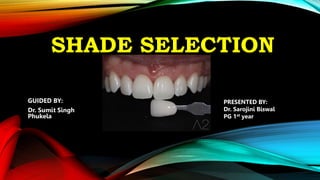
- 1. SHADE SELECTION GUIDED BY: Dr. Sumit Singh Phukela PRESENTED BY: Dr. Sarojini Biswal PG 1st year
- 2. CONTENTS 1. COLOR COMPOSITION 2. COLOR PERCEPTION 3. COLOR SCIENCE IN CERAMICS • TRANSLUCENCY • OPALIZERS IN CERAMICS • OPALESCENCE • TYNDALL EFFECT • COUNTER OPALESCENCE 4. SHADE SELECTION • VISUAL SHADE MATCHING • SHADE MATCHING GUIDELINES • VITA CLASSICAL • VITA 3D MASTER • DIGITAL SHADE MATCHING • COLORIMETERS • SPECTROPHOTOMETERS • LIMITATIONS • DIGITAL CAMERAS 5. SHADE MAPPING 6. COMMUNICATION WITH THE LABORATORY
- 3. COLOR COMPOSITION Color cannot exist without light. Color is simply the perception of light reflected from an object. The reflected light rays stimulate sensors in the human retina and send signals to the visual cortex of the brain. The appearance of the natural tooth is a result of our perception of light rays bounced back from the teeth. The three factors involved in color perception are light, observer, and object.
- 4. Physical Properties Of Light: Light is electromagnetic radiation(a form of energy) that travels in space at speed of 3*108m/s in the form of waves and photons(packets of energy). Light is usually referred to as visible light which has a wavelength ranging from 380-780nm and is perceivable by the human eyes. The perception of color depends on light interacting with an object, this light striking an object is termed incident light.
- 5. Depending on the property of the object the incident light can be reflected or transmitted through the object. This interaction is responsible for the appearance of the object. Optics Of Light: The physical property of the object determines the way the light incident on the object is emitted from it. Light interacts with an object in various ways:- REFLECTION:- Mirror-like reflection of incident light from a smooth surface. Specular (mirror-like) reflections are perceived as gloss/luster in natural teeth, especially when they are wet. A rough tooth surface would produce diffuse reflected light.
- 6. ABSORPTION:- Light that enters an opaque body and does not emerge from it is said to have undergone absorption. The absorbed light is converted to heat. SCATTERING:- Light that gets deflected before absorption is called scattered light. TRANSMISSION:- Natural teeth are not entirely opaque but translucent. This results in light being partly absorbed. The part of the light which is not absorbed is transmitted through the tooth. This transmitted light can undergo scattering before emerging from the tooth.
- 7. NEWTON’S COLOR EXPERIMENT Issac Newton believed that while the light was polychromatic and could be divided into its constituent colors. He proved his hypotheses by using a glass prism to split white light into its constituent colors which were red, orange, yellow, green, blue, indigo, and violet. He extended this experiment by using a second glass prism to combine these constituent colors black into white color. He described red, green, and blue as primary colors. Combining them results in white color. If two of these primary colors are mixed, they form secondary colors. Secondary colors are yellow, cyan, and magenta.
- 8. Based on this discovery, Newton created the color wheel with 6 component colors in which each primary color was separated by a secondary color. The color wheel provides information that constant colors can be obtained by color mixing.
- 9. COLOR MIXING There are 2 types of color mixing: Additive Color Mixing:- New colors are produced by mixing two or more primary colors. When all primary colors (RGB) are mixed they produce white color, associated with television and computer displays. Color is obtained by emitted light. Subtractive Color Mixing:- white is taken as the base and colors are added to produce subtractive colors CMYK system, i.e., (White+Cyan+ Magenta= Blue)
- 10. DIMENSIONS OF COLOR: Standard values to represent color are required for • Reference • Communication • Easy script • Accuracy • Reproducibility ALBERT H MUNSELL proposed a color space that describes three dimensions of color:- HUE:- denotes the dominant color of the object. VALUE:- denotes the lightness and darkness of the object. CHROMA:- denotes the degree of saturation of the color
- 11. COLOR MEASUREMENT SYSTEM: MUNSELL SYSTEM:- There are independent dimensions represented cylindrically with the value measured from 0 to 10 vertically, hue measured in degrees outward from the neutral. Hue follows an order of red> yellow> green> blue> purple with intermediate shades of yellow-red, green- yellow, blue-green, purple-blue, and red-purple. Value is denoted from 0 to 10 with 0 indicating pure black and 10 indicating pure white. Black white and greys between them are called neutral colors and colors with hue are called chromatic colors.
- 12. Chroma represents the degree of departure from the neutral color. Colors with low chroma are said to be ‘weak’ and colors with high chroma are termed as ‘strong’or ‘vivid’. Similar colors can be compared with Nickerson’s color difference formula : I= Ca/5 + 2/H + 6/V + 3/C
- 13. OSTWALD COLOR SYSTEM:- Similar to the Munsell system, it uses dominance, wave-length, and purity instead of hue, value, and chroma. CHROMATICITY SYSTEMS:- Based on Newton’s circle these systems are instrumental methods of color measurement. Colors are plotted in a chromaticity graph. CIE XYZ SYSTEM:- This system is based on Trichromatic theory which states that a color can be matched by mixing the three primary wavelengths. The amount of primaries required to produce a given color is known as the Tristimulus values of the color and are specified as X, Y, and Z.
- 14. The color of the object depends on the SED of the light source(S), reflectance characteristics of the sample (R), and the sensitivity of the observer(Y). These coordinates are used together to describe the color of an object in a 3-dimensional manner. However this model is non-uniform in that equal changes in X, and Y, do not correspond to the same perceived visual difference.
- 15. CIE L*a*b* COLOR SPACE:- Introduced in 1976. This system transforms the tristimulus values of X, Y, and Z into cartesian coordinates (L*a*b). L* denotes lightness on a scale of 0-100 (black-white). The hue and chroma are represented by a* (red- green axis) and b* (blue-yellow axis). This system can also be used to quantify color differences between the two objects by the formula /E = _/( /L)2 + ( / a*)2 + ( / b*)2. A ( /E) value of 1 or greater denotes a perceivable color difference in color between two objects.
- 16. when the value of /E values of 3.7 results in a clinically perceivable color difference in dental restorations. This quantification aids in fabricating restorations of optimum aesthetics by ensuring the color difference in restorations is kept to a minimum. CIE L*C*h* COLOR SPACE:- This system is similar to the CIE L*a*b* color space with the difference being that the Cartesian coordinates (L*a*b*) are transformed into cylindrical coordinates (L*C*h*).
- 17. L represents the lightness while C represents chroma and h represents the hue. Color differences between two objects can be calculated by the formula /E = _/(/L)2 + (/C*)2 + (/H*)2 Both CIE L*a*b* and CIE L*C*h* systems are devices independent. Therefore these systems can be used for dental shade matching procedures without the need to calibrate digital monitors and displays.
- 18. COLOR PERCEPTION As light hits the eye, an image is formed. The human eye/brain comprising the visual apparatus is responsible for visual perception. The amount of light entering the eye is controlled by the iris which dilates or constricts depending on the level of illumination. The filtered light then hits the retina which consists of light-sensitive receptors called rods and cones.
- 19. RODS:- Highly light sensitive (requires less light intensity) Responsible for scotopic(low light) vision. Monochromatic vision Located in the periphery of the retina Ranges in the numbers from 90 to 120 million CONES:- Low light sensitivity (requires highly intense light) 3 types of receptors: -S (short wavelength)-400-500 nm wavelength (blue). -M (medium wavelength)- 450-630nm wavelength (green). - L (Long wavelength)- 500 – 700nm wavelength (red) Receptors are present in the ratio of 8:4:1(L: M:S) Consists of a photopsin (specific wavelength) and 11- cis retinal Located in the fovea centralis. Range in the numbers from 6 to 7 million
- 20. Upon interaction with light, signals are sent to the thalamus from the rods and cones and then sent to the visual cortex in the occipital lobe of the brain. Color perception is a psychological process. Hence it can differ between individuals. Accurate color measurement can be made with colorimeters. However, these instruments are less accurate in the evaluation of curved or rough surfaces.
- 21. COLOR SCIENCE IN CERAMICS HUE:- Most commonly used shade guide for shade matching is the VITA Classical shade guide. This guide depicts hue variation:- A- Reddish brown B- Orange yellow C- Greenish grey D- Pinkish grey Hue selection should be done under appropriate lighting with a color temperature of 5500K.
- 22. VALUE:- Most important factor of color determination. Varies with the intensity of light; should be examined under average or dim light. • Average light- 5000K • Dim light- 3000K A Grey background can be placed during intraoral photography to aid in value estimation.
- 23. CHROMA:- Pigmented portion of hue. Defined as the “Amount of pigments contained in a hue shade”. VITA guide- 4 chroma levels are available; A1 – A4 with A4 indicating deeper chroma. Yamamoto (1992):- Color desaturation occurs in the layered ceramic build-up. Ubassy (1993):- Use neutral ceramic powder to desaturate the basic color powder.
- 24. TRANSLUCENCY:- This is a difficult parameter to quantify and is an important factor to be assessed during shade assessment. Teeth in younger individuals exhibit increased translucency in enamel. Teeth in older individuals exhibit increased chromaticity of dentin. Sekine et al described 3 types of translucency: Type A- Mild Translucency Type B- Streak translucency Type C- Incisal + Proximal translucency
- 25. Translucency scale –(1-5) 1- low translucency; 5- High translucency For example- Type B3 denotes moderate translucency with incisal streaks. Reference photographs during laboratory steps are preferable. It is important to note that shade guides are made using different ceramics than the layering powder; this could lead to more errors due to metamerism effects(due to layers).
- 26. OPALIZERS IN CERAMICS:- Pure ceramic is as transparent as glass. Opaque pigments are added to produce translucency. Opalizers in ceramics : OPACIFIER REFLECTIVITY INDEX TiO2 2.52 ZrO2 2.2 Sn2O2 2.0 Particle sizes less than the wavelength of approaching light produce an opalizing effect.
- 27. OPALESCENCE: Scattering occurs when the wavelength of white light strikes an object with a matching or lesser particle size than the incident light. For example- white light has a wavelength of 400-700 nm (0.4-0.7 microns). When white light is incident on a tooth with enamel structure of 0.4 microns or less, the shorter end of the wavelength (400 nm- blue) is reflected by the particles whereas the longer wavelength is transmitted and absorbed and this produces the bluish tint of illuminated natural teeth.
- 28. Seen in natural teeth and opal stone. This effect occurs due to the particular type of diffraction related to the presence of very fine and perfectly homogenous particles.
- 29. TYNDALL EFFECT:- Responsible for blue streaks during the day and orange streaks during the evening. Tooth absorbs the orange yellow and reflects low wavelength light (since OH apatite crystal particle size is smaller, larger wavelengths are not reflected). The transmitted light appears orange-yellow in color because smaller wavelength rays are filtered by the reflective nature.
- 30. Tetracycline-stained teeth do not exhibit any opalescence due to their opaque nature. New pigments to mimic stains or opalescence effects are available. Repeated firing increases the risk of homogenization since pigments within the ceramic matrix could get disintegrated. Zirconium particles can be added to avoid homogenization since they can disintegrate beyond 700 C.
- 31. COUNTER OPALESCENCE:- Incisal translucency appears blue-colored. Proximal contacts are opacified. Hence, reflected light is transmitted again through overlaying ceramic. Thus producing brownish yellow shades. Precautions to be taken for counter opalescence: Avoid thin ceramics Use opaque dentin shades. Use darker opaque material.
- 32. SHADE SELECTION:- 1) VISUAL SHADE MATCHING:- Visual shade matching involves the use of commercially available color standards to compare and match the color of the target tooth. Shade guides from various manufacturers are used to match teeth shades. Shade matching with commercial shade guides is at best an arbitrary method of shade matching since they do not cover the entirety of the tooth color space.
- 33. Other factors such as tooth texture, color gradation, and translucency cannot be matched with any currently available shade guide. Shade tab arrangement is also very important in shade selection with shades arranged according to the value resulting in more accurate matching. Most commonly used shade guides VITA Classical (Vita Zahnfabrik, Bad Sackingen, Germany) and Chromascop(Ivoclar-Vivadent, Amherst, New York) do not have value measurements in their shade guides causing subtle variations in shades of restorations.
- 34. Another factor to consider is that shade tabs are made up of a single uniform shade but a natural tooth has a gradation in color schemes with the cervical portion of the tooth being darker and the incisal portion being highly translucent. Such variations are not accounted for in the shade guide systems. An attempt to cover the entirety of the natural tooth color space resulted in the Vita-pan 3D Master system.
- 35. Spectrophotometric studies show that this system covers the natural tooth color in a visually equidistant manner. This shade selection system groups the shade tabs according to value and not hue as with the Vita Classical and chromascop systems Visual shade matching is also highly dependent on the experience and training of the clinician. Studies show that clinicians with an understanding of color science and shade matching guidelines tend to select shade more accurately than operators with no training.
- 36. For all their limitations, the majority of the dental profession uses shade guides to select shades of restorations due to their ease of use, low cost, and most laboratories are equipped with a common shade guide. SHADE MATCHING GUIDELINES:- Controlled lighting conditions are recommended in the dental operatory. A color temperature of 5500K and a color rendering index of >90 are recommended.
- 37. The patient should be in an upright position with the teeth at the dentist’s eye level Any cosmetics such as lipstick should be removed, and bright- colored clothing should be draped Shade selection should be performed before the tooth becomes dehydrated.
- 38. If this is not available in the dental operatory, then shade selection should be performed in natural daylight at around noon since a diffuse skylight is lesser at this time of the day. The operatory ceilings, walls, countertops, and cabinets should be in pastel shades and neutral grey color. Photographs of teeth and shade tabs should be made with a neutral color background. A grey card with 18% grey color saturation is recommended to reduce inconsistencies in shade selection.
- 39. The value should be selected in the first; squint test can be can be performed to decrease the amount of light entering the eyes. Hue and Chroma are to be checked next with color matching not done for more than 10 seconds to avoid eye fatigue. Gazing at an achromatic surface (neutral grey) helps in replenishing the photopigments of the eye.
- 40. VITA CLASSICAL Shades are clustered according to Hue with tabs grouped as A(red-yellow), B(yellow), C(Grey), and D(red-yellow-grey). Chroma is designated with numerical values of 1,2,3 and 4 with lesser values representing lesser saturation. There is no value measurement in this shade system. Hue is selected first followed by Chroma.
- 41. Rearranging the shade tabs in this system according to the Value (lighter to darker) results in more accurate shade matching. The shade tabs arranged according to value are in the order of B1, A1, B2, D2, A2, C1, C2, D4, A3, D3, B3, A3.5, B4, C3, A4, C4
- 42. VITA 3D MASTER:- The manufacturer groups the tabs according to value with numerical designations of 1, 2, 3, 4, and 5. Chroma is evaluated next with the tabs in each value group divided into 1, 2, and 3. Hue is evaluated finally with tabs designated as L (light), M (medium), and R(reddish). The value and hue are the same for all tabs in the M group with only chroma varying.
- 43. The 3D-master tabs are marked by employing a number-letter– number system. The 1st number indicates value, from 0 being the lightest to 5 being the darkest. The middle letter indicates hue with the L, M, and R corresponding to yellowish, medium, and reddish hue. The 2nd number designates chroma, from the least chromatic 1 to the most chromatic 3. Two varieties of shade guides are available for shade selection in the Vitapan 3D master shade guide: Vitapan 3D master tooth guide (blue chips) Vitapan 3D master color guide (red chips)
- 46. Intermediate shades such as 1.5 can be given for more precise shades. DIGITAL SHADE MATCHING:- Color measuring instruments mimic the functioning of the human eye in that the software algorithm uses red, green, and blue as primary colors. These instruments measure color as a function of additive color mixing. VITA EASYSHADE V
- 49. COLORIMETERS:- Colorimeters were the first type of color measuring instrument to be used for objective shade assessment. They use a light detector (photocell) coupled with multiple filters to provide standard illumination and a photodetector. The filters act as analog function generators that limit spectral characteristics to that match the three response curves of the observer. The accuracy of the colorimeter is dependent on the accuracy of the filters in producing light with spectral characteristics matching that of the observer. DIGITAL COLORIMETER
- 50. The ability of the colorimeters to match the standard observer functions with filters is inferior to that of spectrophotometers. SPECTROPHOTOMETERS:- These instruments measure the spectral reflectance or transmission of objects. Consists of a light source, monochromator, and photodetector. The light sources used may be tungsten halogen bulbs or xenon flash tubes The light is resolved into its individual wavelengths before or after it falls on the sample using the monochromator(prism).
- 51. The latest instrument use photodiodes as photodetectors with the advantage of a separate diode element for each wavelength. This allows for simultaneous integration of all wavelengths. Spectrophotometers are most accurate for shade matching and are currently regarded as the gold standard in dentistry.
- 52. PROBLEMS DURING SHADE MATCHING:- Color vision problems/color blindness: It is the absence of one or more photosensitive pigments. Other color vision defects include achromatism, dichromatism, and trichromatism Age: There is a yellowish coloring of the lens and cornea, with age causing bias in vision Fatigue: Fatigue can lead to compromised visual accuracy, thus causing inappropriate shade selection Emotions: Our emotions influence pupil dilatation and constriction that helps to discriminate between colors Medications: The use of caffeine, drugs, and alcohol affects our color perception
- 53. Binocular difference: This is the difference in perception between the left and right eye Environmental influences: The background or surroundings next to the tooth can affect the color perceived by the tooth. This is why it is advised to remove any makeup and use a neutral gray background to reduce the environment's influence.
- 54. DIGITAL CAMERAS:- Digital photography is rapidly gaining prominence as an effective shade matching aid. Digital imaging provides information on tooth shade, shape, texture, and color distribution. Intraoral image with reference shade tabs positioned next to the tooth aid in shade matching and effective communication of the determined shade to the lab technician. Most digital still or video cameras acquire red, blue, and green color information with color reproduction as a function of the additive color. Advantages of digital imaging include ease of access, storage, and communication.
- 55. Digital photography yields intricate details in an effective manner. The aspect ratio of the sensor influences image quality with full-frame and half-frame sensors having better image quality compared to compact cameras. It has been proved that the use of ring flashes with digital results in over-exposed images which would result in higher value measurement during shade matching and less reliable results. Commercially available imaging software (Adobe Photoshop CS5 and above, Corel PaintShop pro) can be used to measure color differences to aid in shade matching.
- 56. COMMUNICATION: SHADE MAPPING:- Shade mapping is recommended for customized shade matching for each individual. The tooth is divided into nine segments by three sets of equidistant vertical and horizontal lines. The character of the tooth(craze lines, hypocalcification, proximal darkening, and translucency) in each segment is noted to replicate it in the restoration.
- 58. COMMUNICATION WITH THE LAB:- The focus of shade matching in prosthodontics has always been the evaluation of a shade followed by the evaluation of the final result. Most clinicians do not account for the shade duplication process in the lab with ceramic formulations varying for each restoration. Another factor that is observed is that the Lab technician does not view the clinical scenario and thus communication of intricate details is highly critical to obtain an optimal aesthetic result.
- 59. ADVANTAGES:- • Accurate visualization of form and texture. • Easier storage • Easier communication with digital data. DISADVANTAGES:- • Needs a high-resolution camera. • Requires some training in imaging software. • Requires some training in handling cameras.
- 60. CONCLUSION The proper shade selection is an essential basic step to achieve a perfect esthetic. The color of the teeth is influenced by many factors causing subjective differences. The variation in natural teeth and ceramics material is the main reason for shade selection to be time-consuming and skill- demanding..
- 61. REFERENCES • Binu George “ Textbook Of Complete Denture Prosthodontics” • Sheldon Winkler “Essentials of Complete Denture Prosthodontics” 3rd edition. • Zarb GA, Bolender CL, Hickey JC, Carlsson GE: Boucher’s Prosthodontic Treatment of Edentulous Patients ed 12. • Fundamentals of fixed prosthodontics by Shillinburg 3rd edition. • Joiner A. Tooth colour : a review of the literature. J Dent. 2004;32:3-12 • Wee AG. Color accuracy of commercial digital cameras for use in dentistry. Dent Master. 2006;22(6):553-9 • Preston JD. Current status of shade selection and color matching. Quintes Intern. 1958;16(1):642-8 • Heffernan MJ, Aquilino SA, Diaz-Arnold AM, et al. Relative translucency of six all-ceramic systems. Part 1: core materials. J ProsthetDent. 2002;88(1):4-9. • Nickerson D. History of the Munsell color system and its scientific application. J Opt Soc Am. 1940;30:575-86. • CIE 1976 color-difference formulae. Color Res Appl. 1977;2(1):7-11.