Download to read offline


![UREDNI[TVO
Glavni i odgovorni urednik:
Prim. dr Avdo ]erani}
Pomo}nik glavnog i odgovornog urednika:
dr D`enana Detanac
Tehni~ki urednik:
dr D`email Detanac
Nau~ni savet Me|unarodni nau~ni savet
Prof. dr Aleksandar Karamarkovi} (Srbija) Prof. dr Ivan Damjanov (SAD)
Prof. dr Branka Nikoli} (Srbija) Prof. dr Milan R.Kne`evi} ([panija)
Prof. dr Radivoj Koci} (Srbija) Prof. dr Ino Hused`inovi} (Hrvatska)
Prof. dr Ivan Dimitrijevi} (Srbija) Prof. dr Anastasika Poposka (Makedonija)
Prof. dr Stojan Sekuli} (Srbija) Prof. dr Sergio Zylbersztejn (Brazil)
Prof. dr Marina Savin (Srbija) Prof. dr Beniamino Palmieri (Italija)
Prof. dr Milica Berisavac (Srbija) Prof. dr Sahib H. Muminagi} (Bosna i Hercegovina)
Prof. dr Milan Kne`evi} (Srbija) Prof. dr Osman Sinanovi} (Bosna i Hercegovina)
Prof. dr Milo{ Jovanovi} (Srbija) Prof. dr Selma Uzunovi}-Kamberovi} (Bosna i Hercegovina)
Prof. dr Sne`ana Jan~i} (Srbija) Prof. dr Agima Ljaljevi} (Crna Gora)
Prof. dr ^edomir S. Vu~eti} (Srbija) Prof. dr Suada Helji} (Bosna i Hercegovina)
Prof. dr Slobodan Obradovi} (Srbija) Prof. dr Milica Martinovi} (Crna Gora)
Prof. dr Slobodan Grebeldinger (Srbija) Prof. dr Nermina Had`igrahi} (Bosna i Hercegovina)
Prof. dr Slobodan M. Jankovi} (Srbija) Prof. dr Miralem Musi} (Bosna i Hercegovina)
Prof. dr @ivan Maksimovi} (Srbija) Prof. dr Spase Jovkovski (Makedonija)
Prof. dr Zlata Janji} (Srbija) Prof. dr Evangelos J. Giamarellos-Bourboulis (Gr~ka)
Prof. dr Svetislav Milenkovi} (Srbija) Prof. dr Paolo Pelosi (Italija)
Prof. dr Radmilo Jankovi} (Srbija) Prof. dr Zsolt Molnar (Ma|arska)
Lektor za engleski jezik
Selma Mehovi}
Dizajn
Prim. dr Avdo ]erani}
Izdava~
Udru`enje lekara Sanamed, Novi Pazar
^ASOPIS IZLAZI TRI PUTA GODI[NJE
Adresa uredni{tva
„SANAMED“, Ul. Palih boraca 52, 36300 Novi Pazar, Srbija
email: sanamednp2006ªgmail.com, www.sanamed.rs
[tampa
„ProGraphico“, Novi Pazar
Tira`
500
Pretplata
Godi{nja pretplata: 4000 din. za doma}e ustanove; 1500 din. za pojedince; za inostranstvo 75 eura (u dinarskoj
protivrednosti po kursu na dan uplate). Pretplatu vr{iti na ra~un 205-185654-03, Komercijalna banka. Za sve do-
datne informacije kontaktirati Uredni{tvo.
ISSN-1452-662X](https://image.slidesharecdn.com/24625161-2322-46cf-8dff-e69346491ecb-150121105753-conversion-gate02/85/Sanamed-9-3-2014-3-320.jpg)
![EDITORIAL BOARD
Editor-in-chief:
Prim. dr Avdo ]erani}
Associate Editor:
dr D`enana Detanac
Technical Editor:
dr D`email Detanac
Scientific council International scientific council
Prof. dr Aleksandar Karamarkovi} (Serbia) Prof. dr Ivan Damjanov (USA)
Prof. dr Branka Nikoli} (Serbia) Prof. dr Milan R.Kne`evi} (Spain)
Prof. dr Radivoj Koci} (Serbia) Prof. dr Ino Hused`inovi} (Croatia)
Prof. dr Ivan Dimitrijevi} (Serbia) Prof. dr Anastasika Poposka (R. Macedonia)
Prof. dr Stojan Sekuli} (Serbia) Prof. dr Sergio Zylbersztejn (Brazil)
Prof. dr Marina Savin (Serbia) Prof. dr Beniamino Palmieri (Italy)
Prof. dr Milica Berisavac (Serbia) Prof. dr Sahib H. Muminagi} (Bosnia and Herzegovina)
Prof. dr Milan Kne`evi} (Serbia) Prof. dr Osman Sinanovi} (Bosnia and Herzegovina)
Prof. dr Milo{ Jovanovi} (Serbia) Prof.drSelmaUzunovi}-Kamberovi}(BosniaandHerzegovina)
Prof. dr Sne`ana Jan~i} (Serbia) Prof. dr Agima Ljaljevi} (Montenegro)
Prof. dr ^edomir S. Vu~eti} (Serbia) Prof. dr Suada Helji} (Bosnia and Herzegovina)
Prof. dr Slobodan Obradovi} (Serbia) Prof. dr Milica Martinovi} (Montenegro)
Prof. dr Slobodan Grebeldinger (Serbia) Prof. dr Nermina Had`igrahi} (Bosnia and Herzegovina)
Prof. dr Slobodan M. Jankovi} (Serbia) Prof. dr Miralem Musi} (Bosnia and Herzegovina)
Prof. dr @ivan Maksimovi} (Serbia) Prof. dr Spase Jovkovski (R. Macedonia)
Prof. dr Zlata Janji} (Serbia) Prof. dr Evangelos J. Giamarellos-Bourboulis (Greece)
Prof. dr Svetislav Milenkovi} (Serbia) Prof. dr Paolo Pelosi (Italy)
Prof. dr Radmilo Jankovi}(Serbia) Prof. dr Zsolt Molnar (Hungary)
English language editor
Selma Mehovi}
Design
Prim dr Avdo ]erani}
Publisher
Association of medical doctors “Sanamed”, Novi Pazar
ISSUED three times a year.
Editorial address
“SANAMED”, Ul. Palih boraca 52, 36 300 Novi Pazar, Serbia
email: sanamednpªgmail.com, www.sanamed.rs
Print
“ProGraphico”, Novi Pazar
Circulation
500
Subscription
Annual subscriptions: 4000 RSD for domestic institutions and 1500 RSD for individuals. For readers abroad, an-
nual subscription is 75 Euro (in Dinar equivalent at the exchange rate on the day of payment). For further instruc-
tions and informations, contact Editorial Board.
ISSN-1452-662X](https://image.slidesharecdn.com/24625161-2322-46cf-8dff-e69346491ecb-150121105753-conversion-gate02/85/Sanamed-9-3-2014-4-320.jpg)
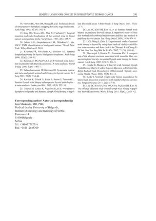
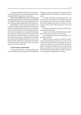
![SADR@AJ
• RE^ UREDNIKA.......................................................................................................................................
• ORIGINALNI NAU^NI RAD
• DEJSTVO HRONI^NOG TROVANJA OLOVOM NA VREDNOST
KRVNOG PRITISKAKOD DECE.............................................................................................................
@ivkovi} Jovan,
1
Savi} Zoran
2
1
Medicinski fakultet Univerziteta u Pri{tini, sa sedi{tem u Kosovskoj Mitrovici
2
Zdravstveni centar Kosovska Mitrovica
• STRU^NI RAD
• KORELACIJAIZMEU PORODI^NOG NASILJANAD @ENAMAI NEUROTICIZMA.....................
Numanovi} S. Almedina,
1
Jovi~i} M. Milena
2
1
Internacionalni univerzitet Novi Pazar, Novi Pazar, Srbija
2
Klini~ki centar Kragujevac, Klinika za psihijatriju, Kragujevac, Srbija
• PRIKAZ SLU^AJA
• RANA DIJAGNOSTIKA KRANIOSINOSTOZE KOD ODOJ^ADI
U PRIMARNOJ ZDRAVSTVENOJ ZA[TITI ...........................................................................................
Skoric Jasmina
Dom zdravlja „Simo Milo{evi}“, Slu`ba za zdravstvenu za{titu dece i omladine ^ukarica, Beograd, Srbija
• KOKAINSKAKARDIOMIOPATIJA— PRIKAZ SLU^AJA...................................................................
Georgiev Antonio,
1
Zhivadinovik Julija
2
1
Univerzitetska Klinika za kardiologiju, Univerzitet „Sv. ]irilo i Metodije“, Medicinski fakultet, Skoplje, R. Makedonija
2
Institut za anatomiju, Univerzitet „Sv. ]irilo i Metodije“, Medicinski fakultet, Skoplje, R. Makedonija
• REVIJALNI RAD
• KONCEPT STRA@ARSKIH LIMFNIH NODUSA
KOD DIFERENTOVANOG TIROIDNOG KARCINOMA .......................................................................
Markovi} Ivan, D`odi} Radan
Medicinski fakultet Univerziteta u Beogradu, Institut za onkologiju i radiologiju Srbije, Beograd, Srbija
• SISTEMSKE BOLESTI KOJE UTI^U NAZUBNU PATOLOGIJU..........................................................
Kne`evi} R. Milan,
1
An|eli} S. Gordana,
2
Kne`evi} M. Milena
3
1
FEBOMFS, University Las Palmas, Spain
2
Institute for Medical Research, Military Medical Academy, Belgrade, Serbia
3
Medical School, University Las Palmas, Spain
• ETNI^KA PRIPADNOST I DIABETES TIP 2 KOD AZIJSKIH
INDIJSKIH MIGRANATAU OKLANDU, NOVI ZELAND.....................................................................
Jowitt Ljiljana
University of Auckland, Department of Surgery, Auckland, New Zealand
• UPUTSTVO AUTORIMA..........................................................................................................................
Broj 9(3)/2014](https://image.slidesharecdn.com/24625161-2322-46cf-8dff-e69346491ecb-150121105753-conversion-gate02/85/Sanamed-9-3-2014-6-320.jpg)
![RIJE^ UREDNIKA
Uzimaju}i papir da napi{em par re~enica ~ini
mi i obavezu, i zadovoljstvo da ne{to ka`em i osvr-
nem se saradnicima uredni{tva sa iskrenim i du`nim
po{tovanjem, jer oni daju poseban ton ovome {to svi
skupa radimo.
Kako sam po struci hirurg i slikar, podsjeti me to
na jedan davni period iza nas. Postoji negde zapisa-
no da „zlatno doba hirurgije je period poslije 1870.
godine, kada se posebno izu~ava anatomija“. Jedan
paradoks tog vremjena i vremjena u kome `ivimo,
izaziva ~u|enje. Tada su se umjetnici, i to sa velikim
problemima interesovali za ljudsko tijelo, njegov iz-
gled i proporcije. „Tako je Donatelo (1386–1486),
prvi slikar koji je radio disekcije ljudskog tijela. Leo-
nardo da Vin~i (1452–1519), je zasnovao portretsku i
fiziolo{ku anatomiju i tvrdio je da je sam uradio tri-
desetak disekcija ljudskog tijela.“
Paradoks se ogleda u tome da su se ljudi koji
nikada nisu imali na umu da se bave medicinom ba-
vili ljudskim tijelom ne bi li dali svoj doprinos nauci
radi bli`ih spoznaja u re{avanju svih tajni za to do-
ba, u korist ~ovjeka, u borbi za o~uvanje zdravlja.
Danas, kada se tajne u medicini mijenjaju iz nedje-
lje u nedjelju, i kada postoje uslovi sa najsavreme-
nijim pristupom u tehnici i kada znaju za lije~enje
obolelih, toliko postoji nezainteresovanost, i to ba{
kod onih koji su najpozvaniji da se uklju~e u razne
programe i postanu deo medicinske nauke za do-
brobit dru{tva. Tragi~no je {to je opao taj tonus i
mnogi lekari zaustavljaju sebe u daljem radu na pr-
voj stepenici obrazovanja, na koju stanu po zavr{e-
noj specijalizaciji.
Pa dobro, nijesu motivisani da pi{u, da stvara-
ju, ali je jo{ tragi~nije {to neki ba{ tada pomisle da
oni ve} sve znaju i da su se ve} umorili za dalje usa-
vr{avanje. Neki su i opasni po druge jer su se uvrsti-
li u one ljude koji „ne znaju, a misle da znaju“, i
njih se treba kloniti.
Takvi pojedinci su izbrisali hijerarhiju, oni bi
da gaze po u~iteljima, misle da je njih sam Bog po-
slao da re{avaju probleme, a pacijent im je no}na
mora, i on je taj koji mora da trpi, ~eka i tra`i spas.
[ta bi rekao Leonardo da Vin~i kada bi video hirur-
ga koji ne zna anatomiju a ide da operi{e ~ovjeka?
To je taj paradoks za koji ne znam da li sam ga
plasti~no prikazao, ali znam da sam ga upotrijebio
namjerno, `ele}i da se osvrnem na na{ rad od prvog
broja SANAMED-a, kada smo ga izdavali jednom
godi{nje i koji je od tada sazreo i stao u red ~asopi-
sa starih nekoliko decenija i sada ve} izlazi tri puta
godi{nje, a po kvalitetu mnogi nam ve} i zavide, i
ve}ina nam ~estita na njegovom uzrastu.
Po ko zna koji put moram da zahvalim na{im
saradnicima, a posebno sada{njoj ekipi mladih le-
kara koji svojim entuzijazmom potvr|uju, kroz kva-
litet onoga {to ~ine, da je vjera u ~ovjeka vrijedna
samo onda kad, „zna{ da zna, a on zna da zna“ i to-
ga treba slijediti.
Sam `ivot predstavlja vje~itu borbu da bi se sti-
glo na vrh. Ako ste rije{ili da budete u~esnik u toj bor-
bi, budite ponosni {to imate izazov, bez obzira {to }ete
do tog vrha biti mnogo puta povre|eni i neshva}eni.
U na{em ~asopisu ima mnogo mesta za takve
entuzijaste.
Koristim priliku da svima ~estitam Novu Godi-
nu i po`elim puno uspeha u profesionalnom i privat-
nom `ivotu.
Srda~an pozdrav,
Prim. dr Avdo ]erani}
glavni i odgovorni urednik](https://image.slidesharecdn.com/24625161-2322-46cf-8dff-e69346491ecb-150121105753-conversion-gate02/85/Sanamed-9-3-2014-9-320.jpg)


![tion, but recurrence is reported if the patient relapses
into cocaine use (5).
Efforts to assist the patient with their drug addic-
tion should be a part of every treatment plan. Hospitali-
zation for detoxification may be necessary, particularly
if other drugs also are being abused. Outpatient treat-
ment of drug dependence is strongly advised. Absti-
nence from cocaine use and long time follow up is
mandatory.
CONCLUSION
This is the first publication of cocaine-related car-
diomyopathy in our country. Physicians usually don’t
consider the possibility of cocaine use of their patients.
Many cocaine users have little or no idea of the risks
associated with its use. So, patients, health care work-
ers and the public should be educated about the dan-
gers and the considerable risks of cocaine use.
Abbreviations
ACE — Angiotensin converting enzyme
BNP — Natriuretic peptide
BUN — Blood urea nitrogen
ECG — Electrocardiography
EF — Ejection fraction
ESC — European Society of Cardiology
HR — Heart rate
MI — Myocardial infarction
MRA — Mineral corticoid receptor antagonist
ST — ECG between the end of the S wave (the J
point) and the beginning of the T wave
236 Georgiev Antonio, Zhivadinovik Julija
Sa`etak
KOKAINSKAKARDIOMIOPATIJA— PRIKAZ SLU^AJA
Georgiev Antonio,
1
Zhivadinovik Julija
2
1
Univerzitetska Klinika za kardiologiju, Univerzitet „Sv. ]irilo i Metodije“, Medicinski fakultet, Skoplje, R. Makedonija
2
Institut za anatomiju, Univerzitet „Sv. ]irilo i Metodije“, Medicinski fakultet, Skoplje, R. Makedonija
Kokain je druga naj~e{}a nezakonita droga koja
se koristi i naj~e{}i uzrok smrti zbog droge. Upotreba
kokaina daje akutne i hroni~ne komplikacije, koje
mogu uklju~ivati bilo koji sistem organa, naj~e{}e
kardiovaskularni sistem. Kardiomiopatija mo`e biti
izazvana konzumiranjem kokaina. Ovaj ~lanak pred-
stavlja slu~aj dvadeset~etvorogodi{njeg mu{karca s
reverzibilnom kokainskom kardiomiopatijom, {to
predstavlja prvi takav u Republici Makedoniji. Nalazi
klini~kog pregleda, laboratorijskih analiza, rendgen-
grafije srca i plu}a, ultrazvuka srca i tretman su prika-
zani.
Klju~ne re~i: kokain, kokainska kardiomiopatija,
dijagnoza, le~enje.
References
1. Finkel JB, Marhefka GD. Rethinking cocaine-associa-
ted chest pain and acute coronary syndromes. Mayo Clin Proc.
2011; 86(12): 1198–207.
2. Egred M, Davis GK. Cocaine and the heart. Postgrad
Med J. 2005; 81(959): 568–71.
3. SAMHSA— Substance abuse and mental health service
administration. Sharp decline also continues for methampheta-
mine use. US Department of Health and Human Services
(DHS); Office of Applied Studies Results from the 2008 Natio-
nal Survey on Drug Use and Health: National Findings.
http://oas.samhsa.gov/nsduh/2k8nsduh/2k8Results.cfm. 2009.
4. Phillips K, Luk A, Soor GS, Abraham JR, Leong S, Bu-
tany J. Cocaine cardiotoxicity: a review of the pathophysiology,
pathology, and treatment options. Am J Cardiovasc Drugs.
2009; 9(3): 177–96.
5. Schwartz BG, Rezkalla S, Kloner RA. Cardiovascular
effects of cocaine. Circulation. 2010; 122(24): 2558–69.
6. Awtry EH, Philippides GJ. Alcoholic and cocaine-asso-
ciated cardiomyopathies. Prog Cardiovasc Dis. 2010; 52(4):
289–99.
7. Freeman K, Feldman JA. Cocaine, myocardial infarc-
tion, and b-blockers: time to rethink the equation? Ann Emerg
Med. 2008; 51(2): 130–4.
8. Bhargava S, Arora RR. Cocaine and cardiovascular
complications. Am J Ther. 2011; 18(4): e95–e100.
9. Velasquez EM, Anand RC, Newman WP 3rd, Richard
SS, Glancy DL. Cardiovascular complications associated with
cocaine use. J La State Med Soc. 2004; 156(6): 302–10.
10. Chokshi SK, Moore R, Pandian NG, Isner JM. Revers-
ible cardiomyopathy associated with cocaine intoxication. Ann
Intern Med. 1989; 111(12): 1039–40.
11. Virmani R, Robinowitz M, Smialek JE, Smyth DF.
Cardiovascular effects of cocaine: an autopsy study of 40 pati-
ents. Am Heart J. 1989; 115(5): 1068–76.
12. Tazelaar HD, Karch SB, Stephens BG, Billingham
ME. Cocaine and the heart. Hum Pathol. 1987; 18(2): 195–9.
13. Robledo-Carmona J, Ortega-Jimenez MV, Garcia-Pi-
nilla JM, Cabra B, de Teresa E. Severe Cardiomyopathy Associ-
ated to Cocaine Abuse. Int J Cardiol. 2006; 112(1): 130–1.
14. Rangel C, Shu RG, Lazar LD, Vittinghoff E, Hsue PY,
Marcus GM. Beta-blockers for chest pain associated with recent
cocaine use. Arch Intern Med 2010; 170(10): 874–9.](https://image.slidesharecdn.com/24625161-2322-46cf-8dff-e69346491ecb-150121105753-conversion-gate02/85/Sanamed-9-3-2014-32-320.jpg)


![UPUTSTVO AUTORIMA
SANAMED je medicinski ~asopis osnovan 2006.
godine. ^asopis objavljuje: originalne nau~ne i stru~ne
~lanke, prikaze bolesnika, revijske radove, pisma ured-
niku, ~lanke iz istorije medicine, prikaz objavljenih
knjiga i druge medicinske informacije.
Rukopise slati na adresu:
Prim. dr Avdo ]erani},
(za Sanamed)
Ul. Palih boraca 52, 36300 Novi Pazar
Email: sanamednp2006ªgmail.com
www.sanamed.rs
Prispeli rukopis Ure|iva~ki odbor {alje recenzen-
tima radi stru~ne procene. Ukoliko recenzenti predlo`e
izmene ili dopune, kopija recenzije se dostavlja autoru
s molbom da unese tra`ene izmene u tekst rada ili da
argumentovano obrazlo`i svoje neslaganje s primed-
bama recenzenta. Kona~nu odluku o prihvatanju rada
za {tampu donosi glavni i odgovorni urednik.
Za objavljene radove se ne ispla}uje honorar, a
autorska prava se prenose na izdava~a. Rukopisi i pri-
lozi se ne vra}aju. Za reprodukciju ili ponovno obja-
vljivanje nekog segmenta rada publikovanog u Sana-
medu neophodna je saglasnost izdava~a.
^asopis se {tampa na srpskom jeziku, sa kratkim
sadr`ajem prevedenim na engleski jezik. Radovi stra-
nih autora se {tampaju na engleskom jeziku sa kratkim
sadr`ajem na srpskom i engleskom jeziku.
OP[TA UPUTSTVA
Rukopis treba poslati u tri primerka, otkucan jedno-
strano na beloj hartiji formata A4. Tekst rada kucati u pro-
gramu za obradu teksta Word, latinicom, sa dvostrukim
proredom, isklju~ivo fontom Times New Roman i veli~i-
nom slova 12 ta~aka (12 pt). Sve margine podesiti na 25
mm, a tekst kucati sa levim poravnanjem i uvla~enjem
svakog pasusa za 10 mm, bez deljenja re~i (hifenacije).
Rukopis mora biti organizovan na slede}i na~in:
naslovna strana, sa`etak na srpskom jeziku, sa`etak na
engleskom jeziku, klju~ne re~i, uvod, cilj rada, bole-
snici i metodi/materijal i metodi, rezultati, diskusija,
zaklju~ak, literatura, tabele, legende za slike i slike.
Svaki deo rukopisa (naslovna strana, itd.) mora
po~eti na posebnoj strani. Sve strane moraju biti nume-
risane po redosledu, po~ev od naslovne strane. Prezime
prvog autora se mora otkucati u gornjem desnom uglu
svake stranice. Podaci o kori{}enoj literaturi u tekstu
ozna~avaju se arapskim brojevima u zagradama, i to
onim redosledom kojim se pojavljuju u tekstu.
Obim rukopisa. Celokupni rukopis rada, koji ~i-
ne naslovna strana, kratak sadr`aj, tekst rada, spisak li-
terature, svi prilozi, odnosno potpisi za njih i legenda
(tabele, slike, grafikoni, sheme, crte`i), naslovna stra-
na i sa`etak na engleskom jeziku, mora iznositi za ori-
ginalni rad, saop{tenje, rad iz istorije medicine i pre-
gled literature do 5.000 re~i, a za prikaz bolesnika, rad
za praksu, edukativni ~lanak do 3.000 re~i; radovi za
ostale rubrike moraju imati do 1.500 re~i.
Provera broja re~i u dokumentu mo`e se izvr{iti u
programu Word kroz podmeni Tools-Word Count ili Fi-
le-Properties-Statistics.
Sva merenja, izuzev krvnog pritiska, moraju biti
izra`ena u internacionalnim SI jedinicama, a ako je
neophodno, i u konvencionalnim jedinicama (u zagra-
di). Za lekove se moraju koristiti generi~ka imena. Za-
{ti}ena imena se mogu dodati u zagradi.
Savetujemo autore da sa~uvaju bar jednu kopiju
rukopisa za sebe. SANAMED nije odgovoran ako se
rukopis izgubi u po{ti.
Naslovna strana. Naslovna strana sadr`i naslov ra-
da, kratak naslov rada (do 50 slovnih mesta), puna prezi-
mena i imena svih autora, naziv i mesto institucije u ko-
joj je rad izvr{en, zahvalnost za pomo} u izvr{enju rada
(ako je ima), obja{njenje skra}enica koje su kori{}ene u
tekstu (ako ih je bilo) i u donjem desnom uglu ime i
adresu autora sa kojim }e se obavljati korespondencija.
Naslov rada treba da bude sa`et, ali informativan.
Ako je potrebno, mo`e se dodati i podnaslov.
Kratak naslov treba da sadr`i najbitnije informaci-
je iz punog naslova rada, ali ne sme biti du`i od 50
slovnih mesta.
Ako je bilo materijalne ili neke druge pomo}i u iz-
radi rada, onda se mo`e sa`eto izre}i zahvalnost osoba-
ma ili institucijama koje su tu pomo} pru`ile.](https://image.slidesharecdn.com/24625161-2322-46cf-8dff-e69346491ecb-150121105753-conversion-gate02/85/Sanamed-9-3-2014-61-320.jpg)

![INSTRUCTIONS TO AUTHORS
SANAMED is a medical journal, published since
2006. The journal publishes: original papers, case re-
ports, review articles, letters to the Editor, other articles
and information concerned with practice and research
in medicine.
Address manuscripts to:
Prim. dr Avdo ]erani},
(for Sanamed)
Ul. Palih boraca 52, 36300 Novi Pazar
Email sanamednp2006ªgmail.com
www.sanamed.rs
Arrived manuscript is sent to reviewers for expert
assessment by the Editorial Board. If reviewers propo-
se changes or amendments, copies of reviews are sub-
mitted to authors with a request to enter the required
changes to the text or explain its disagreement with the
remarks of the reviewer. The final decision of accep-
tance for publishing is given by Editor in chief.
There are no paid royalties for published works,
and copyrights are transferred to publisher. Manu-
scripts are not returned. To reproduce or republish any
part of paper in SANAMED approval of publishers is
required.
The journal is published in Serbian, with the sum-
mary translated into English. Works of foreign authors
are published in English with a summary in English
and Serbian.
GENERAL GUIDELINES
The manuscript should be submitted in triplicate,
typed on one side of A4 white paper. Text of the paper
should be typed in a word processing program Word,
written in Latin, double-spaced, only in Times New Ro-
man font size 12 points. All margins should be set at 25
mm, and the text should be typed with the left align-
ment and paragraph indentations of 10 mm, without di-
viding the words.
The manuscript should be arranged as following:
title page, abstract, key words, introduction, patients and
methods/material and methods, results, discussion, con-
clusion, references, tables, figure legends and figures.
Each manuscript component (title page, etc.) be-
gins on a separate page. All pages are numbered consec-
utively beginning with the title page. The first author’s
last name is typed at the top right corner of each page.
References in the text are designated with Arabic
numerals in parentheses, and the order in which they
appear in the text.
Manuscript volume. The complete manuscript,
which includes title page, short abstract, text of the ar-
ticle, literature, all figures and permisions for them and
legends (tables, images, graphs, diagrams, drawings),
title page and abstract in English, can have the length
up to 5000 words for original paper, report, paper on
the history of medicine and literature overview, while
for patient presentation, practice paper, educative arti-
cle it can be up to 3000 words, and other papers can be
up to 1500 words.
The word count check in a document can be done
in Word processor program in submenu Tools Word Co-
unt or File Properties Statistics.
All measurements, except blood pressure, are re-
ported in the System International (SI) and, if neces-
sary, in conventional units (in parentheses). Generic
names are used for drugs. Brand names may be inser-
ted in parentheses.
Authors are advised to retain extra copies of the
manuscript. SANAMED is not responsible for the loss
of manuscripts in the mail.
Title page. The title page contains the title, short
title, full names of all the authors, names and full loca-
tion of the department and institution where work was
performed, acknowledgments, abbreviations used,
and name of the corresponding author. The title of the
article is concise but informative, and it includes ani-
mal species if appropriate. A subtitle can be added if
necessary.
Ashort title of less than 50 spaces, for use as a run-
ning head, is included.
Abrief acknowledgment of grants and other assis-
tance, if any, is included.
A list of abbreviations used in the paper, if any, is
included. List abbreviations alphabetically followed](https://image.slidesharecdn.com/24625161-2322-46cf-8dff-e69346491ecb-150121105753-conversion-gate02/85/Sanamed-9-3-2014-65-320.jpg)
![61
SANAMED / glavni i odgovorni urednik Avdo ]erani}. —
God. 1, br. 1 (2006)– . — Novi Pazar : Udru`enje lekara Sana-
med, 2006– (Novi Pazar : ProGraphico). — 30 cm
Tri puta godišnje. - Drugo izdanje na drugom medijumu: Sana-
med (Online) = ISSN 2217-8171
ISSN 1452-662X = Sanamed
COBISS.SR-ID 135154444
CIP — Katalogizacija u publikaciji
Narodna biblioteka Srbije, Beograd](https://image.slidesharecdn.com/24625161-2322-46cf-8dff-e69346491ecb-150121105753-conversion-gate02/85/Sanamed-9-3-2014-68-320.jpg)


This document provides information about the editorial board and international scientific council of the journal "Sanamed". It lists the editor-in-chief, associate editor, technical editor, and over 50 professors and doctors that make up the national and international scientific councils. It also provides basic publication details such as the address, email, website, print details, circulation, subscription costs, and ISSN number.


