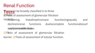
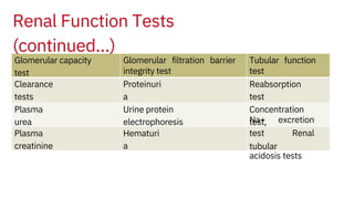
2. Learning Objectives:
assessment of renal function and their
interpretation
To explain the biochemical basis of different tests done
for the
•Erythropoietin (by interstitial
cells)
Kidneys perform two major
functions:
A.Formation of urine as the waste
product B.Production of hormones, e.g.
• Renin
•Calcitriol by the proximal convoluted
tubule
3.
4. Filtrate is processed by tubules to form on an average 1.5 L of urine
daily.
Formation of urine involves filtration of about 180 L of plasma daily at
the
glomeruli which allows the passage of water, dissolved solutes and
small molecules of molecular weight 35,000 and below.
Most cells and larger molecules are retained.
Glomerular filtrate is similar to plasma in osmolality and composition of
small
molecules like sugar, urea, creatinine, electrolytes, etc. but is devoid of
most
enzymes, proteins and hormones.
5. Necessary compounds are retained, and waste products are
excreted.
In this process, tubules reabsorb substances from filtrate (urea) and
secrete many substances into the filtrate.
In this process tubules perform the very important function of water,
electrolytes
and acid base balance.
6. proteinuria, to warrant further renal
investigations.
b)Monitoring the progression of renal disease
c)Monitoring and adjusting the dose of potentially renal toxic
drugs
Real function evaluation is required in a number of clinical
situations, e.g.
Routine urine examination is usually the first test undertaken to
assess the
mainly excreted by the
kidney.
a)Assessment of the extent of renal
damage
renal function and very often it gives some important information,
like
Assessment of Renal
Function
7. These can be broadly classified in to three
groups:
2)Tests of assessment of glomerular filtration
barrier. 3)Tests of assessment of tubular function.
Assuming thatallnephronsare functioningequally and
declineinrenal functionis duetocomplete functionallossof
nephronsrather than
compromisedfunction
s.
1)Tests of assessment of glomerular filtration
capacity.
Renal Function
Tests
8. Glomerular capacity
test
Glomerular filtration barrier
integrity test
Tubular function
test
Clearance
tests
Plasma
creatinine
Hematuri
a
Proteinuri
a
Reabsorption
test
Na+ excretion
test Renal
tubular
acidosis tests
Plasma
urea
Urine protein
electrophoresis
Concentration
test,
Renal Function Tests
(continued…)
9. secrete
1) Substance only
filtered 2) Substance
filtered and
Clearance = GFRInulin
Clearance < GFRUrea
As already discussed, clearance is measure of GFR. Under
ideal
reabsorbed
3) Substance filtered
and
Clearance > GFRCreatinine
conditions, it is equal to GFR. It varies according to the
renal metabolism of the substance as given below.
Renal Function Tests
(continued…)
Glomerular Filtration Capacity
Tests
Clearance
test
10. Renal Function Tests
(continued…)
No endogenous compound fulfils these criteria.
Ideally, clearance of a substance, which is only filtered but neither reabsorbed or
Creatinine, though filtered and secreted by tubules, gives a good estimate of GFR at low
plasma creatinine level.
This is because about 10% of urinary creatinine comes from tubular secretion and there
is an equal (about 10%) overestimation of plasma creatinine due to interfering
substance
secreted, is equal to GFR.
Inulin and iothalamate are used for research purposes for accurate GFR
measurement.
11. Renal Function Tests
(continued…)
But at a higher plasma creatinine level, the tubular secretion increases more
than
the interference from other substances in the plasma.
This leads to 20-30% or more GFR value at high creatinine level.
Cystatin C, a low molecular weight protein, filtered by the glomerulus is another
marker of GFR.
12. Renal Function Tests
(continued…)
Plasma Creatinine and Urea
levels
Creatinine is more sensitive than urea.
These are fairly good indicators of GFR under normal conditions,
A significant increase in plasma creatinine above normal shows at least
50% decrease in GFR.
without significant alteration of liver function and metabolic state.
A rise creatinine level correlates well with moderate decrease in GFR from
75% to 25% of normal.
Further reduction in GFR does not correlate well with plasma levels.
14. Renal Function Tests
(continued…)
Evaluation of Filtration
Barrier
Proteinuri
a
This is the first sign of glomerular injury before a decrease in GFR.
✓
A total urinary protein excretion of
or
albumin>30 mg/day is indicative of glomerular damage.
✓
Protein to creatinine ratio (mg of protein/mg of creatinine) in spot urine sample is a good
substitute for 24 hour urinary protein estimation.
✓
Microalbuminuria (30-300 mg/day) is the earliest sign of renal damage due to microvascular
changes, as seen in diabetes mellitus and hypertension.
>150 mg/day,
15. Renal Function Tests
(continued…)
Evaluation of Filtration Barrier
(continued…)
Hematuri
a
The intact glomerulus does not allow the passage of RBCs.
But with severe glomerular damage, RBC leakage occurs.
Hence detection of microscopic hematuria or RBC casts confirms glomerular damage,
again an early sign before overt decrease in GFR.
16. Renal Function Tests
(continued…)
Evaluation of Filtration Barrier
(continued…)
Urinary Protein
Electrophoresis
In minimal glomerular damage, low molecular weight proteins such as albumin, α1-
antitrypsin and transferrin are excreted into the urine (selective proteinuria). In severe
damage, albumin and other proteins like immunoglobulins also appear. Similarly, in
tubular defects, other small molecular weight proteins appear which can be detected
by electrophoresis in the α-globulin region. These are not normally seen in the urine.
17. Renal Function Tests
(continued…)
Tubular
Functions
Reabsorption
test
Glucose, phosphate, uric acid, bicarbonate and K+ are reabsorbed in the proximal
convoluted tubule. Low blood levels of all these (except glucose) indicate tubular
The same is true of glycosuria with normal blood glucose level. β2-microglobulin
detection by electrophoresis in urine is also a good indicator because,
damage
.
after filtration, it is normally totally reabsorbed.
18. Renal Function Tests
(continued…)
Tubular Functions
(continued…)
Renal Concentration Ability or Water Deprivation
Test
fluid intake is withheld overnight, or preferably for 18 hours.
Measurement of urine osmolality is considered better than specific gravity, though the
latter can also be used.
The urinary osmolality varies very widely, from 50 m osm/Kg in conditions of excessive
fluid intake, to 1200 m osm/Kg.
A random urine sample has a value of 850-900 m osm/Kg. In the water deprivation test,
19. Renal Function Tests
(continued…)
Tubular Functions
(continued…)
Renal Concentration Ability or Water Deprivation Test
(continued…)
nephrogenic diabetes insipidus (lack of response to ADH) the osmolality rarely exceeds
300 m osm/Kg.
The first urine sample in the morning is collected and osmolality is measured.
If it exceeds 850 m osm/Kg, the renal concentration ability is considered normal.
In conditions resulting in low ADH activity (hypothalamic or pituitary disorders) or in
20. Renal Function Tests
(continued…)
Tubular Functions
(continued…)
Specific
Gravity
Under normal conditions, measurement of specific gravity also shows changes parallel
to osmolality.
Under standard physiological conditions, urine specific gravity of 1.010 corresponds
to an osmolality of 300 m osm/Kg.
However, it should not be used if there is proteinuria or glycosuria, which increase the
urine density.
Also intravenous administration of radio-opaque iodinated compounds affects the
correlation between specific gravity and osmolality.
21. Renal Function Tests
(continued…)
Tubular Functions
(continued…)
Sodium Excretion
Test
The rest is excreted through skin and
feces.
Impaired tubular function also leads to a decrease in the renal tubular capacity to
conserve or excrete sodium as per the body requirement.
The patient is given an average diet (as in hospital) containing about 100 m mols of
+
few days.
It should be about 15 m mols/day less than the dietary intake (85 mM/day or 3.3 gm of
Na
salt), implying that about 85% sodium is excreted in the urine.
(5 gm salt/day) for about a week and urinary sodium is estimated over the next
22. Renal Function Tests
(continued…)
Tubular Functions
(continued…)
Sodium Excretion Test
(continued…)
Then give the patient a salt free diet as per hospital recommendation (average Na+
content 10 mM or 0.4 gm of NaCl/day for the next few days).
Again determine the Na+excretion in the urine.
It should be decreased to about 10 mM/day.
The kidneys fail to conserve sodium in chronic renal failure, Addison’s disease and
severe uncontrolled diabetes mellitus with glycosuria.
23. Renal Function Tests
(continued…)
Tubular Functions
(continued…)
Sodium Excretion Test
(continued…)
Ammonium chloride loading
test:
This test utilizes H+secretion ability of the distal tubule.
If the urinary pH is less than 5.5, this test is not needed.
The patient is given a load of NH4Cl (100 mg/Kg body weight) and the urine pH is
measured in urine samples, collected hourly up to 8 hours.
In normal persons, at least one of the samples should show a pH of 5.5 or less.
In renal tubular acidosis, this pH does not fall below 6.5.
25. Renal Failure
•Renal failure refers to a severe decline in real function, leading to:
✓
✓
✓
retentionofnitrogenouswasteproducts,
disturbanceofextracellularfluidvolumeand
acid base balance.
27. •Uremia: When azotemia is associated with clinical symptoms and
multi organ failure, it is called uremia.
•Renal failure can be acute or chronic depending on the time of onset.
28. ACUTE RENAL FAILURE
•This is defined as the sudden decline in renal function over days and
weeks leading to the accumulation of nitrogenous waste products and
other disturbances.
•It is usually associated with oliguria or anuria.
•The basic pathology is severe decrease in glomerular filtration due to pre-
renal, renal and post-renal causes.
30. Pre-renal
causes
•It is seen in conditions like:
Severe diarrhea, as in cholera
Severe prolonged vomiting, as in intestinal
obstruction
Burns and shock
Severe hemorrhage, e.g. accident/trauma
Fluid depletion, e.g. diabetic ketoacidosis
➢
➢
➢
➢
➢
32. Post-renal
causes
•GFR is decreased due to obstruction to flow of glomerular
filtrate •Stones in kidney, ureter and bladder
•Tumors in kidney and bladder
•Urethral strictures
•This obstruction leads to increase in intra-renal pressure and
hydronephrosis ( filtrate
accumulation in kidneys). The increased pressure in renal capillaries and
glomerulus
precipitates failure.
•Prostate
enlargement
33. Clinical signs and
symptoms
•Colicky
pain
•Decreased skin
turgor
•Thirst, nausea,
vomiting
•Dizziness,
weakness
•Postural
hypotension
•Dry mucous membranes and tongue
•Pain in the flanks in cases with renal
ischemia
34. Investigatio
ns
•
20 mmol/L in ARF due to renal
causes.
renal artery stenosis, etc. They may involve radiological
investigation.
Tests of renal functions like blood levels of urea, creatinine,
electrolytes
•Investigations related to the underlying possible cause like stones,
tumors,
etc. These are significantly and persistently increased.
Hyperkalemia is also common.
•Urinary sodium excretion is important in differentiating between
pre-renal
and renal causes. It is less than 10 mmol/L in prerenal ARF and
more than
35. Approach to
Treatment
•
Maintaining fluid
volume
•Renal support in the form of
dialysis
usually reversible. Renal function is
regained.
•Correction of electrolyte disturbances, like
hyperkalemia
•Treatment of the underlying cause
-Renal replacement therapy like dialysis is temporary and patients
are able
to live without it after recovery.
-Acute renal failure, if treated in time, in a well equipped hospital,
36. CHRONIC RENAL FAILURE
•This is mostly due to chronic kidney diseases resulting in gradual
loss of
nephron function, leading to end stage renal disease (ESRD).
•ESRD makes the patient permanently dependent on renal
replacement
therapy –either dialysis or renal transplant.
•Chronic kidney disease (CKD) is defined as a renal disease of
more than
three months’ duration, leading to gradual decrease in GFR.
37. CHRONIC RENAL FAILURE (contd..)
•Based on the associated risk factors and GFR, it is divided into 5
stages:
CKD with milddecrease
Moderatedecrease
Severedecrease
Stages of CKD
I CKD with renal
damage
Normal
GFR
II
II
I
IV Renal
failure
in GFR
in GFR
in GFR
90 mL/min/1.73 m2
60-89 mL/min/1.73
m2 30-59
mL/min/1.73 m2
15-29 mL/min/1.73
38. CHRONIC RENAL FAILURE (contd..)
•
Risk factors include:
-Old age and family history of renal disease
-Hypertension
-Diabetes mellitus
-Autoimmune diseases
-Past H/o renal failure
•GFR is calculated by an equation dependent on plasma creatinine,
weight,
height,
etc.
39. CHRONIC RENAL FAILURE (contd..)
Causes
:
CockroftGault Formula
•Creatinine clearance = (140 ‒ age) X body weight
72 X Plasma creatinine (mg/dL)
•For women the value obtained is multiplied by 0.85.
•All kidney diseases, if untreated, can lead to renal failure ultimately.
•Hypertension and diabetes are two most common causes of renal
failure
worldwide.
40. CHRONIC RENAL FAILURE (contd..)
Abnormalities of CRF
1
.
6
.
3.Metabolic acidosis
Oedema with oliguria or
anuria
Hypertriglyceridemia, ↓ HDL-
cholesterol
2
.
4
.
5
.
Anemiadue to erythropoietin deficiency
Osteomalacia/rickets due to calcitriol
deficiency
Electrolyte disturbances: hyperkalemia,
hypernatremia, hyperphosphatemia, and
hypocalcemia.
41. CHRONIC RENAL FAILURE (contd..)
Abnormalities of CRF
(contd..)
7.Protein-calorie malnutrition
8.Heart failure and cardiomyopathy
9.Hypercoagulation and accelerated atherosclerosis
10.Bleeding tendency
11.Susceptibility to infections
12.Secondary hyperparathyroidism due to
hypocalcemia
42. CHRONIC RENAL FAILURE (contd..)
Biochemical
Investigations
Serial measurement of
•Blood urea and creatinine and GFR,
electrolytes
•Bicarbonate
•Proteins in blood and urine to evaluate
proteinuria
•Other relevant investigations for cause and manifestations like
diabetes, anemia, hyperlipidemia, coagulation alterations, etc.
•Ca and
phosphate
•Alkaline phosphatase for metabolic bone
disease
++
43. CHRONIC RENAL FAILURE (contd..)
Treatment
Approach
•Protein restriction –Usually not done in stage I and II, but in later stages.
This is
•Slowing of diabetes and hypertensive nephropathy by maintaining good control
of
diabetes ad hypertension
•Real replacement therapy in the form of dialysis or renal
transplantation
required because urea is derived from proteins and is excreted by
kidneys. •Treatment of anaemia with iron and erythropoietin
•Treatment of bone disease with calcitriol
•Treatment of electrolyte disturbance