Rare standanrds studies
•
0 likes•199 views

This document describes a method developed by the CHARISMA project to isolate rare colorant compounds from the plants weld (Reseda luteola L.) and madder (Rubia tinctorum L.) using preparative liquid chromatography. Several rare standards were obtained and most were identified using mass spectrometry and NMR spectroscopy. The isolated pure standards will be shared with partners in the CHARISMA project to aid in the identification of colorants in cultural heritage objects.
Recommended

Recommended
More Related Content
Featured
Featured (20)
Rare standanrds studies
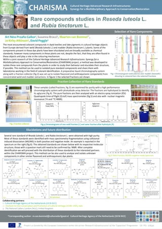
- 1. CHARISMA Cultural Heritage Advanced Research Infrastructures: Synergy for a Multidisciplinary Approach to Conservation/Restoration Rare compounds studies in Reseda luteola L. and Rubia tinctorum L. Selection of Rare Components Art Néss Proaño Gaibor1, Susanna Bracci2, Maarten van Bommel1*, Jo Kirby Atkinson3, David Peggie3 The most encountered colorant compounds in dyed textiles and lake pigments in cultural heritage objects from Europe derived from weld (Reseda luteola L.) and madder (Rubia tinctorum L.) plants. Some of the components present in these dye plants have been elucidated and are broadly available as chemical standards; however many components in these plants are not, despite the fact, that they are often found in these objects and play a role in the colouring mechanism. Within a joint research of the Cultural Heritage Advanced Research Infrastructures: Synergy for a Multidisciplinary Approach to Conservation/Restoration (CHARISMA) project, a method was developed to isolate these rare compounds from the plants in order to study their behavior and elucidate their structure, if possible. This method can be used to isolated pure standard components and share them with laboratories working in the field of colorant identification. A preparative liquid chromatography system along with a fraction collector (fig.2) was set up to isolate flavonoid and anthraquinonic components from Fig.1 Chromatogram weld (above) and madder (beneath) concentrated weld and madder extractions. In figure 1 the selected fractions are shown. showing in coloured bars the selected fractions Fraction Collection of Rare Standards These samples (called fractions, fig.3) are examined for purity with a high performance chromatography system with photodiode array detector. The fractions are hydrolyzed to identify its aglycone (fig.4). The pure fractions are then analyzed with an electro spray ionization (ESI) Quadrapole time-of-flight (Q-tof) mass spectrometer (fig.5) and also with nuclear magnetic resonance (1H and 13C-NMR). 0.30 Fig.3 Collected fractions 12.626 0.22 Apigetrin Aglycone 16.006 0.20 Apigenin 0.25 0.18 Yield 98% Absorption at 350 nm Absorption at 350 nm 0.16 0.20 0.14 0.12 0.15 0.10 0.08 0.10 0.06 12.910 16.008 0.04 17.420 12.773 18.615 0.05 0.02 0.00 0.00 6.00 7.00 8.00 9.00 10.00 11.00 12.00 13.00 14.00 15.00 16.00 17.00 18.00 19.00 20.00 10.00 11.00 12.00 13.00 14.00 15.00 16.00 17.00 18.00 19.00 20.00 Retention time (min) Retention time (min) Fig.2 fraction collector Fig.4 Chromatograms of one weld fraction (L) and same fraction after hydrolysis (R) Fig.5 ESI Q-Tof with CapLC Elucidations and future distribution Several rare standard of Reseda luteola L. and Rubia tinctorum L. were obtained with high purity. Most of these standards were identified with mass spectrometry fragmentation using collisional- induced dissociation (MS/MS) in both positive and negative mode. An example is reported in the spectrum on the right (fig.6). The obtained standards are shown below with its respective molecular structure, those with a question mark still need to be confirmed by NMR. After complete identification we will proceed with the distribution of these standards to the interested partners within the CHARISMA project. This method can be also used to analyse and isolate unknown components in other similar flavonoid and anthraquinonic dye plants. Fig.6 Weld Fraction; MS/MS negative mode Spectrum Apigenin-6,8-di-C-glucoside Luteolin 7-O-glucoside Apigetrin Luteolin-4’-O-glucoside? Luteolin Apigenin Luteolin-3',7-diglucoside C27H30O15 C21H20O11 C21H20O10 R1:OH R2: O-glycose C15H10O6 C15H10O5 C27H30O16 Mw:594.1585 g/mol Mw:448.1006 g/mol Mw:448.1006 Luteolin-3’-O-glucoside? Mw:286.048 g/mol Mw: 270.053 g/mol Mw:610.1534 g/mol OH OH OH OH g/mol R1: O-glucose R2: OH OH R1 HO HO OH OH C21H20O11 OH OH OH HO Mw:448.1006 g/mol R2 O OH HO HO HO O O O O O O O O HO O HO O O HO HO HO O OH HO O O HO O HO OH HO OH HO O HO OH OH O OH OH O OH O OH O OH O HO OH W1 W2 W3 W4 W5/6 W7 HO OH O OH O W8 Xantho-purpurin? Rubiadin 3-O-primeveroside Alizarin Ruberythric acid Lucidin 3-O-b-primeveroside Purpurin Rubiadin Nordamnacanthal? C14H8O4 C26H28O13 C14H8O4 C15H8O5 C25H26O13 C26H28O14 C14H8O5 C15H10O4 Mw:240.2151 g/mol Mw:548.1530 g/mol Mw:240.2151 Mw:268.2255 g/mol Mw:534.4736 g/mol Mw:564.4999 g/mol Mw:256.2145 g/mol Mw:254.2420 g/mol O OH O OH g/mol O OH O OH O OH O OH O OH O OH O O OH O OH H O O O O OH OH OH O OH OH O HO O OH O OH OH O OH O This fractions result was not conclusive , HO OH O OH nordanmacanthal is one atomic mass O OH HO O HO O unit higher than the mass spectrum in O Fraction M2 was not elucidated. OH O O OH O positive mode. Negative mode did not HO show any result. M6 HO M1a M1b OH M2 hydrolized M3 HO OH M4 M5 M7 Collaborating partners: 1 - Cultural Heritage Agency of the Netherlands (OCW-RCE) 2 – Institute for the Conservation and Promotion of Cultural Heritage (ICVBC-CNR), Italy 3 - The National Gallery London (NGL), United Kingdom *Corresponding author: m.van.bommel@cultureelerfgoed.nl Cultural Heritage Agency of the Netherlands (OCW-RCE)