SlideShare a Scribd company logo
1 of 72
Download to read offline
Alyaa H. Al-Mossawi
Author
Dr. Zahraa S. Al-Garawi
Prepared by
Mustansiriyah University
Baghdad 2018
Qualitative tests of amino
acids and proteins and
enzyme kinetics
Qualitative Tests of Amino
Acids and Proteins,
and Enzyme Kinetics
‫إ‬
‫عداد‬
‫هاشم‬ ‫علياء‬
‫الموسوي‬
‫حياتية‬ ‫كيمياء‬ .‫م‬
‫وتاليف‬ ‫إعداد‬
‫الكرعاوي‬ ‫سالم‬ ‫زهراء‬ .‫د‬
‫حياتية‬ ‫كيمياء‬ .‫م‬ .‫أ‬
z.mosin@uomustansiriyah.edu.iq
‫المستنصرية‬ ‫الجامعة‬
‫مقابل‬ ‫فلسطين‬ ‫شارع‬ ،‫واالستنساخ‬ ‫للطباعة‬ ‫حسنين‬ ‫مكتبة‬ ‫في‬ ‫المؤلف‬ ‫هذا‬ ‫طبع‬ ‫تم‬
.‫المستنصرية‬ ‫الجامعة‬
‫الهاتف‬ ‫رقم‬
07707643338
‫البريد‬
‫االلكتروني‬
doublem985@gmail.com
‫او‬ ‫والحذف‬ ‫التعديل‬ ‫يجوز‬ ‫وال‬ ‫والناشر‬ ‫للمؤلفة‬ ‫محفوظة‬ ‫والنشر‬ ‫التأليف‬ ‫حقوق‬
.‫واالستالل‬ ‫اإلضافة‬
‫شباط‬ ‫في‬ ‫االولى‬ ‫الطبعة‬
٢٠١٨
‫ايلو‬ ‫في‬ ‫ّث‬‫د‬ُ‫ح‬
‫ل‬
٢٠١٨
‫ب‬
‫غداد‬
‫وتقديـــــــــــر‬ ‫شكــــــــــر‬
‫الى‬ ‫واالمتنان‬ ‫بالشكر‬ ‫نتقدم‬
‫قسم‬ ‫في‬ ‫األعزاء‬ ‫األساتذة‬
‫الكيمياء‬
،
‫المستنصرية‬ ‫الجامعة‬
‫سموم‬ ‫فالح‬ .‫د‬.‫م‬.‫أ‬
‫الفرطوسي‬
‫(رئيس‬
)‫حياتية‬ ‫الكيمياء‬ ‫فرع‬
‫ح‬ .‫د‬.‫م‬.‫أ‬ ‫و‬
‫سنين‬
ّ‫ي‬‫الق‬ ‫المقترحات‬ ‫وتقديم‬ ‫والمساندة‬ ‫للتأييد‬ ،‫البيرماني‬ ‫كامل‬
‫مة‬
‫هذا‬ ‫إلنجاز‬
‫ال‬ ‫المؤلف‬
‫منهجي‬
.
Contents
Subject Page
no.
Preface 6
Qualitative tests of amino acids
Amino acids 8
1. Solubility test 12
2. Ninhydrin Reaction 14
3. Xanthoproteic Reaction 18
4. Millon`s Reaction 21
5. Acree-Rosenheim Reaction 24
6. Pauly recation 27
7. Lead Sulfide Test 30
8. Nitroprusside test 33
9. Sakaguchi`
s Test 35
Qualitative tests of proteins
1. Biuret test 38
2. Precipitation reactions 39
a) Denaturation of proteins 43
b) Precipitation by heavy metals 44
c) Precipitation by organic acids 45
d) Precipitation by neutral salts 46
e) Precipitation by alcohol 50
f) Heller`
s test 51
g) Precipitation by Esbach`
s
reactions
52
Enzymes
Enzymes and enzyme kinetics 55
1. Calculation of substrate
concentration
59
2. Calculation of enzyme velocity 59
3. Determination of the type of
inhibition
63
References 67
Preface
This handbook is for the 3rd year students of
the Chemistry Department at Mustansiriyah
University. It has the syllabus for the second
course of the Biochemistry lab. The
handbook contains some qualitative tests for
detecting the amino acids and proteins, as
well as, the enzyme kinetic experiments,
which are currently available in our lab. The
qualitative tests are commonly used to detect
different types of amino acids and proteins
either separately or within a mixture.
The last part of this handbook contains the
enzyme kinetics experiments, which are used
to determine the kinetic parameters of the
7
enzyme alkaline phosphatase in the presence
and absence of inhibitor.
The test methods in this handbook have been
qualified as standard methods using valid
reagents. Some methods here employ toxic
or very acidic solutions which need to be
handled with care. Safety protectors such as
eye wears, gloves, lab coats are important for
students and lab staff to do each experiment.
Using this handbook by the students is
necessary to follow the experiments, to
complete the homework and to study for the
exams.
Dr. Zahraa
https://uomustansiriyah.edu.iq/e-learn/profile.php?id=4801
8
Qualitative tests of amino acids
Amino acids
An amino acid (R-CH-NH2COOH) contains
an amino group and a carboxylic acid group
as side chains. It is the building block of all
proteins and is linked with other amino acids
as a chain by the peptide bonds (CONH-) to
form the primary structure of a protein, see
Figure 1.
Fig 1. Structure of amino acids. The right-hand
side figure shows the peptide bond.
Carbon
9
There are 20 naturally occurring amino acids,
10
which vary from one to another according to
the type of their side chains, see Figure 2.
Amino acids are amphoteric with an
extremely high melting point (usually
exceeding 200°C), react as amines in some
reactions, but as carboxylic acids in others.
At a certain pH, the number of the positive
charges (from protonated ammonium
groups) are equal to the number of the
negative charges (from deprotonated
carboxylate groups), therefore, no net charge
will be observed. This point is named as “the
isoelectric point” and the amino acid will
exist as zwitter ion. Herein, the Isoelectric
point (Ip) is the pH value at which the
concentration of anionic and cationic groups
is equal (i.e. the net charge of this molecule
equal to zero). It is the point where the
molecule does not move to either the cathode
or the anode when it is put in an electric field.
11
Because its solubility will be minimum, it is
possible to precipitate at this point.
Amino acids are responded to all typical
reactions associated with compounds
containing carboxylic acid and amino
groups, usually under conditions where the
zwitter ions form is present in only small
quantities. Amino acids are chiral molecules
(except glycine) and exhibit an optical
activity due to the presence of an asymmetric
α-carbon atom.
Amino acids with an L-configuration are
present in all naturally occurring proteins,
whereas those with D-form are found in
antibiotics and in bacterial cell walls.
Amino acids are classified into groups
according to their side chains, see Figure 2.
12
The amino acids tests are:
1. Solubility test
The solubility of amino acids largely depends
on the pH of the solution. Any change in the pH
value causes structural changes in the amino
acids and alters the relative solubility of the
molecule.
In acidic solutions, both amino and
carboxylic groups are protonated, whereas, in
basic solutions, both groups are deprotonation.
13
Materials
Different amino acid solution: Gly, Lys and
Glu.
Solvents: H2O, HCl, NaOH and Chloroform.
Method
Mix a small amount of each amino acid
solution with few mls of each solvent, record
the results.
Questions?
1. What is the best solvent of amino acids?
Why?
2. Can you dissolve Gly in water?
3. Can Lys be dissolved in NaOH?
4. What is the difference you can see when
dissolve Lys and Glu in chloroform?
5. Do most amino acids dissolve in water?
14
2. Ninhydrin reaction
Ninhydrin [tri-ketohydrindene hydrate] is a
powerful oxidizing agent used for detecting
ammonia or primary and secondary amines.
In a range of pH around 4-8, all α-L-amino
acids react with ninhydrin to give a purple
colored product (diketohydrin). Ninhydrin
Fig 3. The ninhydrin coupling reaction with amino acids and with Pro.
15
interacts with amino acids causes
decarboxylation and deamination (liberation of
CO2 and ammonia) to produce the purple
Rhuemann,
s compound and aldehyde, see
Figure 3.
The amino acids Pro and hydroxyl Pro react
with ninhydrine, but they give a yellow colored
complex instead of a purple one.
Primary amines and ammonia react similarly,
but without the liberation of CO2. Different
molecules such as peptides and proteins are
also able to positively react with ninhydrin test.
Mechanically, one reduced ninhydrin molecule
condenses with another non-reduced ninhydrin
molecule and ammonia to form a violet –blue
or purple condensation product, see Figure 4.
16
Caution
Ninhydrin is a strong oxidizing agent;
therefore, it should be handled with care. Apart
from that, eyewear, gloves and hood are
required. If accidentally get in touch with skin,
extremely wash with water. However, the
resulting stains are temporary and will be
eliminated within 24 hours.
Fig 4. The mechanism of ninhydrin coupling reaction
with amino acids.
17
Method
1. Add 1ml of each of the test solutions to 2
drops of ninhydrin solution.
2. Boil the mixture over a water bath for 2 min.
3. Allow to cool and observe the resulted blue
color.
Questions?
1. Which of the following gives a positive
ninhydrin test?
Phospholipid, sucrose, galactose, Pro,
Albumin.
2. What is ninhydrin reaction? How it is
useful?
3. How dose methylamine (CH3NH2) reacts
with ninhydrin test?
4. What kinds of protection you should use
when doing ninhydrin experiment?
5. Why Pro and hydroxyl Pro give yellow
color with ninhydrin?
18
3. Xanthoproteic reaction
This test is used to differentiate between
aromatic amino acids. Treating aromatic amino
acids with concentrated nitric acid leads to the
nitration of the aromatic ring and formation of
a yellow nitro-product (nitro derivatives), see
Figure 5.
When a strongly basic solution is added, the
color of the product turns to be darker (from
Fig 5. The Xanthoproteic reaction with aromatic
amino acids.
19
yellow to orange) due to the ionization of the
petrolic group.
Caution
Concentrated HNO3 is a toxic and corrosive
substance that can cause severe burns and
discolor the skin. Gloves, safety glasses and a
fume hood are required. Avoid inhaling vapors
and ingesting the compound.
Method
1. Add 1m of concentrated HNO3 to 1ml of
the test sample.
2. Heat the mixture and cool it.
3. Slowly add (40% w/v in a water solution)
NaOH until the mixture becomes alkaline (use
litmus strip to indicate the pH) and color-
change is noted. Changing the color from
yellow to orange indicates the presence of an
aromatic amino acid.
20
Questions?
1. Which of the following gives positive test
with Xanthoprotic reaction?
Trp, Cys, Ala, Gly, Tyr, Thr
2. What is HNO3 doing for the reaction?
3. what is the color of a positive reaction with
Xanthoprotic?
4. Phe does not react with Xanthoprotic, why?
21
4. Millon`s reaction
The test was developed by the French
chemist Auguste Million. Million`s reagent
is a mixture of mercuric nitrate/nitric acid
and water.
This test is specific for Tyr, the only amino
acid contains a phenol group (a hydroxyl
group attached to a benzene ring). However,
all phenols give positive results with
Million`s test.
The reaction occurs in 2 steps: first, the
phenolic group of Tyr nitrates by HNO3, then
in the presence of Hg(NO3)2, the nitrated Tyr
forms a reddish-brown solution or a yellow
precipitate of a nitrated Tyr, which is a
positive test, Figure 6.
22
Method
1. To 1ml of amino acid solution, add 1ml of
Million`
s reagent in a test tube
2. Warm the tube in a boiling water bath for
10 min.
3. A reddish-brown color is a
positive reaction.
Fig 6. The Millon reaction with a phenolic amino acid.
23
Questions?
1. Why Millon`s reagent gives a reddish color
with phenolic amino acids?
2. Which test is more specific for Tyr?
Millon`s or Xanthoprotic test? Why?
3. If you increase the concentration of Tyr,
will that increase the depth of the product?
4. What Hg is doing for the reaction?
5. What is Millon reagent?
24
5. Acree-Rosenheim reaction
Acree-Rosenheim is another test for detecting
Trp in a protein solution. Tryptophan is mixed
with formaldehyde (CH2O). Concentrated
sulfuric acid is added to form two layers. A
violet zone in the junction point of two layers
is the positive result, see Figure 7. Similar
result obtained by the Hopkins-Cole reaction
which is frequently used to detect Trp in a
protein solution. In the Hopkins-Cole reaction,
when a glyoxylic acid CHOCOOH is added to
a protein solution (containing Trp) in the
presence of H2SO4, a violet ring will form.
+ H2-C=O → a violet ring
H2SO4 Conc.
Fig 7. Acree-Rosenheim reaction with Trp.
25
Method
(Acree-Rosenheim Reaction)
1. Add 1 ml of amino solution to 1ml of
CH2O.
2. Concentrated H2SO4 (1 ml) is then added
to form two layers.
3. Watch the development of a purple
colored ring between the two layers as
positive test for Trp appearance.
Questions?
1. What is Acree-Rosenheim reagent?
2. What is the difference between Acree-
Rosenheim and Hopkins-Cole
reaction?
3. If the protein solution contains a
mixture of Trp, phe and Tye, which
amino acids form the purple ring more
quickly?
26
4. How would the phenolic amino acid
be distinguished by Acree-Rosenheim
reaction?
5. If you have Phe, which of the
following test is the best to detect?
Ninhydrine, Million, Xanthoproteic,
Hopkins-Cole?
27
6. Pauly reaction
Pauly reaction was firstly described by the
German chemist Hermann Pauly. It is used to
detect Tyr or His amino acids by a coupling
reaction with diazotized sulfanilic acid under
Diazonium compound
Sulfanilic acid
Tyrosine
Fig 8. Pauly reaction with Tyr.
28
alkaline conditions. As a result, a red colored
solution is produced.
Principally, diazotized sulfanilic acid is
resulted by a reaction between the
sulfanilic acid and sodium nitrite and
sodium carbonate to form the diazonium
component. Then, the diazonium
components react with the imidazole ring
of His or phenolic group of Tyr to form a
dark red compound, see Figure 8.
Method
1. Cool down a test tube using a cool
icebox.
2. Add 1ml of sulfanilic acid to the cool
test tube, remain in the icebox.
3. Add to the mixture 1ml of NaNO3, let
it cool for 3min.
4. Add 2ml of Na CO3.
5. Observe the color.
29
Questions?
1. How does the Pauly test work?
2. Which amino acid gives a positive
product with Pauly reagent? and which
gives negative product?
3. How does His react with Pauly?
30
7. Lead sulfide test
This is specific for sulfhydryl group (-SH).
Sulphur containing amino acids, such as Cys
and Cyst is converted into sodium sulfide
(Na2S) when boiled with 40 % NaOH. The
reaction depends on a partial conversion of
the organic sulfur into inorganic sulfide
Na2S, which can be detected using sodium
plumbate (Na2pb(OH)4: lead acetate solution
Pb(CH3COO)2 in an alkaline media). Na2S
precipitates as a black lead sulfide, see Figure
9. Methionine or any other Meth containing-
proteins such as Casein and Gelatin, gives a
negative result with this test.
Protien-SH + 2 NaOH → Na2S
40% Na-Sulfide
Na2S + Pb (CH3COO)2 → PbS + 2 CH3COONa
NaOH
Fig 9. Lead sulphide reaction with sulphur-containing amino acids.
31
Method
1. Prepare sodium plumbate (Na2Pb(OH)4
solution as following:
Add 5ml of 40% NaOH to 2ml of dilute
Pb(CH3COO)2. A white precipitate of
Lead hydroxide Pb(OH)2 will be formed.
2. Boil solution (1) to completely dissolve
Pb(OH)2 and form Na2Pb(OH)4.
3. Add few drops of 40% NaOH to 2ml of
amino acid solution and boil the solution for
2min.
4. Cooldown solution (3) and add few drops of
solution (2), then watch the development of
a brown color or precipitate (PbS) as an
indication of the (-SH) group.
Questions?
1. If you have a mixture of Cys, Cyst, Meth,
Gly and Gln, do you expect the reaction
will be positive with Lead sulfide test?
32
2. Why do you use sodium plumbate in lead
sulfide teat?
3. How can you know that the Lead sulfide
test is positive?
4. Can you use this test for most of amino
acids?
5. How the Lead acetate test works?
33
8. Nitroprusside test
This test is specific for Cys which has a free
sulfhydryl group (-SH) that is able to react
with the nitroprusside in the presence of
excess ammonia (NH4OH).
Principally, the free thiol group of Cys gives
a red color with sodium nitroprusside in the
presence of NH4OH, see Figure 10. To
compare: do the test to Cys, Cystaein and
Meth.
Nitroprusside test is commonly used to detect
the ketones in urine.
Na2(CN)5Fe (NO)2 H2O + HS-CH2CHNH2COOH → Red complex
Na-Nitroprosside Cys
NH4OH
Fig 10. Na-nitroprusside reaction with Cys amino acid.
34
Method
1. Add 0.5ml Na-nitroprusside solution
to 2ml of amino acid solution and
shack well.
2. Add 0.5ml NH4OH and watch the
change in the color.
Questions?
1. Do you think there is a difference
between Lead acetate and
Nitroprosside test? What is it?
35
9. Sakaguchi`
test
Sakaguchi`
s test is specific for the guanidine
group-containing amino acids, such as Arg.
Guanidine group interacts with α-naphthol
and alkaline hypobromite to produce a red-
colored complex.
Method
1. To 1ml of amino acid solution, add 2
ml of 40% NaOH and mix well.
2. Add 2ml of α-naphthol solution and
mix well.
3. Add 2 drops of sodium hypobromite
(BrNaO) solution, mix well and record
the results (a red colored complex).
36
Questions?
1. What is the principle of Sakaguchi`
test?
2. Why is this test specific for Arg?
3. What is the alkaline medium that you
need for this test?
4. What is the positive result of this test?
5. What the will be result if used tertiary
amine with Sakaguchi reagents?
37
Qualitative Tests of Proteins
38
Proteins
Proteins are polypeptides consist of many
amino acids bound together via peptide
bonds. Amino acids interact with each other
via different forces to build up the final three-
dimensional (3D) structure of the protein.
These forces are responsible for the stability
and rigidity of the protein, which are: van der
Waals force between temporary dipoles,
ionic interactions between charged groups,
salt-bridges and polar-polar interactions.
Proteins regulate a variety of activities in all
the known living organisms, ranging from
the replication of a genetic code to
transporting oxygen. Proteins exist as a 3D
39
tertiary and quaternary substance. The
functional properties depend on the 3D
structure, which arises when particular
sequences of amino acids in a polypeptide
chain fold to generate linear chains have
compact domains with specific structures.
These folded domains either serve as
modules for larger polypeptide assemblies,
or they support specific catalytic or binding
sites for other functional substances or metal
ions.
Here are the main tests for detecting the
presence of a protein in a solution:
1. Biuret test
Biuret test is used to indicate the presence of
peptide bonds within the protein molecule.
This test could be useful to quantitatively
know the concentration of the protein, where
the intensity of the product`s color increases
40
linearly with the concentration of the protein.
In addition, according to the Beer-Lambert
law, the optical density at 540 nm
wavelength is directly proportionated to the
protein concentration.
41
Biuret reagent (H2N-CO-NH-CO-NH2)
results from a coupling reaction of two urea
molecules. It reduces Cu(II) to Cu (I) and
Fig 11. a) Reaction of Biuret with Cu(II), b) interaction of
Biuret reagent with the peptide bond.
a
b
42
form a red colored Cu-complex as a positive
result for C=O-NH bond involvement, see
Figure 11a.
Because Biuret has a bond (C=O-NH) similar
to the peptide bond, it is the most useful test
for samples with two peptide bonds and
above, see Figure 11b. Buffers, such as Tris
and ammonia interfere with this assay.
Method
1. Add 1 ml of 40% NaOH to 1 ml
protein sample to make it alkaline.
2. Add 5 ml Biuret reagent to solution (1)
3. Watch changing the color within 5
min.
43
Questions?
1. If you let your sample to stand 20 min
with Biuret Reagent, what will
happen?
2. What is the component of Biuret
reagent?
3. What is the principle of Biuret
reaction?
4. Which of the following do you expect
will give aa positive result with
Biuret? Glucose? Casein? Glu?
Phospholipid?
5. Why the reagent called Biuret?.
44
2. Precipitation reactions
a. Denaturation of proteins
Denaturation is an irrevocable change in the
protein structure lead to loss of its biological
properties. Because the fold type of the
protein ( -sheet, -helix, random coil)
determines its function, any change of the
tertiary structure will alter its activity.
Temperature and pH are the main causes of
denaturation and precipitation of proteins
because they destroy the weak bonds and
affect the tertiary and secondary, but not the
primary structure.
Method
1. In a water bath, boil 1ml protein of
different protein solutions such as
Bovine albumin and egg globulin.
2. Record your results
45
b. Precipitation by heavy metals
At natural pH 7 or above, proteins are
usually negatively charged. Adding
positively charged metal ions will neutralize
the charges of protein and lead to protein
precipitation. Heavy metals such as Ag+
,
Pb2+
, Hg2+
, etc form a complex with the
alkaline proteins and then precipitate.
NOTE: avoid very high pH to reduce the risk
of metal hydroxides precipitation.
Method
1. To 2 ml of different protein solutions (e.g.,
Bovine albumin, egg white, gelatin, casein),
add few drops of: AgNO3, Lead acetate
Pb(CH3COO)2.to each protein solution.
2. Observe the extent of precipitate in each
experiment.
46
c. Precipitation by organic acids
In acidic medium, proteins are positively
charged. Adding acidic solutions such as
picric acid and Trichloroacetic acid (TCA)
will neutralize the protein charge and
irreversibly form an insoluble solution due to
their high content of negative charges.
Method
1. To 2 ml of protein solution (albumin
solution or egg white solution), add 5-8
drops of picric acid or TCA solution.
2. Note the precipitate in each protein
tube.
3. Slowly add (1 ml) of 40% NaOH
solution and record if increasing the pH
will cause renaturation of the protein
(dissolve the precipitate).
47
d. Precipitation by neutral salts
To investigate the effect of salt
concentrations on the protein solubility, a
series of low concentrations of salt is added
to a protein solution (salting in). low
concentrations will increase the protein
solubility. Salt molecules decrease the
electrostatic energy between the protein
molecules, which increase the solubility and
stability of the protein structure.
Increasing the salt concentration (salting
out) will decrease the protein solubility at
some points. The excess of salt ions (not
bound to the protein) competes with protein
in binding the solvent molecules. This
decrease in salvation allows the proteins to
aggregate and precipitate. Each protein can
be reversibly precipitated at specific salt
concentration.
48
This test is used to separate different proteins
using high salt concentration solution
(oversaturation solutions), which will
precipitate the certain protein due to the
effect of salt ions on the protein solubility.
Proteins precipitate at different salt
concentrations; therefore, a series of salt
concentrations are required to test the
precipitation ability of each protein.
In salting out the experiment, you must
take into account the following:
• The type of the salt (ammonium sulfate
(NH4)2SO4 is commonly used)
• The molecular weight (Mwt) of the
protein: the highest Mwt, the fastest to
precipitate.
• Proteins with high Mwt usually require
the low concentration of salt to precipitate,
and vice versa.
49
• It is a reversible reaction; the protein can
be recovered by dissolving with H2O
• Applied the salting out experiment on a
mixture of protein
Method (salting in)
1. To 2 ml of albumin solution (egg
white) add (1ml) NaCl solution.
2. Add an equivalent amount of
(NH4)2SO4 until you notice over
saturation.
3. Repeat the experiment with other
proteins such as globulin, mucin and
lysozomes. Describe the difference in
the solubility or precipitation ability.
Method (salting out)
1. Add (1ml) globulin solution in (1ml)
0.1 M NaCl.
50
2. Add 1ml globulin solution to 1ml
saturated (NH4)2SO4 and record your
observation.
3. Add 1ml egg albumin to 1ml saturated
(NH4)2SO4 and record your
observation. Add excess amount of
solid (NH4)2SO4 to indicate the
oversaturation.
51
e. Denaturation by alcohol
Some alcoholic reagents such as ethanol or
propanol can disrupt the hydrogen bonding
within the protein structure by intra
hydrogen-bonding with the protein
molecules. For sterilizing purposes, 70%
alcohol is used to denature the protein of the
bacteria because it effectively penetrates the
bacterial cell wall. Whereas, 95% alcohol has
no similar effect, probably because it
coagulates the surface proteins as a crust,
which would prevent the alcohol to penetrate
into the cell wall.
Method
1. Treat 2ml of a protein solution with a
few drops of alcohol.
2. Notice the protein precipitation.
52
f. Denaturation by acids (Heller`
s test)
Nitric acid desaturates proteins and form
a white precipitate.
Method
1. Add 1ml HNO3 (conc). to 1ml of
protein solution.
2. Note the protein precipitate formed.
53
g. Precipitation by Esbach`
s reactions
It is a mixture of dilute acids (picric acid,
citric acid and water) used to quantitatively
detects albumin in the urine. This reagent
precipitates protein where a yellowish to
brown color product is formed depending on
the quantity of the protein.
Method
1. Add 1ml Esbach reagent to 1ml of
protein solution.
2. Note the color of protein precipitate.
Questions?
1. What causes denaturation of protein?
2. If you heat the protein solution up to 70
C, will that affected the tertiary
structure of the protein? The secondary
structure?
54
3. Why is (NH4)2SO4 precipitated
protein?
4. What is slating-out protein? Salting in
protein?
5. Which of the following metals causes
precipitation of the protein? Why?
Hg, Al, Ni, Ca, Zn, Cu, Ag, Au, Pb, Na.
6. What is the difference between organic
acid and normal acid in terms of
precipitation the protein? Which one
causes reversible precipitation?
7. Do you think that absolute ethanol is
better than 70% ethanol to precipitate
albumin?
8. What is Heller reagent?
9. Do you think that adding HNO3 will
cause reversible denaturation?
10. what is the best test to distinguish
Albumin? Globulin?
55
Enzyme kinetics
Enzymes
56
The Enzyme is a biological molecule with an
active site to bind a substrate and accelerate
a chemical reaction.
Determination of enzyme kinetics
The aim of this practice is to experimentally
obtain data for the kinetics of an enzymatic
reaction in the presence and absence of an
inhibitor. This data is then will be used to
produce Lineweaver-Burke plots in the
presence and absence of an inhibitor to
determine Km, Vmax and the type of
inhibition.
In this experiment, the alkaline phosphatase
enzyme (ALP) catalyzes the hydrolyses of
the substrate p-nitrophenyl phosphate (p-
NPP) in alkaline medium to produce p-
nitrophenol (p-NP) and phosphoric acid, as
following
57
The product p-NP is yellow under the
alkaline pH of the assay. Therefore, the rate
of the enzyme-catalyzed reaction can be
followed by recording the absorption
readings at 405nm of the amount of product
accumulated as a function of time (min).
In the second part of the experiment, you will
determine the type of inhibition when Na-
pyrophosphate is added to the enzyme
reaction mixture.
p-nitrophenyl phosphatephosphate p-nitrophenol
58
Reagents
Substrate: 0.02 M p-NPP (stock solution)
Buffer: 0. L M-glycine/NaOH buffer pH 10.4
Enzyme: ALP (2 mg/ml in 0.1M-
glycine/NaOH buffer pH 10.4)
Inhibitor: 100 mM Na-pyrophosphate or 200
mM imidazole
Method
Using clean plastic cuvettes, set up reaction
mixtures as instructed in Table 1. Set the
spectrophotometer up using buffer as a blank
(no enzyme and no substrate). Set the blank
to zero at 405nm. Leave the blank inside the
instrument for the rest of the experiment.
Now start the enzyme assays. Do all the
addition to the cuvette and leave the enzyme
solution LAST. Cover the cuvette by a
parafilm and mix by inversion. Remove the
Tetra sodium pyrophosphate
59
parafilm, put the cuvette into the
spectrophotometer and read the absorbance
(Abs) at 1 min. intervals for 6 min.
Repeat tube 1 but without adding the
enzyme, can you record a rate for the
chemical reaction?
1) Calculation of substrate
concentration [S]
Table 1. The reaction mixture of enzyme kinetics without inhibitor I.
Tube no. Buffer
(ml)
Substrate (ml)
0.02 M p-NPP
Na-PP
(I) ml
100 mM
H2O
(ml)
Enzyme
solution(ml)
LAST
Blank 1.9 - - 1.3 -
1 1.9 1.0 - 0.2 0.1
2 1.9 0.8 - 0.4 0.1
3 1.9 0.6 - 0.6 0.1
4 1.9 0.4 - 0.8 0.1
5 1.9 0.2 - 1.0 0.1
6 1.9 0.1 - 1.1 0.1
Non-
Enzyme
rate
1.9 1.0 - 0.3 None
60
Calculate the substrate concentration [S]
from the concentration of the stock solution
of p-NPP (0.02M) and its dilution in each of
the enzyme reaction mixtures (1-6) using the
dilution low N1V1=N2V2.
2) Calculation of the enzyme
velocity V0
The initial velocity V0 is the amount of the
product per a unit of time (min.) and NOT
ITS
CONCENTRATION. Plot the absorbance
reading (Abs) of each enzyme reaction
against time and derive the V0 of the reaction
from the slope as Abs /min.
Convert each Abs/min to mol p-NP (the
product/min) using the extinction coefficient
61
( ) of p-NP (15 mM-1
cm-1
) and the volume of
the assay mixture (3.2 ml) for each tube, as
following:
Abs= * l * C (Beer-Lambert law)
𝐶 =
𝐴𝑏𝑠. 𝑚𝑖𝑛−1
15 𝑚𝑀−1 𝑐𝑚−1 ∗ 1 𝑐𝑚
𝐶 = 𝐴𝑏𝑠. 𝑚𝑖𝑛−1
1
15
∗
𝑚𝑚𝑜𝑙
𝑚𝐿
𝐶 =
𝐴𝑏𝑠. 𝑚𝑖𝑛−1
15
∗
𝑚𝑚𝑜𝑙
1000
3.2
𝐶 =
𝐴𝑏𝑠. 𝑚𝑖𝑛−1
15
∗
3.2
1000
𝑚𝑜𝑙
𝐶 = 𝐴𝑏𝑠. 𝑚𝑖𝑛−1 ∗ 0.21 𝑚𝑜𝑙
62
= ? 𝑚𝑜𝑙. 𝑚𝑖𝑛−1
(Dividing by 15 converts the Abs into
concentration (mM) units; as a M solution is
the gram Mwt (mol) in 1000 ml). The volume
of the assay is 3.2 ml, the amount of product
is determined by multiplying (Abs/min.15)
by the fraction 3.2/1000, assuming that all
[S] is dissociated to product.
(i) V0 against [S]
Plot V0 for each enzyme-reaction (μmol/min)
against each [S] (mM) to obtain the V max.
(ii) Lineweaver-Burke plot to determine
Km and V max
63
Calculate the reciprocals of each of the
values of V0 (1/V) and of [S] and plot 1/V0
against l/[S]. Draw the straight line of best fit.
This line intercepts the l/V axis at l/Vmax
and the 1/[S] axis at -l/Km as in the example
below:
1
𝑉
=
1
𝑉𝑚𝑎𝑥
+
𝐾𝑚
𝑉𝑚𝑎𝑥
[𝑆]
3) Determination of the type of
inhibition
1/Vmax
-1/km
1/V
1/[S]
64
Repeat the experiment in the Table 1 using
0.2 ml of Na- pyrophosphate in each cuvette
as instructed in Table 2. (* Note the volumes
of water is differ than that in Table 1).
Table 2. The reaction mixture of the enzyme kinetics with
inhibitor I.
Tube
no.
Buffer
(ml)
Substrate
0.02 M p-
NPP
Na-
PP (I)
ml
100
mM
H2O
(ml)
Enzyme
solution
LAST
Blank 1.9 - 0.2 1.1 -
1 1.9 1.0 0.2 - 0.1
2 1.9 0.8 0.2 0.2 0.1
3 1.9 0.6 0.2 0.4 0.1
4 1.9 0.4 0.2 0.6 0.1
5 1.9 0.2 0.2 0.8 0.1
6 1.9 1.0 0.2 0.9 0.1
(i) V0 against [S] in the presence of I
65
Plot V0 for each enzyme-reaction (μmol/min)
against each [S] (mM) as used in a and i.
(ii) Lineweaver-Burke plot in the
presence of [I]
Calculate the reciprocals of each of the
values of V0 and of [S] as appeared in Table
3 and plot 1/V0 against l/[S] as you used for
(ii). Draw the straight line of best fit and
indicate the type of the inhibition in terms of
Km and V max.
66
Table 3. Table of the reciprocals values of Vo and S in
the presence and absence of inhibitor I
[S] 1/[S] V
( mole/min)
1/V VI
(inhibitor)
1/VI
? ? ? ? ? ?
? ? ? ? ? ?
? ? ? ? ? ?
? ? ? ? ? ?
? ? ? ? ? ?
? ? ? ? ? ?
? ? ? ? ? ?
Questions?
1. Can you ignore the mixture of tube 1
for the non-enzymatic rate? Why?
2. From your plot, what do you conclude
regarding the effect of increasing [S] on
the velocity?
3. What are factors influencing the initial
velocity Vo?
4. Is the effect of the inhibitor that you
observed representative of
67
competitive? Non-competitive
inhibition? Why?
5. What is the Ki of the inhibitor?
68
References
1- P.M.Swamy, (2008), Laboratory manual of
Biotechnology.pastogi Publication, p.90 .
ISBN 978-81-7133-918-1.
2- R.A.Joshi (2006), Question bank of
Biochemistry. New age Iinternational, p.64.
ISBN 978-81-224-1736-4.
3- Chatterjea, (2004), Textbook of
Biochemistry for dental/nursing/Pharmacy
students. Jaypee Broyhers Publishers, p.51.
ISBN 978-81-8061-204-6.
4- Zellner; et al, (2005), Quantitative validation
of different protein precipitation methods in
proteome analysis of blood platelets,
Electrophoresis, 26(12):2481-9.
Doi: 10.1002/elps.200410262.PMID
158945623.
5- Harrison et al., (2003), Bioseparations
Science and Engineering.Oxford University
Press. New Youk, NY.
6- Shuler et al., (2001), Bioprocess engineering:
Basic concepts (2nd
Edition), Prentice Hall
International.
69
7- Ladisch, (2001), Bio separations
Engineering, John Wiley & Sons, Inc.New
York, NY.
8- Lydersen, (1994), Bioprocess Engineering.
John Wiley & Sons. New York,NK.
9- http://www.chem.boun.edu.tr/wp-
content/uploads/2014/04/Chem-415-
Experiment-2.pdf .
Hotspot on the author
Dr. Zahraa S. Al-Garawi is a prof assist in Biochemistry
and a PhD in Biophysics, Structural biology and
Nanomaterials from University of Sussex, UK. She
previously has wrtten a practical handbook in Clinical
Biochemistry for undergraduates, in two languages, in
Arabic and English. Zahraa has published very high-
quality works in her field. Recently, she published a
scientific textbook about steps for understanding writing
and publishing papers, for researcher and student”
Zahraa and Aliaa collaborate in this handbook to provide
the most trusted methods for amino acid and protein tests,
and the enzyme kinetics. This handbook would be of good
benefits and helpful for undergraduates, chemistry and
biology department.

More Related Content

Similar to Qualitative tests of amino acids and proteins and enzyme kinetics

Biochemical analysis of unknown bacteriaAerotolerance Te
Biochemical analysis of unknown bacteriaAerotolerance TeBiochemical analysis of unknown bacteriaAerotolerance Te
Biochemical analysis of unknown bacteriaAerotolerance Te
ChantellPantoja184
 
LectureNotes_BPharmIIISem_PCISyllabus_UNITII_FatsandOils.pdf
LectureNotes_BPharmIIISem_PCISyllabus_UNITII_FatsandOils.pdfLectureNotes_BPharmIIISem_PCISyllabus_UNITII_FatsandOils.pdf
LectureNotes_BPharmIIISem_PCISyllabus_UNITII_FatsandOils.pdf
ssuser7413b8
 

Similar to Qualitative tests of amino acids and proteins and enzyme kinetics (12)

Effect of solvents on formation of disulphide bond in peptides: A comparativ...
	Effect of solvents on formation of disulphide bond in peptides: A comparativ...	Effect of solvents on formation of disulphide bond in peptides: A comparativ...
Effect of solvents on formation of disulphide bond in peptides: A comparativ...
 
Biochemical analysis of unknown bacteriaAerotolerance Te
Biochemical analysis of unknown bacteriaAerotolerance TeBiochemical analysis of unknown bacteriaAerotolerance Te
Biochemical analysis of unknown bacteriaAerotolerance Te
 
Lec.1 aminoacid.chem
Lec.1 aminoacid.chemLec.1 aminoacid.chem
Lec.1 aminoacid.chem
 
Lec.1 a.a.chem
Lec.1 a.a.chemLec.1 a.a.chem
Lec.1 a.a.chem
 
Lab Document_Lipids.docx
Lab Document_Lipids.docxLab Document_Lipids.docx
Lab Document_Lipids.docx
 
Amino acids
Amino acidsAmino acids
Amino acids
 
paper1-published
paper1-publishedpaper1-published
paper1-published
 
Biochemistry and-human-nutrition
Biochemistry and-human-nutritionBiochemistry and-human-nutrition
Biochemistry and-human-nutrition
 
Chemistry of amino acids
Chemistry of amino acidsChemistry of amino acids
Chemistry of amino acids
 
LectureNotes_BPharmIIISem_PCISyllabus_UNITII_FatsandOils.pdf
LectureNotes_BPharmIIISem_PCISyllabus_UNITII_FatsandOils.pdfLectureNotes_BPharmIIISem_PCISyllabus_UNITII_FatsandOils.pdf
LectureNotes_BPharmIIISem_PCISyllabus_UNITII_FatsandOils.pdf
 
Asymmetric synthesis
Asymmetric synthesis Asymmetric synthesis
Asymmetric synthesis
 
Paper 3
Paper 3Paper 3
Paper 3
 

Recently uploaded

Presentation Vikram Lander by Vedansh Gupta.pptx
Presentation Vikram Lander by Vedansh Gupta.pptxPresentation Vikram Lander by Vedansh Gupta.pptx
Presentation Vikram Lander by Vedansh Gupta.pptx
gindu3009
 
Formation of low mass protostars and their circumstellar disks
Formation of low mass protostars and their circumstellar disksFormation of low mass protostars and their circumstellar disks
Formation of low mass protostars and their circumstellar disks
Sérgio Sacani
 
GUIDELINES ON SIMILAR BIOLOGICS Regulatory Requirements for Marketing Authori...
GUIDELINES ON SIMILAR BIOLOGICS Regulatory Requirements for Marketing Authori...GUIDELINES ON SIMILAR BIOLOGICS Regulatory Requirements for Marketing Authori...
GUIDELINES ON SIMILAR BIOLOGICS Regulatory Requirements for Marketing Authori...
Lokesh Kothari
 
Bacterial Identification and Classifications
Bacterial Identification and ClassificationsBacterial Identification and Classifications
Bacterial Identification and Classifications
Areesha Ahmad
 
Biogenic Sulfur Gases as Biosignatures on Temperate Sub-Neptune Waterworlds
Biogenic Sulfur Gases as Biosignatures on Temperate Sub-Neptune WaterworldsBiogenic Sulfur Gases as Biosignatures on Temperate Sub-Neptune Waterworlds
Biogenic Sulfur Gases as Biosignatures on Temperate Sub-Neptune Waterworlds
Sérgio Sacani
 

Recently uploaded (20)

9654467111 Call Girls In Raj Nagar Delhi Short 1500 Night 6000
9654467111 Call Girls In Raj Nagar Delhi Short 1500 Night 60009654467111 Call Girls In Raj Nagar Delhi Short 1500 Night 6000
9654467111 Call Girls In Raj Nagar Delhi Short 1500 Night 6000
 
Presentation Vikram Lander by Vedansh Gupta.pptx
Presentation Vikram Lander by Vedansh Gupta.pptxPresentation Vikram Lander by Vedansh Gupta.pptx
Presentation Vikram Lander by Vedansh Gupta.pptx
 
Formation of low mass protostars and their circumstellar disks
Formation of low mass protostars and their circumstellar disksFormation of low mass protostars and their circumstellar disks
Formation of low mass protostars and their circumstellar disks
 
Botany 4th semester series (krishna).pdf
Botany 4th semester series (krishna).pdfBotany 4th semester series (krishna).pdf
Botany 4th semester series (krishna).pdf
 
Hire 💕 9907093804 Hooghly Call Girls Service Call Girls Agency
Hire 💕 9907093804 Hooghly Call Girls Service Call Girls AgencyHire 💕 9907093804 Hooghly Call Girls Service Call Girls Agency
Hire 💕 9907093804 Hooghly Call Girls Service Call Girls Agency
 
Zoology 4th semester series (krishna).pdf
Zoology 4th semester series (krishna).pdfZoology 4th semester series (krishna).pdf
Zoology 4th semester series (krishna).pdf
 
GUIDELINES ON SIMILAR BIOLOGICS Regulatory Requirements for Marketing Authori...
GUIDELINES ON SIMILAR BIOLOGICS Regulatory Requirements for Marketing Authori...GUIDELINES ON SIMILAR BIOLOGICS Regulatory Requirements for Marketing Authori...
GUIDELINES ON SIMILAR BIOLOGICS Regulatory Requirements for Marketing Authori...
 
Nanoparticles synthesis and characterization​ ​
Nanoparticles synthesis and characterization​  ​Nanoparticles synthesis and characterization​  ​
Nanoparticles synthesis and characterization​ ​
 
COST ESTIMATION FOR A RESEARCH PROJECT.pptx
COST ESTIMATION FOR A RESEARCH PROJECT.pptxCOST ESTIMATION FOR A RESEARCH PROJECT.pptx
COST ESTIMATION FOR A RESEARCH PROJECT.pptx
 
Chemistry 4th semester series (krishna).pdf
Chemistry 4th semester series (krishna).pdfChemistry 4th semester series (krishna).pdf
Chemistry 4th semester series (krishna).pdf
 
FAIRSpectra - Enabling the FAIRification of Spectroscopy and Spectrometry
FAIRSpectra - Enabling the FAIRification of Spectroscopy and SpectrometryFAIRSpectra - Enabling the FAIRification of Spectroscopy and Spectrometry
FAIRSpectra - Enabling the FAIRification of Spectroscopy and Spectrometry
 
Bacterial Identification and Classifications
Bacterial Identification and ClassificationsBacterial Identification and Classifications
Bacterial Identification and Classifications
 
Pulmonary drug delivery system M.pharm -2nd sem P'ceutics
Pulmonary drug delivery system M.pharm -2nd sem P'ceuticsPulmonary drug delivery system M.pharm -2nd sem P'ceutics
Pulmonary drug delivery system M.pharm -2nd sem P'ceutics
 
Vip profile Call Girls In Lonavala 9748763073 For Genuine Sex Service At Just...
Vip profile Call Girls In Lonavala 9748763073 For Genuine Sex Service At Just...Vip profile Call Girls In Lonavala 9748763073 For Genuine Sex Service At Just...
Vip profile Call Girls In Lonavala 9748763073 For Genuine Sex Service At Just...
 
9999266834 Call Girls In Noida Sector 22 (Delhi) Call Girl Service
9999266834 Call Girls In Noida Sector 22 (Delhi) Call Girl Service9999266834 Call Girls In Noida Sector 22 (Delhi) Call Girl Service
9999266834 Call Girls In Noida Sector 22 (Delhi) Call Girl Service
 
CELL -Structural and Functional unit of life.pdf
CELL -Structural and Functional unit of life.pdfCELL -Structural and Functional unit of life.pdf
CELL -Structural and Functional unit of life.pdf
 
Biogenic Sulfur Gases as Biosignatures on Temperate Sub-Neptune Waterworlds
Biogenic Sulfur Gases as Biosignatures on Temperate Sub-Neptune WaterworldsBiogenic Sulfur Gases as Biosignatures on Temperate Sub-Neptune Waterworlds
Biogenic Sulfur Gases as Biosignatures on Temperate Sub-Neptune Waterworlds
 
Forensic Biology & Its biological significance.pdf
Forensic Biology & Its biological significance.pdfForensic Biology & Its biological significance.pdf
Forensic Biology & Its biological significance.pdf
 
Botany 4th semester file By Sumit Kumar yadav.pdf
Botany 4th semester file By Sumit Kumar yadav.pdfBotany 4th semester file By Sumit Kumar yadav.pdf
Botany 4th semester file By Sumit Kumar yadav.pdf
 
GBSN - Microbiology (Unit 2)
GBSN - Microbiology (Unit 2)GBSN - Microbiology (Unit 2)
GBSN - Microbiology (Unit 2)
 

Qualitative tests of amino acids and proteins and enzyme kinetics

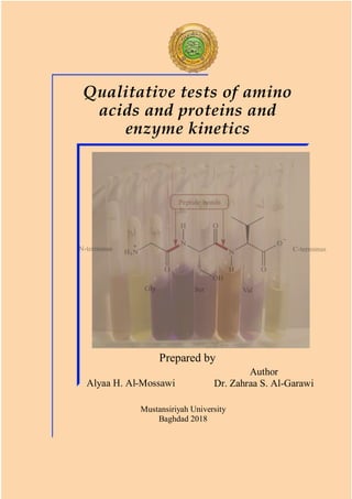
  • 1. Alyaa H. Al-Mossawi Author Dr. Zahraa S. Al-Garawi Prepared by Mustansiriyah University Baghdad 2018 Qualitative tests of amino acids and proteins and enzyme kinetics
  • 2. Qualitative Tests of Amino Acids and Proteins, and Enzyme Kinetics ‫إ‬ ‫عداد‬ ‫هاشم‬ ‫علياء‬ ‫الموسوي‬ ‫حياتية‬ ‫كيمياء‬ .‫م‬ ‫وتاليف‬ ‫إعداد‬ ‫الكرعاوي‬ ‫سالم‬ ‫زهراء‬ .‫د‬ ‫حياتية‬ ‫كيمياء‬ .‫م‬ .‫أ‬ z.mosin@uomustansiriyah.edu.iq ‫المستنصرية‬ ‫الجامعة‬
  • 3. ‫مقابل‬ ‫فلسطين‬ ‫شارع‬ ،‫واالستنساخ‬ ‫للطباعة‬ ‫حسنين‬ ‫مكتبة‬ ‫في‬ ‫المؤلف‬ ‫هذا‬ ‫طبع‬ ‫تم‬ .‫المستنصرية‬ ‫الجامعة‬ ‫الهاتف‬ ‫رقم‬ 07707643338 ‫البريد‬ ‫االلكتروني‬ doublem985@gmail.com ‫او‬ ‫والحذف‬ ‫التعديل‬ ‫يجوز‬ ‫وال‬ ‫والناشر‬ ‫للمؤلفة‬ ‫محفوظة‬ ‫والنشر‬ ‫التأليف‬ ‫حقوق‬ .‫واالستالل‬ ‫اإلضافة‬ ‫شباط‬ ‫في‬ ‫االولى‬ ‫الطبعة‬ ٢٠١٨ ‫ايلو‬ ‫في‬ ‫ّث‬‫د‬ُ‫ح‬ ‫ل‬ ٢٠١٨ ‫ب‬ ‫غداد‬
  • 4. ‫وتقديـــــــــــر‬ ‫شكــــــــــر‬ ‫الى‬ ‫واالمتنان‬ ‫بالشكر‬ ‫نتقدم‬ ‫قسم‬ ‫في‬ ‫األعزاء‬ ‫األساتذة‬ ‫الكيمياء‬ ، ‫المستنصرية‬ ‫الجامعة‬ ‫سموم‬ ‫فالح‬ .‫د‬.‫م‬.‫أ‬ ‫الفرطوسي‬ ‫(رئيس‬ )‫حياتية‬ ‫الكيمياء‬ ‫فرع‬ ‫ح‬ .‫د‬.‫م‬.‫أ‬ ‫و‬ ‫سنين‬ ّ‫ي‬‫الق‬ ‫المقترحات‬ ‫وتقديم‬ ‫والمساندة‬ ‫للتأييد‬ ،‫البيرماني‬ ‫كامل‬ ‫مة‬ ‫هذا‬ ‫إلنجاز‬ ‫ال‬ ‫المؤلف‬ ‫منهجي‬ .
  • 5. Contents Subject Page no. Preface 6 Qualitative tests of amino acids Amino acids 8 1. Solubility test 12 2. Ninhydrin Reaction 14 3. Xanthoproteic Reaction 18 4. Millon`s Reaction 21 5. Acree-Rosenheim Reaction 24 6. Pauly recation 27 7. Lead Sulfide Test 30 8. Nitroprusside test 33 9. Sakaguchi` s Test 35 Qualitative tests of proteins 1. Biuret test 38 2. Precipitation reactions 39 a) Denaturation of proteins 43 b) Precipitation by heavy metals 44 c) Precipitation by organic acids 45
  • 6. d) Precipitation by neutral salts 46 e) Precipitation by alcohol 50 f) Heller` s test 51 g) Precipitation by Esbach` s reactions 52 Enzymes Enzymes and enzyme kinetics 55 1. Calculation of substrate concentration 59 2. Calculation of enzyme velocity 59 3. Determination of the type of inhibition 63 References 67
  • 7. Preface This handbook is for the 3rd year students of the Chemistry Department at Mustansiriyah University. It has the syllabus for the second course of the Biochemistry lab. The handbook contains some qualitative tests for detecting the amino acids and proteins, as well as, the enzyme kinetic experiments, which are currently available in our lab. The qualitative tests are commonly used to detect different types of amino acids and proteins either separately or within a mixture. The last part of this handbook contains the enzyme kinetics experiments, which are used to determine the kinetic parameters of the
  • 8. 7 enzyme alkaline phosphatase in the presence and absence of inhibitor. The test methods in this handbook have been qualified as standard methods using valid reagents. Some methods here employ toxic or very acidic solutions which need to be handled with care. Safety protectors such as eye wears, gloves, lab coats are important for students and lab staff to do each experiment. Using this handbook by the students is necessary to follow the experiments, to complete the homework and to study for the exams. Dr. Zahraa https://uomustansiriyah.edu.iq/e-learn/profile.php?id=4801
  • 9. 8 Qualitative tests of amino acids Amino acids An amino acid (R-CH-NH2COOH) contains an amino group and a carboxylic acid group as side chains. It is the building block of all proteins and is linked with other amino acids as a chain by the peptide bonds (CONH-) to form the primary structure of a protein, see Figure 1. Fig 1. Structure of amino acids. The right-hand side figure shows the peptide bond. Carbon
  • 10. 9 There are 20 naturally occurring amino acids,
  • 11. 10 which vary from one to another according to the type of their side chains, see Figure 2. Amino acids are amphoteric with an extremely high melting point (usually exceeding 200°C), react as amines in some reactions, but as carboxylic acids in others. At a certain pH, the number of the positive charges (from protonated ammonium groups) are equal to the number of the negative charges (from deprotonated carboxylate groups), therefore, no net charge will be observed. This point is named as “the isoelectric point” and the amino acid will exist as zwitter ion. Herein, the Isoelectric point (Ip) is the pH value at which the concentration of anionic and cationic groups is equal (i.e. the net charge of this molecule equal to zero). It is the point where the molecule does not move to either the cathode or the anode when it is put in an electric field.
  • 12. 11 Because its solubility will be minimum, it is possible to precipitate at this point. Amino acids are responded to all typical reactions associated with compounds containing carboxylic acid and amino groups, usually under conditions where the zwitter ions form is present in only small quantities. Amino acids are chiral molecules (except glycine) and exhibit an optical activity due to the presence of an asymmetric α-carbon atom. Amino acids with an L-configuration are present in all naturally occurring proteins, whereas those with D-form are found in antibiotics and in bacterial cell walls. Amino acids are classified into groups according to their side chains, see Figure 2.
  • 13. 12 The amino acids tests are: 1. Solubility test The solubility of amino acids largely depends on the pH of the solution. Any change in the pH value causes structural changes in the amino acids and alters the relative solubility of the molecule. In acidic solutions, both amino and carboxylic groups are protonated, whereas, in basic solutions, both groups are deprotonation.
  • 14. 13 Materials Different amino acid solution: Gly, Lys and Glu. Solvents: H2O, HCl, NaOH and Chloroform. Method Mix a small amount of each amino acid solution with few mls of each solvent, record the results. Questions? 1. What is the best solvent of amino acids? Why? 2. Can you dissolve Gly in water? 3. Can Lys be dissolved in NaOH? 4. What is the difference you can see when dissolve Lys and Glu in chloroform? 5. Do most amino acids dissolve in water?
  • 15. 14 2. Ninhydrin reaction Ninhydrin [tri-ketohydrindene hydrate] is a powerful oxidizing agent used for detecting ammonia or primary and secondary amines. In a range of pH around 4-8, all α-L-amino acids react with ninhydrin to give a purple colored product (diketohydrin). Ninhydrin Fig 3. The ninhydrin coupling reaction with amino acids and with Pro.
  • 16. 15 interacts with amino acids causes decarboxylation and deamination (liberation of CO2 and ammonia) to produce the purple Rhuemann, s compound and aldehyde, see Figure 3. The amino acids Pro and hydroxyl Pro react with ninhydrine, but they give a yellow colored complex instead of a purple one. Primary amines and ammonia react similarly, but without the liberation of CO2. Different molecules such as peptides and proteins are also able to positively react with ninhydrin test. Mechanically, one reduced ninhydrin molecule condenses with another non-reduced ninhydrin molecule and ammonia to form a violet –blue or purple condensation product, see Figure 4.
  • 17. 16 Caution Ninhydrin is a strong oxidizing agent; therefore, it should be handled with care. Apart from that, eyewear, gloves and hood are required. If accidentally get in touch with skin, extremely wash with water. However, the resulting stains are temporary and will be eliminated within 24 hours. Fig 4. The mechanism of ninhydrin coupling reaction with amino acids.
  • 18. 17 Method 1. Add 1ml of each of the test solutions to 2 drops of ninhydrin solution. 2. Boil the mixture over a water bath for 2 min. 3. Allow to cool and observe the resulted blue color. Questions? 1. Which of the following gives a positive ninhydrin test? Phospholipid, sucrose, galactose, Pro, Albumin. 2. What is ninhydrin reaction? How it is useful? 3. How dose methylamine (CH3NH2) reacts with ninhydrin test? 4. What kinds of protection you should use when doing ninhydrin experiment? 5. Why Pro and hydroxyl Pro give yellow color with ninhydrin?
  • 19. 18 3. Xanthoproteic reaction This test is used to differentiate between aromatic amino acids. Treating aromatic amino acids with concentrated nitric acid leads to the nitration of the aromatic ring and formation of a yellow nitro-product (nitro derivatives), see Figure 5. When a strongly basic solution is added, the color of the product turns to be darker (from Fig 5. The Xanthoproteic reaction with aromatic amino acids.
  • 20. 19 yellow to orange) due to the ionization of the petrolic group. Caution Concentrated HNO3 is a toxic and corrosive substance that can cause severe burns and discolor the skin. Gloves, safety glasses and a fume hood are required. Avoid inhaling vapors and ingesting the compound. Method 1. Add 1m of concentrated HNO3 to 1ml of the test sample. 2. Heat the mixture and cool it. 3. Slowly add (40% w/v in a water solution) NaOH until the mixture becomes alkaline (use litmus strip to indicate the pH) and color- change is noted. Changing the color from yellow to orange indicates the presence of an aromatic amino acid.
  • 21. 20 Questions? 1. Which of the following gives positive test with Xanthoprotic reaction? Trp, Cys, Ala, Gly, Tyr, Thr 2. What is HNO3 doing for the reaction? 3. what is the color of a positive reaction with Xanthoprotic? 4. Phe does not react with Xanthoprotic, why?
  • 22. 21 4. Millon`s reaction The test was developed by the French chemist Auguste Million. Million`s reagent is a mixture of mercuric nitrate/nitric acid and water. This test is specific for Tyr, the only amino acid contains a phenol group (a hydroxyl group attached to a benzene ring). However, all phenols give positive results with Million`s test. The reaction occurs in 2 steps: first, the phenolic group of Tyr nitrates by HNO3, then in the presence of Hg(NO3)2, the nitrated Tyr forms a reddish-brown solution or a yellow precipitate of a nitrated Tyr, which is a positive test, Figure 6.
  • 23. 22 Method 1. To 1ml of amino acid solution, add 1ml of Million` s reagent in a test tube 2. Warm the tube in a boiling water bath for 10 min. 3. A reddish-brown color is a positive reaction. Fig 6. The Millon reaction with a phenolic amino acid.
  • 24. 23 Questions? 1. Why Millon`s reagent gives a reddish color with phenolic amino acids? 2. Which test is more specific for Tyr? Millon`s or Xanthoprotic test? Why? 3. If you increase the concentration of Tyr, will that increase the depth of the product? 4. What Hg is doing for the reaction? 5. What is Millon reagent?
  • 25. 24 5. Acree-Rosenheim reaction Acree-Rosenheim is another test for detecting Trp in a protein solution. Tryptophan is mixed with formaldehyde (CH2O). Concentrated sulfuric acid is added to form two layers. A violet zone in the junction point of two layers is the positive result, see Figure 7. Similar result obtained by the Hopkins-Cole reaction which is frequently used to detect Trp in a protein solution. In the Hopkins-Cole reaction, when a glyoxylic acid CHOCOOH is added to a protein solution (containing Trp) in the presence of H2SO4, a violet ring will form. + H2-C=O → a violet ring H2SO4 Conc. Fig 7. Acree-Rosenheim reaction with Trp.
  • 26. 25 Method (Acree-Rosenheim Reaction) 1. Add 1 ml of amino solution to 1ml of CH2O. 2. Concentrated H2SO4 (1 ml) is then added to form two layers. 3. Watch the development of a purple colored ring between the two layers as positive test for Trp appearance. Questions? 1. What is Acree-Rosenheim reagent? 2. What is the difference between Acree- Rosenheim and Hopkins-Cole reaction? 3. If the protein solution contains a mixture of Trp, phe and Tye, which amino acids form the purple ring more quickly?
  • 27. 26 4. How would the phenolic amino acid be distinguished by Acree-Rosenheim reaction? 5. If you have Phe, which of the following test is the best to detect? Ninhydrine, Million, Xanthoproteic, Hopkins-Cole?
  • 28. 27 6. Pauly reaction Pauly reaction was firstly described by the German chemist Hermann Pauly. It is used to detect Tyr or His amino acids by a coupling reaction with diazotized sulfanilic acid under Diazonium compound Sulfanilic acid Tyrosine Fig 8. Pauly reaction with Tyr.
  • 29. 28 alkaline conditions. As a result, a red colored solution is produced. Principally, diazotized sulfanilic acid is resulted by a reaction between the sulfanilic acid and sodium nitrite and sodium carbonate to form the diazonium component. Then, the diazonium components react with the imidazole ring of His or phenolic group of Tyr to form a dark red compound, see Figure 8. Method 1. Cool down a test tube using a cool icebox. 2. Add 1ml of sulfanilic acid to the cool test tube, remain in the icebox. 3. Add to the mixture 1ml of NaNO3, let it cool for 3min. 4. Add 2ml of Na CO3. 5. Observe the color.
  • 30. 29 Questions? 1. How does the Pauly test work? 2. Which amino acid gives a positive product with Pauly reagent? and which gives negative product? 3. How does His react with Pauly?
  • 31. 30 7. Lead sulfide test This is specific for sulfhydryl group (-SH). Sulphur containing amino acids, such as Cys and Cyst is converted into sodium sulfide (Na2S) when boiled with 40 % NaOH. The reaction depends on a partial conversion of the organic sulfur into inorganic sulfide Na2S, which can be detected using sodium plumbate (Na2pb(OH)4: lead acetate solution Pb(CH3COO)2 in an alkaline media). Na2S precipitates as a black lead sulfide, see Figure 9. Methionine or any other Meth containing- proteins such as Casein and Gelatin, gives a negative result with this test. Protien-SH + 2 NaOH → Na2S 40% Na-Sulfide Na2S + Pb (CH3COO)2 → PbS + 2 CH3COONa NaOH Fig 9. Lead sulphide reaction with sulphur-containing amino acids.
  • 32. 31 Method 1. Prepare sodium plumbate (Na2Pb(OH)4 solution as following: Add 5ml of 40% NaOH to 2ml of dilute Pb(CH3COO)2. A white precipitate of Lead hydroxide Pb(OH)2 will be formed. 2. Boil solution (1) to completely dissolve Pb(OH)2 and form Na2Pb(OH)4. 3. Add few drops of 40% NaOH to 2ml of amino acid solution and boil the solution for 2min. 4. Cooldown solution (3) and add few drops of solution (2), then watch the development of a brown color or precipitate (PbS) as an indication of the (-SH) group. Questions? 1. If you have a mixture of Cys, Cyst, Meth, Gly and Gln, do you expect the reaction will be positive with Lead sulfide test?
  • 33. 32 2. Why do you use sodium plumbate in lead sulfide teat? 3. How can you know that the Lead sulfide test is positive? 4. Can you use this test for most of amino acids? 5. How the Lead acetate test works?
  • 34. 33 8. Nitroprusside test This test is specific for Cys which has a free sulfhydryl group (-SH) that is able to react with the nitroprusside in the presence of excess ammonia (NH4OH). Principally, the free thiol group of Cys gives a red color with sodium nitroprusside in the presence of NH4OH, see Figure 10. To compare: do the test to Cys, Cystaein and Meth. Nitroprusside test is commonly used to detect the ketones in urine. Na2(CN)5Fe (NO)2 H2O + HS-CH2CHNH2COOH → Red complex Na-Nitroprosside Cys NH4OH Fig 10. Na-nitroprusside reaction with Cys amino acid.
  • 35. 34 Method 1. Add 0.5ml Na-nitroprusside solution to 2ml of amino acid solution and shack well. 2. Add 0.5ml NH4OH and watch the change in the color. Questions? 1. Do you think there is a difference between Lead acetate and Nitroprosside test? What is it?
  • 36. 35 9. Sakaguchi` test Sakaguchi` s test is specific for the guanidine group-containing amino acids, such as Arg. Guanidine group interacts with α-naphthol and alkaline hypobromite to produce a red- colored complex. Method 1. To 1ml of amino acid solution, add 2 ml of 40% NaOH and mix well. 2. Add 2ml of α-naphthol solution and mix well. 3. Add 2 drops of sodium hypobromite (BrNaO) solution, mix well and record the results (a red colored complex).
  • 37. 36 Questions? 1. What is the principle of Sakaguchi` test? 2. Why is this test specific for Arg? 3. What is the alkaline medium that you need for this test? 4. What is the positive result of this test? 5. What the will be result if used tertiary amine with Sakaguchi reagents?
  • 39. 38 Proteins Proteins are polypeptides consist of many amino acids bound together via peptide bonds. Amino acids interact with each other via different forces to build up the final three- dimensional (3D) structure of the protein. These forces are responsible for the stability and rigidity of the protein, which are: van der Waals force between temporary dipoles, ionic interactions between charged groups, salt-bridges and polar-polar interactions. Proteins regulate a variety of activities in all the known living organisms, ranging from the replication of a genetic code to transporting oxygen. Proteins exist as a 3D
  • 40. 39 tertiary and quaternary substance. The functional properties depend on the 3D structure, which arises when particular sequences of amino acids in a polypeptide chain fold to generate linear chains have compact domains with specific structures. These folded domains either serve as modules for larger polypeptide assemblies, or they support specific catalytic or binding sites for other functional substances or metal ions. Here are the main tests for detecting the presence of a protein in a solution: 1. Biuret test Biuret test is used to indicate the presence of peptide bonds within the protein molecule. This test could be useful to quantitatively know the concentration of the protein, where the intensity of the product`s color increases
  • 41. 40 linearly with the concentration of the protein. In addition, according to the Beer-Lambert law, the optical density at 540 nm wavelength is directly proportionated to the protein concentration.
  • 42. 41 Biuret reagent (H2N-CO-NH-CO-NH2) results from a coupling reaction of two urea molecules. It reduces Cu(II) to Cu (I) and Fig 11. a) Reaction of Biuret with Cu(II), b) interaction of Biuret reagent with the peptide bond. a b
  • 43. 42 form a red colored Cu-complex as a positive result for C=O-NH bond involvement, see Figure 11a. Because Biuret has a bond (C=O-NH) similar to the peptide bond, it is the most useful test for samples with two peptide bonds and above, see Figure 11b. Buffers, such as Tris and ammonia interfere with this assay. Method 1. Add 1 ml of 40% NaOH to 1 ml protein sample to make it alkaline. 2. Add 5 ml Biuret reagent to solution (1) 3. Watch changing the color within 5 min.
  • 44. 43 Questions? 1. If you let your sample to stand 20 min with Biuret Reagent, what will happen? 2. What is the component of Biuret reagent? 3. What is the principle of Biuret reaction? 4. Which of the following do you expect will give aa positive result with Biuret? Glucose? Casein? Glu? Phospholipid? 5. Why the reagent called Biuret?.
  • 45. 44 2. Precipitation reactions a. Denaturation of proteins Denaturation is an irrevocable change in the protein structure lead to loss of its biological properties. Because the fold type of the protein ( -sheet, -helix, random coil) determines its function, any change of the tertiary structure will alter its activity. Temperature and pH are the main causes of denaturation and precipitation of proteins because they destroy the weak bonds and affect the tertiary and secondary, but not the primary structure. Method 1. In a water bath, boil 1ml protein of different protein solutions such as Bovine albumin and egg globulin. 2. Record your results
  • 46. 45 b. Precipitation by heavy metals At natural pH 7 or above, proteins are usually negatively charged. Adding positively charged metal ions will neutralize the charges of protein and lead to protein precipitation. Heavy metals such as Ag+ , Pb2+ , Hg2+ , etc form a complex with the alkaline proteins and then precipitate. NOTE: avoid very high pH to reduce the risk of metal hydroxides precipitation. Method 1. To 2 ml of different protein solutions (e.g., Bovine albumin, egg white, gelatin, casein), add few drops of: AgNO3, Lead acetate Pb(CH3COO)2.to each protein solution. 2. Observe the extent of precipitate in each experiment.
  • 47. 46 c. Precipitation by organic acids In acidic medium, proteins are positively charged. Adding acidic solutions such as picric acid and Trichloroacetic acid (TCA) will neutralize the protein charge and irreversibly form an insoluble solution due to their high content of negative charges. Method 1. To 2 ml of protein solution (albumin solution or egg white solution), add 5-8 drops of picric acid or TCA solution. 2. Note the precipitate in each protein tube. 3. Slowly add (1 ml) of 40% NaOH solution and record if increasing the pH will cause renaturation of the protein (dissolve the precipitate).
  • 48. 47 d. Precipitation by neutral salts To investigate the effect of salt concentrations on the protein solubility, a series of low concentrations of salt is added to a protein solution (salting in). low concentrations will increase the protein solubility. Salt molecules decrease the electrostatic energy between the protein molecules, which increase the solubility and stability of the protein structure. Increasing the salt concentration (salting out) will decrease the protein solubility at some points. The excess of salt ions (not bound to the protein) competes with protein in binding the solvent molecules. This decrease in salvation allows the proteins to aggregate and precipitate. Each protein can be reversibly precipitated at specific salt concentration.
  • 49. 48 This test is used to separate different proteins using high salt concentration solution (oversaturation solutions), which will precipitate the certain protein due to the effect of salt ions on the protein solubility. Proteins precipitate at different salt concentrations; therefore, a series of salt concentrations are required to test the precipitation ability of each protein. In salting out the experiment, you must take into account the following: • The type of the salt (ammonium sulfate (NH4)2SO4 is commonly used) • The molecular weight (Mwt) of the protein: the highest Mwt, the fastest to precipitate. • Proteins with high Mwt usually require the low concentration of salt to precipitate, and vice versa.
  • 50. 49 • It is a reversible reaction; the protein can be recovered by dissolving with H2O • Applied the salting out experiment on a mixture of protein Method (salting in) 1. To 2 ml of albumin solution (egg white) add (1ml) NaCl solution. 2. Add an equivalent amount of (NH4)2SO4 until you notice over saturation. 3. Repeat the experiment with other proteins such as globulin, mucin and lysozomes. Describe the difference in the solubility or precipitation ability. Method (salting out) 1. Add (1ml) globulin solution in (1ml) 0.1 M NaCl.
  • 51. 50 2. Add 1ml globulin solution to 1ml saturated (NH4)2SO4 and record your observation. 3. Add 1ml egg albumin to 1ml saturated (NH4)2SO4 and record your observation. Add excess amount of solid (NH4)2SO4 to indicate the oversaturation.
  • 52. 51 e. Denaturation by alcohol Some alcoholic reagents such as ethanol or propanol can disrupt the hydrogen bonding within the protein structure by intra hydrogen-bonding with the protein molecules. For sterilizing purposes, 70% alcohol is used to denature the protein of the bacteria because it effectively penetrates the bacterial cell wall. Whereas, 95% alcohol has no similar effect, probably because it coagulates the surface proteins as a crust, which would prevent the alcohol to penetrate into the cell wall. Method 1. Treat 2ml of a protein solution with a few drops of alcohol. 2. Notice the protein precipitation.
  • 53. 52 f. Denaturation by acids (Heller` s test) Nitric acid desaturates proteins and form a white precipitate. Method 1. Add 1ml HNO3 (conc). to 1ml of protein solution. 2. Note the protein precipitate formed.
  • 54. 53 g. Precipitation by Esbach` s reactions It is a mixture of dilute acids (picric acid, citric acid and water) used to quantitatively detects albumin in the urine. This reagent precipitates protein where a yellowish to brown color product is formed depending on the quantity of the protein. Method 1. Add 1ml Esbach reagent to 1ml of protein solution. 2. Note the color of protein precipitate. Questions? 1. What causes denaturation of protein? 2. If you heat the protein solution up to 70 C, will that affected the tertiary structure of the protein? The secondary structure?
  • 55. 54 3. Why is (NH4)2SO4 precipitated protein? 4. What is slating-out protein? Salting in protein? 5. Which of the following metals causes precipitation of the protein? Why? Hg, Al, Ni, Ca, Zn, Cu, Ag, Au, Pb, Na. 6. What is the difference between organic acid and normal acid in terms of precipitation the protein? Which one causes reversible precipitation? 7. Do you think that absolute ethanol is better than 70% ethanol to precipitate albumin? 8. What is Heller reagent? 9. Do you think that adding HNO3 will cause reversible denaturation? 10. what is the best test to distinguish Albumin? Globulin?
  • 57. 56 The Enzyme is a biological molecule with an active site to bind a substrate and accelerate a chemical reaction. Determination of enzyme kinetics The aim of this practice is to experimentally obtain data for the kinetics of an enzymatic reaction in the presence and absence of an inhibitor. This data is then will be used to produce Lineweaver-Burke plots in the presence and absence of an inhibitor to determine Km, Vmax and the type of inhibition. In this experiment, the alkaline phosphatase enzyme (ALP) catalyzes the hydrolyses of the substrate p-nitrophenyl phosphate (p- NPP) in alkaline medium to produce p- nitrophenol (p-NP) and phosphoric acid, as following
  • 58. 57 The product p-NP is yellow under the alkaline pH of the assay. Therefore, the rate of the enzyme-catalyzed reaction can be followed by recording the absorption readings at 405nm of the amount of product accumulated as a function of time (min). In the second part of the experiment, you will determine the type of inhibition when Na- pyrophosphate is added to the enzyme reaction mixture. p-nitrophenyl phosphatephosphate p-nitrophenol
  • 59. 58 Reagents Substrate: 0.02 M p-NPP (stock solution) Buffer: 0. L M-glycine/NaOH buffer pH 10.4 Enzyme: ALP (2 mg/ml in 0.1M- glycine/NaOH buffer pH 10.4) Inhibitor: 100 mM Na-pyrophosphate or 200 mM imidazole Method Using clean plastic cuvettes, set up reaction mixtures as instructed in Table 1. Set the spectrophotometer up using buffer as a blank (no enzyme and no substrate). Set the blank to zero at 405nm. Leave the blank inside the instrument for the rest of the experiment. Now start the enzyme assays. Do all the addition to the cuvette and leave the enzyme solution LAST. Cover the cuvette by a parafilm and mix by inversion. Remove the Tetra sodium pyrophosphate
  • 60. 59 parafilm, put the cuvette into the spectrophotometer and read the absorbance (Abs) at 1 min. intervals for 6 min. Repeat tube 1 but without adding the enzyme, can you record a rate for the chemical reaction? 1) Calculation of substrate concentration [S] Table 1. The reaction mixture of enzyme kinetics without inhibitor I. Tube no. Buffer (ml) Substrate (ml) 0.02 M p-NPP Na-PP (I) ml 100 mM H2O (ml) Enzyme solution(ml) LAST Blank 1.9 - - 1.3 - 1 1.9 1.0 - 0.2 0.1 2 1.9 0.8 - 0.4 0.1 3 1.9 0.6 - 0.6 0.1 4 1.9 0.4 - 0.8 0.1 5 1.9 0.2 - 1.0 0.1 6 1.9 0.1 - 1.1 0.1 Non- Enzyme rate 1.9 1.0 - 0.3 None
  • 61. 60 Calculate the substrate concentration [S] from the concentration of the stock solution of p-NPP (0.02M) and its dilution in each of the enzyme reaction mixtures (1-6) using the dilution low N1V1=N2V2. 2) Calculation of the enzyme velocity V0 The initial velocity V0 is the amount of the product per a unit of time (min.) and NOT ITS CONCENTRATION. Plot the absorbance reading (Abs) of each enzyme reaction against time and derive the V0 of the reaction from the slope as Abs /min. Convert each Abs/min to mol p-NP (the product/min) using the extinction coefficient
  • 62. 61 ( ) of p-NP (15 mM-1 cm-1 ) and the volume of the assay mixture (3.2 ml) for each tube, as following: Abs= * l * C (Beer-Lambert law) 𝐶 = 𝐴𝑏𝑠. 𝑚𝑖𝑛−1 15 𝑚𝑀−1 𝑐𝑚−1 ∗ 1 𝑐𝑚 𝐶 = 𝐴𝑏𝑠. 𝑚𝑖𝑛−1 1 15 ∗ 𝑚𝑚𝑜𝑙 𝑚𝐿 𝐶 = 𝐴𝑏𝑠. 𝑚𝑖𝑛−1 15 ∗ 𝑚𝑚𝑜𝑙 1000 3.2 𝐶 = 𝐴𝑏𝑠. 𝑚𝑖𝑛−1 15 ∗ 3.2 1000 𝑚𝑜𝑙 𝐶 = 𝐴𝑏𝑠. 𝑚𝑖𝑛−1 ∗ 0.21 𝑚𝑜𝑙
  • 63. 62 = ? 𝑚𝑜𝑙. 𝑚𝑖𝑛−1 (Dividing by 15 converts the Abs into concentration (mM) units; as a M solution is the gram Mwt (mol) in 1000 ml). The volume of the assay is 3.2 ml, the amount of product is determined by multiplying (Abs/min.15) by the fraction 3.2/1000, assuming that all [S] is dissociated to product. (i) V0 against [S] Plot V0 for each enzyme-reaction (μmol/min) against each [S] (mM) to obtain the V max. (ii) Lineweaver-Burke plot to determine Km and V max
  • 64. 63 Calculate the reciprocals of each of the values of V0 (1/V) and of [S] and plot 1/V0 against l/[S]. Draw the straight line of best fit. This line intercepts the l/V axis at l/Vmax and the 1/[S] axis at -l/Km as in the example below: 1 𝑉 = 1 𝑉𝑚𝑎𝑥 + 𝐾𝑚 𝑉𝑚𝑎𝑥 [𝑆] 3) Determination of the type of inhibition 1/Vmax -1/km 1/V 1/[S]
  • 65. 64 Repeat the experiment in the Table 1 using 0.2 ml of Na- pyrophosphate in each cuvette as instructed in Table 2. (* Note the volumes of water is differ than that in Table 1). Table 2. The reaction mixture of the enzyme kinetics with inhibitor I. Tube no. Buffer (ml) Substrate 0.02 M p- NPP Na- PP (I) ml 100 mM H2O (ml) Enzyme solution LAST Blank 1.9 - 0.2 1.1 - 1 1.9 1.0 0.2 - 0.1 2 1.9 0.8 0.2 0.2 0.1 3 1.9 0.6 0.2 0.4 0.1 4 1.9 0.4 0.2 0.6 0.1 5 1.9 0.2 0.2 0.8 0.1 6 1.9 1.0 0.2 0.9 0.1 (i) V0 against [S] in the presence of I
  • 66. 65 Plot V0 for each enzyme-reaction (μmol/min) against each [S] (mM) as used in a and i. (ii) Lineweaver-Burke plot in the presence of [I] Calculate the reciprocals of each of the values of V0 and of [S] as appeared in Table 3 and plot 1/V0 against l/[S] as you used for (ii). Draw the straight line of best fit and indicate the type of the inhibition in terms of Km and V max.
  • 67. 66 Table 3. Table of the reciprocals values of Vo and S in the presence and absence of inhibitor I [S] 1/[S] V ( mole/min) 1/V VI (inhibitor) 1/VI ? ? ? ? ? ? ? ? ? ? ? ? ? ? ? ? ? ? ? ? ? ? ? ? ? ? ? ? ? ? ? ? ? ? ? ? ? ? ? ? ? ? Questions? 1. Can you ignore the mixture of tube 1 for the non-enzymatic rate? Why? 2. From your plot, what do you conclude regarding the effect of increasing [S] on the velocity? 3. What are factors influencing the initial velocity Vo? 4. Is the effect of the inhibitor that you observed representative of
  • 68. 67 competitive? Non-competitive inhibition? Why? 5. What is the Ki of the inhibitor?
  • 69. 68 References 1- P.M.Swamy, (2008), Laboratory manual of Biotechnology.pastogi Publication, p.90 . ISBN 978-81-7133-918-1. 2- R.A.Joshi (2006), Question bank of Biochemistry. New age Iinternational, p.64. ISBN 978-81-224-1736-4. 3- Chatterjea, (2004), Textbook of Biochemistry for dental/nursing/Pharmacy students. Jaypee Broyhers Publishers, p.51. ISBN 978-81-8061-204-6. 4- Zellner; et al, (2005), Quantitative validation of different protein precipitation methods in proteome analysis of blood platelets, Electrophoresis, 26(12):2481-9. Doi: 10.1002/elps.200410262.PMID 158945623. 5- Harrison et al., (2003), Bioseparations Science and Engineering.Oxford University Press. New Youk, NY. 6- Shuler et al., (2001), Bioprocess engineering: Basic concepts (2nd Edition), Prentice Hall International.
  • 70. 69 7- Ladisch, (2001), Bio separations Engineering, John Wiley & Sons, Inc.New York, NY. 8- Lydersen, (1994), Bioprocess Engineering. John Wiley & Sons. New York,NK. 9- http://www.chem.boun.edu.tr/wp- content/uploads/2014/04/Chem-415- Experiment-2.pdf .
  • 71.
  • 72. Hotspot on the author Dr. Zahraa S. Al-Garawi is a prof assist in Biochemistry and a PhD in Biophysics, Structural biology and Nanomaterials from University of Sussex, UK. She previously has wrtten a practical handbook in Clinical Biochemistry for undergraduates, in two languages, in Arabic and English. Zahraa has published very high- quality works in her field. Recently, she published a scientific textbook about steps for understanding writing and publishing papers, for researcher and student” Zahraa and Aliaa collaborate in this handbook to provide the most trusted methods for amino acid and protein tests, and the enzyme kinetics. This handbook would be of good benefits and helpful for undergraduates, chemistry and biology department.