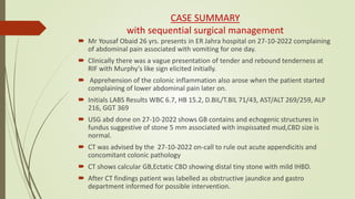
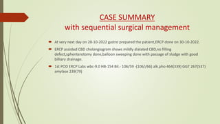
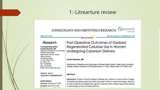
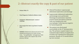
Post Operative Pyrexia/Fever
Morbidity Meeting
Derpartment of General Surgery Unit B2
Under Supervised by Dr Baky Hassan
Coordinator By Dr Kotab
Peer Support Dr.Omer Yousaf
Learning objectives
 Case summary.
 The story of this patient from a sequential process to reach the final diagnosis and surgical
management planning.
 We will discuss Post operative pyrexia its Definition ,Common and Un-common causes.
 How we digged up that probable aetiology and causative agent of this particular case.
 Today we will discuss an unexpected causative agent of postoperative fever, But it's a common
surgical adjunct frequently used in different surgical procedures.
 That surgical adjunct types/classification and mechanism of action.
 Literature review as referance.
 Last but not least we will conclude our discussion with an important take-home message.
CASE SUMMARY
with sequential surgical management
 Mr Yousaf Obaid 26 yrs. presents in ER Jahra hospital on 27-10-2022 complaining
of abdominal pain associated with vomiting for one day.
 Clinically there was a vague presentation of tender and rebound tenderness at
RIF with Murphy's like sign elicited initially.
 Apprehension of the colonic inflammation also arose when the patient started
complaining of lower abdominal pain later on.
 Initials LABS Results WBC 6.7, HB 15.2, D.BIL/T.BIL 71/43, AST/ALT 269/259, ALP
216, GGT 369
 USG abd done on 27-10-2022 shows GB contains and echogenic structures in
fundus suggestive of stone 5 mm associated with inspissated mud,CBD size is
normal.
 CT was advised by the 27-10-2022 on-call to rule out acute appendicitis and
concomitant colonic pathology
 CT shows calcular GB,Ectatic CBD showing distal tiny stone with mild IHBD.
 After CT findings patient was labelled as obstructive jaundice and gastro
department informed for possible intervention.
CASE SUMMARY
with sequential surgical management
 At very next day on 28-10-2022 gastro prepared the patient,ERCP done on 30-10-2022.
 ERCP assisted CBD cholangiogram shows mildly dialated CBD,no filling
defect,sphenterotomy done,balloon sweeping done with passage of sludge with good
billiary drainage.
 1st POD ERCP Labs wbc-9.0 HB-154 Bil.- 106/59 -(106//66) alk.pho 464(339) GGT 267(537)
amylase 239(79)
CASE SUMMARY
with sequential surgical management
 Now it was our turn,time for our intervention and that is laparoscopic cholecystectomy at
the same admission since the patient was well-optimised pre-operatively,we did it on the
7th of November 2022.
 Per-Operative the calot’s triangle easily achieved, dissection was difficult, since the GB was
intrahepatic. Raw are bleeding from GB bed.
 Monopolar diathermy coagulation done with spray mood followed by placement of
haemostatic agent (surgical snow) to have better haemostasis.
 The lap chole was declared complete and started to roll back the procedure e.g. deflation of
pneumoperitoneum and port removal was done, No Drain placed insitu.
CASE SUMMARY cont.
with sequential surgical management
 In routine laparoscopic surgery patients are discharged on the first day, but our
patient was unable to do so as he was suffering from severe fever e.g. 38 c or 104 F
,Pulse 86 BPM Which is got worse and worse in the next coming up post-operative
days.
 Our patient was young having now obvious co-morbidity e.g. Non HTNsive and
Non-DM, No Hx of allergy, No transfusion ,No Hx of anaesthesia related fever
(malignant hyperthermia), no past and recent sig medical hx and surgical hx.
Since Our patient was young having now
obvious co-morbidity e.g.
 Non HTNsive and Non-DM,
 No Hx of allergy,
 No transfusion ,
 No Hx of anaesthesia related fever
malignant hyperthermia,
 No past and recent sig medical hx
and surgical hx.
The following work-ups did not show
any significant results
 Septic screening
 CBC
 Blood culture
 Urine RE , Urine C/S
 Sputum c/s
 X-ray-chest
We evaluated postoperative fever; a helpful mnemonic is the "four Ws”
Work up for Post Operative Fever
Workup details
Since we didn’t find any clue following so we
assumed ??
Pathophysiology of fever
 Fever, or pyrexia, is the elevation of an individual's core body temperature above
a 'set-point' regulated by the body's thermoregulatory centre in the
hypothalamus.
 This increase in the body's 'set-point' temperature is often due to a physiological
process brought about by infectious causes or non-infectious causes such as
inflammation, malignancy, or autoimmune processes.
 These processes involve the release of immunological mediators, which trigger
the thermoregulatory centre of the hypothalamus, leading to an increase in the
body's core temperature.
definition & types of postoperative fever
cont.
 Pyrexia (fever) refers to a raised body temperature, typically greater than 37.5c. It
is common in surgical patients, either normal immediate post-operative response
or as feature of a specific post-operative complication.
 Whilst infection is regularly the suspected cause, other conditions must be
considered when approaching the surgical patient with pyrexia.
Fever occurs immediately after surgery or within
hours on postoperative days (POD) 0 or 1.
• Malignant hyperthermia: high-grade fever (greater than
40 C), occurs shortly after inhalational anesthetics or
muscle relaxant (e.g., halothane or succinylcholine), may
have a family history of death after anesthesia.
• Bacteremia: High-grade fever (greater than 40 C)
occurring 30 to 40 minutes after the beginning of the
procedure (e.g., Urinary tract instrumentation in the
presence of infected urine).
• Management includes blood cultures three times and
starting empiric antibiotics.
• Gas gangrene of the wound: High-grade fever
(greater than 40 C) occurring after gastrointestinal
(GI) surgery due to contamination with Clostridium
perfringens; severe wound pain; treat with surgical
debridement and antibiotics.
• Febrile non-hemolytic transfusion reaction: Fevers,
chills, and malaise 1 to 6 hours after surgery (without
hemolysis). Management: Stop transfusion (rule out
hemolytic transfusion reaction) and give antipyretics
(avoid aspirin in the thrombocytopenic patient).
Post Operative Pyrexia/Fever
 The most common cause of pyrexia in the post-operative patient is infection.
• Day 1-2 – consider a respiratory source (or body’s routine response to surgery)
• Day 3-5 – consider a respiratory or urinary tract source
• Day 5-7 – consider a surgical site infection or abscess/collection formation
• Any day post-operatively – consider infected IV lines or central lines as a source
 The investigation of the infection source should also be tailored to the patient.
 For example, in a patient who has undergone a bowel resection and
anastomosis, anastomotic leak is an important differential to be considered and
should be investigated as a matter of urgency.
Other Causes of Pyrexia
 Other causes of post-operative pyrexia include:
• Iatrogenic – which may include a drug-induced reaction (e.g. antibiotics or
anaesthetic agents) or from a transfusion reaction
• Venous thromboembolism – although rare, a PE or DVT can cause a low grade
fever without any other overt clinical features
• Secondary to prosthetic implantation – with any foreign body, for example after
an AAA repair, a low-grade fever may be evident
Investigations
 A septic screen is essential in investigating the surgical patient with pyrexia. In
most cases, the source is obvious and your screen can be tailored accordingly,
yet in a less clear presentation a wider screen is indicated; this can include:
• Blood tests – FBC, CRP, U&Es, LFTs, clotting
• Urine dipstick +/- urine MCS
• Cultures – blood, urine, sputum, and wound swab
• Imaging – Plain film chest radiograph, specific cross-sectional imaging
 If the source cannot be identified through the septic screen, more detailed
investigations may be required
Management
 Any identified infection should be treated empirically with antibiotics, pending
sensitivity results from any cultures taken. Empirical antibiotic regimes will vary
depending on local sensitivities and patient allergies, therefore following local
hospital guidance is advised.
 If no infectious cause can be identified, starting empirical antibiotics straight away
is not always essential. First look for non-infectious causes and consult a senior
colleague and a microbiologist for further advice.

Types of local heamstatic Agents
Classification by mechanism of action
Haemostatic materials are grouped into three categories based on
their mechanism of action:
 factor concentrators:- Factor concentrators work by absorbing
water from the blood and concentrating the blood components at
the injury site
 procoagulants:- Procoagulants take action through supplementing
coagulation factors and activating the blood coagulation cascade.
 mucoadhesive agents :- agents provide a physical barrier to blood
flow by cross-linking blood components
1:-Litrearture review
2:-Abstract exactly the copy & past of our patient
 Patient: Male, 51
 Final Diagnosis: Oxidised cellulose retain
 Symptoms: Abdominal pain • nausea •
vomiting
 Past surgical Hx: Had a Lap.Chole 25 moths
back
 WORKUP :Abdominal ultrasound and CT scan
showed the presence of a cystic circular
mass, “neo-gallbladder homogeneous fluid
content, close to the surgical clips of the
previous GB bed
 Clinical Procedure: Laparoscopic
abdominal exploration and drainage
 Laparoscopic abdominal exploration
and drainage were performed.
Histological examination reported
protein-based amorphous material
with rare lymphocytes and
macrophages. Culturing was negative
for bacterial growth
 The patient was discharged
uneventfully on the 4th postoperative
day. The primary surgical report was
evaluated with evidence of application
of Gelita-Cel® Standard for hemostatic
purposes. Results of 12-month follow-
up were normal.
2:-Abstract exactly the copy & paste of our patient
We reviewed the literature and retrieved a total of 28 papers comprising 38 cases of OC retention. The details of cases are shown in Table 2. Surgical sites included:
brain (6), cervical spine (1), thoracic spine (1), thorax (10), abdomen (12), and pelvis (8). Twenty-nine patients received Surgicel ®, 2 received Oxycel®, 1 received
Surgiflo® (Johnson and Johnson, Somerville, NJ, USA), and 6 received non-specified OC (Table 2). Here, we report the first case of complications due to Gelita-
Cel® retention.
Conclusions
 Oxidise cellulose should be removed when
haemostasis is obtained.
 Drain should place in situ in vicinity of place
haemostatic to drain the inflammatory fluid.
 If necessary, only a small quantity of OC should be
placed in situ and it should be documented in the
surgical report and the discharge document in
order to correctly inform the patient.
 Accurate surgical history evaluation should always
be performed
 multidisciplinary case evaluation between surgeons
and radiologists should be done to achieve a more
accurate diagnosis and prevent unnecessary revision
surgery or further medical and/or surgical
interventions.
 Since we raised this issue regarding complication
following the use surgicalsnow/Gelita-Cel®
 Our aim here is not to criticize the use of OC/surgical
snow as haemostatic agent – it has well-documented
effectiveness in surgery – but rather to provide some
practical suggestions on how to avoid the severe
complications reported in the literature.
That's it all about this morbidity meeting
Please Shares your Experience &
comments regarding Heamostatic
agents induced Post Operative
Fever

post operative fever.pptx.....................

  • 1.
    Post Operative Pyrexia/Fever MorbidityMeeting Derpartment of General Surgery Unit B2 Under Supervised by Dr Baky Hassan Coordinator By Dr Kotab Peer Support Dr.Omer Yousaf
  • 2.
    Learning objectives  Casesummary.  The story of this patient from a sequential process to reach the final diagnosis and surgical management planning.  We will discuss Post operative pyrexia its Definition ,Common and Un-common causes.  How we digged up that probable aetiology and causative agent of this particular case.  Today we will discuss an unexpected causative agent of postoperative fever, But it's a common surgical adjunct frequently used in different surgical procedures.  That surgical adjunct types/classification and mechanism of action.  Literature review as referance.  Last but not least we will conclude our discussion with an important take-home message.
  • 3.
    CASE SUMMARY with sequentialsurgical management  Mr Yousaf Obaid 26 yrs. presents in ER Jahra hospital on 27-10-2022 complaining of abdominal pain associated with vomiting for one day.  Clinically there was a vague presentation of tender and rebound tenderness at RIF with Murphy's like sign elicited initially.  Apprehension of the colonic inflammation also arose when the patient started complaining of lower abdominal pain later on.  Initials LABS Results WBC 6.7, HB 15.2, D.BIL/T.BIL 71/43, AST/ALT 269/259, ALP 216, GGT 369  USG abd done on 27-10-2022 shows GB contains and echogenic structures in fundus suggestive of stone 5 mm associated with inspissated mud,CBD size is normal.  CT was advised by the 27-10-2022 on-call to rule out acute appendicitis and concomitant colonic pathology  CT shows calcular GB,Ectatic CBD showing distal tiny stone with mild IHBD.  After CT findings patient was labelled as obstructive jaundice and gastro department informed for possible intervention.
  • 4.
    CASE SUMMARY with sequentialsurgical management  At very next day on 28-10-2022 gastro prepared the patient,ERCP done on 30-10-2022.  ERCP assisted CBD cholangiogram shows mildly dialated CBD,no filling defect,sphenterotomy done,balloon sweeping done with passage of sludge with good billiary drainage.  1st POD ERCP Labs wbc-9.0 HB-154 Bil.- 106/59 -(106//66) alk.pho 464(339) GGT 267(537) amylase 239(79)
  • 5.
    CASE SUMMARY with sequentialsurgical management  Now it was our turn,time for our intervention and that is laparoscopic cholecystectomy at the same admission since the patient was well-optimised pre-operatively,we did it on the 7th of November 2022.  Per-Operative the calot’s triangle easily achieved, dissection was difficult, since the GB was intrahepatic. Raw are bleeding from GB bed.  Monopolar diathermy coagulation done with spray mood followed by placement of haemostatic agent (surgical snow) to have better haemostasis.  The lap chole was declared complete and started to roll back the procedure e.g. deflation of pneumoperitoneum and port removal was done, No Drain placed insitu.
  • 6.
    CASE SUMMARY cont. withsequential surgical management  In routine laparoscopic surgery patients are discharged on the first day, but our patient was unable to do so as he was suffering from severe fever e.g. 38 c or 104 F ,Pulse 86 BPM Which is got worse and worse in the next coming up post-operative days.  Our patient was young having now obvious co-morbidity e.g. Non HTNsive and Non-DM, No Hx of allergy, No transfusion ,No Hx of anaesthesia related fever (malignant hyperthermia), no past and recent sig medical hx and surgical hx.
  • 7.
    Since Our patientwas young having now obvious co-morbidity e.g.  Non HTNsive and Non-DM,  No Hx of allergy,  No transfusion ,  No Hx of anaesthesia related fever malignant hyperthermia,  No past and recent sig medical hx and surgical hx. The following work-ups did not show any significant results  Septic screening  CBC  Blood culture  Urine RE , Urine C/S  Sputum c/s  X-ray-chest
  • 8.
    We evaluated postoperativefever; a helpful mnemonic is the "four Ws” Work up for Post Operative Fever
  • 9.
  • 10.
    Since we didn’tfind any clue following so we assumed ??
  • 11.
    Pathophysiology of fever Fever, or pyrexia, is the elevation of an individual's core body temperature above a 'set-point' regulated by the body's thermoregulatory centre in the hypothalamus.  This increase in the body's 'set-point' temperature is often due to a physiological process brought about by infectious causes or non-infectious causes such as inflammation, malignancy, or autoimmune processes.  These processes involve the release of immunological mediators, which trigger the thermoregulatory centre of the hypothalamus, leading to an increase in the body's core temperature.
  • 12.
    definition & typesof postoperative fever cont.  Pyrexia (fever) refers to a raised body temperature, typically greater than 37.5c. It is common in surgical patients, either normal immediate post-operative response or as feature of a specific post-operative complication.  Whilst infection is regularly the suspected cause, other conditions must be considered when approaching the surgical patient with pyrexia.
  • 13.
    Fever occurs immediatelyafter surgery or within hours on postoperative days (POD) 0 or 1. • Malignant hyperthermia: high-grade fever (greater than 40 C), occurs shortly after inhalational anesthetics or muscle relaxant (e.g., halothane or succinylcholine), may have a family history of death after anesthesia. • Bacteremia: High-grade fever (greater than 40 C) occurring 30 to 40 minutes after the beginning of the procedure (e.g., Urinary tract instrumentation in the presence of infected urine). • Management includes blood cultures three times and starting empiric antibiotics. • Gas gangrene of the wound: High-grade fever (greater than 40 C) occurring after gastrointestinal (GI) surgery due to contamination with Clostridium perfringens; severe wound pain; treat with surgical debridement and antibiotics. • Febrile non-hemolytic transfusion reaction: Fevers, chills, and malaise 1 to 6 hours after surgery (without hemolysis). Management: Stop transfusion (rule out hemolytic transfusion reaction) and give antipyretics (avoid aspirin in the thrombocytopenic patient).
  • 14.
    Post Operative Pyrexia/Fever The most common cause of pyrexia in the post-operative patient is infection. • Day 1-2 – consider a respiratory source (or body’s routine response to surgery) • Day 3-5 – consider a respiratory or urinary tract source • Day 5-7 – consider a surgical site infection or abscess/collection formation • Any day post-operatively – consider infected IV lines or central lines as a source  The investigation of the infection source should also be tailored to the patient.  For example, in a patient who has undergone a bowel resection and anastomosis, anastomotic leak is an important differential to be considered and should be investigated as a matter of urgency.
  • 15.
    Other Causes ofPyrexia  Other causes of post-operative pyrexia include: • Iatrogenic – which may include a drug-induced reaction (e.g. antibiotics or anaesthetic agents) or from a transfusion reaction • Venous thromboembolism – although rare, a PE or DVT can cause a low grade fever without any other overt clinical features • Secondary to prosthetic implantation – with any foreign body, for example after an AAA repair, a low-grade fever may be evident
  • 16.
    Investigations  A septicscreen is essential in investigating the surgical patient with pyrexia. In most cases, the source is obvious and your screen can be tailored accordingly, yet in a less clear presentation a wider screen is indicated; this can include: • Blood tests – FBC, CRP, U&Es, LFTs, clotting • Urine dipstick +/- urine MCS • Cultures – blood, urine, sputum, and wound swab • Imaging – Plain film chest radiograph, specific cross-sectional imaging  If the source cannot be identified through the septic screen, more detailed investigations may be required
  • 17.
    Management  Any identifiedinfection should be treated empirically with antibiotics, pending sensitivity results from any cultures taken. Empirical antibiotic regimes will vary depending on local sensitivities and patient allergies, therefore following local hospital guidance is advised.  If no infectious cause can be identified, starting empirical antibiotics straight away is not always essential. First look for non-infectious causes and consult a senior colleague and a microbiologist for further advice. 
  • 18.
    Types of localheamstatic Agents
  • 19.
    Classification by mechanismof action Haemostatic materials are grouped into three categories based on their mechanism of action:  factor concentrators:- Factor concentrators work by absorbing water from the blood and concentrating the blood components at the injury site  procoagulants:- Procoagulants take action through supplementing coagulation factors and activating the blood coagulation cascade.  mucoadhesive agents :- agents provide a physical barrier to blood flow by cross-linking blood components
  • 20.
  • 21.
    2:-Abstract exactly thecopy & past of our patient  Patient: Male, 51  Final Diagnosis: Oxidised cellulose retain  Symptoms: Abdominal pain • nausea • vomiting  Past surgical Hx: Had a Lap.Chole 25 moths back  WORKUP :Abdominal ultrasound and CT scan showed the presence of a cystic circular mass, “neo-gallbladder homogeneous fluid content, close to the surgical clips of the previous GB bed  Clinical Procedure: Laparoscopic abdominal exploration and drainage  Laparoscopic abdominal exploration and drainage were performed. Histological examination reported protein-based amorphous material with rare lymphocytes and macrophages. Culturing was negative for bacterial growth  The patient was discharged uneventfully on the 4th postoperative day. The primary surgical report was evaluated with evidence of application of Gelita-Cel® Standard for hemostatic purposes. Results of 12-month follow- up were normal.
  • 22.
    2:-Abstract exactly thecopy & paste of our patient
  • 23.
    We reviewed theliterature and retrieved a total of 28 papers comprising 38 cases of OC retention. The details of cases are shown in Table 2. Surgical sites included: brain (6), cervical spine (1), thoracic spine (1), thorax (10), abdomen (12), and pelvis (8). Twenty-nine patients received Surgicel ®, 2 received Oxycel®, 1 received Surgiflo® (Johnson and Johnson, Somerville, NJ, USA), and 6 received non-specified OC (Table 2). Here, we report the first case of complications due to Gelita- Cel® retention.
  • 25.
    Conclusions  Oxidise celluloseshould be removed when haemostasis is obtained.  Drain should place in situ in vicinity of place haemostatic to drain the inflammatory fluid.  If necessary, only a small quantity of OC should be placed in situ and it should be documented in the surgical report and the discharge document in order to correctly inform the patient.  Accurate surgical history evaluation should always be performed  multidisciplinary case evaluation between surgeons and radiologists should be done to achieve a more accurate diagnosis and prevent unnecessary revision surgery or further medical and/or surgical interventions.  Since we raised this issue regarding complication following the use surgicalsnow/Gelita-Cel®  Our aim here is not to criticize the use of OC/surgical snow as haemostatic agent – it has well-documented effectiveness in surgery – but rather to provide some practical suggestions on how to avoid the severe complications reported in the literature.
  • 26.
    That's it allabout this morbidity meeting Please Shares your Experience & comments regarding Heamostatic agents induced Post Operative Fever