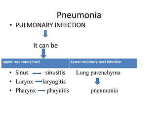
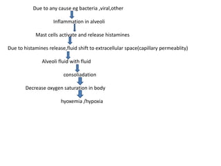
Pneumonia
ANY INFECTION IN LUNG
PARENCHYMA
Pneumonia
• PULMONARY INFECTION
It can be
• Sinus sinusitis Lung parenchyma
• Larynx laryngitis
• Pharynx phaynitis pneumonia
upper respiratory tract Lower resiratory tract infection
PATHOPHYSIOLOGY
• Occurs when local and systemic defense is weak
1. Local factors
• Loss or suppression of cough reflex coma ,anesthesia
,drug,chest pain
• Dysfunction of mucociliary apparatus-smoking ,inhalation of
toxic gases ,viral disease,genetic disorder
• Accumulation of thick secretion-cystic fibrosis
• Interferance with phagocytic function-alcohol tobacco
,smoking
• Pulmonary congestion and edema
2. Systemic factors
• Defect in innate immunity-neutrophils,macrophage,complement deficiency
• Defect in humoral (antibody)
• Defectin MY-88 gene-control toll like recepyors gene
• Defect in cell mediated immune response
3.Other reason
• Flu candems other infection supervenes
• Infection from other routes - hematogenous
• Transmission
• Most commanly transmitted by droplet spred fron close contact with source
• Incudtion period typically 1-3 days
Due to any cause eg bacteria ,viral,other
Inflammation in alveoli
Mast cells activate and release histamines
Due to histamines release,fluid shift to extracellular space(capillary permeablity)
Alveoli fluid with fluid
consoliadation
Decrease oxygen saturation in body
hyoxemia /hypoxia
Classification of Pneumonias
According to involment of lungs-
1. Lobar pneumonia-when entire lobe is affected
2. Bronchopneumonia-Patchy involved in lower lobes
According to clinical setting-
1. Community acquired pneumonia(CAP)
2. Hospital Acquird pneumonia (HAP)
According to microscpy-
1. Typical pneumonia
2. Atypical pneumonia
According to clinical setting-
Community acquired pneumonia-
Etiology- bacterial ,viral, fugal
Bacterial- streptoccus pneumonia/pneumococcous
-hemophillus influenzae
-moraxella catarrhalis
-staphylococcus aureus
-klebsiella pneumoniae
-pseudomonas aeruginosa
-mycoplasma pneumoniae
Risk factor –increased age,Chronic diseases,Diebetes
Viral pneumoniae-covid -19
-adenovirus
-rhinovirus
-varicella virusage
Risk factor-old age ,immunosuppressed,other chronic disease
Hospital acquired pneumonia-
Etiology-pseudomonas,acinetobactor
-Enterobacteriacae
-Staphaureus
According to involment of lungs-
1. Lobar pneumonia-diffuse inflammation affecting the part or the
entire lobe
-usally develops in healthy indviduals
-stages of pneumonia –
1.congestion-alveolar fluid with bacteria and neutrophils
2.red hepatization- rbc,wbc,fibrin,solid consistency of lung
3.gray hepatization-lysis of rbc,fibrin
4.resolution-neutrophils/macrophage
-comman caustive orgnisms-pneumococci,pnerumoniae
Bronchopneumonia-patchy area of acute suppurative infection of terminal bronchioles
-comman in infants and old age
-usally develop in infants and old age
-usally accompany pre exitisg diseases
pathological features – patchy consolidation,bronchi,bronchiolesand
adjacent alveolar spaces are filled with exudates rich in neutrophils
-comman causative organism-pseudomonas,haemophilusi nfluenza
According to microscopy
Typical pneumonia-
-involment of alveoli
-etiology-bacteria
-clinical feature- productive cough,sputum present
-ausculation-finding wheeze ,bronchial sound
-on chest x ray- consolidation(solidifiction)
-wbc count-increase
-treatment-antibiotics
Atypical pneumonia-
-involment of alveoli and interstitium
-etiology-viral,chlamydia,mycoplasma
-clinical features—small amount of sputu,
-ausculation-less finding wheeze ,bronchial sound
-on chest x ray-no finding
-wbc count- normal
-treatment-supportive treatment antiviral
Pneumonia causing microbes –
Virus-influenza
Bacteria-streptococcus pneumoniae hemophilus influenzae,staphylococcus aures,mycoplasma
pneumoniae,clamydophila pneumoniae,legionella pneumoniae
Fungi-coccidoidomycosis,histoplasmosis,blastomycosis,cryptococcus
Mycobacteria-mycobacterium tuberculosis
Preventing pneumonia-
-hand washing
-lowering indoor pollution
-adequate nutrition
Symptoms-
Diagnostic evaluation-
• sputumfor culture and sensitivity
•Chest x ray
•Bronchoscopy
•Abg analysis
•Complete blood count
•Ct scan
Medical management-
1. Oxygen therapy
2. Use of antibiotic therapy for treatment-
ampicillan,erythromycin
3.Bronchodilators –allow the airway to widan and
makesbreath easiear
4. Analgesics – use to reduce chest pain
5. Prevention of pneumonia by vaccination

Pneumonia presentation college level by pankaj

  • 1.
  • 2.
    Pneumonia • PULMONARY INFECTION Itcan be • Sinus sinusitis Lung parenchyma • Larynx laryngitis • Pharynx phaynitis pneumonia upper respiratory tract Lower resiratory tract infection
  • 3.
    PATHOPHYSIOLOGY • Occurs whenlocal and systemic defense is weak 1. Local factors • Loss or suppression of cough reflex coma ,anesthesia ,drug,chest pain • Dysfunction of mucociliary apparatus-smoking ,inhalation of toxic gases ,viral disease,genetic disorder • Accumulation of thick secretion-cystic fibrosis • Interferance with phagocytic function-alcohol tobacco ,smoking • Pulmonary congestion and edema
  • 4.
    2. Systemic factors •Defect in innate immunity-neutrophils,macrophage,complement deficiency • Defect in humoral (antibody) • Defectin MY-88 gene-control toll like recepyors gene • Defect in cell mediated immune response 3.Other reason • Flu candems other infection supervenes • Infection from other routes - hematogenous • Transmission • Most commanly transmitted by droplet spred fron close contact with source • Incudtion period typically 1-3 days
  • 5.
    Due to anycause eg bacteria ,viral,other Inflammation in alveoli Mast cells activate and release histamines Due to histamines release,fluid shift to extracellular space(capillary permeablity) Alveoli fluid with fluid consoliadation Decrease oxygen saturation in body hyoxemia /hypoxia
  • 6.
    Classification of Pneumonias Accordingto involment of lungs- 1. Lobar pneumonia-when entire lobe is affected 2. Bronchopneumonia-Patchy involved in lower lobes According to clinical setting- 1. Community acquired pneumonia(CAP) 2. Hospital Acquird pneumonia (HAP) According to microscpy- 1. Typical pneumonia 2. Atypical pneumonia
  • 7.
    According to clinicalsetting- Community acquired pneumonia- Etiology- bacterial ,viral, fugal Bacterial- streptoccus pneumonia/pneumococcous -hemophillus influenzae -moraxella catarrhalis -staphylococcus aureus -klebsiella pneumoniae -pseudomonas aeruginosa -mycoplasma pneumoniae Risk factor –increased age,Chronic diseases,Diebetes Viral pneumoniae-covid -19 -adenovirus -rhinovirus -varicella virusage Risk factor-old age ,immunosuppressed,other chronic disease
  • 8.
    Hospital acquired pneumonia- Etiology-pseudomonas,acinetobactor -Enterobacteriacae -Staphaureus Accordingto involment of lungs- 1. Lobar pneumonia-diffuse inflammation affecting the part or the entire lobe -usally develops in healthy indviduals -stages of pneumonia – 1.congestion-alveolar fluid with bacteria and neutrophils 2.red hepatization- rbc,wbc,fibrin,solid consistency of lung 3.gray hepatization-lysis of rbc,fibrin 4.resolution-neutrophils/macrophage -comman caustive orgnisms-pneumococci,pnerumoniae
  • 9.
    Bronchopneumonia-patchy area ofacute suppurative infection of terminal bronchioles -comman in infants and old age -usally develop in infants and old age -usally accompany pre exitisg diseases pathological features – patchy consolidation,bronchi,bronchiolesand adjacent alveolar spaces are filled with exudates rich in neutrophils -comman causative organism-pseudomonas,haemophilusi nfluenza
  • 10.
    According to microscopy Typicalpneumonia- -involment of alveoli -etiology-bacteria -clinical feature- productive cough,sputum present -ausculation-finding wheeze ,bronchial sound -on chest x ray- consolidation(solidifiction) -wbc count-increase -treatment-antibiotics Atypical pneumonia- -involment of alveoli and interstitium -etiology-viral,chlamydia,mycoplasma -clinical features—small amount of sputu, -ausculation-less finding wheeze ,bronchial sound -on chest x ray-no finding -wbc count- normal -treatment-supportive treatment antiviral
  • 11.
    Pneumonia causing microbes– Virus-influenza Bacteria-streptococcus pneumoniae hemophilus influenzae,staphylococcus aures,mycoplasma pneumoniae,clamydophila pneumoniae,legionella pneumoniae Fungi-coccidoidomycosis,histoplasmosis,blastomycosis,cryptococcus Mycobacteria-mycobacterium tuberculosis Preventing pneumonia- -hand washing -lowering indoor pollution -adequate nutrition
  • 12.
  • 13.
    Diagnostic evaluation- • sputumforculture and sensitivity •Chest x ray •Bronchoscopy •Abg analysis •Complete blood count •Ct scan
  • 14.
    Medical management- 1. Oxygentherapy 2. Use of antibiotic therapy for treatment- ampicillan,erythromycin 3.Bronchodilators –allow the airway to widan and makesbreath easiear 4. Analgesics – use to reduce chest pain 5. Prevention of pneumonia by vaccination