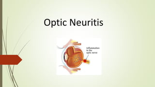
2. Definition: inflammation of the optic nerve
Most common cause: multiple sclerosis
Less common causes include parameningeal,
meningeal, or intraocular inflammation associated with
viral infections or postviral syndromes
(e.g., tuberculosis, syphilis, Lyme disease, viral infections)
Rare causes include toxins (eg, methanol, ethambutol),
neurosyphilis, and vitamin B12 deficiency.
3. Clinical Features
Vision impairment:
blurry vision, sudden vision loss, color
blindness, visual field defects (e.g., central
scotoma)
Retrobulbar pain (increased pain caused
by eye movements)
Unilateral impairment of visual acuity
occurs over hours to days, becoming
maximal within 2 weeks.
Visual loss is associated with headache,
globe tenderness, or eye pain in more than
90% of patients; the pain is typically
exacerbated by eye movement.
4. Visual field
testing
Visual field testing demonstrates a central scotoma (blind
spot) associated with decreased visual acuity.
Examination of the fundus shows unilateral disk swelling
when the nerve head is involved, but is normal when the
inflammatory process is posterior to the optic disk
(retrobulbar neuritis), as is most common in
demyelinating disease.
The pupils are equal in size but show less pronounced
constriction in response to illumination of the affected eye
(relative afferent pupillary defect; Marcus Gunn pupil).
Recovery of vision begins in a few weeks and may
progresses for a year. Normal vision returns in the
majority of cases.
5. Diagnostics
Swinging-flashlight test: relative afferent pupillary defect
Ophthalmoscopy
-Retrobulbar neuritis: normal ophthalmoscopic finding
-Papillitis: poorly defined papilla, hyperemia, hemorrhage at the border of
the papilla
Visual evoked potential (VEP)
-Assess conduction disorders of the optic nerve