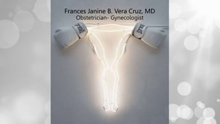
2. 1. PATHOLIGIC CASES
a. OB
a. Abortion
b. Ectopic Pregnancy
c. Molar pregnancy
b. Gynecologic
a. Pelvic inflammatory diseas
b. Cervical pathology
c. Ovarian New growth
d. Adenomyosis and Myoma
e. Pediatric
2.HOSPITAL EXPOSURE
3. HOSPITAL EXPOSURE
• Scrubs (with cap, mask and OR shoes)
• Stethoscope
• BP app
• Thermometer
• Tape measure
• Alcohol
• Snacks and water
4. VAGINITIS
• Normal physiologic vaginal discharge:
• cervical and vaginal epithelial cells, normal bacterial flora, water,
electrolytes, other chemicals
• pH 4.0
• Lactobacilli, S. epidermidis, E.coli, diphtheroids, streptococci
5. VAGINITIS
• Three common infections infections of the vagina are produced by:
• Fungus (candidiasis)
• Protozoon (trichomonas)
• Synergistic bacterial infection (bacterial vaginosis)
• Symptoms associated with vaginal infection:
• Vaginal discharge, superficial dyspareunia, dysuria, odor, vulvar
burning
6. BACTERIAL VAGINOSIS
• Risk factors
▫ New or multiple sexual partners
▫ Women who have sex with women
▫ Douching at least monthly or within the prior 7 days
▫ Social stressors
• Associated with
▫ Upper tract infections (endomyometritis, PID)
▫ Vaginal cuff cellulitis
▫ In pregnancy – preterm rupture of the membranes and endomyometritis;
7. BACTERIAL VAGINOSIS
Criteria: Amsel’s Clinical Criteria
• Homogenous vaginal discharge
• pH ≥ 4.5
• Amine-like odor when mixed with KOH (whiff test)
• Wet smear demonstrates clue cells greater in
number than 20% of the of vaginal epithelial cells
*** 3 out of 4 criteria is sufficient for diagnosis
8. Condition
Symptoms and
Signs[*]
Findings on
Examination[*] pH Wet Mount Comment
Bacterial
vaginosis[†]
Increased
discharge
(white, thin)
Thin, whitish gray
homogeneous
discharge,
sometimes frothy
>4.5 Clue cells (>20%)
shift in flora
Greatly
decreased
lactobacilli
Increased odor Amine odor after
adding potassium
hydroxide to wet
mount
Greatly
increased cocci,
bacilli small
curved rods
Candidiasis Increased
discharge
(white, thick)[‡]
Thick, curdy
discharge
<4.5 Hyphae or spores Can be mixed
infection with
bacterial
vaginosis, T.
vaginalis, or
both, and have
higher pH
Pruritus Vaginal erythema
Dysuria
Burning
Typical Features of Vaginitis
9. Trichomoniasi
s
Increased
discharge (yellow,
frothy)
Yellow, frothy
discharge with or
without vaginal or
cervical erythema
>4.5 Motile trichomonads More symptoms
at higher
vaginal pH
Increased odor Increased white
cells
Pruritus
Dysuria
Condition
Symptoms and
Signs[*]
Findings on
Examination[*] pH Wet Mount Comment
10.
11.
12.
13.
14.
15. ABORTION
- Loss of fetus less than 20 weeks age of gestation or a birthweight less
than 500g.
16. Basis for Diagnosis
• Amenorrhea
• Positive pregnancy test
• Vaginal bleeding
• Uterine contraction
18. CERVIX HISTORY
THREATENED closed Spotting or bleeding Can be accompanied by
uterine contractions, low
back pains.
INCOMPLETE open With passage of meaty material Palpable placental tissues
per OS
COMPLETE closed With passage of meaty material Uterus is small
INEVITABLE open Ruptured amniotic fluid Ruptured amniotic fluid as
visualized, or tested via
Ferning’s test, test for pH
(>7), TVS
MISSED closed Spotting or bleeding Uterus is small for age
SEPTIC any History of instrumentation or induced
abortion.
Accompanied by fever and signs of shock
Uterine tenderness
20. TYPES
TUBAL Fallopian tubes. Most common site is the ampullary area.
INTERSTITIAL/ CORNUAL
PREGNANCY
Within the interstitial portion of the FT
ABDOMINAL Primary – the 1st and only implantation occurs on a
peritoneal surface. Secondary – implantation originally in
the tubal ostia, subsequently aborted and then
reimplanted into the peritoneal surface
CERVICAL Cervical canal
LIGAMENTOUS A secondary form of EP in which a primary tubal
pregnancy erodes into the mesosalpinx and is located
between the leaves of the broad ligament
HETEROTOPIC A condition in which ectopic and intrauterine pregnancies
coexist
OVARIAN A condition in which an EP implants within the ovarian
cortex
21.
22. FINDINGS
• The uterus may be slightly enlarged and soft
• Uterine and cervical motion tenderness may suggest peritoneal
inflammation
• An adnexal mass may be palpated
• Uterine contents may be present in the vagina due to shedding
of endometrial lining stimulated by an ectopic pregnancy
23. MOLAR PREGNANCY
Hydatidiform moles (HM) are abnormal conceptions with excessive
placental, and little or no fetal, development. Grossly, a HM resembles
a bunch of grapes, with or without fetal components. It is subdivided
into:
1. complete hydatidiform mole (CHM)
2. partial hydatidiform mole (PHM)
• based on morphologic, cytogenetic, and clinicopathologic features.
24. AUGUST 29, 2017
MJ
Dr. Vera Cruz
25YO
G3P1(1011)
Patient was
amenorrheic for 18
weeks, with no
associated breast
tenderness, nausea or
vomiting. (+)
Pregnancy test
2 days PTA, patient noted vaginal
spotting, no other symptoms noted.
A few hours prior to admission,
patient consulted for pre-natal check-
up by which she was given an UTZ
request. UTZ revealed the possibility
of a molar pregnancy hence consult at
our institution.
Ultrasound done revealed Enlarged
Anteverted Uterus.
Endometrial Mass Consider
Gestational Trophoblastic Disease
Probably Hydatidiform
Mole
Theca Lutein Cysts.
GTD 18 1/7 weeks AOG
Pertinent PE
Abdominal exam:
Abdomen soft, nontender, no
scars
Internal exam:
Cervix closed, no cervical
motion tenderness, uterus
enlarged to 20 weeks size, no
adnexal mass/ tenderness, no
bleeding
Admit patient
For serum beta-hcg
For CBC, UA, blood chem,
PT/PTT, chest xray
For tvs-utz
For whole abdomen utz
Initial BHCG- 1,894,360.00
mIU/ml