Download to read offline
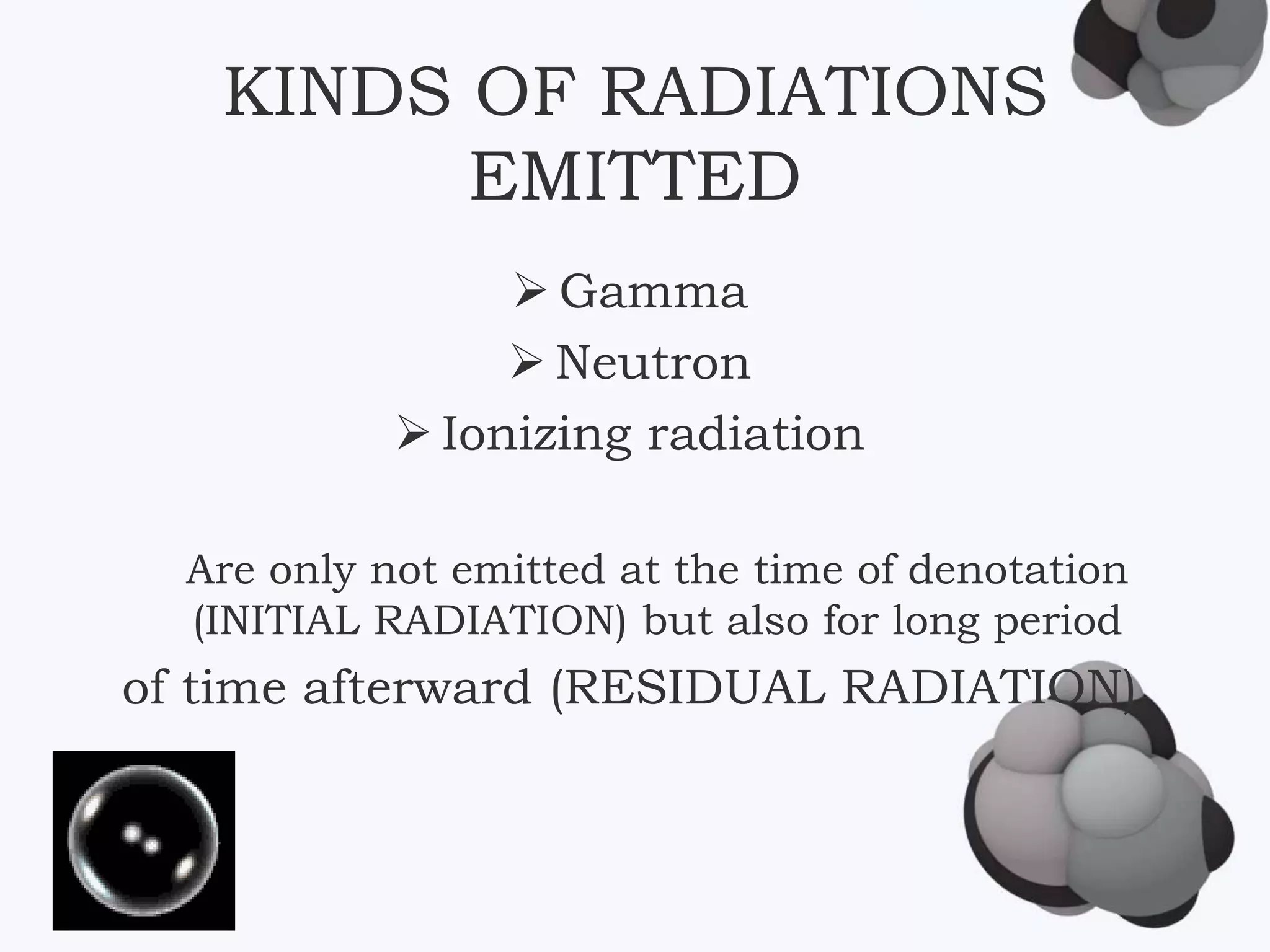
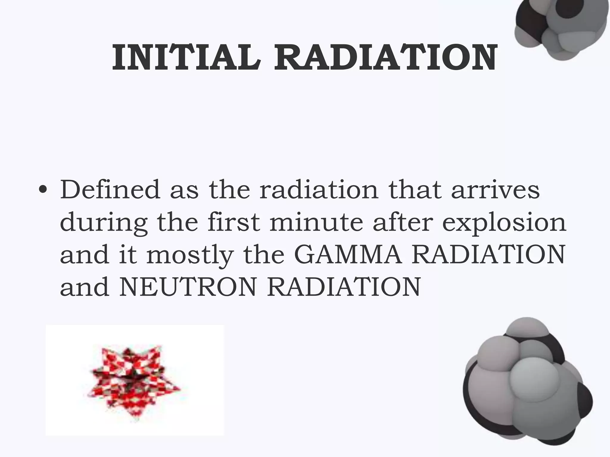


The document discusses various topics related to nuclear physics and radiation, including: 1) The different types of radiation emitted from nuclear explosions, including gamma, neutron, and ionizing radiation. Residual radiation comes from weapon debris, fission products, and radiated soil in the case of a ground burst. 2) The effects of radiation exposure on humans, including increased risk of cancer, hair loss, organ damage, and reduced immunity. Different organs are affected at different radiation dose levels. 3) Applications of radiation in medicine, including radiation therapy to treat cancer, nuclear medicine scans using radiotracers, and total body irradiation used before bone marrow transplants. 4) Risks of residual nuclear fall


