2. Photo courtesy of the NIDA Web site. From
A Slide Teaching Packet: The Brain and the
Actions of Cocaine, Opiates, and Marijuana.
3. Your Brain on DrugsYour Brain on Drugs
1-2 Min 3-4 5-6
6-7 7-8 8-9
9-10 10-20 20-30
Photo courtesy of Nora Volkow, Ph.D. Mapping cocaine
binding sites in human and baboon brain in vivo. Fowler JS,
Volkow ND, Wolf AP, Dewey SL, Schlyer DJ, Macgregor RIR,
Hitzemann R, Logan J, Bendreim B, Gatley ST. et al.
Synapse 1989;4(4):371-377.
4. Your Brain After DrugsYour Brain After Drugs
Normal
Cocaine Abuser (10
days)
Cocaine Abuser (100 days)
Photo courtesy of Nora Volkow, Ph.D. Volkow ND, Hitzemann R, Wang C-I, Fowler IS, Wolf AP, Dewey SL. Long-term frontal brain metabolic changes
in cocaine abusers. Synapse 11:184-190, 1992; Volkow ND, Fowler JS, Wang G-J, Hitzemann R, Logan J, Schlyer D, Dewey 5, Wolf AP. Decreased
dopamine D2 receptor availability is associated with reduced frontal metabolism in cocaine abusers. Synapse 14:169-177, 1993.
6. Di Chiara et al., Neuroscience, 1999.,Fiorino and Phillips, J. Neuroscience, 1997.
Natural Rewards Elevate
Dopamine Levels
0
50
100
150
200
0 60 120 180
Time (min)
%ofBasalDAOutput
NAc shell
Empty
Food Sex
Box Feeding
100
150
200
DAConcentration(%Baseline)
Sample
Number
1 2 3 4 5 6 7 8
Female Present
7. 0
100
200
300
400
500
600
700
800
900
1000
1100
0 1 2 3 4 5 hr
%ofBasalRelease
DA
DOPAC
HVA
Accumbens
Amphetamine
0
100
200
300
400
0 1 2 3 4 5 hr
%ofBasalRelease
DA
DOPAC
HVA
Accumbens
Cocaine
Time After Drug
Morphine
0
100
150
200
250
0 1 2 3 hr
Time After Drug
%ofBasalRelease
Accumbens
Caudate
Nicotine
Di Chiara and Imperato, PNAS, 1988
Effects of Drugs on Dopamine Release
%ofBasalRelease
0
100
150
200
250
0 1 2 3 4 5 hr
Accumbens
0.5
1.0
2.5
10
Dose
mg/kg
mg/kg
mg/kg
mg/kg
8. Control Addicted
Dopamine D2 Receptors are Decreased by Addiction
Functionally…
DAD2ReceptorAvailability
Cocaine
Alcohol
Heroin
Meth
9. The Memory of DrugsThe Memory of Drugs
Nature VideoNature Video Cocaine VideoCocaine Video
Front of BrainFront of Brain
Back of BrainBack of Brain
AmygdalaAmygdala
not lit upnot lit up
AmygdalaAmygdala
activatedactivated
Photo courtesy of Anna Rose Childress, Ph.D.
10. Rats Exposed to Nicotine in Adolescence
Self-Administer More Nicotine
Than Rats First Exposed as Adults
Collins et al, Neuropharmacology, 2004, Levin et al, Psychopharmacology, 2003
Editor's Notes
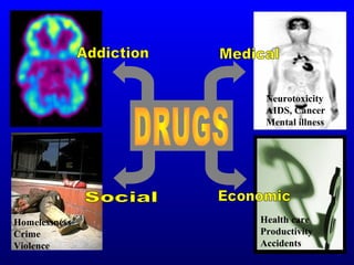
The effects of drug abuse are wide ranging and affect people of all ages . Besides addiction, drug abuse is linked to a variety of health problems, including HIV/AIDS, cancer, heart disease, and many more. It is also linked to homelessness, crime, and violence. Thus, addiction is costly to both individuals and society.
Slide 4: The brain is your body’s “Command Central” Your brain controls more than the way you think. The brain controls our physical sensations and body movements. How we understand what we see, hear, smell, taste, and touch. Our sense of balance and coordination. Memory. Feelings of pleasure and reward. The ability to make judgments. When we catch a football, dance, jog, speak, sing, laugh, whistle, smile, cry—that’s our brain receiving, processing, and sending out messages to different parts of our body. When we feel good for whatever reason—laughing with a friend or seeing a good movie or eating our favorite ice cream—the brain’s reward system is activated. As we said before, the reward system is the part of the brain that makes you feel good. The reward system is a collection of neurons that release dopamine, a neurotransmitter. When dopamine is released by these neurons, a person feels pleasure. Scientists have linked dopamine to most drugs of abuse—cocaine, marijuana, heroin, alcohol, and nicotine. These drugs all activate the reward system and cause neurons to release large damaging amounts of dopamine. Over time, drugs change this part of the brain. As a result, things that used to make you feel good—like eating ice cream, skateboarding, or getting a hug—no longer feel as good.
Slide 7: This is literally the brain on drugs When someone gets “high” on cocaine, where does the cocaine go in the brain? With the help of a radioactive tracer, this PET scan shows us a person’s brain on cocaine and the area of the brain, highlighted in yellow, where cocaine is “binding” or attaching itself. This PET scan shows us minute by minute, in a time-lapsed sequence, just how quickly cocaine begins affecting a particular area of the brain. We start in the upper left hand corner. You can see that 1 minute after cocaine is administered to this subject nothing much happens. All areas of the brain are functioning normally. But after 3 to 4 minutes [the next scan to the right], we see some areas starting to turn yellow. These areas are part of a brain structure called the striatum [stry-a-tum] that is the main target in the brain bound and activated by cocaine. At the 5- to 8-minute interval, we see that cocaine is affecting a large area of the brain. After that, the drug’s effects begin to wear off. At the 9- to 10-minute point, the high feeling is almost gone. Unless the abuser takes more cocaine, the experience is over in about 20 to 30 minutes. Scientists are doing research to find out if the striatum produces the “high” feeling and controls our feelings of pleasure and motivation. One of the reasons scientists are curious about specific areas of the brain affected by drugs, such as cocaine, is to develop treatments for people who become addicted to these drugs. Scientists hope to find the most effective way to change an addicted brain back to normal functioning.
Slide 8: Long-term effects of drug abuse The images in these PET scans, which depict brain glucose utilization (a marker of brain activity), show that once the brain becomes addicted to a drug like cocaine, it is affected for a long, long time. In other words, once addicted, the brain is literally changed. Let’s see how. In this slide, increasing amounts of brain function are measured by yellow or red. The top row shows a normal functioning brain without drugs. You can see a lot of brain activity. In other words, there is a lot of yellow and red color. The middle row shows a cocaine addict’s brain after 10 days without any cocaine use at all. What is happening here? [Pause for response.] Less yellow and red means less normal activity occurring in the brain—even after the cocaine abuser has abstained from the drug for 10 days. The third row shows the same addict’s brain after 100 days without any cocaine. We can see a little more yellow and red, so there is some improvement— more brain activity—at this point. But the addict’s brain is still not back to a normal level of functioning. . . more than 3 months later. Scientists are concerned that there may be areas in the brain that never fully recover from drug abuse and addiction.
Slide 9: Drugs have long-term consequences Here is another example of what science has shown us about the long-term effects of drugs. What this PET scan shows us is how just 10 days of drug use can produce very dramatic and long term changes in the brain activity of a monkey. The drug used in this experiment was amphetamine, or what some people call “speed.” Remember the previous slide showed us what the brain of a chronic cocaine abuser looks like. This slide shows us what using a drug like amphetamine can do, in only 10 days, to the brain of a monkey. This slide also gives us a better idea of what methamphetamine, a drug similar in structure, can do to the brain. Methamphetamine use is becoming increasingly popular in certain areas of the country. The top row shows us, in yellow and red, normal brain activity. The second row shows us that same brain 4 weeks after being given amphetamine for 10 days. There is a dramatic decrease in brain activity. This decreased brain activity continues for up to 1 year after amphetamine use. These continuous brain changes often trigger other changes in social and emotional behavior too, including a possible increase in aggressiveness, feelings of isolation, and depression.
Natural rewards stimulate dopamine neurotransmission. Eating something that you enjoy or being stimulated sexually can cause dopamine levels to increase. In these graphs, dopamine is being measured inside the brains of animals. Its increase is shown in response to food or sex cues. This basic mechanism of controlled dopamine release and reuptake has been carefully shaped and calibrated by evolution to reward normal activities critical for our survival.
Nearly all drugs of abuse increase dopamine neurotransmission. This slide shows the increase in brain dopamine (DA) levels (measured in animals) following exposure to various drugs of abuse. All of the drugs depicted in this slide have different mechanisms of action, however they all increase activity in the brain reward pathway by increasing dopamine neurotransmission. It is because drugs activate these brain regions usually more effectively and for longer periods of time than natural rewards that they have an inherent risk of being abused.
Repeated drug exposure also changes brain function. Positron emission tomography (PET) images show similar changes in brain dopamine receptors resulting from addiction to different substances. Dopamine D2 receptors are one of five types of receptors that bind dopamine in the brain. The brain images on the left are those of controls, while those on the right are from individuals addicted to cocaine, methamphetamine, alcohol, or heroin. The striatum (which contains the reward and motor circuitry) shows up as bright red and yellow in the controls (in the left column), indicating numerous D2 receptors. Conversely, the brains of addicted individuals (in the right column) show a less intense signal, indicating lower levels of D2 receptors. This reduction likely stems from repeated over-stimulation of the dopamine receptors. Brain adaptations such as this contribute to the compulsion to abuse drugs.
Slide 10: The memory of drugs This slide demonstrates something really amazing—how just the mention of items associated with drug use may cause an addict to crave or desire drugs. This PET scan is part of a scientific study that compared recovering addicts, who had stopped using cocaine, with people who had no history of cocaine use. The study hoped to determine what parts of the brain are activated when drugs are craved. For this study, brain scans were performed while subjects watched two videos. The first video, a nondrug presentation, showed nature images—mountains, rivers, animals, flowers, trees. The second video showed cocaine and drug paraphernalia, such as pipes, needles, matches, and other items familiar to addicts. This is how the memory of drugs works: The yellow area on the upper part of the second image is the amygdala (a-mig-duh-luh), a part of the brain’s limbic system, which is critical for memory and responsible for evoking emotions. For an addict, when a drug craving occurs, the amygdala becomes active and a craving for cocaine is triggered. So if it’s the middle of the night, raining, snowing, it doesn’t matter. This craving demands the drug immediately. Rational thoughts are dismissed by the uncontrollable desire for drugs. At this point, a basic change has occurred in the brain. The person is no longer in control. This changed brain makes it almost impossible for drug addicts to stay drug-free without professional help. Because addiction is a brain disease.
Animal studies show a difference in response to drugs of abuse in early-exposed “adolescents.” Rats first exposed to nicotine as adolescents self-administer nicotine more often and in higher total doses per session than rats first exposed as adults. Corresponding and persistent changes in the brain were also found in those animals that were exposed early to nicotine.