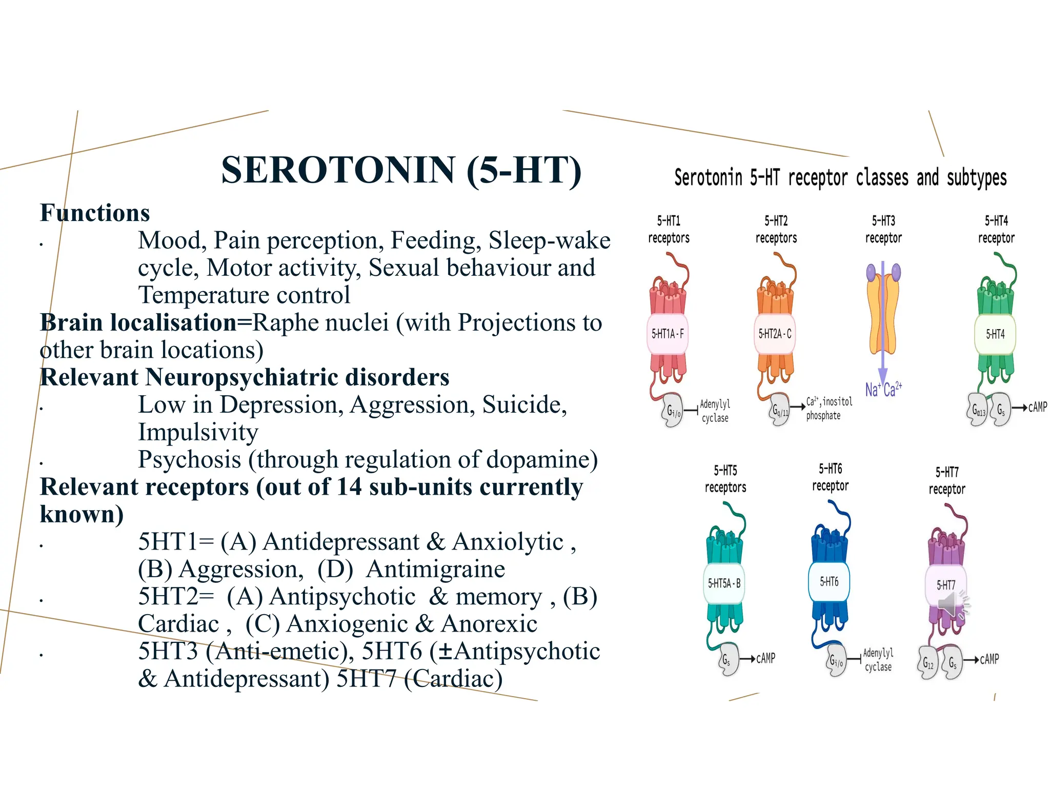
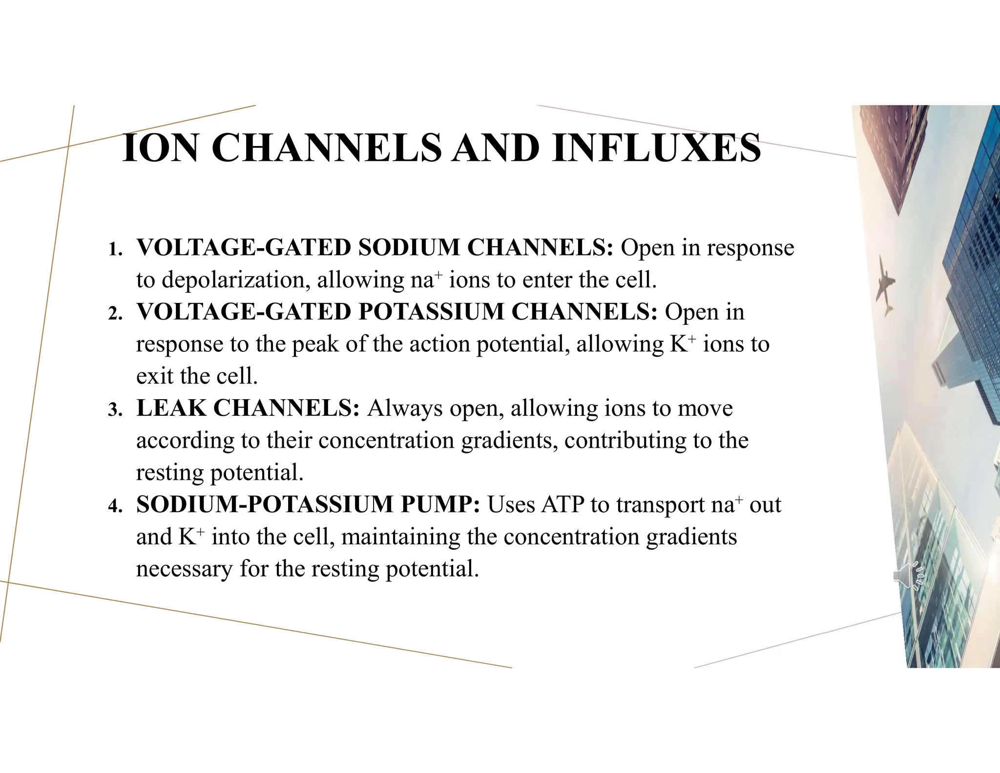
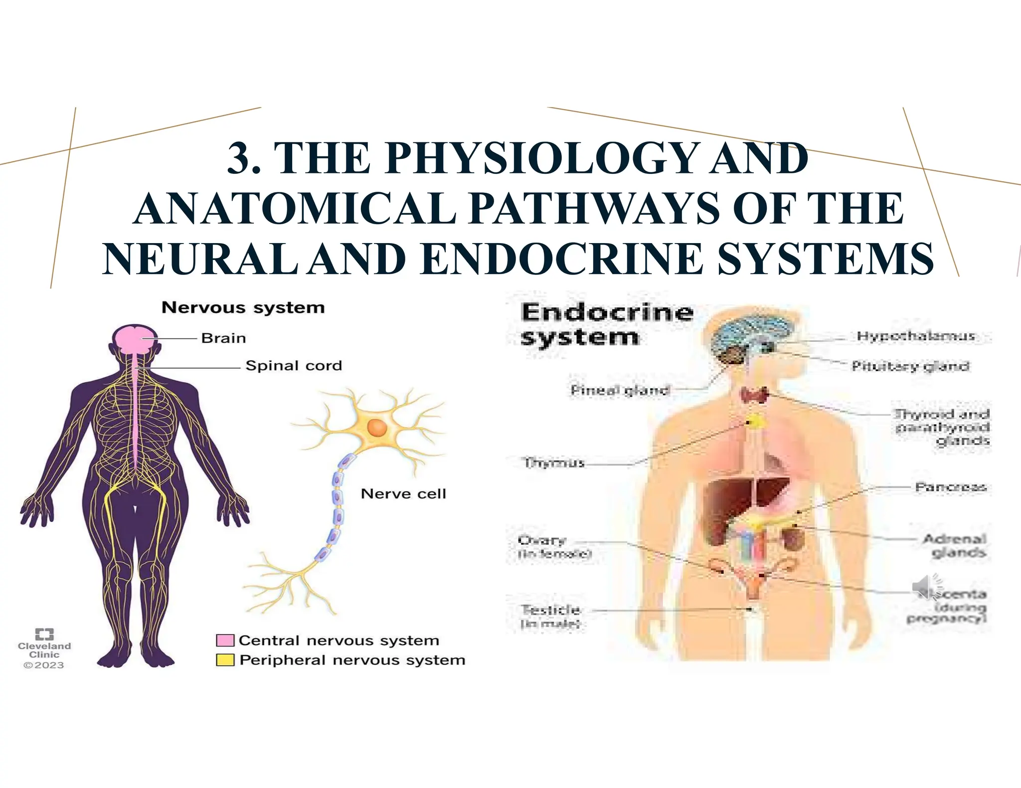
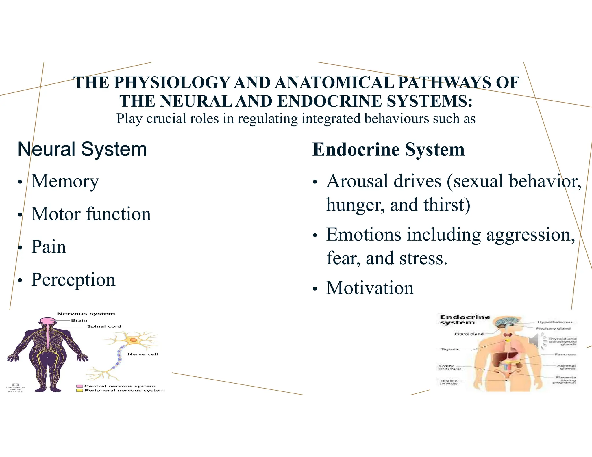
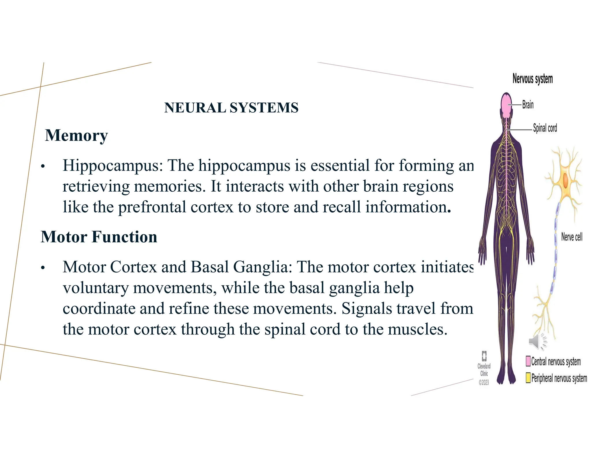
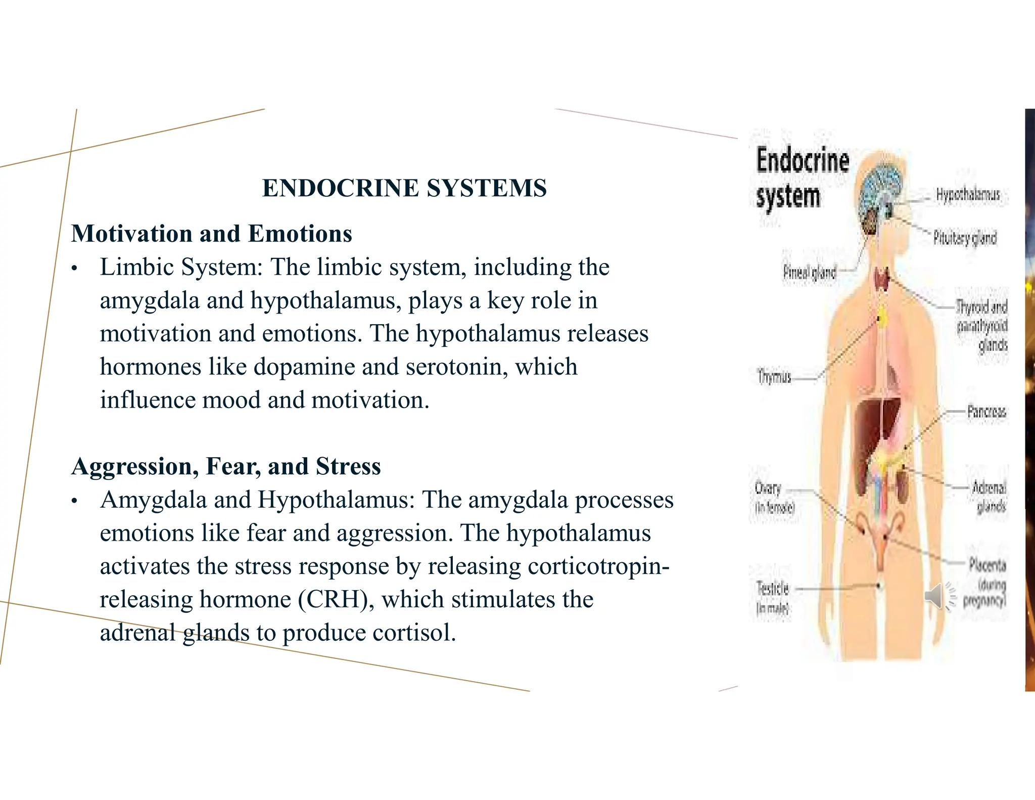
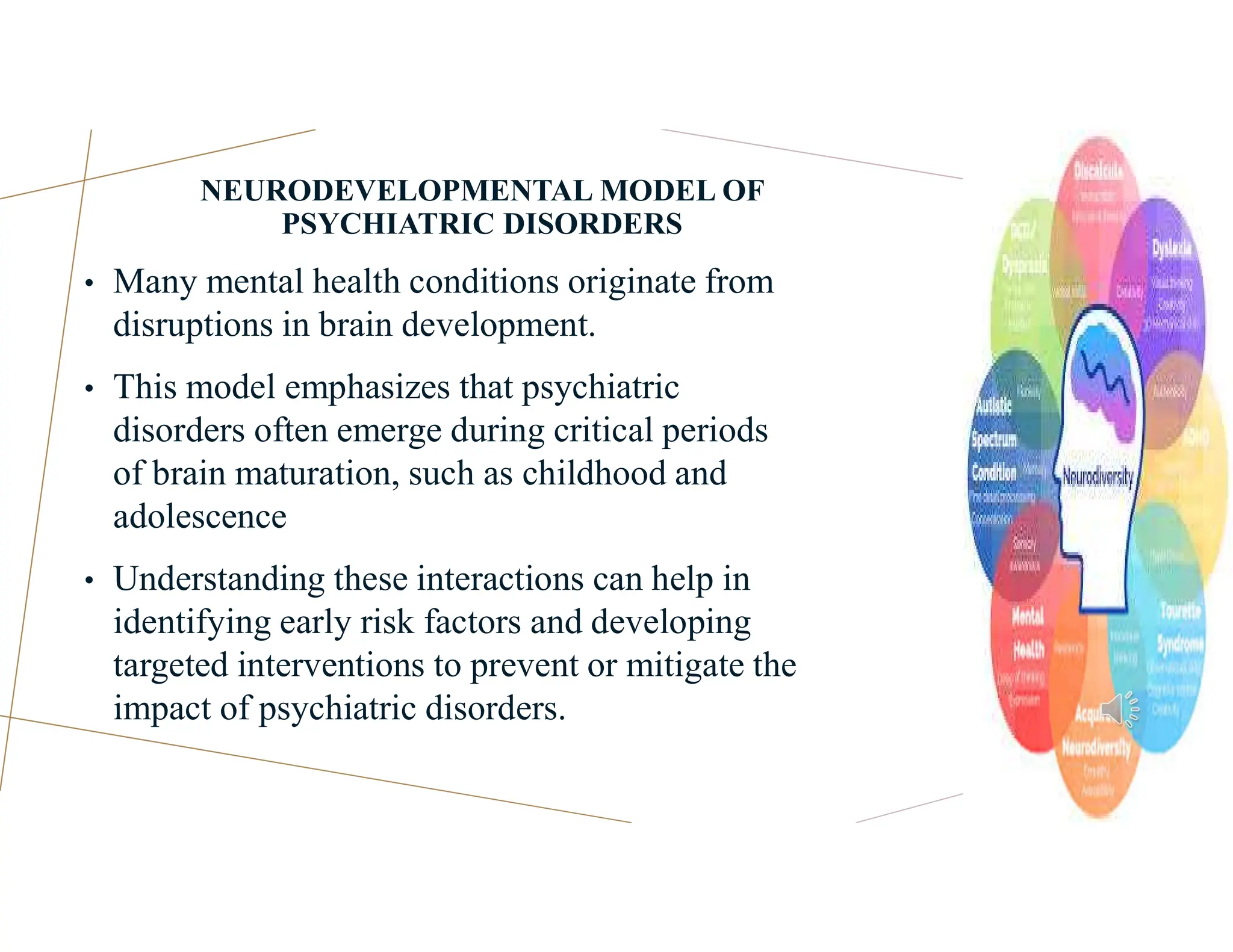
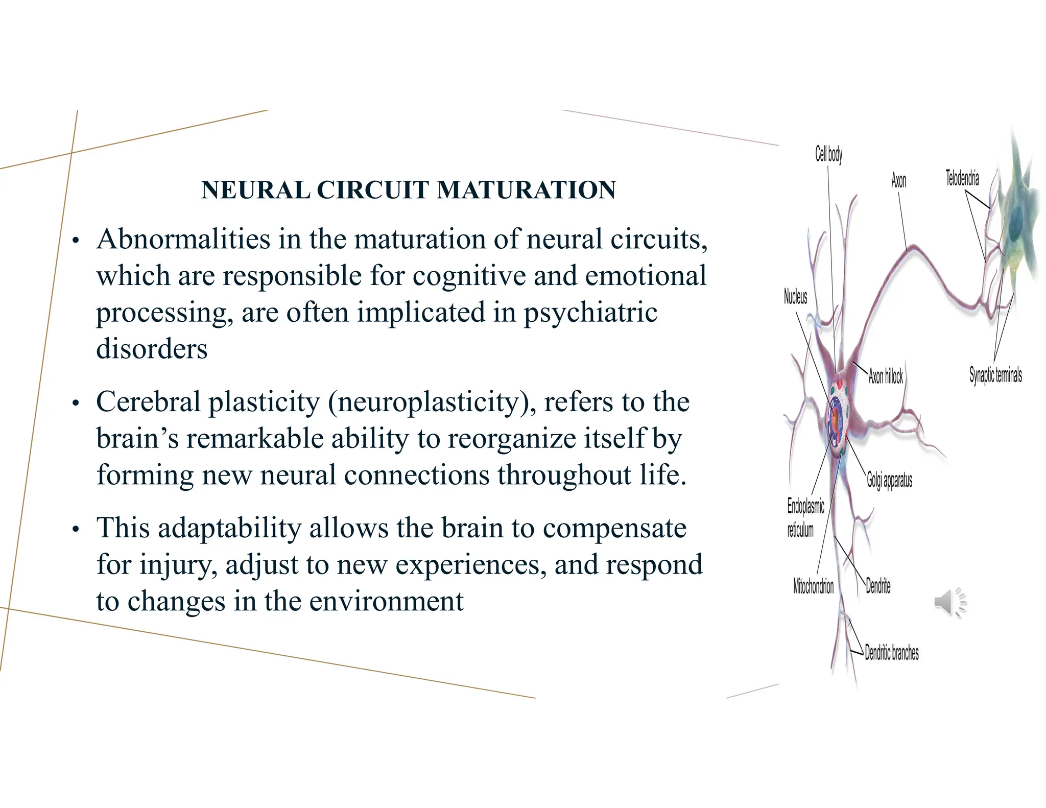
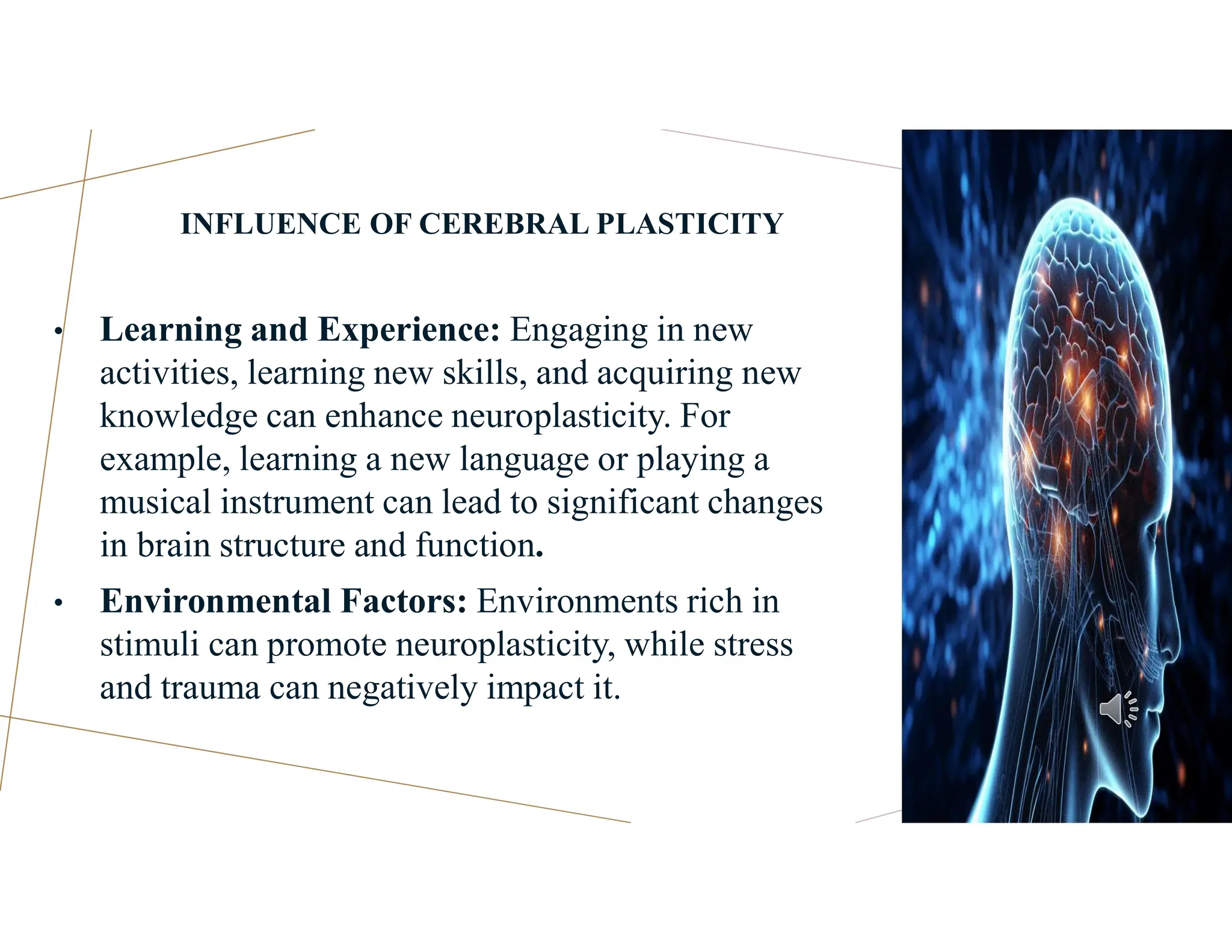
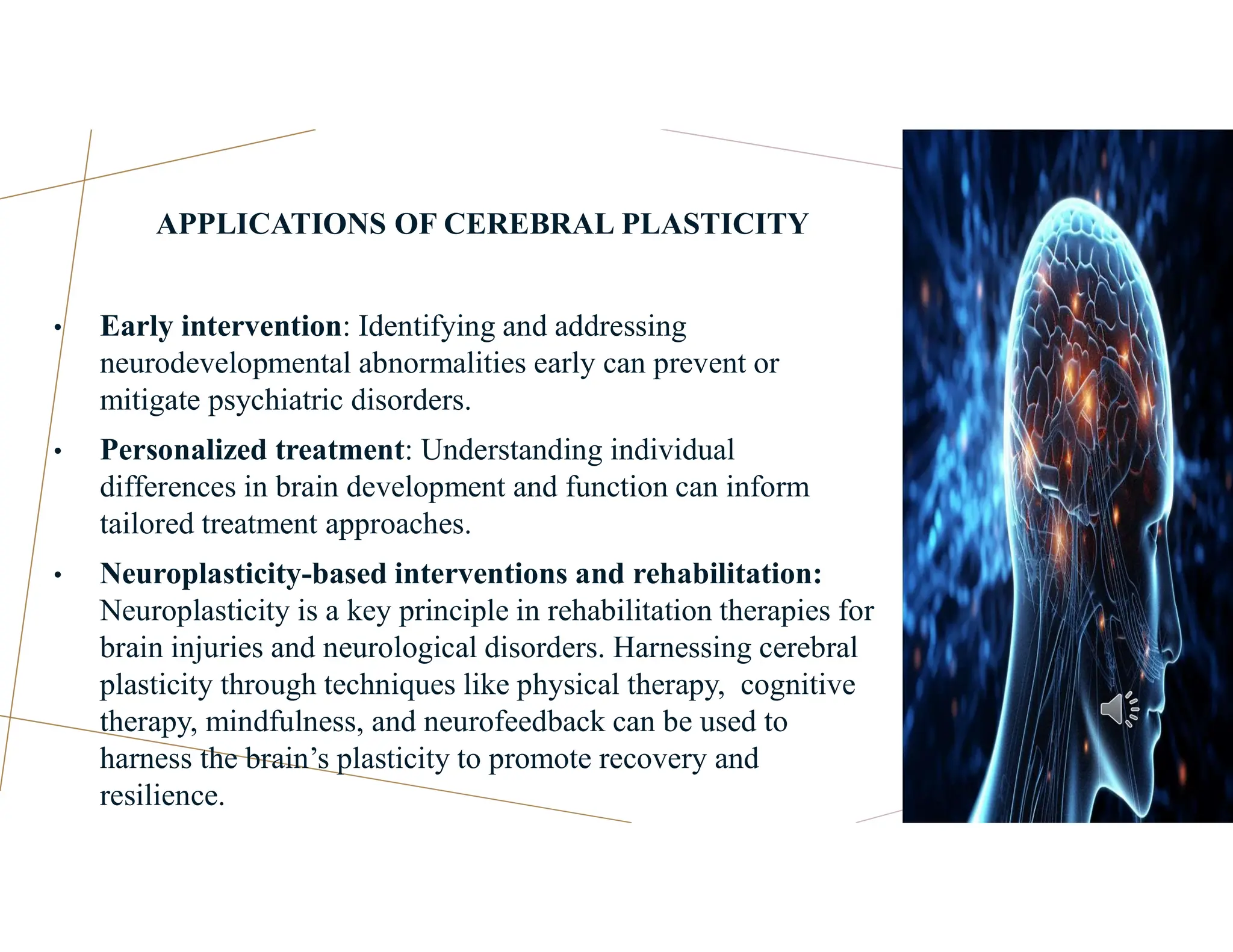
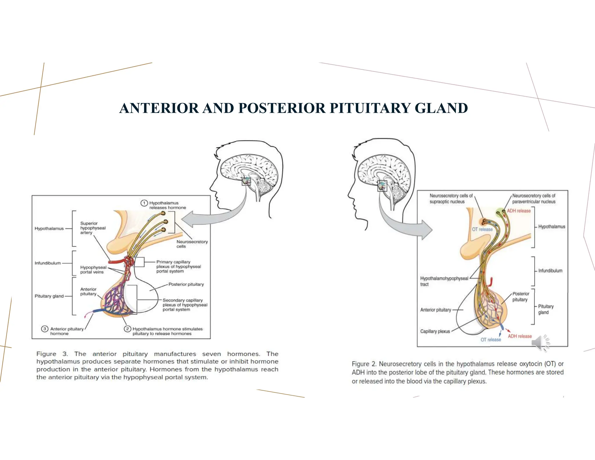
NEUROPHYSIOLOGICAL
PROCESSES
Prof. Adesanmi Akinsulore (MBChB, MPH, FWACP,
FMCPsych)
Primaries Revision Course of the Faculty of Psychiatry, West African
College of Physicians
8th August 2024
AGEN
DA
OUTLINE
1) Basic knowledge of physiology of the neurons
2) Basic knowledge of resting & action potential.
3) Physiology and anatomical pathways involved in the
neural and endocrine systems. The disturbance of
Neuronal functions with relevance to organic and non
– organic psychiatric disorders.
4) Neurodevelopmental model of psychiatric disorders
and cerebral plasticity
5) Neuroendocrine system
6) Physiology of arousal and sleep
7) Normal electroencephalogram (EEG) and evoked
response techniques
1. BASIC KNOWLEDGE OF
PHYSIOLOGY OF THE NEURONS
NEURON
‱ Neurons are the fundamental units of the brain and nervous
system, responsible for receiving sensory input, sending motor
commands, and transforming and relaying electrical signals.
Each neuron consists of three main parts:
 Cell Body (Soma): Contains the nucleus and is responsible
for maintaining the cell’s health.
 Dendrites: Branch-like structures that receive messages from
other neurons.
 Axon: A long, slender projection that transmits electrical
impulses away from the cell body to other neurons or
muscles.
SYNAPSES
‱ Synapses are the junctions where neurons communicate with
each other or with other target cells. There are two main types of
synapses:
‱ Chemical Synapses: Use neurotransmitters to send signals
across a synaptic cleft. This type is predominant in the nervous
system.
‱ Electrical Synapses: Allow direct passage of ions and small
molecules between neurons through gap junctions, enabling
faster communication.
RECEPTORS
‱ Receptors are proteins on the surface of neurons that bind to
neurotransmitters and initiate a response in the target cell. There are
two main types of receptors:
‱ Ionotropic Receptors: These are ligand-gated ion channels that open
in response to neurotransmitter binding, allowing ions to flow into or
out of the neuron, leading to rapid changes in membrane potential.
‱ Metabotropic Receptors: These are G-protein-coupled receptors
that activate second messenger systems, leading to slower but longer-
lasting effects on the neuron.
‱ Autoreceptors: Special types of receptors located in the postsynaptic
neurone and involved in negative feedback mechanism for cessation of
neuronal actions
IONOTROPIC RECEPTORS
(LIGAND-GATED)
Also known as Ion Channel Receptors
‱ Allows for unidirectional flow of ions
‱ Results in quick response
‱ Can have excitatory or inhibitory
effects
‱ e.g., GABAA, NMDA, 5HT3
METABOTROPIC
RECEPTORS
‱ Also called G-protein coupled receptors
(Serpentine receptor)
‱ Acts via Adenylase cyclase (Gi-Inhibitory,
Gs -Excitatory), and Phospholipase C
‱ Requires a second messenger (e.g., ATP or
AMP)
‱ Relatively slower response
‱ Longer lasting effect
‱ e.g., Dopamine, All 5HT (except 5HT3),
Neuropeptides and Opiods receptors
AUTORECEPTORS
‱ An autoreceptor is a receptor that
when bound by ligand reduces
release of that ligand into the
synapse.
‱ The α2 receptor is a classic
example of an autoreceptor, as when
it is bound by noradrenaline (NA) it
inhibits NA release.
NEUROTRANSMITTERS
Neurotransmitters are chemicals that transmit signals across a synapse from one
neuron to another. The process involves several steps:
1. Synthesis: Neurotransmitters are synthesized in the neuron’s cell body or axon
terminal. For example, acetylcholine is synthesized from choline and acetyl-
CoA.
2. Storage: Once synthesized, neurotransmitters are stored in synaptic vesicles
within the axon terminal.
3. Release: When an action potential reaches the axon terminal, it triggers the
influx of calcium ions, causing the vesicles to fuse with the presynaptic
membrane and release neurotransmitters into the synaptic cleft.
4. Binding: Neurotransmitters diffuse across the synaptic cleft and bind to specific
receptors on the postsynaptic membrane, initiating a response in the target cell.
5. Termination: The action of neurotransmitters is terminated by reuptake into the
presynaptic neuron, enzymatic degradation, or diffusion away from the synapse.
NEUROTRANSMITTERS
Synthesis of Neurotransmitters
 Occurs in the presynaptic neuron
 Involves:
o Amino acid precursors (e.g.,
tyrosine, tryptophan)
o Enzymes (e.g., tyrosine
hydroxylase, tryptophan
hydroxylase)
o Packaging into vesicles for
release
Uptake of Neurotransmitters
 Occurs in the presynaptic neuron
after release
 Involves:
o Reuptake transporters (e.g.,
dopamine transporter,
serotonin transporter)
o Enzymatic degradation (e.g.,
monoamine oxidase)
o Recycling or degradation of
neurotransmitters
NEUROTRANSMITTERS - EXAMPLES
Amino Acids
 Inhibitory: Gamma Amino Butyric Acid
(GABA)
 Excitatory: Glutamate (NMDA),
Aspartate, Glycine and Taurine
Biogenic Amines
 Cathecolamines: Dopamine,
Adrenaline, Nor-adrenaline
 Indolamine: Serotonine (5-HT)
 Others: Acetylcholine, Histamine
Peptides: Glucagon, Insulin,
substance-P, and Cholecystokinnin
Neucleotides
 Adenosine
 Adenosine triphosphate (ATP)
Others:
 Gases (e.g., Nitreous Oxide,
Carbon monoxide), Amantadines
(Endogenous cannabinoids), and
Eicosannoids (e.g.,
Prostaglandins, Prostacyclin)
GAMMAAMINO BUTYRIC ACID (GABA)
Function
‱ Produce calming effect
‱ Control of anxiety, fear, stress and seizure
‱ Mediate action of Benzodiazepines,
Barbiturates, and Alcohol
Brain localization: GABAergic neurons are
located in the hippocampus, thalamus, basal
ganglia, hypothalamus, and brainstem.
Receptors
‱ GABA-A = Chloride ion channel linked
‱ GABA-B = G-protein coupled
GLUTAMATE
Function
‱ Intermediary metabolism and protein
synthesis
‱ Memory and learning
Relevant Neuropsychiatric disorders:
Implicated in
‱ Epilepsy, toxic effects of stroke, head and
spinal cord trauma, Schizophrenia,
Huntington’s, Parkinson’s, or Alzheimer’s
disease, amyotrophic lateral sclerosis, and
AIDS dementia
Receptors
ACETYLCHOLINE
‱ An ester of choline and acetic acid
‱ plays a role in memory, motivation, learning,
attention, arousal, involuntary muscle movement
and involved in promoting REM sleep.
Brain localisation
‱ Nucleus Basalis of Meynert (with Projections to
cerebral and Limbic cortices
‱ Others=Renshaw cells, Striatum, Medial septal
nucleus, Reticular formation
Relevant Neuropsychiatric disorders
‱ Low in Alzheimer’s type dementia
‱ Imbalanced in Parkinson’s disease
Receptors
‱ Nicotinic= Ion Channels
‱ Muscarinic (M1-M5)= G-protein coupled
DOPAMINE
Function
‱ Arousal
‱ Motivation
‱ Motor movement
‱ Novelty seeking
‱ Reward mechanism (addiction)
Brain localisation/Pathways
‱ Long pathways= Nigrostriatal (movement), mesolimbic and
mesocortical (Psychosis)
‱ Short pathways= Tuberoinfundibular (prolactin),
Incertohypothalamic (Sexual behaviour)
‱ Ultra-short pathways= Amacrine retinal cells, Olfactory
system
Clinically relevant disorders
‱ Low=Parkinson’s diseases (Nigrostriatal), Anhedonia and
Negative symptoms of psychoses (mesocortical)
‱ High= Positive psychotic symptoms (Mesolimbic area)
NORADRENALINE
Function
‱ Anxiety
‱ Arousal
‱ Autonomic mediation
‱ Mood regulation
Brain localization: Locus cereleus (with projections to
other brain areas)
Relevant Neuropsychiatric Disorders
‱ Low in Depression
‱ Affected in anxiety disorders
Receptors
‱ Alpha (α)
1= Phospholipase-C linked Postsynaptic
2= G-Protein linked (inhibitory) pre-synaptic
Autoreceptor
‱ Beta (ÎČ)= G-Protein linked (stimulatory)
regulator of α
1= Affinity for norepinephrine
2= Affinity for epinephrine
SEROTONIN (5-HT)
Functions
‱ Mood, Pain perception, Feeding, Sleep-wake
cycle, Motor activity, Sexual behaviour and
Temperature control
Brain localisation=Raphe nuclei (with Projections to
other brain locations)
Relevant Neuropsychiatric disorders
‱ Low in Depression, Aggression, Suicide,
Impulsivity
‱ Psychosis (through regulation of dopamine)
Relevant receptors (out of 14 sub-units currently
known)
‱ 5HT1= (A) Antidepressant & Anxiolytic ,
(B) Aggression, (D) Antimigraine
‱ 5HT2= (A) Antipsychotic & memory , (B)
Cardiac , (C) Anxiogenic & Anorexic
‱ 5HT3 (Anti-emetic), 5HT6 (±Antipsychotic
& Antidepressant) 5HT7 (Cardiac)
OTHER NEUROTRANSMITTERS AND THEIR FUNCTIONS
 Substance P: Pain sensation, Low in
Huntington chorea
 Neurotensin: Antidopaminergic (Reward
mechanism, ±Benefit psychosis)
 Cholecystokinnins: Pathogenesis of
Schizophrenia
 Neuropeptide Y, Leptin, & Ghrelin:
weight gain
 Endogenous Opiods: Regulation of Pain,
Anxiety, memory
 Somatostatin: Inhibition of growth
hormones
2. BASIC KNOWLEDGE OF RESTING &
ACTION POTENTIAL
RESTING & ACTION POTENTIAL
Resting Potential
‱ The resting potential is the stable, negative
electric charge of a neuron's cell membrane
when it is not actively transmitting a signal.
‱ This potential is maintained by a higher
concentration of potassium ions (K+) inside
the cell and a higher concentration of
sodium ions (Na+) outside the cell.
For a typical neurone, this is -30mV to -90mV
(Average = -70mV)
- 70 mV neuronal membrane = Polarised
. More positive voltage= Depolarised
.More negative voltage= Hyperpolarised
Action Potential
‱ An action potential is a rapid, temporary
change in the membrane potential that
travels along the axon of a neuron.
‱ It is initiated when the neuron receives a
strong enough stimulus, causing the
membrane potential to become less
negative (depolarize) and reach a
threshold, usually around -55 mV1.
ACTION POTENTIALS
1. DEPOLARIZATION: Voltage-gated sodium channels open,
allowing naâș ions to rush into the cell, making the inside more
positive.
2. REPOLARIZATION: At the peak of the action potential (around
+30 mv), sodium channels close and voltage-gated potassium
channels open, allowing Kâș ions to flow out of the cell, restoring the
negative charge inside.
3. HYPERPOLARIZATION: The membrane potential temporarily
becomes more negative than the resting potential due to the continued
outflow of Kâș ions.
4. RETURN TO RESTING POTENTIAL: The sodium-potassium
pump and leak channels restore the resting potential.
ION CHANNELS AND INFLUXES
1. VOLTAGE-GATED SODIUM CHANNELS: Open in response
to depolarization, allowing naâș ions to enter the cell.
2. VOLTAGE-GATED POTASSIUM CHANNELS: Open in
response to the peak of the action potential, allowing Kâș ions to
exit the cell.
3. LEAK CHANNELS: Always open, allowing ions to move
according to their concentration gradients, contributing to the
resting potential.
4. SODIUM-POTASSIUM PUMP: Uses ATP to transport naâș out
and Kâș into the cell, maintaining the concentration gradients
necessary for the resting potential.
ION CHANNELS AND INFLUXES
 Sodium-potassium pump: Maintains
the resting potential by pumping
sodium ions out and potassium ions
into the cell.
 Voltage-gated sodium channels: Open
during depolarization, allowing
sodium influx.
 Voltage-gated potassium channels:
Open during repolarization, allowing
potassium efflux.
‱ The resting potential is the stable negative
charge of a neuron's cell membrane.
‱ An action potential is a brief electrical
impulse generated by ion influxes and
effluxes through specialized channels.
‱ Understanding these concepts is crucial for
grasping how neurons communicate and
process information.
3. THE PHYSIOLOGYAND
ANATOMICAL PATHWAYS OF THE
NEURALAND ENDOCRINE SYSTEMS
THE PHYSIOLOGYAND ANATOMICAL PATHWAYS OF
THE NEURALAND ENDOCRINE SYSTEMS:
Play crucial roles in regulating integrated behaviours such as
Neural System
‱ Memory
‱ Motor function
‱ Pain
‱ Perception
Endocrine System
‱ Arousal drives (sexual behavior,
hunger, and thirst)
‱ Emotions including aggression,
fear, and stress.
‱ Motivation
NEURAL SYSTEMS
Memory
‱ Hippocampus: The hippocampus is essential for forming and
retrieving memories. It interacts with other brain regions
like the prefrontal cortex to store and recall information.
Motor Function
‱ Motor Cortex and Basal Ganglia: The motor cortex initiates
voluntary movements, while the basal ganglia help
coordinate and refine these movements. Signals travel from
the motor cortex through the spinal cord to the muscles.
NEURAL SYSTEMS
Pain
‱ Nociceptors: Specialized sensory receptors called nociceptors
detect painful stimuli and send signals through the spinal cord
to the brain. The thalamus and somatosensory cortex process
these signals, resulting in the perception of pain.
Perception
‱ Sensory Pathways: Sensory information from the
environment is detected by sensory receptors and transmitted
to the brain via afferent neurons. The primary sensory cortex
processes this information, allowing us to perceive stimuli
like touch, sound, and sight.
ENDOCRINE SYSTEMS
Arousal Drives (Sexual Behavior, Hunger, and
Thirst):
‱ Hypothalamus: The hypothalamus regulates
arousal drives by releasing hormones that
influence sexual behavior, hunger, and thirst.
‱ For example, it releases gonadotropin-
releasing hormone (GnRH) to stimulate sexual
behavior and orexin to regulate hunger
ENDOCRINE SYSTEMS
Motivation and Emotions
‱ Limbic System: The limbic system, including the
amygdala and hypothalamus, plays a key role in
motivation and emotions. The hypothalamus releases
hormones like dopamine and serotonin, which
influence mood and motivation.
Aggression, Fear, and Stress
‱ Amygdala and Hypothalamus: The amygdala processes
emotions like fear and aggression. The hypothalamus
activates the stress response by releasing corticotropin-
releasing hormone (CRH), which stimulates the
adrenal glands to produce cortisol.
INTEGRATED PATHWAYS
Neural-Endocrine Interaction
‱ Hypothalamic-Pituitary-Adrenal
(HPA) Axis: This pathway involves
the hypothalamus, pituitary gland, and
adrenal glands.
‱ It regulates the body’s response to
stress by releasing cortisol, which
helps manage stress and maintain
homeostasis
Communication Between Systems
‱ Neurotransmitters and Hormones: The
nervous system uses neurotransmitters
like dopamine and serotonin for rapid
communication,
‱ while the endocrine system uses
hormones like cortisol and adrenaline for
longer-lasting effects
4. NEURODEVELOPMENTAL MODEL
OF PSYCHIATRIC DISORDERS AND
CEREBRAL PLASTICITY
.
NEURODEVELOPMENTAL MODEL OF
PSYCHIATRIC DISORDERS
‱ Many mental health conditions originate from
disruptions in brain development.
‱ This model emphasizes that psychiatric
disorders often emerge during critical periods
of brain maturation, such as childhood and
adolescence
‱ Understanding these interactions can help in
identifying early risk factors and developing
targeted interventions to prevent or mitigate the
impact of psychiatric disorders.
EARLY-LIFE EXPERIENCES
‱ Adverse experiences during critical
developmental periods can have long-lasting
effects on brain structure and function,
potentially leading to mental health conditions.
‱ Schizophrenia: Abnormalities in prenatal and
perinatal development, leading to altered neural
circuits and connectivity, contribute to the
development of schizophrenia.
‱ Depression: Early life stress and adversity can
shape the development of neural circuits,
increasing the risk of depression.
GENETIC AND ENVIRONMENTAL INTERACTIONS
‱ Both genetic predispositions and environmental
factors, such as prenatal stress, infections, and
early-life adversity, interact to influence brain
development and the risk of psychiatric disorders
‱ Genetic mutations or variations can predispose
individuals to psychiatric disorders, but
environmental influences can trigger or exacerbate
these conditions.
‱ For instance, maternal infections during pregnancy
have been linked to an increased risk of
schizophrenia in offspring
NEURAL CIRCUIT MATURATION
‱ Abnormalities in the maturation of neural circuits,
which are responsible for cognitive and emotional
processing, are often implicated in psychiatric
disorders
‱ Cerebral plasticity (neuroplasticity), refers to the
brain’s remarkable ability to reorganize itself by
forming new neural connections throughout life.
‱ This adaptability allows the brain to compensate
for injury, adjust to new experiences, and respond
to changes in the environment
CEREBRAL PLASTICITY
Types of Plasticity:
‱ Structural Plasticity: Changes in the physical
structure of the brain, such as the growth of new
neurons (neurogenesis) and the formation of new
synapses.
‱ Functional Plasticity: The brain’s ability to move
functions from damaged areas to undamaged areas.
This is often seen in stroke recovery, where other
parts of the brain take over functions previously
managed by the affected area.
MECHANISMS OF CEREBRAL PLASTICITY
‱ Synaptic Plasticity: The strengthening or
weakening of synapses, which are the connections
between neurons. This process is crucial for
learning and memory.
‱ Cortical Remapping: The brain’s ability to
reorganize itself by mapping functions from one
area to another, often in response to injury or
sensory loss
INFLUENCE OF CEREBRAL PLASTICITY
‱ Learning and Experience: Engaging in new
activities, learning new skills, and acquiring new
knowledge can enhance neuroplasticity. For
example, learning a new language or playing a
musical instrument can lead to significant changes
in brain structure and function.
‱ Environmental Factors: Environments rich in
stimuli can promote neuroplasticity, while stress
and trauma can negatively impact it.
APPLICATIONS OF CEREBRAL PLASTICITY
‱ Early intervention: Identifying and addressing
neurodevelopmental abnormalities early can prevent or
mitigate psychiatric disorders.
‱ Personalized treatment: Understanding individual
differences in brain development and function can inform
tailored treatment approaches.
‱ Neuroplasticity-based interventions and rehabilitation:
Neuroplasticity is a key principle in rehabilitation therapies for
brain injuries and neurological disorders. Harnessing cerebral
plasticity through techniques like physical therapy, cognitive
therapy, mindfulness, and neurofeedback can be used to
harness the brain’s plasticity to promote recovery and
resilience.
5. NEUROENDOCRINE SYSTEM
.
THE NEUROENDOCRINE SYSTEM
‱ Complex network that bridges the nervous system and the
endocrine system.
Plays a crucial role in maintaining homeostasis and regulating
various physiological processes in the body, such as:
‱ Growth
‱ Metabolism
‱ Reproduction
‱ Stress response
.
COMPONENTS OF NEUROENDOCRINE SYSTEM
1. Hypothalamus
‱ Often referred to as the brain’s
relay center,
‱ Receives signals from different
parts of the brain and translates
them into hormonal signals.
‱ Produces hormones like oxytocin
and vasopressin, which are then
transported to the pituitary gland.
.
COMPONENTS OF NEUROENDOCRINE SYSTEM
2. Pituitary Gland
‱ This gland is divided into three
lobes: anterior, intermediate, and
posterior.
‱ The hypothalamus controls the
anterior pituitary’s hormone
secretion by sending releasing
factors.
‱ The posterior pituitary stores and
releases hormones produced by
the hypothalamus.
.
ANTERIOR AND POSTERIOR PITUITARY GLAND
.
COMPONENTS OF NEUROENDOCRINE SYSTEM
3. Neuroendocrine Cells
‱ These specialized cells can act as neurons,
responding to neural inputs, and as
hormonal secretors, releasing hormones
directly into the bloodstream.
‱ This dual function allows the
neuroendocrine system to respond swiftly to
environmental changes while ensuring
widespread and lasting effects
.
FUNCTION OF NEUROENDOCRINE SYSTEM
Regulation of Hormone Secretion:
 The neuroendocrine system ensures that the body
maintains hormonal balance by controlling the
timing and amount of hormone release.
 This is crucial for processes like glucose
management, where insulin and glucagon levels are
adjusted in response to blood sugar levels.
.
FUNCTION OF NEUROENDOCRINE SYSTEM
Coordination of Bodily Functions
 By managing the communication between
the nervous and endocrine systems, the
neuroendocrine system helps coordinate
various bodily functions, ensuring that
processes like metabolism, reproduction, and
stress responses are harmonized.
.
THE HYPOTHALAMUS AND
PITUITARY GLAND
.
PITUITARY HORMONES
.
THE RELEASE FACTORS AND FEEDBACK
CONTROL MECHANISMS
‱ Essential components of the neuroendocrine system,
‱ Ensuring precise regulation of hormone levels in the body.
‱ Release factors are hormones produced by the hypothalamus that stimulate or
inhibit the secretion of hormones from the anterior pituitary gland.
‱ Feedback control is a regulatory mechanism in which the output of a system
influences its own activity.
‱ In the context of the neuroendocrine system, feedback control ensures that
hormone levels remain within a narrow, optimal range.
THE RELEASE FACTORS
TWO MAIN TYPES OF FEEDBACK CONTROL
‱ Negative Feedback: This is the most common form of feedback control. When
the levels of a hormone rise above a certain threshold, the hypothalamus and
pituitary gland reduce the secretion of releasing factors and hormones to bring
the levels back down.
‱ For example, high levels of thyroid hormones (T3 and T4) inhibit the release of
TRH and TSH, reducing further production of thyroid hormones
‱ Positive Feedback: This is less common but occurs in certain situations. In
positive feedback, an increase in hormone levels triggers further release of that
hormone.
‱ An example is the release of oxytocin during childbirth.
6. PHYSIOLOGY OFAROUSALAND
SLEEP
.
AROUSAL
‱ State of being awake and responsive to stimuli.
‱ Involves various neural circuits and neurotransmitters that keep the brain alert
and ready to respond to the environment.
‱ Reticular Activating System (RAS): Located in the brainstem, the RAS plays
a crucial role in maintaining wakefulness and alertness by sending signals to
the cerebral cortex.
‱ Neurotransmitters such as norepinephrine, dopamine, serotonin, and
acetylcholine are involved in promoting wakefulness. These neurotransmitters
help modulate brain activity and responsiveness.
‱ Thalamus: Acts as a relay station, transmitting sensory information to the
cerebral cortex, which helps in maintaining alertness.
SLEEP
‱ A complex and dynamic process that involves multiple
stages and brain regions. It is regulated by two main
processes:
‱ Circadian Rhythm: This is the body’s internal clock,
primarily regulated by the suprachiasmatic nucleus
(SCN) in the hypothalamus. It controls the timing of
sleep and wakefulness over a 24-hour period2.
‱ Homeostatic Sleep Drive: This refers to the pressure
to sleep that builds up the longer you stay awake. It
ensures that you get enough sleep to recover and
function properly3
SLEEP STAGES
‱ Divided into Non-Rapid Eye Movement (NREM) and Rapid Eye Movement
(REM) sleep with each type having distinct physiological characteristics:
NREM Sleep:
‱ Stage 1: Light sleep, where you drift in and out of sleep. Muscle activity slows,
and you can be easily awakened.
‱ Stage 2: Eye movement stops, and brain waves become slower with occasional
bursts of rapid waves called sleep spindles.
‱ Stage 3: Deep sleep, characterized by very slow brain waves called delta
waves. It is difficult to wake someone during this stage.
REM Sleep:
‱ This stage is characterized by rapid eye movements, increased brain activity,
and vivid dreams. The body becomes almost paralyzed to prevent acting out
dreams.
‱ REM sleep is crucial for cognitive functions like memory consolidation and
learning.
7. NORMAL
ELECTROENCEPHALOGRAM (EEG)
.
ELECTROENCEPHALOGRAM (EEG)
‱ An EEG is a non-invasive test that measures electrical activity in the brain. It is commonly
used to diagnose and monitor conditions affecting the brain, such as epilepsy, sleep disorders,
and brain injuries.
Key Features:
‱ Wave Patterns: EEG records brain wave patterns, which are categorized into different
frequency bands:
‱ Delta (ή): < 4 Hz, associated with deep sleep.
‱ Theta (ξ): 4–8 Hz, linked to light sleep and relaxation.
‱ Alpha (α): 8–13 Hz, observed when a person is awake but relaxed, especially with closed
eyes.
‱ Beta (ÎČ): 13–30 Hz, related to active thinking and focus (mostly recorded in frontal lobe
while awake)
‱ Gamma (γ): > 30 Hz, associated with high-level cognitive functions.
EVOKED RESPONSE (ER)
‱ ER are brain responses that are directly related to specific sensory,
cognitive, or motor events.
‱ They are extracted from the EEG by averaging the brain’s reaction to
repeated stimuli.
Types of Evoked Responses:
‱ Visual Evoked Potentials (VEPs): Measure the brain’s response to
visual stimuli, such as flashing lights or patterns.
‱ Auditory Evoked Potentials (AEPs): Assess the brain’s response to
sounds, like clicks or tones.
‱ Somatosensory Evoked Potentials (SEPs): Evaluate the brain’s
reaction to tactile or electrical stimulation of the skin.
EVOKED RESPONSE (ER) TECHNIQUES
‱ Stimulus Presentation: A specific stimulus (visual,
auditory, or tactile) is presented repeatedly.
‱ Signal Averaging: The EEG signals following each stimulus
are averaged to enhance the evoked response and reduce
background noise.
‱ Analysis: The averaged response is analyzed to assess the
functioning of specific neural pathways and brain regions.
CLINICALAPPLICATIONS EVOKED RESPONSE (ER)
:
‱ Neurological Assessment: Evoked potentials
are used to diagnose and monitor conditions
like multiple sclerosis, optic neuritis, and
auditory pathway disorders.
‱ Intraoperative Monitoring: These
techniques help monitor the integrity of neural
pathways during surgeries, reducing the risk of
damage.
APPLICATION OF EEG
:
Valuable tool in the investigation of cerebral pathologies
‱ Encephalopathy: EEG can reveal diffuse slowing of brain
waves, which is indicative of encephalopathy, a condition that
affects brain function1.
‱ Brain Tumors: Abnormal EEG patterns can help localize brain
tumors, especially when combined with imaging techniques
like MRI2.
‱ Stroke: EEG may show focal slowing or epileptiform activity
in areas affected by stroke, aiding in the assessment of brain
damage2.
APPLICATION OF EEG
:
Valuable tool in the investigation of Seizure disorder
‱ Diagnosis: EEG can capture abnormal electrical discharges
(spikes and sharp waves) that are characteristic of epilepsy.
‱ Classification: Different types of seizures (e.g., focal,
generalized) produce distinct EEG patterns, helping in the
classification and treatment planning.
‱ Monitoring: Continuous EEG monitoring is used in
intensive care units to detect non-convulsive seizures and
status epilepticus.
APPLICATION OF EEG
:
Valuable tool in the investigation of psychiatric
disorders
‱ Schizophrenia: Patients with schizophrenia may exhibit abnormal
EEG patterns, such as reduced alpha activity and increased delta
and theta activity.
‱ Depression: EEG can show altered brain wave patterns in
individuals with major depressive disorder, which may help in
understanding the neurophysiological basis of the condition.
‱ ADHD: Children with Attention Deficit Hyperactivity Disorder
(ADHD) often display increased theta/beta ratio on EEG, which can
aid in diagnosis and treatment monitoring.
EFFECTS OF DRUGS ON EEG
‱ Antiepileptic Drugs (AEDs): These drugs can normalize
abnormal EEG patterns in epilepsy patients, reducing the frequency
and severity of seizures.
‱ Psychotropic Medications: Drugs used to treat psychiatric
disorders, such as antidepressants and antipsychotics, can alter EEG
patterns. For example, benzodiazepines increase beta activity, while
antipsychotics may reduce alpha activity.
‱ Anesthetics: EEG monitoring during anesthesia helps in
assessing the depth of anesthesia and preventing intraoperative
awareness
THANK YOU FOR
LISTENING
Prof. Adesanmi Akinsulore
Department of Mental Health
Obafemi Awolowo University, Ile-Ife, Nigeria
sanmilore@oauife.edu.ng

neurophysiology and neuroanatomy humansB

  • 1.
    NEUROPHYSIOLOGICAL PROCESSES Prof. Adesanmi Akinsulore(MBChB, MPH, FWACP, FMCPsych) Primaries Revision Course of the Faculty of Psychiatry, West African College of Physicians 8th August 2024
  • 2.
    AGEN DA OUTLINE 1) Basic knowledgeof physiology of the neurons 2) Basic knowledge of resting & action potential. 3) Physiology and anatomical pathways involved in the neural and endocrine systems. The disturbance of Neuronal functions with relevance to organic and non – organic psychiatric disorders. 4) Neurodevelopmental model of psychiatric disorders and cerebral plasticity 5) Neuroendocrine system 6) Physiology of arousal and sleep 7) Normal electroencephalogram (EEG) and evoked response techniques
  • 3.
    1. BASIC KNOWLEDGEOF PHYSIOLOGY OF THE NEURONS
  • 4.
    NEURON ‱ Neurons arethe fundamental units of the brain and nervous system, responsible for receiving sensory input, sending motor commands, and transforming and relaying electrical signals. Each neuron consists of three main parts:  Cell Body (Soma): Contains the nucleus and is responsible for maintaining the cell’s health.  Dendrites: Branch-like structures that receive messages from other neurons.  Axon: A long, slender projection that transmits electrical impulses away from the cell body to other neurons or muscles.
  • 5.
    SYNAPSES ‱ Synapses arethe junctions where neurons communicate with each other or with other target cells. There are two main types of synapses: ‱ Chemical Synapses: Use neurotransmitters to send signals across a synaptic cleft. This type is predominant in the nervous system. ‱ Electrical Synapses: Allow direct passage of ions and small molecules between neurons through gap junctions, enabling faster communication.
  • 6.
    RECEPTORS ‱ Receptors areproteins on the surface of neurons that bind to neurotransmitters and initiate a response in the target cell. There are two main types of receptors: ‱ Ionotropic Receptors: These are ligand-gated ion channels that open in response to neurotransmitter binding, allowing ions to flow into or out of the neuron, leading to rapid changes in membrane potential. ‱ Metabotropic Receptors: These are G-protein-coupled receptors that activate second messenger systems, leading to slower but longer- lasting effects on the neuron. ‱ Autoreceptors: Special types of receptors located in the postsynaptic neurone and involved in negative feedback mechanism for cessation of neuronal actions
  • 7.
    IONOTROPIC RECEPTORS (LIGAND-GATED) Also knownas Ion Channel Receptors ‱ Allows for unidirectional flow of ions ‱ Results in quick response ‱ Can have excitatory or inhibitory effects ‱ e.g., GABAA, NMDA, 5HT3
  • 8.
    METABOTROPIC RECEPTORS ‱ Also calledG-protein coupled receptors (Serpentine receptor) ‱ Acts via Adenylase cyclase (Gi-Inhibitory, Gs -Excitatory), and Phospholipase C ‱ Requires a second messenger (e.g., ATP or AMP) ‱ Relatively slower response ‱ Longer lasting effect ‱ e.g., Dopamine, All 5HT (except 5HT3), Neuropeptides and Opiods receptors
  • 9.
    AUTORECEPTORS ‱ An autoreceptoris a receptor that when bound by ligand reduces release of that ligand into the synapse. ‱ The α2 receptor is a classic example of an autoreceptor, as when it is bound by noradrenaline (NA) it inhibits NA release.
  • 10.
    NEUROTRANSMITTERS Neurotransmitters are chemicalsthat transmit signals across a synapse from one neuron to another. The process involves several steps: 1. Synthesis: Neurotransmitters are synthesized in the neuron’s cell body or axon terminal. For example, acetylcholine is synthesized from choline and acetyl- CoA. 2. Storage: Once synthesized, neurotransmitters are stored in synaptic vesicles within the axon terminal. 3. Release: When an action potential reaches the axon terminal, it triggers the influx of calcium ions, causing the vesicles to fuse with the presynaptic membrane and release neurotransmitters into the synaptic cleft. 4. Binding: Neurotransmitters diffuse across the synaptic cleft and bind to specific receptors on the postsynaptic membrane, initiating a response in the target cell. 5. Termination: The action of neurotransmitters is terminated by reuptake into the presynaptic neuron, enzymatic degradation, or diffusion away from the synapse.
  • 11.
    NEUROTRANSMITTERS Synthesis of Neurotransmitters Occurs in the presynaptic neuron  Involves: o Amino acid precursors (e.g., tyrosine, tryptophan) o Enzymes (e.g., tyrosine hydroxylase, tryptophan hydroxylase) o Packaging into vesicles for release Uptake of Neurotransmitters  Occurs in the presynaptic neuron after release  Involves: o Reuptake transporters (e.g., dopamine transporter, serotonin transporter) o Enzymatic degradation (e.g., monoamine oxidase) o Recycling or degradation of neurotransmitters
  • 12.
    NEUROTRANSMITTERS - EXAMPLES AminoAcids  Inhibitory: Gamma Amino Butyric Acid (GABA)  Excitatory: Glutamate (NMDA), Aspartate, Glycine and Taurine Biogenic Amines  Cathecolamines: Dopamine, Adrenaline, Nor-adrenaline  Indolamine: Serotonine (5-HT)  Others: Acetylcholine, Histamine Peptides: Glucagon, Insulin, substance-P, and Cholecystokinnin Neucleotides  Adenosine  Adenosine triphosphate (ATP) Others:  Gases (e.g., Nitreous Oxide, Carbon monoxide), Amantadines (Endogenous cannabinoids), and Eicosannoids (e.g., Prostaglandins, Prostacyclin)
  • 13.
    GAMMAAMINO BUTYRIC ACID(GABA) Function ‱ Produce calming effect ‱ Control of anxiety, fear, stress and seizure ‱ Mediate action of Benzodiazepines, Barbiturates, and Alcohol Brain localization: GABAergic neurons are located in the hippocampus, thalamus, basal ganglia, hypothalamus, and brainstem. Receptors ‱ GABA-A = Chloride ion channel linked ‱ GABA-B = G-protein coupled
  • 14.
    GLUTAMATE Function ‱ Intermediary metabolismand protein synthesis ‱ Memory and learning Relevant Neuropsychiatric disorders: Implicated in ‱ Epilepsy, toxic effects of stroke, head and spinal cord trauma, Schizophrenia, Huntington’s, Parkinson’s, or Alzheimer’s disease, amyotrophic lateral sclerosis, and AIDS dementia Receptors
  • 15.
    ACETYLCHOLINE ‱ An esterof choline and acetic acid ‱ plays a role in memory, motivation, learning, attention, arousal, involuntary muscle movement and involved in promoting REM sleep. Brain localisation ‱ Nucleus Basalis of Meynert (with Projections to cerebral and Limbic cortices ‱ Others=Renshaw cells, Striatum, Medial septal nucleus, Reticular formation Relevant Neuropsychiatric disorders ‱ Low in Alzheimer’s type dementia ‱ Imbalanced in Parkinson’s disease Receptors ‱ Nicotinic= Ion Channels ‱ Muscarinic (M1-M5)= G-protein coupled
  • 16.
    DOPAMINE Function ‱ Arousal ‱ Motivation ‱Motor movement ‱ Novelty seeking ‱ Reward mechanism (addiction) Brain localisation/Pathways ‱ Long pathways= Nigrostriatal (movement), mesolimbic and mesocortical (Psychosis) ‱ Short pathways= Tuberoinfundibular (prolactin), Incertohypothalamic (Sexual behaviour) ‱ Ultra-short pathways= Amacrine retinal cells, Olfactory system Clinically relevant disorders ‱ Low=Parkinson’s diseases (Nigrostriatal), Anhedonia and Negative symptoms of psychoses (mesocortical) ‱ High= Positive psychotic symptoms (Mesolimbic area)
  • 17.
    NORADRENALINE Function ‱ Anxiety ‱ Arousal ‱Autonomic mediation ‱ Mood regulation Brain localization: Locus cereleus (with projections to other brain areas) Relevant Neuropsychiatric Disorders ‱ Low in Depression ‱ Affected in anxiety disorders Receptors ‱ Alpha (α) 1= Phospholipase-C linked Postsynaptic 2= G-Protein linked (inhibitory) pre-synaptic Autoreceptor ‱ Beta (ÎČ)= G-Protein linked (stimulatory) regulator of α 1= Affinity for norepinephrine 2= Affinity for epinephrine
  • 18.
    SEROTONIN (5-HT) Functions ‱ Mood,Pain perception, Feeding, Sleep-wake cycle, Motor activity, Sexual behaviour and Temperature control Brain localisation=Raphe nuclei (with Projections to other brain locations) Relevant Neuropsychiatric disorders ‱ Low in Depression, Aggression, Suicide, Impulsivity ‱ Psychosis (through regulation of dopamine) Relevant receptors (out of 14 sub-units currently known) ‱ 5HT1= (A) Antidepressant & Anxiolytic , (B) Aggression, (D) Antimigraine ‱ 5HT2= (A) Antipsychotic & memory , (B) Cardiac , (C) Anxiogenic & Anorexic ‱ 5HT3 (Anti-emetic), 5HT6 (±Antipsychotic & Antidepressant) 5HT7 (Cardiac)
  • 19.
    OTHER NEUROTRANSMITTERS ANDTHEIR FUNCTIONS  Substance P: Pain sensation, Low in Huntington chorea  Neurotensin: Antidopaminergic (Reward mechanism, ±Benefit psychosis)  Cholecystokinnins: Pathogenesis of Schizophrenia  Neuropeptide Y, Leptin, & Ghrelin: weight gain  Endogenous Opiods: Regulation of Pain, Anxiety, memory  Somatostatin: Inhibition of growth hormones
  • 20.
    2. BASIC KNOWLEDGEOF RESTING & ACTION POTENTIAL
  • 21.
    RESTING & ACTIONPOTENTIAL Resting Potential ‱ The resting potential is the stable, negative electric charge of a neuron's cell membrane when it is not actively transmitting a signal. ‱ This potential is maintained by a higher concentration of potassium ions (K+) inside the cell and a higher concentration of sodium ions (Na+) outside the cell. For a typical neurone, this is -30mV to -90mV (Average = -70mV) - 70 mV neuronal membrane = Polarised . More positive voltage= Depolarised .More negative voltage= Hyperpolarised Action Potential ‱ An action potential is a rapid, temporary change in the membrane potential that travels along the axon of a neuron. ‱ It is initiated when the neuron receives a strong enough stimulus, causing the membrane potential to become less negative (depolarize) and reach a threshold, usually around -55 mV1.
  • 22.
    ACTION POTENTIALS 1. DEPOLARIZATION:Voltage-gated sodium channels open, allowing naâș ions to rush into the cell, making the inside more positive. 2. REPOLARIZATION: At the peak of the action potential (around +30 mv), sodium channels close and voltage-gated potassium channels open, allowing Kâș ions to flow out of the cell, restoring the negative charge inside. 3. HYPERPOLARIZATION: The membrane potential temporarily becomes more negative than the resting potential due to the continued outflow of Kâș ions. 4. RETURN TO RESTING POTENTIAL: The sodium-potassium pump and leak channels restore the resting potential.
  • 23.
    ION CHANNELS ANDINFLUXES 1. VOLTAGE-GATED SODIUM CHANNELS: Open in response to depolarization, allowing naâș ions to enter the cell. 2. VOLTAGE-GATED POTASSIUM CHANNELS: Open in response to the peak of the action potential, allowing Kâș ions to exit the cell. 3. LEAK CHANNELS: Always open, allowing ions to move according to their concentration gradients, contributing to the resting potential. 4. SODIUM-POTASSIUM PUMP: Uses ATP to transport naâș out and Kâș into the cell, maintaining the concentration gradients necessary for the resting potential.
  • 24.
    ION CHANNELS ANDINFLUXES  Sodium-potassium pump: Maintains the resting potential by pumping sodium ions out and potassium ions into the cell.  Voltage-gated sodium channels: Open during depolarization, allowing sodium influx.  Voltage-gated potassium channels: Open during repolarization, allowing potassium efflux. ‱ The resting potential is the stable negative charge of a neuron's cell membrane. ‱ An action potential is a brief electrical impulse generated by ion influxes and effluxes through specialized channels. ‱ Understanding these concepts is crucial for grasping how neurons communicate and process information.
  • 25.
    3. THE PHYSIOLOGYAND ANATOMICALPATHWAYS OF THE NEURALAND ENDOCRINE SYSTEMS
  • 26.
    THE PHYSIOLOGYAND ANATOMICALPATHWAYS OF THE NEURALAND ENDOCRINE SYSTEMS: Play crucial roles in regulating integrated behaviours such as Neural System ‱ Memory ‱ Motor function ‱ Pain ‱ Perception Endocrine System ‱ Arousal drives (sexual behavior, hunger, and thirst) ‱ Emotions including aggression, fear, and stress. ‱ Motivation
  • 27.
    NEURAL SYSTEMS Memory ‱ Hippocampus:The hippocampus is essential for forming and retrieving memories. It interacts with other brain regions like the prefrontal cortex to store and recall information. Motor Function ‱ Motor Cortex and Basal Ganglia: The motor cortex initiates voluntary movements, while the basal ganglia help coordinate and refine these movements. Signals travel from the motor cortex through the spinal cord to the muscles.
  • 28.
    NEURAL SYSTEMS Pain ‱ Nociceptors:Specialized sensory receptors called nociceptors detect painful stimuli and send signals through the spinal cord to the brain. The thalamus and somatosensory cortex process these signals, resulting in the perception of pain. Perception ‱ Sensory Pathways: Sensory information from the environment is detected by sensory receptors and transmitted to the brain via afferent neurons. The primary sensory cortex processes this information, allowing us to perceive stimuli like touch, sound, and sight.
  • 29.
    ENDOCRINE SYSTEMS Arousal Drives(Sexual Behavior, Hunger, and Thirst): ‱ Hypothalamus: The hypothalamus regulates arousal drives by releasing hormones that influence sexual behavior, hunger, and thirst. ‱ For example, it releases gonadotropin- releasing hormone (GnRH) to stimulate sexual behavior and orexin to regulate hunger
  • 30.
    ENDOCRINE SYSTEMS Motivation andEmotions ‱ Limbic System: The limbic system, including the amygdala and hypothalamus, plays a key role in motivation and emotions. The hypothalamus releases hormones like dopamine and serotonin, which influence mood and motivation. Aggression, Fear, and Stress ‱ Amygdala and Hypothalamus: The amygdala processes emotions like fear and aggression. The hypothalamus activates the stress response by releasing corticotropin- releasing hormone (CRH), which stimulates the adrenal glands to produce cortisol.
  • 31.
    INTEGRATED PATHWAYS Neural-Endocrine Interaction ‱Hypothalamic-Pituitary-Adrenal (HPA) Axis: This pathway involves the hypothalamus, pituitary gland, and adrenal glands. ‱ It regulates the body’s response to stress by releasing cortisol, which helps manage stress and maintain homeostasis Communication Between Systems ‱ Neurotransmitters and Hormones: The nervous system uses neurotransmitters like dopamine and serotonin for rapid communication, ‱ while the endocrine system uses hormones like cortisol and adrenaline for longer-lasting effects
  • 32.
    4. NEURODEVELOPMENTAL MODEL OFPSYCHIATRIC DISORDERS AND CEREBRAL PLASTICITY .
  • 33.
    NEURODEVELOPMENTAL MODEL OF PSYCHIATRICDISORDERS ‱ Many mental health conditions originate from disruptions in brain development. ‱ This model emphasizes that psychiatric disorders often emerge during critical periods of brain maturation, such as childhood and adolescence ‱ Understanding these interactions can help in identifying early risk factors and developing targeted interventions to prevent or mitigate the impact of psychiatric disorders.
  • 34.
    EARLY-LIFE EXPERIENCES ‱ Adverseexperiences during critical developmental periods can have long-lasting effects on brain structure and function, potentially leading to mental health conditions. ‱ Schizophrenia: Abnormalities in prenatal and perinatal development, leading to altered neural circuits and connectivity, contribute to the development of schizophrenia. ‱ Depression: Early life stress and adversity can shape the development of neural circuits, increasing the risk of depression.
  • 35.
    GENETIC AND ENVIRONMENTALINTERACTIONS ‱ Both genetic predispositions and environmental factors, such as prenatal stress, infections, and early-life adversity, interact to influence brain development and the risk of psychiatric disorders ‱ Genetic mutations or variations can predispose individuals to psychiatric disorders, but environmental influences can trigger or exacerbate these conditions. ‱ For instance, maternal infections during pregnancy have been linked to an increased risk of schizophrenia in offspring
  • 36.
    NEURAL CIRCUIT MATURATION ‱Abnormalities in the maturation of neural circuits, which are responsible for cognitive and emotional processing, are often implicated in psychiatric disorders ‱ Cerebral plasticity (neuroplasticity), refers to the brain’s remarkable ability to reorganize itself by forming new neural connections throughout life. ‱ This adaptability allows the brain to compensate for injury, adjust to new experiences, and respond to changes in the environment
  • 37.
    CEREBRAL PLASTICITY Types ofPlasticity: ‱ Structural Plasticity: Changes in the physical structure of the brain, such as the growth of new neurons (neurogenesis) and the formation of new synapses. ‱ Functional Plasticity: The brain’s ability to move functions from damaged areas to undamaged areas. This is often seen in stroke recovery, where other parts of the brain take over functions previously managed by the affected area.
  • 38.
    MECHANISMS OF CEREBRALPLASTICITY ‱ Synaptic Plasticity: The strengthening or weakening of synapses, which are the connections between neurons. This process is crucial for learning and memory. ‱ Cortical Remapping: The brain’s ability to reorganize itself by mapping functions from one area to another, often in response to injury or sensory loss
  • 39.
    INFLUENCE OF CEREBRALPLASTICITY ‱ Learning and Experience: Engaging in new activities, learning new skills, and acquiring new knowledge can enhance neuroplasticity. For example, learning a new language or playing a musical instrument can lead to significant changes in brain structure and function. ‱ Environmental Factors: Environments rich in stimuli can promote neuroplasticity, while stress and trauma can negatively impact it.
  • 40.
    APPLICATIONS OF CEREBRALPLASTICITY ‱ Early intervention: Identifying and addressing neurodevelopmental abnormalities early can prevent or mitigate psychiatric disorders. ‱ Personalized treatment: Understanding individual differences in brain development and function can inform tailored treatment approaches. ‱ Neuroplasticity-based interventions and rehabilitation: Neuroplasticity is a key principle in rehabilitation therapies for brain injuries and neurological disorders. Harnessing cerebral plasticity through techniques like physical therapy, cognitive therapy, mindfulness, and neurofeedback can be used to harness the brain’s plasticity to promote recovery and resilience.
  • 41.
  • 42.
    THE NEUROENDOCRINE SYSTEM ‱Complex network that bridges the nervous system and the endocrine system. Plays a crucial role in maintaining homeostasis and regulating various physiological processes in the body, such as: ‱ Growth ‱ Metabolism ‱ Reproduction ‱ Stress response .
  • 43.
    COMPONENTS OF NEUROENDOCRINESYSTEM 1. Hypothalamus ‱ Often referred to as the brain’s relay center, ‱ Receives signals from different parts of the brain and translates them into hormonal signals. ‱ Produces hormones like oxytocin and vasopressin, which are then transported to the pituitary gland. .
  • 44.
    COMPONENTS OF NEUROENDOCRINESYSTEM 2. Pituitary Gland ‱ This gland is divided into three lobes: anterior, intermediate, and posterior. ‱ The hypothalamus controls the anterior pituitary’s hormone secretion by sending releasing factors. ‱ The posterior pituitary stores and releases hormones produced by the hypothalamus. .
  • 45.
    ANTERIOR AND POSTERIORPITUITARY GLAND .
  • 46.
    COMPONENTS OF NEUROENDOCRINESYSTEM 3. Neuroendocrine Cells ‱ These specialized cells can act as neurons, responding to neural inputs, and as hormonal secretors, releasing hormones directly into the bloodstream. ‱ This dual function allows the neuroendocrine system to respond swiftly to environmental changes while ensuring widespread and lasting effects .
  • 47.
    FUNCTION OF NEUROENDOCRINESYSTEM Regulation of Hormone Secretion:  The neuroendocrine system ensures that the body maintains hormonal balance by controlling the timing and amount of hormone release.  This is crucial for processes like glucose management, where insulin and glucagon levels are adjusted in response to blood sugar levels. .
  • 48.
    FUNCTION OF NEUROENDOCRINESYSTEM Coordination of Bodily Functions  By managing the communication between the nervous and endocrine systems, the neuroendocrine system helps coordinate various bodily functions, ensuring that processes like metabolism, reproduction, and stress responses are harmonized. .
  • 49.
  • 50.
  • 51.
    THE RELEASE FACTORSAND FEEDBACK CONTROL MECHANISMS ‱ Essential components of the neuroendocrine system, ‱ Ensuring precise regulation of hormone levels in the body. ‱ Release factors are hormones produced by the hypothalamus that stimulate or inhibit the secretion of hormones from the anterior pituitary gland. ‱ Feedback control is a regulatory mechanism in which the output of a system influences its own activity. ‱ In the context of the neuroendocrine system, feedback control ensures that hormone levels remain within a narrow, optimal range.
  • 52.
  • 53.
    TWO MAIN TYPESOF FEEDBACK CONTROL ‱ Negative Feedback: This is the most common form of feedback control. When the levels of a hormone rise above a certain threshold, the hypothalamus and pituitary gland reduce the secretion of releasing factors and hormones to bring the levels back down. ‱ For example, high levels of thyroid hormones (T3 and T4) inhibit the release of TRH and TSH, reducing further production of thyroid hormones ‱ Positive Feedback: This is less common but occurs in certain situations. In positive feedback, an increase in hormone levels triggers further release of that hormone. ‱ An example is the release of oxytocin during childbirth.
  • 54.
  • 55.
    AROUSAL ‱ State ofbeing awake and responsive to stimuli. ‱ Involves various neural circuits and neurotransmitters that keep the brain alert and ready to respond to the environment. ‱ Reticular Activating System (RAS): Located in the brainstem, the RAS plays a crucial role in maintaining wakefulness and alertness by sending signals to the cerebral cortex. ‱ Neurotransmitters such as norepinephrine, dopamine, serotonin, and acetylcholine are involved in promoting wakefulness. These neurotransmitters help modulate brain activity and responsiveness. ‱ Thalamus: Acts as a relay station, transmitting sensory information to the cerebral cortex, which helps in maintaining alertness.
  • 56.
    SLEEP ‱ A complexand dynamic process that involves multiple stages and brain regions. It is regulated by two main processes: ‱ Circadian Rhythm: This is the body’s internal clock, primarily regulated by the suprachiasmatic nucleus (SCN) in the hypothalamus. It controls the timing of sleep and wakefulness over a 24-hour period2. ‱ Homeostatic Sleep Drive: This refers to the pressure to sleep that builds up the longer you stay awake. It ensures that you get enough sleep to recover and function properly3
  • 57.
    SLEEP STAGES ‱ Dividedinto Non-Rapid Eye Movement (NREM) and Rapid Eye Movement (REM) sleep with each type having distinct physiological characteristics: NREM Sleep: ‱ Stage 1: Light sleep, where you drift in and out of sleep. Muscle activity slows, and you can be easily awakened. ‱ Stage 2: Eye movement stops, and brain waves become slower with occasional bursts of rapid waves called sleep spindles. ‱ Stage 3: Deep sleep, characterized by very slow brain waves called delta waves. It is difficult to wake someone during this stage. REM Sleep: ‱ This stage is characterized by rapid eye movements, increased brain activity, and vivid dreams. The body becomes almost paralyzed to prevent acting out dreams. ‱ REM sleep is crucial for cognitive functions like memory consolidation and learning.
  • 58.
  • 59.
    ELECTROENCEPHALOGRAM (EEG) ‱ AnEEG is a non-invasive test that measures electrical activity in the brain. It is commonly used to diagnose and monitor conditions affecting the brain, such as epilepsy, sleep disorders, and brain injuries. Key Features: ‱ Wave Patterns: EEG records brain wave patterns, which are categorized into different frequency bands: ‱ Delta (ÎŽ): < 4 Hz, associated with deep sleep. ‱ Theta (Ξ): 4–8 Hz, linked to light sleep and relaxation. ‱ Alpha (α): 8–13 Hz, observed when a person is awake but relaxed, especially with closed eyes. ‱ Beta (ÎČ): 13–30 Hz, related to active thinking and focus (mostly recorded in frontal lobe while awake) ‱ Gamma (Îł): > 30 Hz, associated with high-level cognitive functions.
  • 60.
    EVOKED RESPONSE (ER) ‱ER are brain responses that are directly related to specific sensory, cognitive, or motor events. ‱ They are extracted from the EEG by averaging the brain’s reaction to repeated stimuli. Types of Evoked Responses: ‱ Visual Evoked Potentials (VEPs): Measure the brain’s response to visual stimuli, such as flashing lights or patterns. ‱ Auditory Evoked Potentials (AEPs): Assess the brain’s response to sounds, like clicks or tones. ‱ Somatosensory Evoked Potentials (SEPs): Evaluate the brain’s reaction to tactile or electrical stimulation of the skin.
  • 61.
    EVOKED RESPONSE (ER)TECHNIQUES ‱ Stimulus Presentation: A specific stimulus (visual, auditory, or tactile) is presented repeatedly. ‱ Signal Averaging: The EEG signals following each stimulus are averaged to enhance the evoked response and reduce background noise. ‱ Analysis: The averaged response is analyzed to assess the functioning of specific neural pathways and brain regions.
  • 62.
    CLINICALAPPLICATIONS EVOKED RESPONSE(ER) : ‱ Neurological Assessment: Evoked potentials are used to diagnose and monitor conditions like multiple sclerosis, optic neuritis, and auditory pathway disorders. ‱ Intraoperative Monitoring: These techniques help monitor the integrity of neural pathways during surgeries, reducing the risk of damage.
  • 63.
    APPLICATION OF EEG : Valuabletool in the investigation of cerebral pathologies ‱ Encephalopathy: EEG can reveal diffuse slowing of brain waves, which is indicative of encephalopathy, a condition that affects brain function1. ‱ Brain Tumors: Abnormal EEG patterns can help localize brain tumors, especially when combined with imaging techniques like MRI2. ‱ Stroke: EEG may show focal slowing or epileptiform activity in areas affected by stroke, aiding in the assessment of brain damage2.
  • 64.
    APPLICATION OF EEG : Valuabletool in the investigation of Seizure disorder ‱ Diagnosis: EEG can capture abnormal electrical discharges (spikes and sharp waves) that are characteristic of epilepsy. ‱ Classification: Different types of seizures (e.g., focal, generalized) produce distinct EEG patterns, helping in the classification and treatment planning. ‱ Monitoring: Continuous EEG monitoring is used in intensive care units to detect non-convulsive seizures and status epilepticus.
  • 65.
    APPLICATION OF EEG : Valuabletool in the investigation of psychiatric disorders ‱ Schizophrenia: Patients with schizophrenia may exhibit abnormal EEG patterns, such as reduced alpha activity and increased delta and theta activity. ‱ Depression: EEG can show altered brain wave patterns in individuals with major depressive disorder, which may help in understanding the neurophysiological basis of the condition. ‱ ADHD: Children with Attention Deficit Hyperactivity Disorder (ADHD) often display increased theta/beta ratio on EEG, which can aid in diagnosis and treatment monitoring.
  • 66.
    EFFECTS OF DRUGSON EEG ‱ Antiepileptic Drugs (AEDs): These drugs can normalize abnormal EEG patterns in epilepsy patients, reducing the frequency and severity of seizures. ‱ Psychotropic Medications: Drugs used to treat psychiatric disorders, such as antidepressants and antipsychotics, can alter EEG patterns. For example, benzodiazepines increase beta activity, while antipsychotics may reduce alpha activity. ‱ Anesthetics: EEG monitoring during anesthesia helps in assessing the depth of anesthesia and preventing intraoperative awareness
  • 67.
    THANK YOU FOR LISTENING Prof.Adesanmi Akinsulore Department of Mental Health Obafemi Awolowo University, Ile-Ife, Nigeria sanmilore@oauife.edu.ng