2. per P680 during S-state turnover is 1:0:1:2 for the S0-S1, S1-
S2, S2-S3, and S3-(S4)-S0 transitions, respectively (11, 12).
Brettel at al. (13) observed a retardation of electron transfer
to P680
+
in the S2 and S3 states compared to the S0 and S1
states. This effect was also explained in terms of the
appearance of uncompensated positive charge in the S2 state
due to the above-mentioned intrinsic proton release pattern.
However, extrinsic proton release to a solution, determined
on the basis of measurements with sensitive glass electrodes,
deviates from the intrinsic proton release and depends on
pH (14, 15). For example at pH 6.5 the stoichiometry of the
extrinsic proton release was found to be 1.3:0.1:0.95:1.65.
Such a noninteger pattern has been interpreted in terms of a
combination of specific deprotonations and electrostatically
induced pKa shifts of protonatable amino acid residues. The
debate over whether the extrinsic proton release directly
reflects deprotonation events in the immediate vicinity of
the WOC or is related to pKa changes of amino acid residues
located far from the Mn cluster of the WOC remains
unresolved (for a review, see ref 16).
A thorough analysis of the fluorescence yield oscillation
pattern and the flash-induced absorption changes at 515 nm
led Delrieu and Rosengard (10) to the conclusion that the
physical processes underlying these two data sets are
different. Electric field localized in the vicinity of the Mn
cluster of the WOC and Yz, was suggested to influence the
fluorescence yield by a change in the free energy of the PSII
charge separation reactions (17, 18). In contrast, the absorp-
tion changes at 515 nm were suggested to be affected by
electric fields far from the Mn cluster. Delrieu and Rosengard
(10) concluded that S2 associated charge surplus stably
accumulated near the Mn cluster in a population of centers
was responsible for the modulation of the flash-induced Chl
a fluorescence yield measured on a long time scale (t > 80
ms).
In all previous works fluorescence yields have been studied
at certain fixed time delays after excitation flashes. Recently
Shinkarev et al. (19) attempted to extract more information
about the nature of a period four modulation of the Chl a
fluorescence by analyzing its decay in a time interval from
70 µs to 50 ms. Their study revealed the presence of a
quencher of the Chl a fluorescence arising with a delay of
approximately 0.5 ms after excitation. Flash-number de-
pendent changes in the amount of this quencher were
characterized by a periodicity of four, with maxima after
the third and the seventh flashes. It was proposed that the
quencher is a product forming during the S3-(S4)-S0
transition. The concentration of the quencher did not decrease
significantly at least until 50 ms after the excitation.
Accordingly, photosynthetic oxygen could only partly be
responsible for this quenching.
The mechanisms underlying period four oscillations in the
Chl a fluorescence yield are complex, and no agreement has
been achieved in interpretation of the experimental data.
Clarification of the given question would make fluorescence
measurements a powerful tool to study photosynthetic water
oxidation. It is possible to selectively alter individual
processes occurring at the WOC in PSII-enriched membrane
fragments and thereby to examine the effects of these
processes on the fluorescence decay. In this work we have
approached the problem by measuring the decay kinetics of
the flash-induced high fluorescence state of PSII along with
flash-induced oxygen yield and photoinduced changes in the
PSII fluorescence yield in thylakoids and PSII-enriched
membrane fragments with modified WOC. Calculated QA
-
decay was fit with a sum of three exponential decays. The
decay components reflecting reoxidation of QA
-
with dif-
ferent rates and maximum fluorescence yield show different
oscillation patterns and different responses to modifications
of the WOC. Our data do not support the idea that
photosynthetic oxygen or a product forming during the S3-
(S4)-S0 transition is responsible for the period four modula-
tions of fluorescence. Instead, such modulations reflect a
control over the PSII charge separation and QB site properties
by uncompensated positive charge in the S2 and S3 states of
the WOC.
MATERIALS AND METHODS
Thylakoids were isolated from freshly harvested spinach
as described in Whitmarsh and Ort (20) and stored at -80
°C at a concentration of about 2 mg of Chl/mL. For
measurements, the thylakoids were diluted in buffer 1 (0.3
M sucrose, 40 mM Mes-NaOH, pH 6.5, and 35 mM NaCl)
to a final concentration. PSII-enriched membrane fragments
(PSII membranes) were prepared from the spinach using
Triton X-100 (21). After thawing at 4 °C, thylakoids and
PSII membranes were washed and resuspended in buffer 1.
Thawing and washing of the preparations were carried out
under dim green light (20-25 min). Before measurements,
the preparations were dark-adapted for an additional 20 min
at 4 °C. The rate of oxygen evolution under continuous
illumination of PSII membranes with 0.6 mM K3Fe(CN)6
and 0.6 mM DCBQ as electron acceptors was 300-350 µmol
of O2 (mg of Chl)-1 h-1.
To remove the extrinsic 24 and 17 kDa polypeptides, PSII
membranes were diluted in a medium containing 1 M NaCl,
30 mM Mes-NaOH, pH 6.5, and 0.3 M sucrose to a
concentration of 2 mg of Chl/mL (22). After 30 min
incubation at 4 °C under room light the PSII membranes
were pelleted. The pellet was washed twice and resuspended
in buffer 1.
Decay kinetics of the high fluorescence state of PSII
following single-turnover saturating light flashes and pho-
toinduced changes in fluorescence yield were measured with
a pulse amplitude modulated fluorometer (PAM 101, Walz,
Germany). A light-emitting diode (type NSPB, Nichia,
emission maximum at 465 nm, 30 nm half-width) produced
measuring light pulses of 1 µs duration at frequencies of
1.6 or 100 kHz. A short-pass filter (SP 695, Schott) removed
spurious longer wavelength emission of the LED. A PIN-
photodiode (type S 3590-01, Hamamatsu, 10 × 10 mm
active area, 35 MHz bandwidth, 5 nA dark current) was
protected by a long-pass filter (KC-19, Mashpriborintorg,
Russia) against reflected and scattered measuring light. Low-
frequency (1.6 kHz) measuring light switched to high-
frequency (100 kHz) measuring light 2 ms before excitation
flashes and then back after 50 ms according to the following
scheme:
Modulation of PSII Fluorescence by Events at WOC Biochemistry, Vol. 38, No. 33, 1999 10633
3. Signals from the PAM were digitized by a home-built ADC
with data acquisition program (Brock electronics shop). Data
points were sampled every 50 µs for the fluorescence decay
measurements and 10 decays were averaged in each experi-
ment.
Flash-induced O2 evolution was measured with a Clark-
type horizontal electrode covered by a 5 µm Teflon
membrane in a home-built microcell with 5 µL volume and
a 0.3 mm sample thickness (23). A high-accuracy oxygen
polarograph was built according to a scheme described
previously by Meunier and Popovic (24). The difference
current required to keep polarization between working (Pt)
and reference (Ag) electrodes at 700 mV was amplified and
digitized by the above-mentioned ADC with the data
acquisition program.
Flash-induced O2 yield and fluorescence in PSII mem-
branes were measured in the presence of 1 mM K3Fe(CN)6,
1.2 mM DCBQ, or 0.7-2 µM DCBQ, respectively, added
to the assay medium 2 min before light flashes. Concentration
of the preparations was 8-10 µg of Chl/mL for fluorescence
measurements and 300-350 µg of Chl/mL for O2 yield
measurements. All measurements were done at room tem-
perature.
A xenon flash lamp (FX-224, EG&G, Princeton) with a 9
µs fwhm was used in fluorescence and O2 yield measure-
ments. The flash frequency was 0.5 Hz. An incandescent
lamp (KL 1500 Electronic, Walz, Germany) was used in
measurements of photoinduced changes in fluorescence yield.
The sample was illuminated for 1 s with 400 µM/m2s white
light.
Normalized concentration of QA
-
was calculated, assuming
a nonlinear relationship between the fluorescence yield F(t)
and [QA
-
(t)] (25):
Here, Fmax is the maximum fluorescence yield, F0 is the
fluorescence yield before flashes (when all QA is in the
oxidized state), b is the fraction of PSII units which are not
connected via interunit excitation energy transfer, and p is
the interunit excitation energy transfer probability. Values
of b and p were 0.3 and 0.5, respectively, as described
previously (26, 27).
The fluorescence decay kinetics were fit using the MI-
CROCAL ORIGIN 4.1 program. Analysis of oscillation
patterns based on the Kok model (28) was performed using
a genetic algorithm (29). The fit program was based on the
formula
where n is a flash number and R and β are probabilities of
misses and double hits, correspondingly. Simulation of the
QB(QB
-
) binary oscillation pattern was performed using the
formula
Parameter z, which represents a number of closed centers
with reduced QA forming after each flash (30, 31), was used
in the analysis of fluorescence oscillation patterns.
RESULTS
Flash-Number Dependent Changes in the Fluorescence
Yield in Thylakoids and PSII Membranes. The fluorescence
yield, measured in thylakoids at pH 6.5 and plotted as a
function of flash number at various times after the excitation
flashes (Figure 1A), is characterized by a periodicity of four
that reflects the four-step process of water oxidation (7, 9).
The period four oscillation pattern has minima after the third
and the seventh flashes at short times (t < 700 µs) and after
the fourth and the eighth flashes at longer times (t > 700
µs). By using pump and probe fluorometry (19, 32) and fast
repetition rate fluorometry (33), earlier investigators showed
the same modulation pattern of the fluorescence yield in
thylakoids. For measurements of the fluorescence relaxation
kinetics as a function of flash number in PSII membranes,
0.7 µM DCBQ was added to the assay medium. At this
concentration DCBQ acts effectively as an electron acceptor
and does not quench fluorescence directly. In contrast to
thylakoids, the fluorescence yield measured in PSII mem-
branes after the first flash was significantly higher than that
of the second flash (Figure 1B). This difference was also
observed in the absence of DCBQ when the fluorescence
yield was measured only on the first two flashes (data not
shown) and thus not caused by the acceptor. An irregular
period two oscillation in the fluorescence yield was seen in
PSII membranes at all time intervals except the yield
measured 1.99 s after the flashes which had minima after
FIGURE 1: Normalized Chl a fluorescence yield (Yn/Yss) measured
as a function of flash number in thylakoids (10 µg of Chl/mL) (A)
and PSII membranes (8 µg of Chl/mL) (B) at time t after excitation
flashes. t ) 300 µs, 1 ms, 40 ms, and 1.99 s. Dark time between
the flashes was 2 s. The assay medium contained 40 mM Mes-
NaOH, pH 6.5, 35 mM NaCl, and 300 mM sucrose. 0.7 µM DCBQ
(final concentration) was added to the assay medium for PSII
membranes.
F(t) - F0
Fmax - F0
) b[QA
-
(t)] + (1 - b)
(1 - p)[QA
-
(t)]
1 - p[QA
-
(t)]
Si
n+1
) (1 - R - β)Si-1
n
+ RSi
n
+ βSi-2
n
QB(QB
-
)n+1
) (1 - R - β)QB
-
(QB)n
+ RQB(QB
-
)n
+
βQB(QB
-
)n
10634 Biochemistry, Vol. 38, No. 33, 1999 Putrenko et al.
4. the fourth and the eighth light flashes. The binary oscillation
reflects the two-step process of QB reduction (3).
Analysis of the QA
-
Decay Kinetics in Thylakoids and PSII
Membranes. QA
-
reoxidation involves reactions with dif-
ferent rates (32, 34). These reactions may be modulated by
processes occurring at the WOC in different ways. To
separate these reactions and to obtain their modulation
patterns, we analyzed the QA
- decay in thylakoids as well
as PSII membranes with a model function, a sum of
exponential decay components with offset:
Here Ai are amplitudes of decay components, τi are lifetimes
of these components, and [QA
-
(t ) 2 s)] is concentration of
QA
-
, which remains reduced 2 s after the excitation flashes.
In this way, flash-induced changes in amplitudes of the decay
components reflect flash-number dependent difference in a
relative contribution of reactions with different rates to QA
-
reoxidation. It was found that three components were requir-
ed for satisfactory description of the data (Figure 2 and Table
1). A cross-correlation between the model parameters was
observed when the decay kinetics from different flash
numbers were fit independently, assuming free running half-
times. Therefore, in our work we fit the decay kinetics for
different flash numbers simultaneously, assuming the life-
times to be independent of flash number. In this case, reduced
χ2 increased insignificantly in comparison to that generated
for the model with free running halftimes (Table 1).
Flash-number dependent changes in amplitudes of all
components of the QA
-
decay in thylakoids were character-
ized by a periodicity of four (Figure 3A). It is difficult to
assign the oscillation pattern of the fast (t1/2 ) 450 µs) and
the middle (t1/2 ) 2 ms) decay component to flash-dependent
changes in individual S population(s) due to the rates of
S-state transitions (t1/2 ) 30-1300 µs (35)). Most of the
decay of the slow component (half-time of 111 ms) occurs
on a time scale after all S-state transitions have occurred.
Thus, the Kok model can be applied for the analysis of its
oscillation pattern. Assuming the WOC in the S0 and S1 states
to be a more efficient fluorescence quencher than in the S2
and S3 states, we were able to achieve a good fit to the data
(Table 2, Figure 4A).
In comparison to thylakoids, QA
- reoxidation in PSII
membranes proceeds more slowly, and the contribution of
the slow reactions to the reoxidation increases (Figure 3B).
The middle component (t1/2 ) 7.6 ms) was modulated with
a clear period two. The amplitudes of the fast (t1/2 ) 860
µs) and the slow (t1/2 ) 135 ms) decay components were
characterized by complex oscillation patterns that were the
sum of a binary oscillation and a period four oscillation.
Taking into account the effect of the QB redox states on the
fluorescence yield and using the same values of parameters
R and β for both periods, we simulated well the oscillation
pattern of the slow component (Figure 4A). Period two and
period four oscillations obtained by decomposition of the
pattern are shown in Figure 4B. The periodicity of four in
the slow component is similar to the slow component pattern
in thylakoids. This result suggests that the nature of period
four modulation of the fluorescence in thylakoids and PSII
membranes is the same.
Despite some differences in the experimental conditions
(see Materials and Methods), parameters of the Kok model
used for the simulation of the slow component pattern match
well those of the O2 yield pattern in both thylakoids and
PSII membranes (Table 2). This implies that PAM fluorom-
etry is suitable for detecting S-state dependent changes in
the fluorescence, and weak high frequency (100 kHz)
measuring light does not randomize the S populations. It
should also be noted that due to the 2 s dark interval between
flashes, YD can reduce the Mn cluster in the S2 and S3 states
in some centers. Therefore the S0/S1 ratio obtained from the
simulation of the slow decay component and O2 yield
patterns may not correspond to a true S0/S1 ratio calculated
in experiments with closely spaced flashes.
Effect of CCCP on the Oscillation Pattern of the Decay
Components. To prove the effect of S-state turnover on QA
-
,
we used CCCP, a lipophilic uncoupler of photophosphoryl-
ation in chloroplasts (36), known to accelerate the decay of
the S2 and S3 states (37, 38). The presence of 5 µM CCCP
completely inhibited flash-induced O2 production by thyl-
akoids and eliminated period four modulations of the decay
components (Figure 3C). Appearance of a period two
behavior in oscillation patterns of all components and an
increase in the contribution of the slow decay component
(t1/2 ) 59 ms) to QA
-
reoxidation compared to the control
also occurred. The enhanced binary oscillation may be
explained by an additional effect of CCCP on QB/QB
-
distribution in darkness. Addition of 5 µM CCCP to PSII
membranes tended to simplify the binary oscillation pattern
(Figure 3D). No significant changes were seen in the half-
FIGURE 2: Kinetics of QA
- reoxidation, which were calculated from
the decay of PSII high fluorescence state following the first single-
turnover light flash (open circles), and the theoretical fit assuming
three exponential decay components (solid line) in thylakoids (A)
and PSII membranes (B). The half-times were kept constant at 450
µs, 2 ms, and 111 ms and 860 µs, 7.6 ms, and 135 ms for thylakoids
and PSII membranes, respectively. The small panels show the
weighted difference between the experimental data and the theoreti-
cal fit curve. Reduced χ2 values were 1.09 and 1.16 for thylakoids
and PSII membranes, respectively.
[QA
-
(t)] ) ∑
i)1
n
Ai exp(-t/τi) + [QA
-
(t ) 2s)]
Modulation of PSII Fluorescence by Events at WOC Biochemistry, Vol. 38, No. 33, 1999 10635
5. times of the decay components in thylakoids or PSII
membranes in response to CCCP. It is interesting to note
that there was a big difference between the relative contribu-
tion of the fast and the slow decay components observed on
the first and following flashes in PSII membranes in the
presence of CCCP. This indicates a smaller population of
centers, in which QA
-
reoxidation proceeds quickly, in dark-
adapted PSII membranes in comparison to PSII membranes
illuminated with one or more light flashes. Such a difference
could be explained by photoinduced binding of a plasto-
quinone molecule at the QB site in a population of centers
in which presence of CCCP in the dark caused a release of
plastoquinone. These results clearly show that CCCP affects
the QB site prior to illumination. This effect, which is more
pronounced in PSII membranes, is probably due to an
increase in the microviscosity of the thylakoid membrane in
the presence of CCCP (39, 40).
Effect of Modification of the WOC in PSII Membranes on
the Oscillation Patterns of the Decay Components. To
determine which events occurring during the process of water
oxidation modulate the fluorescence yield, we modified the
WOC in PSII membranes in different ways. Changing the
pH of the assay medium allowed us to alter the stoichiometry
of proton release during S-state transitions with a minimal
modification of the acceptor side of PSII. This allowed us
to examine the effects of deprotonation events on the relative
contribution of the QA
-
reoxidation reactions. The oscillation
patterns of the decay components were not altered remark-
ably at pH 5.5 in comparison to the control (Figure 5A).
However, at pH 7.6 the contribution of the periodicity of
four to the oscillation pattern of the fast component
diminished by about 60%, and the slow component was
modulated with a clear period two (Figure 5B). The lifetimes
of the decay components also changed slightly at pH 7.6
compared to the control. Two exponential decay components
were required to model the QA
-
decay in NaCl-treated PSII
membranes (Figure 5C). QA
- reoxidation in such PSII
membranes proceeded mostly through the slow reaction (t1/2
) 235 ms), and period four and period two oscillations in
both decay components were absent.
Modulation of the Fmax Yield. To reveal the effect of events
occurring at the WOC on charge separation in the PSII
reaction center, we measured flash-number dependent changes
in the Fmax yield (Figure 6). The Fmax yield measured in
thylakoids oscillated with a periodicity of four, with minima
after the third and the seventh flashes. In PSII membranes
the Fmax yield showed a complex oscillation pattern including
period two and period four oscillations. The presence of
CCCP eliminated a period four oscillation in the Fmax yield
in thylakoids and PSII membranes and caused appearance
of a weak binary oscillation. No remarkable changes were
observed in the Fmax yield oscillation pattern in PSII
membranes at pH 5.5. At the same time, at pH 7.6 a
periodicity of two dominated the pattern. NaCl-treated PSII
membranes showed neither period four nor period two
oscillations in the Fmax yield. These results show that the
oscillatory behavior of both Fmax and relative contribution
of QA
-
reoxidation reactions are similar in response to the
modifications of the WOC properties.
Influence of Modification of the WOC on the ActiVity of
PSII Membranes. Effects of the applied modification pro-
cedures on electron transport from the donor side of PSII
were determined from measurements of the rise time and
magnitude of variable fluorescence in PSII membranes (Table
3). The magnitude of variable fluorescence was markedly
decreased only in NaCl-treated membranes. This could result
from a strong donor side inhibition, changes in excitation
Table 1: Reduced χ2 Values Generated for Exponential Decay Models of the QA
- Decay in Thylakoids and PSII Membranes
two exponential decay model,
free running half-times
three exponential decay model,
free running half-times
three exponential decay
model, fixed half-times
thylakoids 1.26 1.07 1.09
PSII membranes 1.47 1.14 1.16
FIGURE 3: Components of the QA
- decay as a function of flash
number in thylakoids (A, C) and PSII membranes (B, D) in the
absence (A, B) and in the presence of 5 µM CCCP (C, D). t1/2 )
halftimes of the components (t1/2 ) ln 2(τ)).The O2 yield on the
third light flash in thylakoids and PSII membranes in the presence
of CCCP was inhibited by 100% and 95%, respectively, compared
to the control.
Table 2: Parameters of the Kok Model, the Number of Closed
Reaction Centers (z), and QB/QB
- Populations in the Dark Used for
Simulation of the O2 Yield and the 111 (135) ms Decay Component
Patterns in Thylakoids and PSII Membranes
oscillation pattern S0 S1 R β z QB QB
-
O2 yield in thylakoids 12% 88% 0.11 0.04 a
O2 yield in PSII membranes 17% 83% 0.16 0.06
111 ms componentb
(thylakoids)
13% 87% 0.1 0.05 0.3%
135 ms component
(PSII membranes)
16% 84% 0.12 0.07 0.8% 85% 15%
a Parameters were not required for satisfactory description of the
oscillation patterns. b
The simulation of the decay components patterns
assumed the WOC in the S0 and S1 states to be a more efficient
fluorescence quencher than in the S2 and S3 states. The S2 population
was taken into account in all calculations, but was negligible.
10636 Biochemistry, Vol. 38, No. 33, 1999 Putrenko et al.
6. energy transfer and/or charge separation efficiency, or
formation of a small population of quenching centers. The
rise time of variable fluorescence was increased in the
presence of 5 µM CCCP and in NaCl-treated PSII mem-
branes indicating a significant decrease in donor side electron
transport. The decay of variable fluorescence associated with
reoxidation of the plastoquinone pool was slowed the most
in the presence of CCCP.
No significant inhibition of flash-induced O2 yield was
seen in PSII membranes at pH 5.5 (Figure 7). A decrease in
the yield by 80% was observed in PSII membranes at pH
7.6. Such a decrease correlates with a diminution of the
contribution of the periodicity of four to the oscillation
pattern of the fast component at this pH (compare Figure 5
with Figure 7). However, the flash-induced O2 yield de-
creased to only 60% of its original magnitude after NaCl-
treatment while flash-dependent changes in the amplitudes
of the decay components with periods two and four were
completely eliminated.
DISCUSSION
Origin of the Decay Components. Our studies confirm the
complex kinetics of reoxidation of QA
- in thylakoids and in
PSII membranes. The fastest component of this process, with
half-times of 450 and 860 µs in thylakoids and PSII
membranes, respectively, reflects electron transfer from QA
-
to QB in active reaction centers, with plastoquinone bound
to the QB site before excitation (41). These centers are
identified as type A centers in Figure 8. The middle
component (t1/2 ) 2 (7.6) ms) is likely associated with
reoxidation of QA
-
in centers with an empty QB site in the
dark (41). In such reaction centers (type B) QA
-
reoxidation
would be limited by diffusion of a plastoquinone molecule
to the empty QB site (Figure 8). The half-times of the fast
and the middle decay components presented in this report
do not quite correspond to those previously reported by
Renger et al. (34) for thylakoids and PSII membranes (300
(670) µs and 3.3 (10.5) ms). This could result from
differences in PSII preparation, growth conditions, or spinach
type; however, it may also reflect the assumption in our
FIGURE 4: Comparison of the oscillation patterns of the 111 and 135 ms decay components in thylakoids and PSII membranes, respectively,
with fit (A), using the Kok model and assuming the WOC in S0 and S1 states to be a more efficient quencher than in the S2 and S3 states.
Conditions of the fit procedure are indicated in Table 2. Right panel (B) shows oscillation patterns obtained by decomposition of the 135
ms component pattern on period two and period four behaviors.
FIGURE 5: Components of the QA
- decay as a function of flash number in PSII membranes at pH 5.5 (A), at pH 7.6 (B), and in NaCl-
treated PSII membranes (C). The assay medium with pH 5.5 and 7.6 contained 40 mM MES-NaOH and 10 mM Hepes-NaOH, respectively.
Modulation of PSII Fluorescence by Events at WOC Biochemistry, Vol. 38, No. 33, 1999 10637
7. model that half-times of the decay components were
independent of the flash number and correspond to an
averaged magnitude calculated for all kinetics.
The half-times of the slow component determined in our
work as 111 ms for thylakoids and 135 ms for PSII
membranes significantly vary from those reported by Renger
et al. (34) (590 and 290 ms). It has been proposed that the
slow component reflects the reoxidation of QA
-
by the Mn
cluster of the WOC in the S2 (S3) state in inactive centers,
lacking the capability of QA
-
reoxidation via QB (32, 34).
This conclusion is based on the fact that in the presence of
DCMU a redox reaction between the Mn cluster and QA
-
occurs (t1/2 ) 1.5 s) (42). It was also found that in a fraction
of PSII reaction centers reoxidation of QA
-
was slow, with
a half-time of 1.5 ( 0.3 s (43), and the WOC in these centers
FIGURE 6: Fmax yield as a function of flash number in thylakoids (A) and PSII membranes (B) in the absence (control) and in the presence
of 5 µM CCCP, at pH 5.5 and 7.6 (B), and in NaCl-treated PSII membranes. The assay medium contained 40 mM Mes-NaOH, pH 6.5
(5.5), or 10 mM Hepes-NaOH, pH 7.6, 35 mM NaCl, and 300 mM sucrose. 0.7-2 µM DCBQ (final concentration) was added to the assay
medium for PSII membranes.
Table 3: Rise Time, Decay Rate, and Magnitude of Variable
Fluorescence (Fv/F0) in PSII Membranes at Different pH of the
Assay Medium, in the Presence of 5 µM CCCP or in NaCl-Treated
PSII Membranes
modification rise time (t1/2, ms) decay rate (t1/2, s) Fv/F0
a
pH 6.5 (control) 27 0.6 1
pH 5.5 27 0.6 1
pH 7.6 28 0.8 0.94
5 µM CCCP 39 1 0.97
NaCl treatment 46 0.7 0.78
a
Magnitude of variable fluorescence was set as 1 for the control.
FIGURE 7: Flash-induced O2 yield measured in PSII membranes
(350 µg of Chl/mL) at different values of pH of the assay medium
(5.5, 6.5 (control), and 7.6) and in NaCl-treated PSII membranes.
Dark time between flashes was 2 s. The assay medium contained
40 mM Mes-NaOH, pH 6.5 (5.5), or 10 mM Hepes-NaOH, pH
7.6, 35 mM NaCl, 300 mM sucrose, 1 mM K3Fe(CN)6, and 1.2
mM DCBQ.
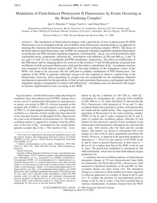
FIGURE 8: Proposed mechanism for period four modulation of the
Chl a fluorescence in PSII membranes at different pH. Centers in
which QA
- reoxidation proceeds with half-times of 860 µs, 7.6 ms,
and 135 ms are identified as type A, B, and C centers, respectively.
In type A centers the QB site is occupied by plastoquinone before
excitation. In type B centers this site is empty in the dark, and
QA
- reoxidation is limited by diffusion of plastoquinone to the QB
site. Type C centers are considered as inactive centers with an empty
QB site, and therefore, QA
- is reoxidized by cyt b559. KPC represents
the rate constant of primary charge separation. Numbers with the
S states signify values of uncompensated positive charge in these
states. According to the proposed mechanism, appearance of the
uncompensated positive charge in the S2 and S3 states (except type
B centers) results in conformational rearrangements in the PSII
reaction center which affect both charge separation reactions and
plastoquinone-binding properties of the QB site (see text). Type A
and C centers also differ in values of the uncompensated positive
charge in the S2 and S3 states at pH 5.5-6.5 and 7.6.
10638 Biochemistry, Vol. 38, No. 33, 1999 Putrenko et al.
8. functioned at physiological rates in the presence of exogenous
quinone acceptors (44). However, the electron acceptor for
QA
-
reoxidation in such centers was not identified. Our data
do not support the conclusion stated above for the following
reasons. First, the halftimes of the QA
-
reoxidation in the
inactive centers and back-reaction between QA
- and the Mn
cluster are significantly slower than those found in the present
study. Second, if the slow decay component reflected a redox
reaction with a participation of the Mn cluster of the WOC
in the S2 or S3 states, addition of CCCP should eliminate
the contribution of this component. Instead of this, addition
of CCCP caused an increase in the contribution of the slow
component. Meanwhile, the half-time of the slow component
in thylakoids (111 ms) is close to that of the reduction of
cyt b559, measured in thylakoids at pH 7.8 (t1/2 ) 100 ms)
(45). On the basis of comparisons of the half-time of cyt
b559 reduction and maximum halftime for the reduction of
the plastoquinone pool (6-10 ms), Whitmarsh and Cramer
(45) concluded that cyt b559 is an acceptor for no more than
1 of every 10 electrons accepted by the pool. There are
controversies concerning the source of the electrons for
oxidized cyt b559. Since DCMU concentrations which ap-
peared to block electron transport from water to methyl
viologen were found to only partially inhibit the rate of cyt
b559 photoreduction, Samson and Fork (46) suggested reduced
QA
- as a reductant to cyt b559. At the same time, Buser et al.
(47) found that DCMU inhibited the reduction of cyt b559
under conditions where the plastoquinone pool and QA were
reduced. Thus, they concluded that QB
•-(H+) or QBH2 was
the most likely source of the reduction of the oxidized cyt
b559. In contrast to thylakoids, the halftime of the reduction
of cyt b559 in PSII membranes was reported to be drastically
slower (70 s at pH 8.0) (47). However, the alkaline pH of
the assay medium in that study, although not effecting flash-
induced oxygen yield in thylakoids (15), would strongly
inhibit the oxygen flash yield in PSII membranes (pH 7.6)
(Figure 7). Therefore, one can expect the rate of the
reoxidation of cyt b559 to be different in PSII membranes at
pH 6.5 and 8.0 due to damage of the donor side. Physical
differences between active and inactive reaction centers could
be related to heterogeneity exhibited by cyt b559 (48). We
speculate that the slow decay component reflects the reoxi-
dation of QA
- by cyt b559 in inactive centers with modified
QB site. This assumption can be supported by the fact that
the population of PSII centers with the slow QA
-
reoxidation
rate (type C centers in Figure 8) is smaller in thylakoids than
in PSII membranes and increases in the modified PSII
membranes. However, further studies are required to confirm
this suggestion.
Analysis of the QA
-
decay in thylakoids and PSII
membranes shows that type B centers in these preparations
differ in properties of the WOC. Thus the amount of such
centers in PSII membranes as opposed to thylakoids was not
S-state dependent and they seemed to be incapable of O2
production. Such a difference is likely due to damage of these
centers during preparation of PSII membranes. There is also
variance in the WOC properties between type A and type C
centers in PSII membranes at different pH. On the basis of
these results, one can consider QB site properties to be related
somehow to capacity of the WOC.
Interestingly, the period two changes in the amount of the
type A centers and the type B and type C centers are opposite
in phase. It is not possible to observe this in control
thylakoids due to the absence of a binary oscillation in the
fluorescence; however, the same phase shift was also seen
in thylakoids in the presence of CCCP. These results imply
that the proportion of active and inactive PSII reaction centers
is dependent on the redox state of QB.
Which Process Is Responsible for Period Four Oscillation
in PSII Fluorescence? The rate of reduction of P680
+
is
dependent on S state (t1/2 ) 23 ns in the S0 and S1 states and
a biphasic reduction with t1/2 ) 50 and 260 ns in the S2 and
S3 states (13)) which results in S-state dependent changes
in the concentration of P680
+ during a microsecond flash.
As P680
+
is a known quencher of fluorescence, this could
result in period four oscillations in fluorescence yield.
However, this possibility can be ruled out as the period four
oscillation is still present at times longer than 5 ms after
excitation even though P680
+
is fully reduced to P680 on this
time scale (49).
Oxygen, another known quencher of fluorescence, has also
been proposed to contribute to period four oscillations in
fluorescence yield (19). However, oxygen does not yet appear
at 100 µs after excitation, even though period four oscillations
do, and oxygen reaches equilibration between its concentra-
tion in local and bulk phases in a few milliseconds (50) so
could not explain the persistence of period four oscillations
at longer times. In addition, the removal of the extrinsic 17
and 24 kDa proteins, which inhibits the flash-induced O2
yield by only 40%, resulted in complete elimination of period
four oscillations in fluorescence.
Even though oxygen is not likely the direct quencher of
fluorescence, in our studies using various procedures to alter
donor side capacity, we have found that a decrease in the
oxygen yield does correlate with a diminution of the period
four modulation. For example the period four changes in
the amount of type A centers is significantly decreased at
pH 7.6 as is the oxygen yield. Plijter et al. (51) reported
that PSII membranes at pH 8.3 still exhibited a period four
oscillation in absorption changes at 345 nm, which are
attributed to the oxidation of the Mn cluster, even though
oxygen evolution was completely inhibited at this pH. A 1.5
ms phase of the 345 nm absorption changes related to the
reduction of the Mn cluster during the S3-(S4)-S0 transition
slowed to 3.2 ms at pH 8.3. Putting together our results
(Table 3 and Figure 5) and results presented above, one can
conclude that the four-step oxidation of the Mn cluster alone
is not sufficient to produce modulation of the PSII fluores-
cence. Normal chemistry, proceeding at the WOC and
resulting in oxidation of water and formation of O2, appears
to be required to observe a period four in the fluorescence.
If oxygen itself is not responsible, it is reasonable to suggest
that deprotonation events occurring during water oxidation
are responsible for the period four modulation of both PSII
charge separation and QB site properties.
How Proton Release Could Affect the PSII Fluorescence.
It is widely accepted that the absence of proton release during
the S1-S2 transition results in the appearance of uncompen-
sated positive charge in the S2 and S3 states (11, 13). Several
studies indicate the influence of an external electric field on
fluorescence (17, 18). The oscillation patterns of the slow
decay component observed in thylakoids and PSII mem-
branes are explained well with respect to the presence of
charge surplus in the S2 and S3 states. However, it is most
Modulation of PSII Fluorescence by Events at WOC Biochemistry, Vol. 38, No. 33, 1999 10639
9. unlikely that this charge surplus directly affects the QB site.
We suggest that conformational rearrangements of protein-
(s) in the reaction center induced by the appearance of the
uncompensated positive charge influence QB site properties.
This suggestion is supported by recent work of Christen and
Renger (52). They concluded that Si state dependence of
proton/deuterium exchange effect on the fast P680
+
reduction
kinetics is not easily reconcilable with a simple electrostatic
effect caused by a single localized charge and may rather
reflect structural differences of the WOC in redox states S0,
S1 versus S2, S3. However, the Fmax yield may be directly
influenced by the uncompensated charge.
In accordance with pH-dependent pattern of the extrinsic
proton release accompanying the S-state transitions of the
WOC (14), the predicted stoichiometry of uncompensated
charge is 0:-0.75:+0.25:+0.25 at pH 5.5, 0:-0.3:+0.6:
+0.65 at pH 6.5, and 0:-0.05:+0.5:+0.5 at pH 7.6 for the
S0, S1, S2, and S3 states, correspondingly. However, no
correlation is observed between the pH-dependent changes
in the given stoichiometry and changes in the period four
modulation of the fluorescence at the same pH (Figures 3B
and 5A,B). This implies that the extrinsic proton release does
not affect the given processes. It has been suggested that
the extrinsic proton release is related to amino acid pKa shifts
indirectly caused by events at the WOC (53, 54). Our data
are consistent with this idea if the fluorescence modulation
is proposed to reflect intrinsic deprotonation events. It is
reasonable to expect that deprotonation of a protonatable
group, which is not occurring or negligible at pH 5.5-6.5
during the S1-S2 transition, could rise significantly at pH
7.6. This suggestion can be supported by the fact that the
extrinsic proton release during the S1-S2 transition and
absorption changes at 435 nm, associated with this release,
are small at pH 5.5-6.5 (0-0.1) but become maximal near
pH 7.65 (approximately 0.5) (14). pH-dependent changes in
the period four modulation of the population of type A
centers suggest that the pK of the proposed protonatable
group in such centers is in the 7.3-7.5 range. Histidine
residues that can titrate in this range have been suggested to
be ligands to the Mn cluster (55, 56). In type C centers the
pK of this protonatable group shifts to approximately 6.9-
7.1. The proposed mechanism for the period four modulation
of the fluorescence at different pH in PSII membranes is
shown in Figure 8. Such a mechanism suggests that depro-
tonation in the vicinity of the Mn cluster at pH 7.6 in the
type A and type C centers, resulting in a decrease in local
uncompensated charge in the higher S2 and S3 states, appears
to disturb the normal chemistry of water oxidation.
Formation of H2O2 attributed to the donor side of PSII in
PSII core complexes at pH 7.6 was reported by Fine and
Frasch (57). However, it is unclear whether uncompensated
positive charge itself and/or conformational rearrangements
of the WOC induced by the charge are sufficient for water
oxidation. Alternatively, disturbance of the normal chemistry
of water oxidation at pH 7.6 may be explained by pH-induced
change in the Mn cluster properties. However this mechanism
would imply that, in contrast to fluorescence, the water
oxidation process is not effected by intrinsic deprotonation
events.
The mechanism proposed in this work to account for a
period four oscillation in the fluorescence is in contradiction
with the study of Delrieu and Rosengard (10). They ascribed
the oscillation pattern of the fluorescence yield measured
80 ms after exitation flashes in thylakoids to flash-dependent
changes in the S2 population. The simulation of the oscillation
pattern with respect to S2-state considered the dark distribu-
tion of S0, S1, S2, and S3 populations and parameters R, β,
and z to be 18, 63, 17, and 2 and 0.025, 0, and 10 (30). The
dark distribution of S populations and the values of the
parameters varied from those generated for O2 yield pattern
(12.5, 79, 8.5, 0; 0.06, 0, 9) within the same preparation.
They also differed considerably from generally accepted ones
(31, 58). These discrepancies do not support simulations of
the fluorescence yield pattern, which assume period four
oscillations arise from the S2 state.
Effect of NaCl Treatment. Interestingly, disappearance of
period four as well as period two modulation of the
fluorescence was observed in NaCl-treated PSII membranes
while the flash-induced O2 yield was inhibited by only 40%.
Elimination of a binary oscillation in the absorption changes
at 350 nm was previously detected in NaCl-washed PSII
membranes in the presence of hydroxylamine as an electron
donor for PSII (59). This effect was explained in terms of
an alteration of the acceptor side of PSII due to removal of
the 17 and 24 kDa proteins. The redox potential of QA/QA
-
shifts from -80 to +65 mV in Ca-depleted PSII membranes
(60). Both of these results imply long-range allosteric
coupling of the WOC to the QA site across the thylakoid
membrane. Recently, using fluorometric and Mo¨ssbauer
spectroscopy, Garbers et al. (61) found a correlation between
electron transfer from QA
- to QB and protein flexibility in
PSII membranes. Thus, elimination of a binary oscillation
in the fluorescence as a result of the removal of the extrinsic
proteins is more likely related to conformation changes in
the QA and/or QB site(s) than to changes in the Mn cluster
properties. The elimination of the period four oscillation by
NaCl treatment supports a mechanism of indirect modulation
of the fluorescence by uncompensated positive charge
through conformational rearrangements of protein(s) in the
PSII reaction center.
CONCLUSIONS
This work shows that the QA
-
decay in thylakoids and
PSII membranes is well described by three exponential decay
components which reflect QA
-
reoxidation in three distinct
types of PSII centers with differing QB site properties. The
Fmax yield pattern and the flash-dependent changes in the
proportion of the PSII centers are characterized by a
periodicity of four. Neither the four-step oxidation of the
Mn cluster, the flash-induced production of photosynthetic
oxygen, nor the extrinsic proton release is found to be
responsible for the period four modulation of the fluores-
cence. We suggest that both PSII charge separation reactions
and properties of the QB site are sensitive to intrinsic
deprotonation events in the immediate vicinity of the Mn
cluster. The mechanism of such a modulation suggests that
appearance of uncompensated positive charge due to the
absence of a deprotonation of a histidine residue during the
S1-S2 transition results in conformational rearrangements
in the PSII reaction center.
ACKNOWLEDGMENT
We thank Dr. G. Ananyev for presenting a scheme of the
microcell for oxygen yield measurements and the Brock
10640 Biochemistry, Vol. 38, No. 33, 1999 Putrenko et al.
10. electronics and mechanic shops for their excellent technical
support.
REFERENCES
1. Dau, H. (1994) Photochem. Photobiol. 60, 1-23.
2. Lavergne, J., and Trissl, H.-W. (1995) Biophys. J. 68, 2474-
2492.
3. Bowes, J. M., and Crofts, A. R. (1980) Biochim. Biophys. Acta
590, 373-384.
4. Crofts, A. R., and Wraight, C. A. (1983) Biochim. Biophys.
Acta 726, 149-185.
5. Van Gorkom, H. J., Pulles, M. P., Haveman, J., and Den Haan,
G. A. (1976) Biochim. Biophys. Acta 423, 217-226.
6. Robinson, H. H., and Crofts, A. (1983) FEBS Lett. 153, 221-
226.
7. Joliot, P., and Joliot, A. (1971) in Proceeding of the II
International Congress on Photosynthesis Research (Forti, G.,
Avron, M., and Melandri, Eds.) Vol. I, pp 26-38, Dr W Junk
Publishers, The Hague.
8. Delosme, R. (1971) in Proceeding of the II International
Congress on Photosynthesis Research (Forti, G., Avron, M.,
and Melandri, Eds.) Vol. I, pp 187-195, Dr W Junk
Publishers, The Hague.
9. Zankel, K. L. (1973) Biochim. Biophys. Acta 325, 138-148.
10. Delrieu, M. J., and Rosengard, F. (1993) Photosynth. Res. 37,
205-215.
11. Saygin, O¨ ., and Witt, H. T. (1984) FEBS Lett. 197, 224-226.
12. Witt, H. T., Schlodder, E., Brettel, K., and Saygin, O¨ . (1986)
Photosynth. Res. 10, 453-471.
13. Brettel, K., Schlodder, E., and Witt, H. T. (1984) Biochim.
Biophys. Acta 766, 403-415.
14. Rappaport, F., and Lavergne, J. (1991) Biochemistry 30,
10004-10012.
15. Jahns, P., and Junge, W. (1992) Biochemistry 31, 7398-7403.
16. Debus, R. J. (1992) Biochim. Biophys. Acta 1102, 269-352.
17. Dau, H., and Sauer, K. (1991) Biochim. Biophys. Acta 1098,
49-60.
18. Bulychev, A. A., Niyazova, M. M., and Turovetsky, V. B.
(1986) Biochim. Biophys. Acta 856, 218-225.
19. Shinkarev, V. P., Xu, C. H., Govindjee, and Wraight, C. A.
(1997) Photosynth. Res. 51, 43-49.
20. Whitmarsh, J., and Ort, D. (1984) Arch. Biochem. Biophys.
231, 3378-3389.
21. Berthold, D. A., Babcock, G. T., and Yocum, C. F. (1981)
FEBS Lett. 134, 231-234.
22. Miyao, M., and Murata, N. (1983) Biochim. Biophys. Acta
725, 87-93.
23. Ananyev, G. M., and Dismukes, G. C. (1996) Biochemistry
35, 4102-4109.
24. Meunier, P. C., and Popovic, R. (1988) ReV. Sci. Instrum. 59,
486-491.
25. Joliot, A., and Joliot, P. (1964) C. R. Acad. Sci. Paris 258,
4622-4625.
26. Dohnt, G., and Renger, G. (1984) in AdVances in Photosyn-
thesis Research (Sybesma, C., Ed.) Vol. 1, pp 429-432,
Martinus Nijhoff Dr. W. Junk Publisher, The Haag, The
Netherlands.
27. Forbush, B., and Kok, B. (1968) Biochim. Biophys. Acta 162,
243-253.
28. Kok, B., Forbush, B., and McGloin, M. (1970) Photochem.
Photobiol. 11, 457-475.
29. Goldberg, D. E. (1989) Genetic Algorithms in Search,
Optimization and Machine Learning, Addison-Wesley, New
York.
30. Delrieu, M. J., and Rosengard, F. (1988) Biochim. Biophys.
Acta 936, 39-49.
31. Messinger, J., Seaton, G., Wydrzynski, T., Wacker, U., and
Renger, G. (1997) Biochemistry 36, 6862-6873.
32. Cao, J., and Govindjee. (1990) Biochim. Biophys. Acta 1015,
180-188.
33. Kolber, Z. S., Prasil, O., and Falkowski, P. G. (1998) Biochim.
Biophys. Acta 1367, 88-106.
34. Renger, G., Eckert, H.-J., Bergmann, A., Bernarding, J., Liu,
B., Napiwotzki, A., Reifarth, F., and Eichler, H.-J. (1995) Aust.
J. Plant Physiol. 22, 167-181.
35. Dekker, J. P., Plijter, J. J., Ouwehand, L., and Van Gorkom,
H. J. (1984) Biochim. Biophys. Acta 767, 176-179.
36. Avron, M., and Neumann, J. (1968) Annu. ReV. Plant. Physiol.
Plant Mol. Biol. 19, 137-146.
37. Renger, G. (1972) Biochim. Biophys. Acta 256, 428-
439.
38. Ghanotakis, D. F., Yerkes, C., and Babcock, G. T. (1982)
Biochim. Biophys. Acta 682, 21-31.
39. Helgerson, S. L., Cramer, W. A., Harris, J. M., and Lytle, F.
E. (1974) Biochemistry 13, 3057-3061.
40. Cramer, W. A., Horton, P., and Donnell, J. J. (1974) Biochim.
Biophys. Acta 368, 361-370.
41. Crofts, A., Robinson, H. H., and Snozzi, M. (1984) in
AdVances in Photosynthesis Research (Sybesma, C., Ed.) Vol.
1, pp 461-468, Martinus Nijhoff, Dordrecht, The Netherlands.
42. Bennoun, P. (1970) Biochim. Biophys. Acta 216, 357-363.
43. Chylla, R. A., and Whitmarsh, J. (1989) Plant Physiol. 90,
765-772.
44. Graan, T., and Ort, D. (1986) Biochim. Biophys. Acta 852,
320-330.
45. Whitmarsh, J., and Cramer, W. A. (1977) Biochim. Biophys.
Acta 460, 280-289.
46. Samson, G., and Fork, D. (1991) Photosynth. Res. 27, 179-
185.
47. Buser, C. A., Diner, B. A., and Brudvig, G. W. (1992)
Biochemistry 31, 11449-11459.
48. Chylla, R. A., Garab, G., and Whitmarsh, J. (1987) Biochim.
Biophys. Acta 894, 562-571.
49. Witt, H. T. (1991) Photosynth. Res. 29, 55-77.
50. Lavorel, J. (1992) Biochim. Biophys. Acta 1101, 33-40.
51. Plijter, J. J., De Groot, A., Van Dijk, M. A., and Van Gorkom,
H. J. (1986) FEBS Lett. 195, 313-318.
52. Christen, G., and Renger, G. (1999) Biochemistry 38, 2068-
2077.
53. Kretschmann, H., Pauly, S., and Witt, H. T. (1991) Biochim.
Biophys. Acta 1059, 208-214.
54. Renger, G., and Wydrzynski, T. (1991) Biol. Met. 4, 73-80.
55. DeRose, V. J., Yachandra, V. K., McDermott, A. E., Britt, R.
D., Sauer, K., and Klein, M. P. (1991) Biochemistry 30, 1335-
1341.
56. Hoganson, C. W., Ghanotakis, D. F., Babcock, G. T., and
Yocum, C. F. (1989) Photosynth. Res. 22, 285-293.
57. Fine, P. L., and Frasch, W. D. (1992) Biochemistry 31, 12204-
12210.
58. Forbush, B., Kok, B., and McGloin, M. (1971) Photochem.
Photobiol. 14, 307-321.
59. Wensink, J., Dekker, J. P., and Van Gorkom, H. J. (1984)
Biochim. Biophys. Acta 765, 147-155.
60. Krieger, A., Rutherford, A. W., and Johnson, G. N. (1995)
Biochim. Biophys. Acta 1229, 193-201.
61. Garbers, A., Reifarth, F., Kurreck, J., Renger, G., and Parak,
F. (1998) Biochemistry 37, 11399-11404.
BI990518J
Modulation of PSII Fluorescence by Events at WOC Biochemistry, Vol. 38, No. 33, 1999 10641