This review discusses the role of TRIB3, a stress-induced protein, in linking metabolic dysfunction and cancer. TRIB3 regulates multiple stress response pathways including PI3K/AKT/mTOR and autophagy pathways. It does so through its pseudokinase and ubiquitination functions. TRIB3 expression is associated with insulin resistance, diabetes, and atherosclerosis by suppressing insulin signaling. Cancer cells can alter TRIB3 expression or localization to promote cell survival and growth. TRIB3 may act as a switch between homeostasis and disease in metabolic stress responses, making it a potential biomarker and therapeutic target for metabolic diseases and cancer.
Global Medical Cures™ | Antibiotics Resistance Threats in USA
DISCLAIMER-
Global Medical Cures™ does not offer any medical advice, diagnosis, treatment or recommendations. Only your healthcare provider/physician can offer you information and recommendations for you to decide about your healthcare choices.
Impact Assessment of the Community Animal Health System in Mandera West Distr...copppldsecretariat
The pastoralist communities in Kenya’s arid lands rely on their livestock for food and income, and basic veterinary care is one of the best ways to protect livestock assets and pastoralist livelihoods in these areas. This report examines the impact of a privatized, community-based veterinary service in the far northeast of Kenya, and focuses on the outcomes of clinical services provided by community-based animal health workers (CAHWs). Fatality rates in herds in treated by CAHWs using medicines from rural pharmacies were significantly lower than in herds where treatments were provided by untrained livestock keepers. The report adds to the substantial body of evidence already collected in Kenya on the impact and financial rationale for CAHW systems. Although many other countries have now legalized these systems and developed national guidelines for CAHW training, Kenya has yet to officially recognize CAHWs and overall, veterinary services in pastoralist areas often remain in the hands of untrained workers and unlicensed drug vendors.
[ Originally posted on http://www.cop-ppld.net/cop_knowledge_base ]
2. 4.2. Three UPR cascades control the progression of ERS . . . . . . . . . . . . . . . . . . . . . . . . . . . . . . . . . . . . . . . . . . . . . . . . . . . . . . . . . . . . . . . . . . . . . . . . . . . . . . . 00
5. TRIB3 regulates multiple stress response pathways . . . . . . . . . . . . . . . . . . . . . . . . . . . . . . . . . . . . . . . . . . . . . . . . . . . . . . . . . . . . . . . . . . . . . . . . . . . . . . . . . . . . . 00
5.1. Cross-talks between PI3K/AKT/mTOR, autophagy and TRIB3 . . . . . . . . . . . . . . . . . . . . . . . . . . . . . . . . . . . . . . . . . . . . . . . . . . . . . . . . . . . . . . . . . . . . . . 00
5.2. Cross-talks between NF-kB, MAPK and TRIB3 . . . . . . . . . . . . . . . . . . . . . . . . . . . . . . . . . . . . . . . . . . . . . . . . . . . . . . . . . . . . . . . . . . . . . . . . . . . . . . . . . . . 00
6. TRIB3 association with metabolic dysfunctions . . . . . . . . . . . . . . . . . . . . . . . . . . . . . . . . . . . . . . . . . . . . . . . . . . . . . . . . . . . . . . . . . . . . . . . . . . . . . . . . . . . . . . . . . 00
6.1. TRIB3 suppresses insulin signaling and glycogen storage . . . . . . . . . . . . . . . . . . . . . . . . . . . . . . . . . . . . . . . . . . . . . . . . . . . . . . . . . . . . . . . . . . . . . . . . . 00
6.1.1. TRIB3 overexpression in insulin resistance and diabetes . . . . . . . . . . . . . . . . . . . . . . . . . . . . . . . . . . . . . . . . . . . . . . . . . . . . . . . . . . . . . . . . . . . 00
6.1.2. TRIB3 is associated with atherosclerotic plaque rupture . . . . . . . . . . . . . . . . . . . . . . . . . . . . . . . . . . . . . . . . . . . . . . . . . . . . . . . . . . . . . . . . . . . 00
6.1.3. TRIB3 is associated with b-cell dysfunction and death . . . . . . . . . . . . . . . . . . . . . . . . . . . . . . . . . . . . . . . . . . . . . . . . . . . . . . . . . . . . . . . . . . . . 00
6.1.4. The TRIB3 Q84R polymorphism in diabetes and atherosclerosis . . . . . . . . . . . . . . . . . . . . . . . . . . . . . . . . . . . . . . . . . . . . . . . . . . . . . . . . . . . . 00
6.2. TRIB3 is associated with numerous diabetes-associated diseases . . . . . . . . . . . . . . . . . . . . . . . . . . . . . . . . . . . . . . . . . . . . . . . . . . . . . . . . . . . . . . . . . . . 00
6.2.1. Hyper-homocysteinemia . . . . . . . . . . . . . . . . . . . . . . . . . . . . . . . . . . . . . . . . . . . . . . . . . . . . . . . . . . . . . . . . . . . . . . . . . . . . . . . . . . . . . . . . . . . . . . 00
6.2.2. Non-alcoholic fatty liver disease . . . . . . . . . . . . . . . . . . . . . . . . . . . . . . . . . . . . . . . . . . . . . . . . . . . . . . . . . . . . . . . . . . . . . . . . . . . . . . . . . . . . . . . 00
6.2.3. Diabetic nephropathy . . . . . . . . . . . . . . . . . . . . . . . . . . . . . . . . . . . . . . . . . . . . . . . . . . . . . . . . . . . . . . . . . . . . . . . . . . . . . . . . . . . . . . . . . . . . . . . . . 00
7. Linking TRIB3 expression in obesity and cancer . . . . . . . . . . . . . . . . . . . . . . . . . . . . . . . . . . . . . . . . . . . . . . . . . . . . . . . . . . . . . . . . . . . . . . . . . . . . . . . . . . . . . . . . 00
7.1. TRIB3 expression in adipocyte differentiation and visceral obesity . . . . . . . . . . . . . . . . . . . . . . . . . . . . . . . . . . . . . . . . . . . . . . . . . . . . . . . . . . . . . . . . . 00
7.2. Potential of targeting TRIB3 in aggressive cancers . . . . . . . . . . . . . . . . . . . . . . . . . . . . . . . . . . . . . . . . . . . . . . . . . . . . . . . . . . . . . . . . . . . . . . . . . . . . . . . . 00
8. Perspectives . . . . . . . . . . . . . . . . . . . . . . . . . . . . . . . . . . . . . . . . . . . . . . . . . . . . . . . . . . . . . . . . . . . . . . . . . . . . . . . . . . . . . . . . . . . . . . . . . . . . . . . . . . . . . . . . . . . . . . . 00
Acknowledgments . . . . . . . . . . . . . . . . . . . . . . . . . . . . . . . . . . . . . . . . . . . . . . . . . . . . . . . . . . . . . . . . . . . . . . . . . . . . . . . . . . . . . . . . . . . . . . . . . . . . . . . . . . . . . . . . . 00
References . . . . . . . . . . . . . . . . . . . . . . . . . . . . . . . . . . . . . . . . . . . . . . . . . . . . . . . . . . . . . . . . . . . . . . . . . . . . . . . . . . . . . . . . . . . . . . . . . . . . . . . . . . . . . . . . . . . . . . . . 00
1. A long sought-after connection between metabolic
dysfunction and cancer
1.1. Metabolic diseases increase cancer-associated morbidity and
mortality
Chronic metabolic diseases like obesity, type-2 diabetes,
atherosclerosis, and cardiovascular disease (CVD) are becoming
increasingly prevalent worldwide [1e4]. Both hyperinsulinemia
and hyperglycemia contribute to the progression of obesity and
diabetes, and insulin resistance, manifested due to decreased in-
sulin receptor signaling, is the primary risk factor for these meta-
bolic disorders [5,6]. Interestingly, metabolic diseases are also
frequently associated with poorer cancer outcomes [7,8]. In the past
few decades, a number of studies have documented a clear link
between the metabolic syndromes and higher morbidity and
mortality due to different malignancies [9e11]. However, the
crucial mechanism(s) involved in linking these chronic pathologic
manifestations is not properly understood. As early as 1995,
Steenland et al., showed that men with diabetes present with a 39%
higher risk of developing colorectal and prostate cancer [12]. Calle
et al. (2003) published finding from the multicenter Cancer Pre-
vention Study (CPS) which followed more than one million adults
during 1982e1996, and clearly demonstrated that obese men and
women had a 40e80% increased threat of dying from cancers
[13,14]. The danger of having aggressive pancreatic, breast and
colorectal cancers are reported to be amplified in patients with high
body mass index (BMI) and several meta-analyses in patients with
diabetes also showed significantly higher cancer mortality, as
compared with nondiabetic individuals [15,16]. Indeed, the clini-
cally approved glyburide, metformin has provided better clinical
outcome in diabetic patients with advanced cancers [17]. Hence, a
thorough understanding of the long sought-after relationship be-
tween metabolic diseases and cancers will not only provide early
biomarkers for disease progression, but will also elucidate novel
therapeutic targets to decrease cancer-associated complications.
Furthermore, since tremendous increases in metabolic syndrome
are being reported in younger adults [4], which makes them more
susceptible to malignancies later in life, studies on the common
etiologies in metabolic diseases and cancer are garnering a lot of
attention [7,17e22].
Deleterious consequences of chronic inflammation and oxida-
tive stress are known to increase tumor progression and metastasis,
and also facilitate tumor resistance to both chemotherapy and
radiotherapy [7,20,23e28]. Studies have documented that the
visceral adipose tissue secreted inflammatory cytokines can pro-
mote insulin resistance in vascular cells [29e32]. Metabolic com-
plications of insulin resistance make individuals more susceptible
to chronic oxidative stress, neoplastic transformation and aggres-
sive tumor growth. A number of studies have also demonstrated
that second messenger signaling via the insulin and insulin like
growth factor (IGF) receptors play a crucial role in numerous other
chronic diseases such as autoimmunity, arthritis, alzheimer's dis-
ease and aging [33e36]. Thus, it is becoming apparent that the
chronic effects of inflammation in disrupting stress-adaptive
pathways in both normal and malignant cells may influence pro-
gression of these chronic diseases.
1.2. Inflammation: a common etiology in chronic diseases
Obesity induced adipokines, inflammatory cytokines, leptin,
proteolytic enzymes, and endogenous sex steroids, are known to
suppress the anti-inflammatory actions of insulin. The resultant
activation of vascular endothelium and decreased vasodilation of
smooth muscle cells increases blood pressure and causes hyper-
tension [32,37,38]. Increased adhesion of leukocytes and platelets
and the ensuing atherosclerotic thrombus formation further fosters
inflammatory stress and insulin resistance of the vasculature. Ad-
ipose tissue infiltrated macrophages and foam cells can also pro-
duce a state of chronic oxidative stress and inflammation, which
further promote the chronic metabolic dysfunctions [39,40].
Interestingly, the deleterious effects of insulin resistance, inflam-
mation and oxidative stress have also been implicated in both
oncogenic transformation of normal cell [41,42] and in increased
proliferation and metastasis of tumor cells, as shown by us [43] and
others [20,28,44]. Furthermore, since insulin resistance increases
both estrogen and testosterone levels by decreasing SHBG (sex
hormone binding globulin) [45] metabolic diseases can also
augment the growth of endocrine tumors like breast and prostate
cancers [7,23,43]. Therefore, insulin resistance is postulated to be a
common link and comorbidity in both metabolic diseases and
cancer. Indeed, insulin-induced glucose uptake is dysregulated in
D. Mondal et al. / Biochimie xxx (2016) 1e192
Please cite this article in press as: D. Mondal, et al., Tripping on TRIB3 at the junction of health, metabolic dysfunction and cancer, Biochimie
(2016), http://dx.doi.org/10.1016/j.biochi.2016.02.005
3. chronic hyperglycemia and hyperinsulinemia [46,47].
The mitochondrial NAD-dependent deacetylase sirtuin-3
(SIRT3) can help maintain the protective effects of insulin in both
skeletal muscles and adipose tissue [48]. Interestingly, a direct role
of the AKT pseudokinase, TRIB3 in regulating insulin sensitivity and
nutrient metabolism has been documented [49]. It is well estab-
lished that normal cells rely on mitochondrial oxidative phos-
phorylation of glucose to generate adenosine 50-triphosphate
(ATP). However, cancer cells utilize an alternate phenomenon
known as the “Warburg effect”, where aerobic glycolysis and
lactate production is used as the primary source of ATP [50,51]. As
early as in 2006, Schwarzer et al. had shown that TRIB3 can regulate
glucose metabolism and glycolysis, and these studies showed that
TRIB3 functions as an indicator of nutrient starvation via targeting
the PI3K/AKT pathway [52]. Numerous recent studies have also
shown a direct role of both hypoxia [53] and the PI3K/AKT pathway
[54] in tumor growth and therapeutic resistance. As will be dis-
cussed later, TRIB3 is now well-accepted inhibitor of AKT [55,56]
and TRIB3 gene expression is induced following hypoxic stress
[57,58].
Numerous control mechanisms and stress-adaptive pathways
exist in cells to prevent the aberrant intracellular signaling during
insulin resistance, which help maintain homeostasis in both
normal and malignant cells [59e62]. These master regulatory
pathways judiciously balance the nutrient-sensing and stress-
inductive machinery that dictate homeostasis, cellular dysfunc-
tions, or survival growth. Accurate identification of this ‘molec-
ular regulatory switch’ will be of significant importance as both an
effective disease biomarker and a potent therapeutic target. In the
following sections, we will present a number of independent
studies indicating that the mammalian ‘Tribbles’ proteins are at the
nexus of these metabolic pathways and may function as the ‘mo-
lecular regulatory switch’. We will focus on the most multifunc-
tional of these Tribbles proteins, i.e. TRIB3, and present findings
that demonstrate its role in regulating disease progression in both
metabolic syndrome and cancer [63e68].
2. The multifunctional tribbles
2.1. Discovery of mammalian TRIB proteins
Tribbles protein, originally identified in Drosophila melanogaster
(fruit fly), was shown to be an evolutionarily conserved protein that
regulates multiple cellular processes [69,70]. Initial studies showed
that loss of Tribbles can increase proliferation of fruit fly embryos
and its overexpression decreased cell cycle progression enhanced
morphogenesis [71]. Molecular mechanistic studies revealed that
Tribbles overexpression arrested cells in the G2 phase of cell cycle
by enhancing proteasomal degradation of two Drosophila cell cycle
regulating phosphatases, String and Twine [72]. These proteins
were later found to be homologs of mammalian cyclin dependent
kinase, Cdc25 [73]. Since Cdc25 activates the major mitotic kinase
Cdk1, its degradation decreases mitosis and growth of mammalian
cells. Tribbles was also found to increase the ubiquitination and
proteasome-mediated degradation of the Drosophila protein, Slbo
(slow border cells) [74], later identified as a homolog of the
mammalian transcription factor C/EBP (CAAT enhancer binding
protein) [75]. Indeed, multiple C/EBP transcription factors, e.g.
alpha, beta, delta (a, b and d) play crucial roles in lineage specific
differentiation of endothelial, smooth muscle and adipose cells
[76e78]. These C/EBP proteins are also well-known regulators of
cancer cell growth [79,80]. Indeed, Bowers et al. (2002) was the first
to show that the mammalian tribbles homolog, TRIB3 (a.k.a. SKIP3)
is overexpressed in human tumors and is directly associated with
cellular dysfunctions [81].
Mechanistic understanding of the numerous functions of
Drosophila Tribbles fueled intense research in this field, which
facilitated the discovery of three mammalian homologs, TRIB1,
TRIB2 and TRIB3 [82,83]. These early studies on TRIB proteins were
found to decrease cell migration and increase differentiation of
different mammalian cells. Subsequent studies in multiple labora-
tories documented a direct role of mammalian TRIBs in cell prolif-
eration, metabolism, and oncogenic transformation [84e89].
Interestingly, further studies on the TRIB homologs also divulged
significant differences in their amino acid contents and protein
tertiary structures. Furthermore, although the TRIB genes were
found to code for similar functional domains, distinct variations in
their functional activities were clearly evident in different labora-
tories. Homology between TRIB1 and TRIB2 was found to be as high
as 71.3%. However, TRIB3 only showed 53.3% and 53.7% homology
with TRIB1 and TRIB2, respectively [83]. The expression of TRIB
proteins and their subcellular localization also varied in different
tissues, as well as in different disease models
[49,56,64,65,84,86,87]. TRIB1 was observed to preferentially
localize to the nucleus and TRIB2 was usually detected in the
cytoplasm; however, TRIB3 expression was documented in both
cellular compartments. As compared to TRIB1 and TRIB2, both
subcellular localization functional association studies clearly
implicated a more global importance of TRIB3 in different diseases.
This has been presented in two highly cited review articles by
Prudente et al. (2009 2012) [90,91]. Findings within the last
decade emphasized that the multimodal actions of TRIB3 coordi-
nate important metabolic processes including glucose and lipid
metabolism, inflammation, survival, oxidative stress, apoptosis, and
most importantly, tumorigenesis. Indeed, both genotoxic stress and
ER-stress were found to differentially regulate TRIB3 expression
[92]. The crucial importance of TRIB3 is further underscored from
the recent identification of a small molecule ABTL0812 that upre-
gulates TRIB3 and its entry into several antitumor clinical trials [56].
2.2. Differential effects of TRIBs on second messenger signaling
Independent studies on different TRIB isoforms have provided a
general consensus that TRIB protein expression is critically regu-
lated by cellular stress due to either overstimulation or deprivation
of nutrients, like glucose, amino acids and free fatty acids
[83,84,93e98]. Both transcriptional regulation of TRIB genes and
post-translational modification of TRIB proteins have been docu-
mented. In addition, functional association of TRIBs with other
cellular proteins and their differential subcellular localization in
cytosol and nucleus ultimately dictates their actions. Activation of
signaling via inflammatory cytokines such as TNFa (tumor necrosis
factor-alpha), IL-3 (interleukin-3) and HIF-1a (hypoxia inducible
factor-1-alpha) as well as depletion of growth factor signaling from
NGF (nerve growth factor) and IGF-1 (insulin like growth factor-1)
can upregulate the expression of different TRIB isoforms. Indeed,
immune-histochemical (IHC) analysis of tissues from patients with
metabolic diseases revealed increased expression of multiple TRIB
proteins, and most interestingly, both changes in expression (up or
down regulation) and subcellular localization (cytosol vs. nucleus)
was frequently seen [66,67,99]. TRIB1 overexpression has often
been associated with metabolic dysfunctions in vascular tissues
[100]. In endothelial cells, TRIB1 plays a direct role in regulating
both PKB (protein kinase B/AKT) and RAR (retinoic acid receptor)
signaling [101]. In smooth muscle cells, overexpression of TRIB1
inhibits MAPK (mitogen activated protein kinase) mediated acti-
vation of transcription factor AP-1 (activated protein-1) [85].
Furthermore, an important role of TRIB1 in differentiation of M2-
like macrophages was recently shown by Satoh et al. (2013) [102].
Interestingly, although TRIB2 levels were not significantly
D. Mondal et al. / Biochimie xxx (2016) 1e19 3
Please cite this article in press as: D. Mondal, et al., Tripping on TRIB3 at the junction of health, metabolic dysfunction and cancer, Biochimie
(2016), http://dx.doi.org/10.1016/j.biochi.2016.02.005
4. augmented in vascular endothelium obtained from obese or dia-
betic patients, its induction has been associated with both CNS
(central nervous system) dysfunctions and tumorigenesis.
Increased circulating levels of antibodies against TRIB2 were found
in individuals with narcolepsy [103]. TRIB2 expression was also
shown to be augmented in lymphocytes from AML (acute mye-
logenous leukemia) patients [104]. Indeed, TRIB2 has been directly
linked to oncogenic transformation in AML [104], liver cancer [105]
and melanoma [106]. Similar to TRIB2, TRIB3 levels were signifi-
cantly associated with tumor nodes, as compared to the sur-
rounding normal stroma. However, unlike TRIB2, the expression of
TRIB3 was found to be either upregulated or downregulated in both
primary tumors and in different cancer cell lines
[68,89,96e98,107e111].
Remarkably, although TRIB3 message is ubiquitously expressed
in both mesenchymal and hematopoietic cells, TRIB3 protein levels
were more precisely regulated in both a context- and
microenvironment-dependent manner. Brisard et al. (2014) pro-
vided a very intriguing observation in primary oocytes, where dif-
ferential expression and subcellular localization of all three TRIB
proteins was observed during the different pre-ovulatory periods in
cumulus cells [112]. These findings clearly suggested that the
mammalian TRIBs act as a central node involved in fine tuning of
multiple cellular processes, in both normal and transformed cells.
Since TRIB3 is the most well studied member of this family, in the
following sections we will discuss the multimodal actions of TRIB3
and its role in causing different pathophysiologic manifestations.
We will also provide an overview on how TRIB3 expression is
exquisitely regulated during homeostasis and stress. Lastly, we will
also discuss the findings that show TRIB3 as a disease biomarker
and a novel therapeutic target in metabolic diseases and cancer.
3. The mammalian TRIB3 protein
3.1. Multimodal actions of TRIB3
Studies have recognized the crucial functions of TRIB3 in cells of
both mesenchymal [65,91,113e116] and hematopoietic lineages
[94,117e119]. Although the deleterious actions of TRIB3 in meta-
bolic tissues are focused on its potent ability to dysregulate insulin
signaling [65,91,120,121] multiple other functions of TRIB3 are also
evident in diverse cell types, such as apoptosis in both neuronal
cells [52] and pancreatic b-cells [88] and glucose production in
hepatocytes [122,123]. Studies have also shown that TRIB3 can
promote ubiquitination and degradation of different cell cycle
regulatory proteins [124] and this attribute of TRIB3 also dictates its
own degradation via the association with an E3-ubiquitin ligase,
SIAH1 [125]. In contrast, TRIB3 is found to protect HEK293 cells
against the growth inhibitory and cytotoxic effects of the ER-stress
(ERS)-induced transcription factor, ATF4 [126]. Interestingly,
although TRIB3 increased neuronal cell death due to nutrient
deprivation [127] it was associated with increased survival in mast
cells [117]. These dissimilar functions of TRIB3 have been linked to a
cross-talk between TRIB3, AKT and the FoxO regulated signaling
axis [128]. Due to its crucial role in cell cycle, proliferation dif-
ferentiation, recent studies also suggest that TRIB3 may be impor-
tant in maintaining the pluripotency of normal stem cells
[112,129,130] and in the EMT (epithelial-to-mesenchymal transi-
tion) phenotype of cancer stem cells (CSCs) [131e133]. Interest-
ingly, depending on the stage of tumor progression TRIB3
expression varied in different solid tumors, e.g. lung, colon,
esophageal, and breast cancers [68,96,134,135]. These crucial find-
ings on TRIB3 mediated regulation of cell cycle progression and
mitogenesis in both normal and transformed cells have thus
sparked new directions in research to understand its function in
different disease manifestations, as well as its utility as a biomarker
of indolent vs. aggressive disease. The ability of TRIB3 to sensitize
lymphoma cells to sorafenib-induced apoptosis [136], in increasing
the action of autophagy-mediated cell death in glioma cells
[108,137] and in augmenting ERS mediated chemosensitization of
prostate cancer cells to taxols [110] has fostered substantial thera-
peutic implications for this enigmatic protein, as well [56].
3.2. Functional motifs of TRIB3
The TRIB3 mRNA is coded from six exons located in human
chromosome-20 (20p13-p12.2) which generates a 358 amino acid
protein of approximately 65 KDa [138e140]. Protein sequence
analysis showed that TRIB3 has three functional motifs; i.e. a cen-
tral kinase-like domain, and both N-terminal and C-terminal
protein-binding domains with distinct functionalities (Fig. 1).
Earlier studies had focused on the central serine/threonine kinase-
like domain of TRIB3 [138]. Although this region of TRIB3 contains
the kinase catalytic core, it is divergent at the consensus ATP-
binding pocket and thus does not possess any kinase activity
[64,95]. TRIB3 was thus classified as a ‘pseudokinase’ similar to ILKs
(integrin-linked kinases) and JAKs janus tyrosine kinases [141,142].
Studies have shown that TRIB3 can bind to a number of kinase-
dependent proteins and dysregulate their function by negatively
regulating their phosphorylation, thus alter multiple signal trans-
duction pathways [64,95,138e140]. TRIB3 drastically affected cell-
fate determination by negatively affecting the activation of both
PI3K/AKT [67,95] and Notch [107,135], which are crucial survival
pathways in cells undergoing stress. Furthermore, by suppressing
the functional activation of lineage specific transcription factors
like C/EBP (CAAT enhancer binding protein) and PPAR (peroxisome
proliferating activated receptor) TRIB3 was also shown to alter
differentiation of endothelial cells, myocytes and adipocytes
[77,87,94,143,144].
In addition to the well-studied ‘pseudokinase’ domain, both the
N-terminal and C-terminal domains of TRIB3 have also been asso-
ciated with functions that act in additive or synergistic manner to
further fine tune numerous signal transduction cascades. The N-
terminal domain is high in serine and proline content, a charac-
teristic of the PEST sequence [i.e. proline (P), glutamic acid (E),
serine (S), and threonine (T)] which is involved in the degradation
of TRIB3 via both SIAH1 [125] and cdh1 [144]. Interestingly, com-
bined action of this PEST sequence and the ubiquitin ligase function
of TRIB3 can also facilitate proteasomal degradation of multiple
client proteins and crucial cellular transcription factors such as
ATF4, C/EBP, PPAR and IkBa [57,126,143,145e147]. A nuclear locali-
zation signal has also been detected at this N-terminal region of
TRIB3 [96] and thus TRIB3 may be able to alter transcription factor
activity in both the cytosol and nucleus. Interestingly, Hua et al.
(2015) recently reported that TRIB3 is a stress-induced protein that
mediates reciprocal antagonisms between autophagic and protea-
somal degradation systems, which facilitates the connection of
insulin (or IGF-1) signaling to tumor promotion via the induction of
autophagy and lysosomal degradation [148]. This important finding
also documented that TRIB3 interactions with SQSTM1 can
decrease the degradation of this autophagic receptor, and increased
the accumulation of other ubiquitinated proteins. Furthermore,
siRNA mediated decrease in TRIB3 was able to restore autophagy,
and most interestingly, TRIB3 knockdown was successful in atten-
uating tumor growth and metastasis [148]. In contrast to several
previous studies in tumor specimen, where lower TRIB3 and higher
phosphorylated-AKT levels were associated with poor patient
outcome [66,89,134] the above finding by Hua et al. (2015), showed
that both TRIB3 expression and insulin signaling were activated in
cancer patients with a negative prognosis [148]. These investigators
D. Mondal et al. / Biochimie xxx (2016) 1e194
Please cite this article in press as: D. Mondal, et al., Tripping on TRIB3 at the junction of health, metabolic dysfunction and cancer, Biochimie
(2016), http://dx.doi.org/10.1016/j.biochi.2016.02.005
5. also suggested that the blocking of TRIB3-SQSTM1 interactions by
using small molecule inhibitors may be a novel strategy against
cancers, especially in patients with diabetes [148].
The C-terminal domain of TRIB3 contains two conserved se-
quences, i.e. a binding site for mitogen activated protein kinases
(MAPKK) termed the MEK1 domain [95,107,140] and the COP1
domain (constitutive photomorphogenic protein 1) which regulates
ubiquitin ligation of TRIB3 associated proteins [80,105,125,149]. The
MEK1 site mediates interactions with multiple MAPKKs, and a high
throughput kinase inhibitor screen revealed that TRIB3 is a potent
inhibitor of MAPK-ERK/TGFb pathway in breast cancer cells [107]. By
facilitating the ubiquitin binding at the COP1 site, TRIB3 can regulate
proteasomal degradation and half-life of multiple client proteins.
Indeed, TRIB3 was able to suppress insulin-induced adipocyte dif-
ferentiation by negatively regulating PPARg transcriptional activity
[143]. An in vivo study in a rat model of insulin resistance also
showed that knockdown of TRIB3 can improve insulin sensitivity
through PPARg activation [145]. In tumor models of AML [146] and
liver cancer [147] TRIB3 promoted degradation of both C/EBPa and
NF-kB to exert its anti-tumor effects. Interestingly, Aynaud et al.
(2012) also documented that TRIB3 can interact with the DNA
mutator cytidine deaminase APOBEC-3A (A3A) resulting in its
proteasome-independent degradation [148]. Co-transfection of A3A
and TRIB3 expression vectors reduced nuclear DNA editing and
suggested that TRIB3 may be a guardian of genomic integrity, which
is disrupted during the process of oncogenic transformation. There-
fore, the last few years have further illuminated our understanding of
how TRIB3 functions at the juncture of homeostasis, metabolic dis-
ease and cancer.
4. TRIB3, a stress-induced factor
4.1. Metabolic stress: a balance between homeostasis and disease
Microenvironmental stressors, which are activated following
either nutrient excess or nutrient deprivation, can alter cellular
homeostasis [150e153]. Increased protein synthesis is needed to
cope with increased metabolic demand during stress in both
normal and transformed cells, and thus, a coordinated protein
folding by the endoplasmic reticulum (ER) and increased protein
degradation by the proteasome and lysosome are of significant
importance towards maintaining homeostasis [59,119,137,152,153].
The proteolytic systems recognize and destroy misfolded or
damaged proteins and are essential in basic cellular processes
including cell cycle modulation and second messenger signaling.
Although the equilibrium is preserved under physiologic condi-
tions, it is severely affected when unfolded and misfolded proteins
accumulate. Therefore, an adaptive signaling pathway called the
unfolded protein response (UPR) is initiated to re-establish protein
balance in cells undergoing stress. However, compromised ER
function can also initiate signaling networks that suppress the
stress-adaptive mechanisms, which results in cellular dysfunctions
that promote uncontrolled UPR and progression towards ER-stress
(ERS). Prolonged or severe ERS, and an inability of cells to sustain
the UPR, then subverts the survival pathways and initiates pro-
grammed cell death. As will be discussed in the later sections, TRIB3
has a crucial function in regulating whether cells sustain UPR and
promote homeostasis or then progress towards ERS and promote
cell death. Comprehensive reviews on the role of ERS pathways in
metabolic diseases and cancer had been provided before [154,155],
and hence, it is only discussed in brief in the following sections.
Normal cells have a low threshold for stress-adaptation and
show low levels of UPR. Chronic UPR activation results in severe
metabolic dysfunctions in normal cells [59,152]. Therefore, strate-
gies to decrease the constitutive UPR, and its progression towards
ERS, are being investigated as therapy against the metabolic syn-
drome [48,60,66,148]. On the other hand, due to their increased
protein synthesis, aggressive cancer cells have constitutive UPR.
Both UPR and autophagy pathways are well-known to promote
cancer growth, metastatic progression and therapeutic resistance
Fig. 1. Functional domains of TRIB3 protein. (A) The N-terminal domain of TRIB3 contains a protein degradation motif (PEST) and a nuclear localization signal (NLS). The central
domain contains the divergent kinase catalytic core. The C-terminal domain contains a MEK1 binding site and a COP1 biding site. (B) The ‘pseudokinase’ function of TRIB3 inhibits
phosphorylation (activation) of AKT and suppresses the PI3K/AKT/mTOR axis. This function also inhibits IkB phosphorylation and increases nuclear NF-kB levels. The MEK1 binding
motif inhibits numerous MAP-kinases and abrogates the downstream RAS/RAF/MEK/ERK axis. The COP1 motif interacts with E3 ubiquitin ligase to regulate proteasomal degra-
dation of TRIB client proteins, e.g. transcription factors C/EBPa, PPARg and ATF-4.
D. Mondal et al. / Biochimie xxx (2016) 1e19 5
Please cite this article in press as: D. Mondal, et al., Tripping on TRIB3 at the junction of health, metabolic dysfunction and cancer, Biochimie
(2016), http://dx.doi.org/10.1016/j.biochi.2016.02.005
6. [156e158]. However, the exploitation of this overactive UPR to
promote ERS-induced apoptosis in cancer cells is also becoming a
promising approach in multiple tumor models [110,132,133,159].
Since TRIB3 is upregulated by several stress-induced transcription
factors [81,95,160] and since it is notorious in regulating the balance
between UPR and ERS [161], the targeting of TRIB3 may also be a
novel approach [58,110]. Below, we are presenting a short
description of how TRIB3 expression is fine-tuned by cross-talks
with other intracellular signaling pathways, a proper understand-
ing of which will provide better treatment strategies.
4.2. Three UPR cascades control the progression of ERS
Defective proteins are subjected to “proof-reading” and are
rapidly degraded by proteases, and disruption of this protein ho-
meostasis can result in chronic diseases. The stress-adaptive phase
of UPR assists in increased protein folding by numerous chaperone
proteins, enhanced protein degradation by proteasomal machinery,
and decreased protein translation in the rough ER [154,155] (Fig. 2).
Three downstream UPR transducer cascades are initiated following
proteotoxic stress, these are: (i) the ATF6 (activating transcription
factor-6) pathway, (ii) the IRE1 (inositol requiring enzyme-1)
pathway, and (iii) the PERK (protein kinase RNA-like endoplasmic
reticulum kinase) pathway. One of the most crucial molecular
chaperones responsible for proper folding of proteins is Grp78
(Glucose regulated protein 78; a.k.a. BiP) [162]. Under homeostatic
conditions, the ER membrane-associated Grp78 is bound to all
three of the above UPR transducers (ATF6, IRE1 and PERK). How-
ever, following metabolic stress, Grp78/BiP dissociates from these
transducers and initiates the downstream cascades that either
facilitate stress-adaptation or promote stress-induction [154,155].
Dissociation of ATF6 (arm-1) from Grp78 causes its translocation
into the golgi apparatus where it is cleaved to its active form by two
site specific proteases (SP-1 and SP-2). Active ATF6 then translocate
into the nucleus and enhances transcription of multiple chaperones
like Grp78 and Grp94. Therefore, the ATF6 axis primarily functions
in a pro-survival capacity to counteract metabolic stress [163]. In
the second UPR cascade, dissociation from Grp78 activates the
endoribonuclease action of IRE1, which causes the splicing of XBP-1
mRNA (X-box binding protein-1). The spliced XBP1 mRNA codes for
a transcription factor that induces genes needed for increased
protein folding capacity, and thus again help promote cell survival.
In mice, hepatic fatty acid and triglyceride metabolism was shown
to occur through XBP1 [164]. Importantly, Duan et al. (2015)
showed that several miRNAs regulate XBP1 expression and pro-
gression of cardiac hypertrophy and heart failure in vivo [165].
These findings clearly implicated the therapeutic potential of tar-
geting both XBP1 transcription and splicing. Interestingly, spliced
XBP1 was found to suppress intestinal tumorigenesis [133]. Sig-
nificant cross-talk also exists between these first two arms of the
UPR, where the transcription of XBP-1 gene is upregulated by ATF6
and vice versa [166e168]. However, unlike the function of first two
arms of UPR, where the primary goal is to facilitate homeostasis,
the third arm of the UPR is responsible for dictating the stress-
adaptation and stress-susceptibility of cells [169e173]. Accumu-
lating evidences suggest that this third arm is directly involved in
UPR progression to ERS, and this metabolic switch primarily occurs
via the critical regulation TRIB3 protein levels
[48,58,64,66,88e91,120,134,148,160,174]. Numerous studies have
associated TRIB3 with stress-induced cellular dysfunctions and
Fig. 2. Exacerbated UPR causes ERS progression that induces TRIB3 expression. Accumulation of unfolded proteins results in cytotoxicity, unless cellular homeostasis is restored via the
unfolded protein response (UPR). The UPR increases ER chaperones, e.g. Grp78, Grp94, contents to restore normal ER function. Under ER-stress (ERS) conditions, ATF-6, IRE1 and
PERK dissociate from Grp78 and activate multiple downstream pathways, which either enable cell homeostasis or progression of ERS. The PERK-eIF2a pathway suppresses global
protein synthesis, but upregulates both ATF4 and CHOP levels. Thus, prolonged ER-stress overwhelms UPR survival mechanisms to initiate pro-apoptotic pathways by activating the
transcription factors (CHOP and ATF4) that enhance TRIB3 expression.
D. Mondal et al. / Biochimie xxx (2016) 1e196
Please cite this article in press as: D. Mondal, et al., Tripping on TRIB3 at the junction of health, metabolic dysfunction and cancer, Biochimie
(2016), http://dx.doi.org/10.1016/j.biochi.2016.02.005
7. death. The ERS-induced pancreatic b-cell apoptosis via the NF-kB
pathway was shown to be regulated by TRIB3 [64,88]. Furthermore,
in high-fat-fed obese mice, skeletal muscle insulin resistance was
clearly associated with increased TRIB3 expression [48]. The
hypoxia-induced TRIB3 was clearly linked to decreased AKT levels
and good prognosis in breast cancer [89] and colorectal cancer
[134] patients.
In the crucial third arm of UPR, following dissociation from
Grp78, the stress sensing domain of PERK is activated via both auto-
phosphorylation and homo-dimerization (Fig. 2). Activated PERK
then phosphorylates eIF2a (eukaryotic translation initiation factor
2 alpha) which reduce ER protein load and enable homeostasis.
However, although activated eIF2a can inhibit global translation, a
few proteins continue to be synthesized due to their internal
translation initiation sites (cap-independent). These UPR stimu-
lated transcription factors, i.e. ATF4 and CHOP, are responsible for
progression of UPR to ERS via augmenting TRIB3 gene expression
[160,175]. TRIB3 expression is primarily regulated at the level of
transcription by concerted actions of two basic leucine zipper
(bZIP) transcription factors, CHOP and ATF4
[95,96,111,117,128,160,175,176]. Although the translation of ATF4
(activated transcription factor-4) initially increases expression of
chaperone proteins and promote homeostasis, since ATF4 also
promotes the synthesis of another transcription factor, CHOP (C/
EBP homologous protein) [110,160,175] it is the most crucial factor
in dictating TRIB3 expression. In addition, the balance of UPR and
ERS can also be fine-tuned at this juncture via both a negative
feedback loop to suppress ATF4 and CHOP expression and a cross-
talk with other parallel signaling pathways that regulate the
downstream effects [26,66,109,137,161,170,177,178]. Indeed, TRIB3
can regulate both its own degradation [66,74,96,117] as well as the
degradation of both CHOP and ATF4 [57,126,177e179]. Thus, TRIB3
has been considered as both a target as well as a modulator of its
own induction (Fig. 3).
In summary, under conditions of transient or mild stress,
although the coexpression of ATF4 and CHOP increases TRIB3
transcription, TRIB3 also blocks their function on the TRIB3 gene via
a negative feed-back loop. However, under prolonged ERS the
accumulation of ATF4 and CHOP leads to the over-expression of
TRIB3, and TRIB3 sequestration by other proteins and TRIB3-
mediated suppression of AKT (as discussed below) suppresses ho-
meostasis and facilitates cellular dysfunctions and apoptosis. Thus,
the overexpression of TRIB3 trips the switch from survival to death.
5. TRIB3 regulates multiple stress response pathways
5.1. Cross-talks between PI3K/AKT/mTOR, autophagy and TRIB3
In addition to the UPR cascades initiated within the ER, cytosolic
and mitochondrial proteins also provide parallel mechanisms to
adjust intracellular stress in response to multiple exogenous stimuli
(Fig. 4). One of these critical interactions includes TRIB3-mediated
targeting of the PI3K/AKT/mTOR and Autophagy cascades
[180e185]. The PI3K/AKT/mTOR pathway is of crucial importance
in regulating normal metabolic functions [67,128,183] and this
pathway is often activated in aggressive cancer cells [26,40,170].
Briefly, PI3K (phosphatidylinositide 3-kinase) activation phos-
phorylates and activates AKT/PKB (protein kinase-B). The activated
AKT initiates a number of downstream effectors such as CREB
(cAMP response element binding protein) and mTOR (mammalian
target of rapamycin). Interestingly, the first evidence of TRIB3 as a
negative modulator of AKT and mTOR activity was provided by Du
et al.(2003) [123]. The serine/threonine protein kinase mTOR also
plays a direct role in regulating cell survival via autophagy. Similar
to the proteasomal degradation machinery, autophagy is another
protein degradation system in the lysosome, and numerous studies
demonstrate its involvement in pathophysiological processes
[54,56,62,137,170,181]. Similar to the three UPR cascades discussed
above, both mTOR and autophagy pathways can also sense nutrient
levels in cells, by integrating second messenger signaling from
factors like insulin, growth factors, and amino acids. In 2010, Liu
et al. had shown that overexpression of TRIB3 in skeletal muscle
cells of diabetic patients can reduce insulin-stimulated AKT activity
[187] and a follow-up study by this same group corroborated the
crucial role of TRIB3 in regulation of nutrient metabolism during
both short-term fasting or glucose excess [49]. Indeed, the skeletal
muscle is a major site of glucose disposal and one of the major
characteristics of diabetes patients is reduced insulin sensitivity
due to decreased glucose metabolism in skeletal muscles. Glucose
metabolism has been directly linked to both inflammatory diseases
and cancer via the ‘Warburg effect’ [50]. Chronic hypoxia alters
cellular glucose metabolism so cells can adapt to the low oxygen by
increasing HIF (hypoxia-inducible factor). Indeed, hypoxia induced
activation of adipose tissue and endothelial cells are unified
mechanisms for a variety of metabolic disorders [180]. Several
regulators of glycolysis have also been identified as oncogene
candidates, e.g. c-Myc, p53, HIF-1a and Ras, and the interplay be-
tween glycolysis and oncogenic events has been recently reviewed
by Mikawa et al. (2015) [188]. Both hypoxia and PI3K, both regu-
lators of TRIB3, have often been implicated in regulating glycolysis
and the ‘Warburg effect’ in cancer cells [53]. Indeed, a direct link of
TRIB3 to these metabolic pathways has been documented recently
[52]. Schwarzer et al. (2006) showed that TRIB3 expression is
selectively triggered in response to the lack of nutrients like amino
acid and glucose [52]. Similarly, Okamoto et al. (2007) showed that
TRIB3 is a suppressor of PI3K/AKT activity in conditions of fasting
[186]. Thus, TRIB3 plays a direct role in regulating both PI3K/AKT/
mTOR and autophagy.
5.2. Cross-talks between NF-kB, MAPK and TRIB3
A number of investigators have provided evidence that both the
Fig. 3. TRIB3 accumulation tips the balance of cell survival and death. The UPR induced
transcription factors, ATF4 and CHOP increases TRIB3 expression. Under mild or
transient ERS, TRIB3 acts via a negative feedback mechanism to inhibit ATF4 and CHOP,
thereby promoting cell survival. However, under severe or sustained ERS, continued
expression of ATF4 and CHOP leads to the accumulation of TRIB3. Furthermore, TRIB3
mediated suppression of survival pathways and increased degradation of transcription
factors promotes cellular dysfunctions and ultimately results in cell death.
D. Mondal et al. / Biochimie xxx (2016) 1e19 7
Please cite this article in press as: D. Mondal, et al., Tripping on TRIB3 at the junction of health, metabolic dysfunction and cancer, Biochimie
(2016), http://dx.doi.org/10.1016/j.biochi.2016.02.005
8. ‘pseudokinase’ action can regulate the NF-kB signaling pathway as
well, and its MEK1 motif is well-known to negatively regulate the
MAPK axis. The COP1 domain of TRIB3 can also assist in ubiquitin-
mediated degradation of multiple factors involved in both of these
signaling cascades (Fig. 4). Although previous investigations have
suggested that TRIB3 is primarily regulated by ATF4 and CHOP
[57,126,177], a number of studies also show that TRIB3 gene is
transcriptionally activated via the PKC (protein kinase-C) induced
transcription factor NF-kB (nuclear factor kappa of B-cells) as well
as by several of the transcription factors activated via the MAPK
pathway [64,66,68,82,84,96,101,108,117,128,140,145,149]. The
MAPK/ERK pathway (also known as the Ras-Raf-MEK-ERK
pathway) also communicates mitogenic signals from receptors,
e.g. epidermal growth factor receptor (EGFR). Inflammatory cyto-
kines, e.g. IL-1b, IL-3 and IL-6, are known to increase TRIB3
expression. On the other hand, anti-inflammatory agents like
dexamethasone and cAMP, reduced TRIB3 expression via the
transcription factors CREB and FOXO-1 (Forkhead box protein O1)
[117,189,190]. Several studies have also shown that amino acid
excess (or depletion) can similarly increase TRIB3 expression
[191,192]. Indeed, both CHOP and ATF4 are amino acid responsive
genes and contain AARE (amino acid response elements) sequences
in their promoter regions [191]. Carraro et al. (2010) also showed
that the binding of ATF4 to these AARE sequences is crucial in the
transcriptional activation of TRIB3 [192]. These investigators
documented that a leucine deficient diet leads to the induction of
TRIB3. Thus, multiple cellular stressors that activate the PKC and
MAPK pathways can regulate TRIB3 function by controlling both
TRIB3 gene expression and protein stability, and may be a potential
target against both metabolic syndrome and cancer. Indeed, our
previous published study in an aggressive prostate cancer cell line,
C4e2B showed that combined exposure to the anti-HIV drug
Nelfinavir, which induces UPR and autophagy [193,194] and the
phytochemical Curcumin, which suppresses NF-kB [195,196] can
subvert UPR towards ERS, and significantly increased apoptotic cell
death [110]. Interestingly, although combined exposure to these
two agents increased eIF2a and ATF4 expression in both trans-
formed cells (C4e2B) and normal cells (RWPE-1), simultaneous
activation of UPR by Nelfinavir and suppression of NF-kB by Cur-
cumin induced the death sensors CHOP and TRIB3 only in C4e2B
cells, but not in RWPE-1 cells [110]. Similarly, we have recently
shown that the Nelfinavir-mediated ERS can increase TRIB3 levels
in an aggressive multidrug resistant (MDR) breast cancer line (MCF-
7/Dox). Coexposure to Nelfinavir resulted in significant chemo-
sensitization of MCF-7/Dox cells to the anticancer agent, Doxoru-
bicin. Profound increases in in vitro cell death and decreased tumor
growth in in vivo tumor xenografts were documented in these
studies (Accepted Manuscript included in this issue). Therefore,
strategies to induce TRIB3 in cancer cells via targeting the cross-
talks between the UPR cascade with both the NF-kB and MAPK
cascades may have significant potential as promising anti-cancer
treatment approaches.
6. TRIB3 association with metabolic dysfunctions
6.1. TRIB3 suppresses insulin signaling and glycogen storage
Metabolic syndrome results from a failure of uptake, storage and
utilization of excess glucose in the circulation due to dysregulated
insulin receptor (IR) function, which is primarily responsible for
systemic insulin resistance [197] (Fig. 5). Briefly, the insulin re-
ceptor (IR) is composed of two a and b subunits consisting of
extracellular domains, transmembrane and cytoplasmic domains.
Insulin binds to the extracellular subunits of IR and prompts a
Fig. 4. Multimodal actions of TRIB3 at the nexus of multiple signaling nodes. TRIB3 inhibits AKT phosphorylation and suppresses the PI3K/AKT/mTOR pathway. TRIB3 associates with
multiple MAP-Kinases and inhibits the RAS/RAF/MEK/ERK axis. TRIB3 increases ubiquitination of multiple client proteins, e.g. C/EBPa, PPARg, ATF4, and increases their proteasomal
degradation. TRIB3 also activates Caspase-3 to increase apoptotic pathways. There are significant cross-talks between the UPR/ERS, PI3K/AKT/mTOR and RAS/RAF/MEK/ERK
signaling pathways, and their effector proteins are also known to regulate both autophagy and lysosomal degradation of cellular constituents. TRIB3 is situated at the nexus of
multiple signaling nodes and fine-tunes stress-inductive and stress-adaptive mechanisms.
D. Mondal et al. / Biochimie xxx (2016) 1e198
Please cite this article in press as: D. Mondal, et al., Tripping on TRIB3 at the junction of health, metabolic dysfunction and cancer, Biochimie
(2016), http://dx.doi.org/10.1016/j.biochi.2016.02.005
9. conformational change resulting in auto-phosphorylation of tyro-
sine residues. Phosphorylated IR is then recognized by insulin re-
ceptor substrate (IRS) family members, the phosphor-tyrosine
binding (PTB) adapter proteins. Phosphorylated IRS then activates
the regulatory subunit of PI3K (p85) and the catalytic subunit of
PI3K (p110) then phosphorylates phosphatidy-linositol bis-
bisphosphate (PIP2) and results in the formation of PIP3. An
important downstream effector of PIP3 is the transcription factor
AKT (PKB) which then activates multiple downstream cellular
processes such as glucose metabolism, cell proliferation, cell
migration and apoptosis [198,199]. Insulin signaling triggers the
uptake of glucose in the liver, adipose tissue and muscles; where it
is stored as glycogen [200,201]. Therefore, one of the most impor-
tant effects of activated AKT (phosphorylated at both Serine473
and
Threonine308
residues) is the glycogen synthesis cascade. Activated
AKT phosphorylates and inactivates GSK3 (glycogen synthase ki-
nase 3) which inhibits the enzyme glycogen synthase. Indeed,
numerous studies have shown that glycogen synthesis is blocked
by high levels of ERS [202,203] and increased TRIB3 expression
[123,204]. AKT inactivation by TRIB3 dysregulates hepatic glucose
production and thus further promotes insulin resistance. Impor-
tantly, a point mutation in TRIB3 (R84 variant) has often been
associated with impaired glycogen synthesis [205]. The AKT-
suppressive effects of TRIB3 can also block insulin-induced NO
release from endothelial cells and suppress cGMP production and
relaxation of the underlying smooth muscle cells
[85,98,115,199,206]. Interestingly, the same TRIB3 polymorphism
has been associated with decreased NO production, as well [205].
Both the PI3K/AKT and the MEK/ERK signaling pathways are well
established in insulin regulation of smooth muscle cells prolifera-
tion [199] and the silencing of TRIB3 was able to suppress athero-
sclerosis and stabilize plaques in the diabetic mice [115]. Another
important role of insulin is in the stimulation of glucose uptake via
the membrane translocation of glucose transporter, GLUT4. Over-
expression of TRIB3 in skeletal muscle cells can block GLUT4
translocation and suppress insulin-stimulated glucose uptake
[187]. Indeed, it is worth mentioning that the endothelial dys-
functions and dyslipidemia observed with the clinically approved
HIV-1 protease inhibitors like Nelfinavir has also been linked to the
inhibition of both proteasome activity and glucose transport, both
by us [207,208] and others [209]. Thus, TRIB3 is linked to the ART
(antiretroviral therapy) associated metabolic dysfunctions such as
increased endothelial dysfunction, atherosclerosis and lypodys-
trophy, as well.
6.1.1. TRIB3 overexpression in insulin resistance and diabetes
It is well-known that diabetes is manifested due to decreased
insulin-glucose homeostasis in pancreatic islets, vascular endo-
thelial and smooth muscle cells, in both adipocytes and stem cell
progenitors in the adipose depots [2,37,47]. Indeed, TRIB3 has been
associated with all of these cellular dysfunctions [48,66,120]. Since
TRIB3 is also ubiquitously expressed in liver, heart, kidneys, lung,
skin, small intestines and stomach, it may be responsible for sup-
pressed insulin signaling in multiple other tissues, as well. Ampli-
fied UPR and ERS are seen in both liver and adipose tissues of
genetically obese (ob/ob) and diet-induced obese mice [210,211].
Increased levels of Grp78, phospho-eIF2a, spliced XBP1 mRNA, and
CHOP proteins were observed in pancreatic islets from these mice,
Fig. 5. Effect of TRIB3 on insulin signaling and glycogen synthesis. The ER-stress induced protein TRIB3 can inhibit insulin receptor (IR) signaling by suppressing AKT activation
(phosphorylation). The insulin signaling cascade involves the activation of IR, followed by the activation of insulin receptor substrate (IRS1), phosphatidyl-inositol-kinase (PI3K) and
PI3K dependent kinase (PDK1), which causes AKT phosphorylation. The activated AKT can then facilitate glucose transport via the mobilization of glucose transporter-4 (GLUT4) to
the plasma membrane. Activated AKT also activates endothelial nitric oxide synthase enzyme (eNOS) and facilitates the production of nitric oxide (NO) for maintenance of vascular
homeostasis. Activated AKT also increases glycogen synthesis and storage via the phosphorylation of glycogen synthase kinase (GSK3). Thus, the ‘pseudokinase’ function of TRIB3
disrupts multiple downstream effects of insulin signaling by inhibiting AKT phosphorylation.
D. Mondal et al. / Biochimie xxx (2016) 1e19 9
Please cite this article in press as: D. Mondal, et al., Tripping on TRIB3 at the junction of health, metabolic dysfunction and cancer, Biochimie
(2016), http://dx.doi.org/10.1016/j.biochi.2016.02.005
10. and correlated well with the severity of their insulin resistance
[211]. Similarly, mice lacking XBP1 showed chronic hyperglycemia
and increased b-cell loss and clearly implicated a protective role of
XBP1 in insulin resistance [164,167]. The UPR transducers, Grp78,
XBP1s, phospho-eIF2a and phospho-JNK were upregulated in both
liver and adipocytes from insulin-resistant patients, as well [212].
Thus, although all three UPR pathways serve important physiologic
roles in normal glucose homeostasis, prolonged UPR and the
pathophysiologic effects of metabolic diseases are primarily exac-
erbated due to increased TRIB3 levels via third arm of the UPR
(PERK-eIF1a) [64,88,95,115,121]. Interestingly however, a recent
clinical finding by Boden et al. (2014) suggested that insulin resis-
tance is actually associated with diminished ERS responses in adi-
pose tissue of healthy and diabetic subjects [213]. In this respect, it
has been observed that the ATF6 branch of the UPR may be bene-
ficial in augmenting the transcription of gluconeogenic genes and
lowers blood glucose levels in ob/ob mice [167,214]. Since TRIB3 can
regulate the ATF6 arm via suppressing the PERK-eIF2a-ATF4 axis
[174], precise regulation of TRIB3 expression and its downstream
effects may be utterly vital in the progression of insulin resistance
and diabetes.
6.1.2. TRIB3 is associated with atherosclerotic plaque rupture
A number of recent studies have provided evidence that TRIB3
plays a direct role in atherosclerosis progression. Wang et al. (2012)
showed that TRIB3 knockdown in a diabetic mouse model can
significantly decrease blood glucose and increase liver glycogen
levels [115]. In this study, TRIB3 was also shown to play a direct role
in destabilizing atherosclerotic plaques [115]. Phenotypic charac-
teristics of atherosclerotic plaque destabilization, such as fibrous
cap thickness, collagen content and plaque cap-to-core ratio, were
all altered by overexpression of TRIB3. Additionally, TRIB3 silencing
decreased the number and size of aortic plaques. Berisha et al.
(2013) carried out transcriptome analysis of genes in two strains of
mice with atherosclerosis susceptibility [215]. The response to
cholesterol-loading of macrophages (foam cells) from DBA/2 and
ApoE(À/À) mice were tested by gene expression profiling, which
identified three genes known to participate in the ERS stress
response, Ddit3 (CHOP), ATF4 and TRIB3 [215]. Further corrobora-
tive evidence on the role of different TRIB isoforms, changes in their
expression and subcellular localization in atherosclerotic tissues/
cells and development of TRIB3 targeting agents may provide new
and highly promising avenues to suppress both atherosclerosis and
stroke.
6.1.3. TRIB3 is associated with b-cell dysfunction and death
Type-1 Diabetes Mellitus (T1DM) is manifested due to the
pancreas not producing enough insulin and progressive b-cell loss.
Recent evidences clearly implicate a role for TRIB3 in pancreatic b-
cell dysfunctions [88,179]. Qian et al. (2008) carried out studies in
Goto-kakizaki (GK) rats, a model for T1DM with progressive loss of
b-cell function, and showed higher increases in TRIB3 in the hy-
perglycemic rats, as compared to normoglycemic rats [88].
Furthermore, these investigators demonstrated the deleterious ef-
fects of TRIB3-mediated upregulation of caspase-3 activity and
apoptosis, which were precipitated under high glucose concen-
trations. Similar apoptotic death of cardiac myocytes by TRIB3 was
also observed in a rat model of cardiomyopathy by Ti et al. (2011)
[216]. Importantly, strategies towards TRIB3 gene silencing were
able to alleviate diabetic cardiomyopathy in these rats. Zhang et al.
(2013) showed increased TRIB3 expression in the skeletal muscle of
diabetic rats within 10 days of hyperglycemia [217]. Interestingly,
glucose-stimulated TRIB3 expression was dependent on the
nutrient-sensing carbohydrate synthesis pathway, and azaserine,
an inhibitor of the hexosamine biosynthetic pathway, was able to
suppress TRIB3 expression in this model [217]. Thus, strategies that
suppress the deleterious effects of TRIB3 and its interactions with
crucial survival pathways in islet cells may be beneficial.
The above observations in multiple diabetes-associated diseases
have clearly incriminated TRIB3 as a crucial etiologic agent, and
implicated its potential as both a biomarker and pharmacological
target. Below, we discuss findings that associate TRIB3 poly-
morphisms with aggressive disease phenotypes.
6.1.4. The TRIB3 Q84R polymorphism in diabetes and
atherosclerosis
An intriguing observation has been that the Q84R (rs2295490)
genetic polymorphism, which codes for a gain-of-function variant
of TRIB3, can increase the risk of diabetes and atherosclerosis
development. Interestingly, the variant with Arginine at amino acid
84 (R84) is a stronger inhibitor of insulin-mediated AKT activation
as compared with the more frequent Glutamine (Q84) variant. A
number of recent investigations have indeed associated this spe-
cific TRIB3 genotype with the metabolic syndrome [217e223]. This
polymorphism was first linked to impaired insulin-mediated NO
production in human endothelial cells [205] and subsequent find-
ings have linked this variant with both cardiovascular risk [218] and
early-onset diabetes in Caucasians [219]. Several studies have also
suggested that this TRIB3 polymorphism is a risk factor for carotid
atherosclerosis [220,221]. Interestingly, although the wild-type
TRIB3 is known to suppress MAPK signaling [85,107] the Q84R
TRIB3 variant causes enhanced MAPK function in endothelial cells,
and this function was connected to increased intima-media thick-
ness [222]. This further underscored the importance of TRIB3
function, concentration, context dependency and polymorphisms
[49,90]. In a recent review, Prudente et al. (2015) illustrated that the
Q84R polymorphism is relatively common and is frequently asso-
ciated with abnormal insulin signaling, endothelial dysfunction,
pro-atherogenic phenotypes, and other related metabolic abnor-
malities [223]. Findings also implied that other TRIB3 poly-
morphisms may be present in different ethnic population, and may
alter the effects of distinct TRIB3 functional domains that drasti-
cally alter its multimodal effects in regulating both homeostasis
and disease. Interestingly however, despite the clear associations
between TRIB3 and numerous other diabetes-associated diseases
such as hyper-homocysteinemia (HHcy) [113,204], non-alcoholic
fatty liver disease (NAFLD) [223e225], diabetic nephropathy (DN)
[226e228] and visceral obesity [224,229e231]; this Q84R variant is
not currently being used as a prognostic indicator of disease pro-
gression in these diabetes-associated diseases. Furthermore,
despite the increased linkages between diabetes and cancer
[7,9,11e13] and ample evidences documenting the role of TRIB3 as a
common nexus [49,89,90,134], the ability of Q84R or other possible
TRIB3 variants in predicting tumor phenotype, especially in pa-
tients with diabetes, has not been thoroughly investigated.
6.2. TRIB3 is associated with numerous diabetes-associated
diseases
6.2.1. Hyper-homocysteinemia
Hyper-homocysteinemia (HHcy) occurs due to elevated levels of
circulating homocysteine and is often associated with atheroscle-
rosis [232,233]. Similar to hyperglycemic conditions, high concen-
trations of homocysteine can induce TRIB3 expression [113,204].
Interestingly, unlike the role of PKC-induced TRIB3 in inflammatory
stress, the HHcy-mediated induction in TRIB3 was linked to a PKA-
dependent pathway [113]. The transcription factors CREB (cAMP
responsive element binding protein), not ATF4, CHOP or NF-kB, was
found to activate TRIB3 under HHcy conditions [113]. In addition, in
contrast to previous findings on TRIB3 mediated suppression in cell
D. Mondal et al. / Biochimie xxx (2016) 1e1910
Please cite this article in press as: D. Mondal, et al., Tripping on TRIB3 at the junction of health, metabolic dysfunction and cancer, Biochimie
(2016), http://dx.doi.org/10.1016/j.biochi.2016.02.005
11. proliferation, the HHcy-induced augmented TRIB3 levels resulted
in smooth muscle hypertrophy [204]. However, similar to other
systems, TRIB3 silencing had a protective role and decreased hy-
pertrophy. Furthermore, increased TRIB3 levels in HHcy patients
was independent of the ERS transducers PERK or eIF2a, implicating
alternate stress response pathways activated in HHcy that may
enhance TRIB3 expression.
6.2.2. Non-alcoholic fatty liver disease
Insulin resistance also plays an important role in non-alcoholic
fatty liver disease (NAFLD) a major cause of cryptogenic cirrhosis
[234] and liver cancer [235]. Indeed, TRIB3 has been implicated in
both initiation and development of NAFLD primarily via saturated
fatty acid mediated insulin resistance [224]. In a rat model of
NAFLD, Wang et al. (2009) showed that mild to moderate hepatic
steatosis did not produce increases in TRIB3; however, both TRIB3
mRNA expression and protein levels were significantly higher in
rats with fatty hepatitis [225]. Prudente et al. (2015) had also
postulated that the Q84R polymorphism may be linked to these
metabolic alterations, as well [223]. Thus, TRIB3 may utilized as an
early biomarker of cryptogenic cirrhosis and therapeutic targeting
of TRIB3 may be a new strategy against NAFLD, especially in pa-
tients with severe steatosis.
6.2.3. Diabetic nephropathy
Both kidney dysfunctions and nephropathy are observed at a
high percentage of patients with type-2 diabetes [236,237]. Morse
et al. (2010) documented that TRIB3 plays an important role in
diabetic nephropathy in a mouse model [238]. In addition, TRIB3
was up-regulated in kidneys of rats with type-1 diabetes [226].
TRIB3 overexpression also resulted in significant apoptosis in renal
tubular cells [227]. Increased oxidative stress and ER-stress have
been associated with severe loss of podocytes and the resultant
defective kidney function under hyperglycemic conditions.
Although high glucose did not increase TRIB3 expression in normal
podocytes, proliferating podocytes showed significant upregula-
tion of TRIB3. This was especially evident when they were exposed
to hyperglycemic conditions [227]. The induction of ROS (reactive
oxygen species) by H2O2 (hydrogen peroxide) further augmented
TRIB3 expression and nephropathy in activated podocytes. The
chemokine MCP1 (macrophage chemotactic protein-1) contributes
to inflammatory injury associated with nephropathy. Interestingly,
it has been observed that MCP1 is inhibited by TRIB3 [238]. This
suggested that the anti-inflammatory effects of TRIB3 in the kidney
may be a protective mechanism in diabetic nephropathy. A recent
study by Zhang et al. (2015) showed that the Q84R polymorphism is
indeed associated with diabetic nephropathy in Chinese patients
[228]. Although kidney dysfunctions are observed at a significantly
higher rate in African American population, the association of this
Q84R polymorphism may implicate new directions in under-
standing this health disparity and guide better therapeutic options.
7. Linking TRIB3 expression in obesity and cancer
7.1. TRIB3 expression in adipocyte differentiation and visceral
obesity
Increase in visceral adiposity is often accompanied with low
grade inflammation and increases the aggressive behavior of
neoplastic cells [7,11,14,20,30,37,152,224]. Adipose cells behave as
the inflammatory source in systemic inflammation, and hence,
numerous studies have recently addressed the commonalities be-
tween obesity and cancer [17,22,122,210]. It is well accepted that
the balance between lipogenesis and lipolysis is linked to increase
in BMI, and insulin signaling plays a critical role in both
differentiation of pre-adipocytes and fatty acid release from lipid-
laden adipocytes. Recent advances in dissecting the molecular
mechanisms involved in adipogenesis and their lipid metabolism
clearly indicate that the UPR and ERS pathways play central roles
[152,224,238,239]. Stress in the adipocytes can occur due to
nutrient and energy overload, increased demand for protein syn-
thesis and local glucose deprivation. Scheuner et al. (2005) showed
that both UPR function and the secretion capacity of ER are
augmented following continuous increases in blood glucose levels
[240]. Mihai et al. (2015) recently reported that the pre-existence of
mild ERS can predispose adipocytes to an exacerbated response,
especially when they are exposed to inflammatory cytokines like
IL-1b or TNF-a [241]. However, despite these associative evidences,
obesity has not been linked to any specific UPR pathway or to any
distinct UPR transducers, and the importance of TRIB3 has been
largely overlooked in the adipose tissue. A recent in vitro study in
both 3T3-L1 cells and in genetically engineered obese mice clearly
depicted that impaired eIF2a phosphorylation enhances adipocyte
differentiation [242]. In addition, forced production of CHOP, a
downstream target of eIF2a and an upstream regulator of TRIB3,
was able to potently inhibit insulin-induced adipogenesis [242]. In
the Akita mouse model of diabetes, the obesity and hyperglycemia-
induced oxidative stress was accompanied by increased CHOP
expression, and the deletion of CHOP was able to reduce obesity, b-
cell dysfunction and systemic inflammation [243,244]. However,
TRIB3 levels were not measured in these mice. The above studies
also showed that stress signaling via eIF2a and CHOP, but not IRE1a,
suppresses adipogenesis and limits the expansion of fat mass
in vivo. This implicated that several UPR transducers are associated
with obesity-induced dysfunctions. In addition to the role of
insulin-induced glucose uptake via GLUT4 during adipogenic dif-
ferentiation, adipose stem cells also utilize insulin-stimulated p-
AKT for increased proliferation and survival. Appropriate adipo-
genic differentiation also requires temporal increases in lineage
specific transcription factors such as C/EBPa and PPARg. Due to its
suppressive actions on both MAPK signaling [85,107,222] and on
increased proteasomal degradation of C/EBPa and PPARg
[80,143,145], it is likely that TRIB3 plays a direct role in regulating
obesity [242,245]. Studies have also demonstrated a crucial role of
MEK/ERK signaling in regulating both C/EBPb and PPARg expres-
sion during adipogenesis [245]. Indeed, several recent studies have
documented that TRIB3 overexpression is a potent negative regu-
lator of adipogenesis [246] and TRIB3 interactions with PPARg is
involved in inhibiting stem cell differentiation [143]. Under ho-
meostatic conditions, TRIB3 was indeed found to be downregulated
during the clonal expansion phase in mouse 3T3-L1 pre-adipocytes.
However, adipogenesis decreased and lipolysis increased when
TRIB3 is overexpressed. Similarly, inhibition of adipogenesis is also
accompanied by a TRIB3-mediated decrease in phosphorylation of
C/EBPb [74,77]. Studies have shown that TRIB3 can physically
interact with C/EBPb at the repressor domain-1, thereby inhibiting
its DNA binding ability and transactivation function. Both MCP-1
and the MCP-1-induced protein (MCPIP) are known inducers of
adipogenesis. Thus, similar to studies in podocytes, where TRIB3
inhibits the chemokine MIP-1 [238], TRIB3 inhibited both MCP-1
and MCPIP in adipocyte progenitors [247]. However, a precise
regulation of its effects via cross-talk with the MAPK pathway may
also be important [245]. A few recent studies have also implied a
direct role of TRIB3 polymorphisms (especially Q84R) in suscepti-
bility towards increased adiposity and adipokine production
[223,231].
Studies have documented a clear link between obesity and
aggressive phenotype of different cancers [9e11]. The potent anti-
tumor effects of metformin may function via a TRIB3 regulated
mechanism [17,22,210]. The newly developed antitumor drug
D. Mondal et al. / Biochimie xxx (2016) 1e19 11
Please cite this article in press as: D. Mondal, et al., Tripping on TRIB3 at the junction of health, metabolic dysfunction and cancer, Biochimie
(2016), http://dx.doi.org/10.1016/j.biochi.2016.02.005
12. ABTL0812 is known to upregulate TRIB3 [56]. Therefore, targeting
of TRIB3 may be a new direction in both insulin resistance and
cancer research. Furthermore, since the immunomodulatory effects
of adipose tissue is often associated with tumor growth and several
studies show an emerging role of TRIB3 on immune cell functions
[117e119]. Thus, effects of TRIB3 on cancer immune surveillance
may be an unexplored connection between obesity and aggressive
cancers.
7.2. Potential of targeting TRIB3 in aggressive cancers
Tumor cells are influenced by continuous paracrine signals from
the surrounding microenvironments, and their malignant trans-
formation enable a higher threshold stress-adaptation. Indeed,
because of their high proliferation rates, cancer cells require
increased protein assembly capacity in the ER [133,137,248]. Hence,
the combined targeting of UPR/ERS responses and the parallel
survival mechanisms in aggressive cancer cells is becoming a
promising therapeutic approach [110,175,203,249]. In a recent
study, Hou et al. (2015) showed that PERK silencing inhibits growth
of aggressive glioma cells by blocking phospho-AKT levels [250]. A
constitutively active PI3K/AKT/mTOR axis is a key driver in carci-
nogenesis and Everolimus, an mTOR inhibitor, is currently
approved for the treatment of HER2-negative breast cancer [251].
Due to its potent AKT/mTOR suppressive effect, TRIB3 over-
expression may similarly be a potent anti-tumor strategy. In hyp-
oxic tumor microenvironments, autophagy is also activated to
counteract the effects of chronic inflammation and oxidative stress
[252]. Autophagy also facilitates the resistance of tumor cells to
anticancer agents, and the suppression of autophagy has been
beneficial in cancer treatments [26,62,109,253]. In a recent study,
cannabinoids were shown to induce autophagy-mediated cell
death in human glioma cells by stimulating the ERS pathway [137].
This antitumor effect of cannabinoids was also directly linked to
significant TRIB3 induction [161]. In mouse models of hodgkin's
lymphoma, combined exposure to the AKT-inhibitor perifosine and
the RAS/RAF-inhibitor sorafenib could increase tumor cell death
[136]. TRIB3 was identified as the main mediator of the antitumor
activity of perifosine/sorafenib combination [136]. Thus, the tar-
geting of multiple survival pathways by TRIB3 may be very effective
and may also delay drug resistance.
Poor vascularization of tumors, which leads to hypoxic stress
increases TRIB3 gene expression [81,89]. Hypoxia negatively im-
pacts tumor response to therapy by inducing eIF2a phosphoryla-
tion and hypoxia-mediated increase in angiogenesis is essential for
tumor growth [50,254]. Indeed, a highly promising antitumor
strategy has been directed towards the suppression of HIF-1a [255].
In breast cancer patients, Wennemer et al. (2011) showed that
increased TRIB3 expression is directly associated with a better
prognosis in metastatic cancers [89]. However, unlike the previous
studies with colorectal cancer specimen [134], TRIB3 protein levels
did not correlate well with TRIB3 mRNA levels in breast cancer cells
[89]. Intriguingly, the high TRIB3 protein expressing tumors were
found to be hypoxia sensitive and the low TRIB3 expressing tumors
were more hypoxia tolerant [58,89,96]. Furthermore, the siRNA
mediated knockdown of TRIB3 improved hypoxia survival of breast
cancer cells. These findings implicated a TRIB3 targeting strategy to
sensitize tumor cells to hypoxic microenvironments. Simultaneous
targeting of both aggressive tumor cells and the tumor-associated
endothelium may also be a highly viable approach in TRIB3
directed antitumor therapy.
Elevated levels of Grp78 correlate well with higher pathologic
grade, recurrence rate, and poor survival in cancer patients [256].
Increased expression of Grp78 in visceral adipocytes also predicts
endometrial cancer progression and decreased patient survival
[257]. This provides a crucial link between obesity and cancer and
implicates TRIB3 as the crucial metabolic switch and a viable tumor
biomarker [7,14]. Although the overexpression of other UPR
transducers such as XBP1 and ATF6 have been shown in human
cancers including breast and hepatocellular carcinomas [258,259]
the PERK-eIF2a axis, which regulates TRIB3 expression, seems to
play a dominant role in tumor progression [250]. In a transgenic
mouse model overexpressing spliced XBP1, neoplastic trans-
formation of plasma cells and development of multiple myeloma
were also clearly observed [260]. Furthermore, an IRE1a endonu-
clease specific inhibitor, which regulates XBP1 splicing, was shown
to have potent cytotoxic activity in cancer cell [261]. Tumors un-
dergoing nutrient deprivation can activate multiple UPR trans-
ducers, and both IRE1a and eIF2a can increase ATF4 and CHOP to
drive TRIB3 gene expression and death. Thus, studies are focusing
on TRIB3 as a biomarker for aggressive tumors and potentially as a
marker of therapy outcome.
TRIB3 was found to be overexpressed in lung, colorectal, breast
and ovarian tumor models [58,107,134,135]. In addition to the PI3K/
AKT/mTOR cascade, TRIB3 also regulates important cancer
signaling pathways such as MAPK-ERK, TGFb and JAG1/Notch
pathways. Abnormal expression of TRIB3 is a prognostic indicator
of morbidity and mortality in colorectal cancer [134]. Increase in
TRIB3 expression also correlated with poor prognosis in breast
cancer patients [58]. Interestingly however, TRIB3 knockdown
induced apoptosis in human lung cancer cells via the regulation of
Notch 1 expression, which is a crucial regulator of epithelial-
mesenchymal transition (EMT) phenotype [135]. Activated Notch
signaling and growth promoting effects of Notch target genes are
often seen in aggressive cancer cells, and thus, TRIB3 may play a
vital role in tumor progression and metastasis. TGF-b1 also plays a
very important role in tumor progression, and increased TGF-b1
levels can promote tumor invasion. In aggressive breast cancer
cells, a high throughput kinase inhibitor screen revealed that both
TRIB3 and TGFb are Notch regulators [134]. Indeed, TRIB3 plays a
crucial role in regulating TGF-b signaling by physically interacting
with SMAD3, a downstream modulator of TGF-b [99,107]. Hua et al.
(2011) showed that TRIB3 can enhance TGF-b1/SMAD mediated
transcriptional activity and tumor cell survival [99]. These in-
vestigators showed that TRIB3 maintains the mesenchymal status
of HepG2 cells and regulated TGF-b1-induced EMT of hepatocel-
lular carcinoma cells. Zhou et al. (2013) showed that TRIB3
expression is upregulated in non-small cell lung cancer (NSCLC)
and provided a subsequent correlation with EMT phenotype such
as tumor metastasis, disease recurrence and poor patient survival
[135]. A prognostic study using patient tumor specimens also
revealed that the overall survival rate was significantly lower in
patients showing high TRIB3 expression. Miyoshi et al. (2009) also
showed an overexpression of TRIB3 in colorectal cancer and both
TRIB3 mRNA and protein in tumor samples were associated with
poor prognosis [134]. TRIB3 was also found to be an effective
biomarker of erlotinib response in non-small cell lung cancer tu-
mors [262]. In vivo studies showed that siRNA mediated inhibition
of TRIB3 significantly decreases cell growth in seven different
cancer cell lines. Therefore, investigations in different tumor
models suggest that TRIB3 may function as both an oncogene and a
tumor suppressor.
Interestingly, in contrast to the studies showing that UPR is
activated in different human tumors, a recent report suggests that
UPR is actually down-regulated in prostate cancer [263]. The role of
UPR and ERS in cancer cells may thus be more complex than pre-
viously envisioned, and may be dependent upon both tumor ge-
notype and phenotype, as well as on the stressors within the tumor
microenvironments. Further studies looking at TRIB3 expression in
prostate cancer specimen may be of significant benefit in
D. Mondal et al. / Biochimie xxx (2016) 1e1912
Please cite this article in press as: D. Mondal, et al., Tripping on TRIB3 at the junction of health, metabolic dysfunction and cancer, Biochimie
(2016), http://dx.doi.org/10.1016/j.biochi.2016.02.005
13. recognizing indolent vs. aggressive disease. Furthermore, since
steroid hormone action is critically affected by insulin resistance
and since TRIB3 is known to alter insulin signaling, detailed tumor
pathological analysis of TRIB3 expression and its subcellular local-
ization may provide an early biomarker of hormone resistance
phenotype [110,264,265].
A TRIB3 enhancing antitumor agent (ABTL0812) has recently
entered clinical trial and was shown to inhibit the Akt/mTORC1
pathway [56]. Another possible strategy to exploit the actions of
TRIB3 in tumors may be to target the upstream transducers of the
ERS pathway. Indeed, chronic low levels of ERS may increase TRIB3
expression and act in a prosurvival capacity, and it would the
necessary to induce severe ERS that leads to TRIB3 overexpression.
This strategy will be able to subvert the survival mechanisms to-
wards a sustained ERS in tumor cells, which will activate TRIB3 and
the mitochondrial pro-apoptotic caspases [110]. Using cells
cultured from uterine leiomyoma, Xu et al. (2007) showed that the
induction of severe ERS by using Asoprisnil can increase TRIB3
expression in a CHOP-dependent manner, which significantly
induced apoptosis [266]. Suppression of CHOP by RNA interference
was able to reduce this Asoprisnil-induced TRIB3 and decreased
leiomyoma cell death [266]. We have also shown that, by sub-
verting ERS towards apoptosis by Nelfinavir (autophagy inducer)
and Curcumin (NF-kB suppressor) coexposure augments Docetaxel
efficacy in castration resistant prostate cancer (CRPC) cells [110].
Although the basal level of TRIB3 was low in aggressive CRPC cells
(C4e2B), its expression was rapidly increased following the in-
duction of severe ERS by Nelfinavir and Curcumin coexposure and
significantly increased apoptosis even at subtoxic concentrations of
Docetaxel [110]. Interestingly, we also observed that TRIB3 protein
induction was evident within 6 h post drug treatment; which was
significantly earlier than the observed increases in Grp78. These
findings suggested that an alternate downstream UPR effector may
regulate TRIB3. Indeed, our drug combination studies emphasized
that simultaneous targeting of Autophagy and NF-kB pathways
enable a robust and rapid increase in TRIB3, specifically in aggres-
sive cancer cells [148,193,196]. Although Nelfinavir and Curcumin
combination rapidly increased both ATF4 and CHOP expression in
C4e2B cells, studies using the normal prostate cell line, RWPE-1,
showed that only CHOP expression was increased and not ATF4.
This further confirmed an earlier finding by Bromati et al. (2011)
that CHOP and ATF4 coexpression is vital for significant increases in
TRIB3 expression, which is needed to suppress AKT and induce cell
death [160]. Interestingly, ionizing radiation mediated activation of
PERK/eIF2a/ATF4 signaling was similarly found to act via an ERS-
independent pathway in human vascular endothelial cells [174].
Thus, a thorough understanding of pathways that regulate TRIB3
induction and precise regulation of its downstream functions will
provide novel anticancer and therapy-sensitization approaches.
8. Perspectives
Multimodal actions of TRIB3 in regulating cell proliferation,
differentiation and metabolism, implicates its role in sensing,
integrating, and responding to diverse signals that dictate ho-
meostasis. Current evidences clearly show that this TRIB3 balance is
tipped in both metabolic diseases and cancer (Fig. 6). Indeed, more
and more evidences correlate that TRIB3 serves as a switch be-
tween multiple cellular signaling pathways, and its effects may
ultimately be dependent upon the duration and magnitude of the
stress stimulus. However, many unanswered questions remain as to
the mechanisms and consequences of TRIB3 involved processes
that ultimately propel cells towards dysfunctions and death. The
identification of TRIB3 as a crucial factor at the juncture of health,
metabolic diseases and cancer, and the utility of TRIB3 targeted
agents against these diseases will be important for the translation
of novel therapeutic approaches. The promiscuous nature of TRIB3
enables it to bind various proteins and regulate their biological
functions, and thus, TRIB3 plays an important role in insulin
signaling in both normal and cancerous cells. The regulatory effects
of TRIB3 on AKT and MAPK signaling and its role in ubiquitination
and proteasomal degradation of transcription factors such as ATF4,
CHOP, NF-kB, C/EBPa PPARg, further demonstrate its role at the
nexus of proliferation and differentiation in both normal and
dysfunctional cells. Indeed, a better understanding of how the as-
sociations of TRIB3 with different cellular factors are fine-tuned to
enable its crucial functions will be of great importance. Since TRIB3
regulates apoptotic signaling in tumor cells, recent evidences also
suggest its potential as a target in aggressive tumors. Furthermore,
emerging evidences also indicate that the other two family mem-
bers, TRIB1 and TRIB2, may regulate multiple cellular pathways, as
well. Indeed, the cross-talks between these mammalian TRIB pro-
teins will add further complexity in their ability to regulate cellular
functions, but at the same time, this knowledge may also help
develop more precise therapeutic targets. The association of TRIB3
Q84R polymorphism with increased risk of diabetes and athero-
sclerosis also point out its potential as a biomarker in metabolic
diseases. The existence of other variants in TRIB proteins and
delineating their relationship to other diseases will thus be of sig-
nificant clinical benefit, as well. The ability of TRIB variants in
predicting aggressive tumor phenotype needs to be addressed,
especially in regards to the racial disparity observed in many of the
Fig. 6. TRIB3 precariously balances homeostasis, metabolic dysfunction and cancer. TRIB3 causes insulin resistance and facilitates the progression of metabolic syndrome associated
manifestations like obesity, diabetes, hypertension and atherosclerosis. Changes in TRIB3 expression and/or subcellular localization can alter cellular signaling networks that lead to
aggressive phenotype of cancer cells, e.g. proliferation, migration, survival mechanisms, pro-angiogenic effects and drug resistance. Therefore, TRIB3 is a common metabolic ‘switch’
that links health, metabolic dysfunction and cancer.
D. Mondal et al. / Biochimie xxx (2016) 1e19 13
Please cite this article in press as: D. Mondal, et al., Tripping on TRIB3 at the junction of health, metabolic dysfunction and cancer, Biochimie
(2016), http://dx.doi.org/10.1016/j.biochi.2016.02.005
14. aggressive cancers such as prostate and breast cancers. Interest-
ingly, despite decades of research with TRIB3, sex dependent dif-
ferences in TRIB3 expression and function is still an unchartered
territory, and findings on the regulation of TRIBs by steroid hor-
mones need to be more thoroughly addressed. Last but not the
least, although the ageing process is clearly associated with
increased risk of metabolic diseases and cancer, changes in TRIB3
expression in the elderly has not been previously documented.
During the past decade, there have been compelling evidences on
the role of TRIB3 in metabolic diseases and aggressive cancers.
Extensive research on this field has also illuminated our under-
standing of how differences in the ability of both normal cells and
cancer cells to sense and cope with environmental stress can be
ameliorated or exploited for new therapy development. In this
respect, the new discovery of a TRIB3 activating agent ABTL0812
and future evidence of its antitumor efficacy in clinical trials may
provide an impetus towards TRIB3 targeted agents to suppress
metabolic diseases, as well. Most importantly, more TRIB3 centered
research may facilitate novel strategies to disrupt the crucial link
between metabolic diseases and cancer.
Acknowledgments
This work was supported by grants from the Department of
Defense (#PC080811) and funds from the Louisiana Cancer
Research Consortium (LCRC) to DM.
References
[1] R. Samper-Ternent, S. Al Snih, Obesity in older adults: epidemiology and
implications for disability and disease, Rev. Clin. Gerontol. 22 (1) (2012)
10e34.
[2] P.Z. Zimmet, D.J. Magliano, W.H. Herman, J.E. Shaw, Diabetes: a 21st century
challenge, Lancet Diabetes Endocrinol. 2 (1) (2014) 56e64.
[3] S. O'Neill, L. O'Driscoll, Metabolic syndrome: a closer look at the growing
epidemic and its associated pathologies, Obes. Rev. 16 (1) (2015) 1e12.
[4] P.A. Diaz-Valencia, P. Bougneres, A.J. Valleron, Global epidemiology of type 1
diabetes in young adults and adults: a systematic review, BMC Public Health
15 (2015) 255e267.
[5] M. Mansour, The roles of peroxisome proliferator-activated receptors in the
metabolic syndrome, Prog. Mol. Biol. Transl. Sci. 121 (2014) 217e266.
[6] R. Muniyappa, J.R. Sowers, Role of insulin resistance in endothelial
dysfunction, Rev. Endocr. Metab. Disord. 14 (1) (2013) 5e12.
[7] D.P. Rose, P.J. Gracheck, L. Vona-Davis, The interactions of obesity, inflam-
mation and insulin resistance in breast cancer, Cancers (Basel) 7 (4) (2015)
2147e2168.
[8] F. Cantiello, A. Cicione, A. Salonia, R. Autorino, C. De Nunzio, A. Briganti,
G. Gandaglia, P. Dell'Oglio, P. Capogrosso, R. Damiano, Association between
metabolic syndrome, obesity, diabetes mellitus and oncological outcomes of
bladder cancer: a systematic review, Int. J. Urol. 22 (1) (2015) 22e32.
[9] M.H. Faulds, K. Dahlman-Wright, Metabolic diseases and cancer risk, Curr.
Opin. Oncol. 24 (1) (2012) 58e61.
[10] T. Wang, G. Ning, Z. Bloomgarden, Diabetes and cancer relationships,
J. Diabetes 5 (4) (2013) 378e390.
[11] E.J. Gallagher, D. LeRoith, Obesity and diabetes: the increased risk of cancer
and cancer-related mortality, Physiol. Rev. 95 (3) (2015) 727e748.
[12] K. Steenland, S. Nowlin, S. Palu, Cancer incidence in the national health and
nutrition survey I. Follow-up data: diabetes, cholesterol, pulse and physical
activity, Cancer Epidemiol. Biomark. Prev. 4 (8) (1995) 807e811.
[13] E.E. Calle, C. Rodriguez, E.J. Jacobs, M.L. Almon, A. Chao, M.L. McCullough,
H.S. Feigelson, M.J. Thun, The American Cancer Society Cancer Prevention
Study II Nutrition Cohort: rationale, study design, and baseline characteris-
tics, Cancer 94 (9) (2002) 2490e2501.
[14] E.E. Calle, M.J. Thun, Obesity and cancer, Oncogene 23 (38) (2004 Aug 23)
6365e6378.
[15] Z. Ge, Q. Ben, J. Qian, Y. Wang, Y. Li, Diabetes mellitus and risk of gastric
cancer: a systematic review and meta-analysis of observational studies, Eur.
J. Gastroenterol. Hepatol. 23 (12) (2011) 1127e1135.
[16] C. Hope, A. Robertshaw, K.L. Cheung, I. Idris, E. English, Relationship between
HbA(1c) and cancer in people with or without diabetes: a systematic review,
Diabet. Med. (2015), http://dx.doi.org/10.1111/dme.13031.
[17] A. Casadei Gardini, G. Marisi, E. Scarpi, M. Scartozzi, L. Faloppi, N. Silvestris,
G. Masi, C. Vivaldi, O. Brunetti, S. Tamberi, F.G. Foschi, E. Tamburini, E. Tenti,
S. Ricca Rosellini, P. Ulivi, S. Cascinu, O. Nanni, G.L. Frassineti, Effects of
metformin on clinical outcome in diabetic patients with advanced HCC
receiving sorafenib, Expert Opin. Pharmacother. 16 (18) (2015) 2719e2725.
[18] F. Mendonça, R. Soares, Obesity and cancer phenotype: is angiogenesis a
missed link? Life Sci. 139 (2015) 16e23.
[19] S. Ando, I. Barone, C. Giordano, D. Bonofiglio, S. Catalano, The multifaceted
mechanism of leptin signaling within tumor microenvironment in driving
breast cancer growth and progression, Front. Oncol. 4 (2014) 340e348.
[20] E.R. Simpson, K.A. Brown, Obesity and breast cancer: role of inflammation
and aromatase, J. Mol. Endocrinol. 51 (3) (2013) T51eT59.
[21] S. Djiogue, A.H. Nwabo Kamdje, L. Vecchio, M.J. Kipanyula, M. Farahna,
Y. Aldebasi, P.F. Seke Etet, Insulin resistance and cancer: the role of insulin
and IGFs, Endocr. Relat. Cancer 20 (1) (2013) R1eR17.
[22] K. Mahmood, M. Naeem, N.A. Rahimnajjad, Metformin: the hidden chroni-
cles of a magic drug, Eur. J. Intern Med. 24 (1) (2013) 20e26.
[23] G. Gueron, A. De Siervi, E. Vazquez, Advanced prostate cancer: reinforcing
the strings between inflammation and the metastatic behavior, Prostate
Cancer Prost. Dis. 15 (3) (2012) 213e221.
[24] J. Contreras, D.S. Rao, MicroRNAs in inflammation and immune responses,
Leukemia 26 (3) (2012) 404e413.
[25] Y. Wu, F. Liu, Targeting mTOR: evaluating the therapeutic potential of
resveratrol for cancer treatment, Anticancer Agents Med. Chem. 13 (7)
(2013) 1032e1038.
[26] S. Wojcik, Crosstalk between autophagy and proteasome protein degrada-
tion systems: possible implications for cancer therapy, Folia Histochem
Cytobiol. 51 (4) (2013) 249e264.
[27] S. Pal, A. Bhattacharjee, A. Ali, N.C. Mandal, S.C. Mandal, M. Pal, Chronic
inflammation and cancer: potential chemoprevention through nuclear factor
kappa B and p53 mutual antagonism, J. Inflamm. (Lond) 11 (2014) 23e31.
[28] R.L. Blaylock, Cancer microenvironment, inflammation and cancer stem cells:
a hypothesis for a paradigm change and new targets in cancer control, Surg.
Neurol. Int. 6 (2015) 92e98.
[29] S. Hocking, D. Samocha-Bonet, K.L. Milner, J.R. Greenfield, D.J. Chisholm,
Adiposity and insulin resistance in humans: the role of the different tissue
and cellular lipid depots, Endocr. Rev. 34 (4) (2013) 463e500.
[30] F. Molica, S. Morel, B.R. Kwak, F. Rohner-Jeanrenaud, S. Steffens, Adipokines
at the crossroad between obesity and cardiovascular disease, Thromb. Hae-
most. 113 (3) (2015) 553e566.
[31] A. Marcadenti, E.O. de Abreu-Silva, Different adipose tissue depots: meta-
bolic implications and effects of surgical removal, Endocrinol. Nutr. 62 (9)
(2015) 458e464.
[32] G. Lastra, C. Manrique, Perivascular adipose tissue, inflammation and insulin
resistance: link to vascular dysfunction and cardiovascular disease, Horm.
Mol. Biol. Clin. Investig. 22 (1) (2015) 19e26.
[33] M.J. Podolska, M.H. Biermann, C. Mauer€oder, J. Hahn, M. Herrmann, Inflam-
matory etiopathogenesis of systemic lupus erythematosus: an update,
J. Inflamm. Res. 8 (2015) 161e171.
[34] E. Toussirot, F. Michel, D. Binda, G. Dumoulin, The role of leptin in the
pathophysiology of rheumatoid arthritis, Life Sci. 140 (2015) 29e36.
[35] G. Verdile, K.N. Keane, V.F. Cruzat, S. Medic, M. Sabale, J. Rowles,
N. Wijesekara, R.N. Martins, P.E. Fraser, P. Newsholme, Inflammation and
oxidative stress: the molecular connectivity between insulin resistance,
obesity, and alzheimer's disease, Mediat. Inflamm. 2015 (2015)
105828e105842.
[36] C. Franceschi, J. Campisi, Chronic inflammation (inflammaging) and its po-
tential contribution to age-associated diseases, J. Gerontol. A Biol. Sci. Med.
Sci. 69 (Suppl. 1) (2014) S4eS9.
[37] H.J. Yoo, K.M. Choi, Adipokines as a novel link between obesity and
atherosclerosis, World J. Diabetes 5 (3) (2014) 357e363.
[38] L. Badimon, G. Vilahur, LDL-cholesterol versus HDL-cholesterol in the
atherosclerotic plaque: inflammatory resolution versus thrombotic chaos,
Ann. N. Y. Acad. Sci. 1254 (2012) 18e32.
[39] S. Winnik, S. Stein, C.M. Matter, SIRT1-an anti-inflammatory pathway at the
crossroads between metabolic disease and atherosclerosis, Curr. Vasc.
Pharmacol. 10 (6) (2012) 693e696.
[40] B.Z. Shao, B.Z. Han, Y.X. Zeng, D.F. Su, C. Liu, The roles of macrophage
autophagy in atherosclerosis, Acta Pharmacol. Sin. (2016), http://dx.doi.org/
10.1038/aps.2015.87.
[41] F. Sesti, O.E. Tsitsilonis, A. Kotsinas, I.P. Trougakos, Oxidative stress-mediated
biomolecular damage and inflammation in tumorigenesis, Vivo 26 (3) (2012)
395e402.
[42] A. Erol, Systemic DNA damage response and metabolic syndrome as a pre-
malignant state, Curr. Mol. Med. 10 (3) (2010) 321e334.
[43] M.A. Schultz, A.B. Abdel-Mageed, D. Mondal, The nrf1 and nrf2 balance in
oxidative stress regulation and androgen signaling in prostate cancer cells,
Cancers (Basel) 2 (2) (2010) 1354e1378.
[44] L. Muinelo-Romay, L. Alonso-Alconada, M. Alonso-Nocelo, J. Barbazan,
M. Abal, Tumor invasion and oxidative stress: biomarkers and therapeutic
strategies, Curr. Mol. Med. 12 (6) (2012) 746e762.
[45] B. Aydın, S.J. Winters, Sex Hormone Binding Globulin in Children and Ado-
lescents, J. Clin. Res. Pediatr. Endocrinol. (2015 Jan 18), http://dx.doi.org/
10.4274/jcrpe.2764 [Epub ahead of print] PMID: 26761949.
[46] D. Winner, L. Norton, M. Kanat, R. Arya, M. Fourcaudot, A. Hansis-Diarte,
D. Tripathy, R.A. DeFronzo, C.P. Jenkinson, M. Abdul-Ghani, Strong associa-
tion between insulin-mediated glucose uptake and the 2-hour, not the
fasting plasma glucose concentration, in the normal glucose tolerance range,
J. Clin. Endocrinol. Metab. 99 (9) (2014) 3444e3449.
[47] R.E. van Genugten, E.H. Serne, M.W. Heymans, D.H. van Raalte, M. Diamant,
D. Mondal et al. / Biochimie xxx (2016) 1e1914
Please cite this article in press as: D. Mondal, et al., Tripping on TRIB3 at the junction of health, metabolic dysfunction and cancer, Biochimie
(2016), http://dx.doi.org/10.1016/j.biochi.2016.02.005
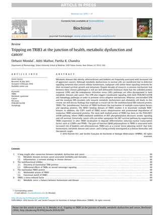
![4.2. Three UPR cascades control the progression of ERS . . . . . . . . . . . . . . . . . . . . . . . . . . . . . . . . . . . . . . . . . . . . . . . . . . . . . . . . . . . . . . . . . . . . . . . . . . . . . . . 00
5. TRIB3 regulates multiple stress response pathways . . . . . . . . . . . . . . . . . . . . . . . . . . . . . . . . . . . . . . . . . . . . . . . . . . . . . . . . . . . . . . . . . . . . . . . . . . . . . . . . . . . . . 00
5.1. Cross-talks between PI3K/AKT/mTOR, autophagy and TRIB3 . . . . . . . . . . . . . . . . . . . . . . . . . . . . . . . . . . . . . . . . . . . . . . . . . . . . . . . . . . . . . . . . . . . . . . 00
5.2. Cross-talks between NF-kB, MAPK and TRIB3 . . . . . . . . . . . . . . . . . . . . . . . . . . . . . . . . . . . . . . . . . . . . . . . . . . . . . . . . . . . . . . . . . . . . . . . . . . . . . . . . . . . 00
6. TRIB3 association with metabolic dysfunctions . . . . . . . . . . . . . . . . . . . . . . . . . . . . . . . . . . . . . . . . . . . . . . . . . . . . . . . . . . . . . . . . . . . . . . . . . . . . . . . . . . . . . . . . . 00
6.1. TRIB3 suppresses insulin signaling and glycogen storage . . . . . . . . . . . . . . . . . . . . . . . . . . . . . . . . . . . . . . . . . . . . . . . . . . . . . . . . . . . . . . . . . . . . . . . . . 00
6.1.1. TRIB3 overexpression in insulin resistance and diabetes . . . . . . . . . . . . . . . . . . . . . . . . . . . . . . . . . . . . . . . . . . . . . . . . . . . . . . . . . . . . . . . . . . . 00
6.1.2. TRIB3 is associated with atherosclerotic plaque rupture . . . . . . . . . . . . . . . . . . . . . . . . . . . . . . . . . . . . . . . . . . . . . . . . . . . . . . . . . . . . . . . . . . . 00
6.1.3. TRIB3 is associated with b-cell dysfunction and death . . . . . . . . . . . . . . . . . . . . . . . . . . . . . . . . . . . . . . . . . . . . . . . . . . . . . . . . . . . . . . . . . . . . 00
6.1.4. The TRIB3 Q84R polymorphism in diabetes and atherosclerosis . . . . . . . . . . . . . . . . . . . . . . . . . . . . . . . . . . . . . . . . . . . . . . . . . . . . . . . . . . . . 00
6.2. TRIB3 is associated with numerous diabetes-associated diseases . . . . . . . . . . . . . . . . . . . . . . . . . . . . . . . . . . . . . . . . . . . . . . . . . . . . . . . . . . . . . . . . . . . 00
6.2.1. Hyper-homocysteinemia . . . . . . . . . . . . . . . . . . . . . . . . . . . . . . . . . . . . . . . . . . . . . . . . . . . . . . . . . . . . . . . . . . . . . . . . . . . . . . . . . . . . . . . . . . . . . . 00
6.2.2. Non-alcoholic fatty liver disease . . . . . . . . . . . . . . . . . . . . . . . . . . . . . . . . . . . . . . . . . . . . . . . . . . . . . . . . . . . . . . . . . . . . . . . . . . . . . . . . . . . . . . . 00
6.2.3. Diabetic nephropathy . . . . . . . . . . . . . . . . . . . . . . . . . . . . . . . . . . . . . . . . . . . . . . . . . . . . . . . . . . . . . . . . . . . . . . . . . . . . . . . . . . . . . . . . . . . . . . . . . 00
7. Linking TRIB3 expression in obesity and cancer . . . . . . . . . . . . . . . . . . . . . . . . . . . . . . . . . . . . . . . . . . . . . . . . . . . . . . . . . . . . . . . . . . . . . . . . . . . . . . . . . . . . . . . . 00
7.1. TRIB3 expression in adipocyte differentiation and visceral obesity . . . . . . . . . . . . . . . . . . . . . . . . . . . . . . . . . . . . . . . . . . . . . . . . . . . . . . . . . . . . . . . . . 00
7.2. Potential of targeting TRIB3 in aggressive cancers . . . . . . . . . . . . . . . . . . . . . . . . . . . . . . . . . . . . . . . . . . . . . . . . . . . . . . . . . . . . . . . . . . . . . . . . . . . . . . . . 00
8. Perspectives . . . . . . . . . . . . . . . . . . . . . . . . . . . . . . . . . . . . . . . . . . . . . . . . . . . . . . . . . . . . . . . . . . . . . . . . . . . . . . . . . . . . . . . . . . . . . . . . . . . . . . . . . . . . . . . . . . . . . . . 00
Acknowledgments . . . . . . . . . . . . . . . . . . . . . . . . . . . . . . . . . . . . . . . . . . . . . . . . . . . . . . . . . . . . . . . . . . . . . . . . . . . . . . . . . . . . . . . . . . . . . . . . . . . . . . . . . . . . . . . . . 00
References . . . . . . . . . . . . . . . . . . . . . . . . . . . . . . . . . . . . . . . . . . . . . . . . . . . . . . . . . . . . . . . . . . . . . . . . . . . . . . . . . . . . . . . . . . . . . . . . . . . . . . . . . . . . . . . . . . . . . . . . 00
1. A long sought-after connection between metabolic
dysfunction and cancer
1.1. Metabolic diseases increase cancer-associated morbidity and
mortality
Chronic metabolic diseases like obesity, type-2 diabetes,
atherosclerosis, and cardiovascular disease (CVD) are becoming
increasingly prevalent worldwide [1e4]. Both hyperinsulinemia
and hyperglycemia contribute to the progression of obesity and
diabetes, and insulin resistance, manifested due to decreased in-
sulin receptor signaling, is the primary risk factor for these meta-
bolic disorders [5,6]. Interestingly, metabolic diseases are also
frequently associated with poorer cancer outcomes [7,8]. In the past
few decades, a number of studies have documented a clear link
between the metabolic syndromes and higher morbidity and
mortality due to different malignancies [9e11]. However, the
crucial mechanism(s) involved in linking these chronic pathologic
manifestations is not properly understood. As early as 1995,
Steenland et al., showed that men with diabetes present with a 39%
higher risk of developing colorectal and prostate cancer [12]. Calle
et al. (2003) published finding from the multicenter Cancer Pre-
vention Study (CPS) which followed more than one million adults
during 1982e1996, and clearly demonstrated that obese men and
women had a 40e80% increased threat of dying from cancers
[13,14]. The danger of having aggressive pancreatic, breast and
colorectal cancers are reported to be amplified in patients with high
body mass index (BMI) and several meta-analyses in patients with
diabetes also showed significantly higher cancer mortality, as
compared with nondiabetic individuals [15,16]. Indeed, the clini-
cally approved glyburide, metformin has provided better clinical
outcome in diabetic patients with advanced cancers [17]. Hence, a
thorough understanding of the long sought-after relationship be-
tween metabolic diseases and cancers will not only provide early
biomarkers for disease progression, but will also elucidate novel
therapeutic targets to decrease cancer-associated complications.
Furthermore, since tremendous increases in metabolic syndrome
are being reported in younger adults [4], which makes them more
susceptible to malignancies later in life, studies on the common
etiologies in metabolic diseases and cancer are garnering a lot of
attention [7,17e22].
Deleterious consequences of chronic inflammation and oxida-
tive stress are known to increase tumor progression and metastasis,
and also facilitate tumor resistance to both chemotherapy and
radiotherapy [7,20,23e28]. Studies have documented that the
visceral adipose tissue secreted inflammatory cytokines can pro-
mote insulin resistance in vascular cells [29e32]. Metabolic com-
plications of insulin resistance make individuals more susceptible
to chronic oxidative stress, neoplastic transformation and aggres-
sive tumor growth. A number of studies have also demonstrated
that second messenger signaling via the insulin and insulin like
growth factor (IGF) receptors play a crucial role in numerous other
chronic diseases such as autoimmunity, arthritis, alzheimer's dis-
ease and aging [33e36]. Thus, it is becoming apparent that the
chronic effects of inflammation in disrupting stress-adaptive
pathways in both normal and malignant cells may influence pro-
gression of these chronic diseases.
1.2. Inflammation: a common etiology in chronic diseases
Obesity induced adipokines, inflammatory cytokines, leptin,
proteolytic enzymes, and endogenous sex steroids, are known to
suppress the anti-inflammatory actions of insulin. The resultant
activation of vascular endothelium and decreased vasodilation of
smooth muscle cells increases blood pressure and causes hyper-
tension [32,37,38]. Increased adhesion of leukocytes and platelets
and the ensuing atherosclerotic thrombus formation further fosters
inflammatory stress and insulin resistance of the vasculature. Ad-
ipose tissue infiltrated macrophages and foam cells can also pro-
duce a state of chronic oxidative stress and inflammation, which
further promote the chronic metabolic dysfunctions [39,40].
Interestingly, the deleterious effects of insulin resistance, inflam-
mation and oxidative stress have also been implicated in both
oncogenic transformation of normal cell [41,42] and in increased
proliferation and metastasis of tumor cells, as shown by us [43] and
others [20,28,44]. Furthermore, since insulin resistance increases
both estrogen and testosterone levels by decreasing SHBG (sex
hormone binding globulin) [45] metabolic diseases can also
augment the growth of endocrine tumors like breast and prostate
cancers [7,23,43]. Therefore, insulin resistance is postulated to be a
common link and comorbidity in both metabolic diseases and
cancer. Indeed, insulin-induced glucose uptake is dysregulated in
D. Mondal et al. / Biochimie xxx (2016) 1e192
Please cite this article in press as: D. Mondal, et al., Tripping on TRIB3 at the junction of health, metabolic dysfunction and cancer, Biochimie
(2016), http://dx.doi.org/10.1016/j.biochi.2016.02.005](data:image/gif;base64,R0lGODlhAQABAIAAAAAAAP///yH5BAEAAAAALAAAAAABAAEAAAIBRAA7)