Mathematical Models For Biological Pattern Formation 2001th Edition Philip Maini
Mathematical Models For Biological Pattern Formation 2001th Edition Philip Maini
Mathematical Models For Biological Pattern Formation 2001th Edition Philip Maini
Mathematical Models For Biological Pattern Formation 2001th Edition Philip Maini
Mathematical Models For Biological Pattern Formation 2001th Edition Philip Maini
1.
Mathematical Models ForBiological Pattern
Formation 2001th Edition Philip Maini download
https://ebookbell.com/product/mathematical-models-for-biological-
pattern-formation-2001th-edition-philip-maini-55925638
Explore and download more ebooks at ebookbell.com
2.
Here are somerecommended products that we believe you will be
interested in. You can click the link to download.
Coupled Mathematical Models For Physical And Biological Nanoscale
Systems And Their Applications 1st Ed Luis L Bonilla
https://ebookbell.com/product/coupled-mathematical-models-for-
physical-and-biological-nanoscale-systems-and-their-applications-1st-
ed-luis-l-bonilla-7149224
Mathematical Models For Decision Making With Multiple Perspectives
Maria Isabel Gomes
https://ebookbell.com/product/mathematical-models-for-decision-making-
with-multiple-perspectives-maria-isabel-gomes-48771668
Mathematical Models For Evacuation Planning In Urban Areas Sarah
Bretschneider
https://ebookbell.com/product/mathematical-models-for-evacuation-
planning-in-urban-areas-sarah-bretschneider-4158480
Mathematical Models For Registration And Applications To Medical
Imaging 1st Edition Stefan Henn
https://ebookbell.com/product/mathematical-models-for-registration-
and-applications-to-medical-imaging-1st-edition-stefan-henn-4200942
3.
Mathematical Models ForEddy Currents And Magnetostatics With Selected
Applications 1st Edition Rachid Touzani
https://ebookbell.com/product/mathematical-models-for-eddy-currents-
and-magnetostatics-with-selected-applications-1st-edition-rachid-
touzani-4380682
Mathematical Models For Society And Biology Edward Beltrami Auth
https://ebookbell.com/product/mathematical-models-for-society-and-
biology-edward-beltrami-auth-4409850
Mathematical Models For Poroelastic Flows 1st Edition Anvarbek
Meirmanov Auth
https://ebookbell.com/product/mathematical-models-for-poroelastic-
flows-1st-edition-anvarbek-meirmanov-auth-4627562
Mathematical Models For Systems Reliability Benjamin Epstein
https://ebookbell.com/product/mathematical-models-for-systems-
reliability-benjamin-epstein-4642048
Mathematical Models For Communicable Diseases Brauer F Castillochavez
C
https://ebookbell.com/product/mathematical-models-for-communicable-
diseases-brauer-f-castillochavez-c-4677816
6.
The IMA Volumes
inMathematics
and its Applications
Volume 121
Series Editor
Willard Miller, Jr.
Springer Science+Business Media, LLC
7.
Institute for Mathematicsand
its Applications
IMA
The Institute for Mathematics and its Applications was estab-
lished by a grant from the National Science Foundation to the University
of Minnesota in 1982. The IMA seeks to encourage the development and
study of fresh mathematical concepts and questions of concern to the other
sciences by bringing together mathematicians and scientists from diverse
fields in an atmosphere that will stimulate discussion and collaboration.
The IMA Volumes are intended to involve the broader scientific com-
munity in this process.
1982-1983
1983-1984
1984-1985
1985-1986
1986-1987
1987-1988
1988-1989
1989-1990
1990-1991
1991-1992
1992-1993
1993-1994
1994-1995
1995-1996
1996-1997
1997-1998
1998-1999
1999-2000
2000-2001
2001-2002
2002-2003
Willard Miller, Jr., Professor and Director
* * * * * * ** * *
IMA ANNUAL PROGRAMS
Statistical and Continuum Approaches to Phase Transition
Mathematical Models for the Economics of Decentralized
Resource Allocation
Continuum Physics and Partial Differential Equations
Stochastic Differential Equations and Their Applications
Scientific Computation
Applied Combinatorics
Nonlinear Waves
Dynamical Systems and Their Applications
Phase Transitions and Free Boundaries
Applied Linear Algebra
Control Theory and its Applications
Emerging Applications of Probability
Waves and Scattering
Mathematical Methods in Material Science
Mathematics of High Performance Computing
Emerging Applications of Dynamical Systems
Mathematics in Biology
Reactive Flows and Transport Phenomena
Mathematics in Multimedia
Mathematics in the Geosciences
Optimization
Continued at the back
8.
Philip K. MainiHans G. Othmer
Editors
Mathematical Models for
Biological Pattern Formation
With 166 Illustrations
Springer
FOREWORD
This 121st IMAvolume, entitled
MATHEMATICAL MODELS
FOR BIOLOGICAL PATTERN FORMATION
is the first of a new series called FRONTIERS IN APPLICATION OF
MATHEMATICS. The FRONTIERS volumes are motivated by IMA pro-
grams and workshops, but are specially planned and written to provide
an entree to and assessment of exciting new areas for the application of
mathematical tools and analysis. The emphasis in FRONTIERS volumes
is on surveys, exposition and outlook, to attract more mathematicians and
other scientists to the study of these areas and to focus efforts on the most
important issues, rather than papers on the most recent research results
aimed at an audience of specialists.
The present volume of peer-reviewed papers grew out of the 1998-99
IMA program on "Mathematics in Biology," in particular the Fall 1998 em-
phasis on "Theoretical Problems in Developmental Biology and Immunol-
ogy." During that period there were two workshops on Pattern Formation
and Morphogenesis, organized by Professors Murray, Maini and Othmer.
James Murray was one of the principal organizers for the entire year pro-
gram.
I am very grateful to James Murray for providing an introduction, and
to Philip Maini and Hans Othmer for their excellent work in planning and
preparing this first FRONTIERS volume.
I also take this opportunity to thank the National Science Foundation,
whose financial support of the IMA made the Mathematics in Biology pro-
gram possible.
Willard Miller, Jr., Professor and Director
v
11.
The editors arepleased to dedicate this volume to Professor James D.
Murray, affectionately known as Jim to his friends. Jim has been a leader in
the mathematical analysis of biological pattern formation for 25 years, and
has influenced it dramatically by his unbending insistence that the problem
is first and foremost a biological one, and therefore the biological details do
really matter. The Centre for Mathematical Biology at Oxford University,
which he founded in 1983, has been a magnet and haven for mathematicians
who were interested in the many aspects of biological pattern formation,
and its success is in no small part due to Jim's warmth and kindness to all,
and his strong support of young researchers.
We wish Jim, and his soulmate Sheila, the best in the coming years.
Philip K. Maini
Hans G. Othmer
12.
CONTENTS
Foreword ............................................................. v
Dedication.......................................................... vii
Biological pattern formation - a marriage of theory
and experiment .. . . . . . . . . . . . . . . . . . . . . . . . . . . . . . . . . . . . . . . . . . . . . . .. . . . . .. 1
J.D. Murray
Spatiotemporal pattern formation in early development:
A review of primitive streak formation and somitogenesis ............. 11
S. Schnell, K.J. Painter, P.K. Maini, and H.G. Othmer
Mathematical modeling of vertebrate limb development. . . . . . . . . . . . . .. 39
Robert H. Dillon
Models for pigment pattern formation in the skin of fishes. . . . . . . . . . .. 59
K.J. Painter
Generic modelling of vegetation patterns. A case study
of Tiger Bush in sub-Saharian Sahel.. ................................ 83
R. Lefever, O. Lejeune, and P. Couteron
Chemical Turing patterns: A model system of a paradigm
for morphogenesis ................................................... 113
David J. Wollkind and Laura E. Stephenson
Beyond spots and stripes: Generation of more complex
patterns by modifications and additions of the basic reaction ........ 143
Hans Meinhardt
Spatiotemporal patterning in models of juxtacrine
intercellular signalling with feedback ................................ 165
Nicholas A.M. Monk, Jonathan A. Sherratt,
and Markus R. Owen
Modelling Dictyostelium discoideum morphogenesis .................. 193
Bakhtier Vasiev and Comelis J. Weijer
ix
13.
x CONTENTS
Modeling branchingand chiral colonial patterning
of lubricating bacteria .............................................. 211
Eshel Ben-Jacob, Inon Cohen, Ido Golding,
and Yonathan Kozlovsky
Modeling self-propelled deformable cell motion
in the Dictyostelium mound; a status report.. . . . . . . . . . . . . . . . . . . . . . .. 255
Wouter-Jan Rappel, Herbert Levine, Alastair Nicol,
and William F. Loomis
A minimal model of locomotion applied to the steady
gliding movement of fish keratocyte cells. . . . . . . . . . . . . . . . . . . . . . . . . . .. 269
A. Mogilner, E. Marland, and D. Bottino
Computer simulations of mechanochemical coupling
in a deforming domain: Applications to cell motion.................. 295
Dean C. Bottino
List of workshop participants ....................................... 315
14.
BIOLOGICAL PATTERN FORMATION- A MARRIAGE
OF THEORY AND EXPERIMENT
J.D. MURRAY'
Abstract. The interdisciplinary challenges to discover the underlying mechanisms
in the generation of biological pattern and form are central issues in development. Here
I briefly discuss a philosophy of such an integrative biology approach. I then describe, by
way of example, the successful use of a very simple model-even linear - for the growth
of brain tumours in an anatomically accurate brain. All of the model parameters are
estimated from experiment and patient data. Even with such a basic model the results
highlight the inadequacies of current medical intervention treatment of brain tumours.
I conclude with some brief general views on the use of models in biology.
Although the biomedical world is in the throes of the genetic revolu-
tion the basic question which genes do not address is the development of
spatio-temporal pattern and form, whether it is the growth of a tumour
or the development of stripes on a fish. During the past 20 some years a
large amount of research in mathematical biology, or biomathematics or
whatever name is given to the application of mathematics to the biomed-
ical sciences, has been devoted to trying to increase our understanding of
the underlying biological processes involved in pattern formation processes.
The relatively few people working in the field in the 1970's has blossomed
into the several thousand who are now actively involved in modelling a vast
and ever widening spectrum of biomedical problems. The collection of pa-
pers in this volume demonstrate not only how powerful such mathematical
models can be, but how far the field has come in even just the past 10 years.
Although we still do not know the complete detailed mechanism involved
in any specific situation I am optimistic that we are approaching the situa-
tion when we shall. Several of the theoretical studies of pattern formation
paradigms, such as the organisation of social amoebae like Dictyostelium
discoideum and bacterial patterns [19, 21], have resulted in major advances
in our understanding and guided the direction of illuminating experimental
programmes. What is, I feel, indisputable, is that major progress has come
about by genuine interaction between the theoreticians and the experimen-
talists. Gone are the days when papers in which functions describing blood
cell density as being "imbedded in some appropriate Banach space" with
statements such as "this will be of great interest to cardiologists" tagged
at the end of a paper replete with theorems and lemmas with as much rel-
evance to biological pattern formation as the length of the the latest pop
singer's earing. The articles in this collection deal with real problems and,
irrespective of the mathematical sophistication involved in the model anal-
yses, relate directly to real biological problems and importantly increase
our basic understanding of the biological processes.
"Department of Applied Mathematics, Box 352420, University of Washingon, Seattle,
Washington 98195-2420.
1
15.
2 J.D. MURRAY
Althoughit is generally accepted, it should perhaps be stressed again
that mathematical descriptions of patterning phenomena are not expla-
nations. One of the principal uses of any theory is in its biological pre-
dictions. From a theoretical point of view, the art of good modelling in
biology relies not only on sufficient mathematical expertise (often not at
all sophisticated), but also on: (i) a sound understanding and appreciation
of the biological problem; (ii) a realistic mathematical representation of the
important biological phenomena; and (iii) a biological interpretation of the
mathematical analysis and results in terms of insights and predictions. Sci-
entifically relevant mathematical or theoretical biology is unquestionably
an interdisciplinary science par excellence.
An important point arising from theoretical models is that any pattern
contains its own history. Consider a simple engineering analogy of our role
in trying to understand a biological process [14]. It is one thing to suggest
that a bridge requires a thousand tons of steel, that any less will result
in too weak a structure, and any more will result in excessive rigidity. It
is quite another matter to instruct the workers on how best to put the
pieces together. In morphogenesis, for example, it is conceivable that the
cells involved in tissue formation and deformation have enough expertise
that given the right set of ingredients and initial instructions they could be
persuaded to construct whatever element one wants. This is the hope of
many who are searching for a full and predictive understanding. However,
it seems very likely that the global effect of all this sophisticated cellular
activity would be critically sensitive to the sequence of events occurring
during development. As scientists we should concern ourselves with how
to take advantage of the limited opportunities we have for communicating
with the workforce so as to direct experiment towards an acceptable end-
product. This is perhaps a little philosophical, but even a cursory look at
many theories in the literature reveal a fixation on simplistic explanations.
On the other hand, in situations which frequently arise especially in medical
problems, the complexity is such that if we wish to be useful we often have
to start with what is clearly an oversimplistic scenario and build into the
models progressively more realism as we discover more about the problem.
There are certainly no ground rules as to how complex or simple a model
has to be to be useful.
None of the individual models that have been suggested for any bio-
logical patterning process, and not even all of them put together, could be
considered a complete model. In the case of some of the widely studied
problems (such as Dictyiostelium discoideum) , each model has shed light
on different aspects of the process and we can now say what the most im-
portant conceptual elements have to be in a complete model. These studies
have served to highlight where our knowledge is deficient and to suggest di-
rections in which fruitful experimentation might lead us. Indeed, a critical
test of these theoretical constructs is in their impact on the experimental
community.
16.
BIOLOGICAL PATTERN FORMATION3
Since the articles in this volume are primarily concerned with biological
as opposed to medical spatial problems, it is perhaps appropriate to briefly
describe a particularly simple model for the highly complex and poorly
understood problem of the growth of human brain tumours (glioblastomas).
The fast pace of medical discoveries, real and spurious, is a fruitful field
for genuine integrative interdisciplinary research. Some of these discoveries
bring new uses for extant theories. For example, the recent experimental
work on the importance of anti-angiogenetic drug [8, 1] for the control of
tumours first suggested by Judah Folkman in the 1970's [6, 7] has brought
the developmental problem of the mechanisms that could be involved in
angiogenesis to the fore [13, 15]: without angiogenesis the tumour cannot
grow.
1. A simple mathematical model for virtual brain tumours
(gliomas) - enhancing medical imaging. Gliomas are particularly
nasty brain tumours that diffuse aggressively, thereby invading the sur-
rounding normal tissue. That the spatial spread involves diffusion is now
fairly generally accepted. Although other processes are probably involved,
diffusion and cell mitosis play major (arguably the major) roles in the
spread of cancer cells. Being a diffusion process there is a long tail where
the cell density is extremely low. There is clearly a threshold level be-
low which even the most sophisticated scans cannot detect in spite of the
continuing development of medical imaging such as enhanced computer-
ized tomography (CT) and magnetic resonance imaging (MRI). At least
one inadequacy of current medical imaging is that even extensive surgi-
cal resection or local irradiation of gliomas, based on where the tumour
"boundary" is as defined by the scans, is followed by tumour recurrence at
or near the edge of the excised tumour [12].
In an attempt to try and get some understanding of the growth of
such tumours, Dr. Elsworth Alvord MD (Pathology, Health Science, Uni-
versity of Washington), myself and several of my graduate students and
post-doctorals over the past six years have looked at some very simple
diffusion models to try and obtain some quantitative estimates of brain
tumour growth, both with and without medical intervention [18, 5, 20, 2].
Perhaps the most damning demonstration of the inadequacies of current
medical treatment has been given by the work [17] with Dr. Alvord and
a former student, Dr. Kristin Swanson. We started with a basic diffusion
model for the cancer cells involving exponential cell growth (justified by
the data on such tumours). The major difference to previous work along
these lines is that the diffusion was simulated within anatomically accurate
heterogeneous brain tissue in three spatial dimensions. The work will be
reported in detail elsewhere. Here I give only a brief sketch of the model
and results, since it highlights the above point that even simple models can
be clinically useful.
17.
4 J.D. MURRAY
Theavailability of the BrainWeb [3] brain atlas database let us de-
fine the gross anatomical boundaries and to vary the degree of motility
of glioma cells in grey or white matter in heterogeneous, anatomically ac-
curate brain tissue. Glioma cells are reported to migrate more rapidly in
white matter than in grey matter [9] so we allow the motility coefficient to
differ depending on the local tissue composition.
Our mathematical model for glioma growth and invasion, including the
differential motility of gliomas in grey and white matter, can be written as
(1)
Be
8t =7 . (D(x)7c) + pc ,
where c(x, t) is the concentration of tumour cells at position x and time
t. D(x), a function of position x in the brain, is the diffusion coefficient
defining the random motility of the glioma cells with D(x) = Dg , Dw,
constants for x in grey and white matter, respectively. p represents the
net proliferation rate of the glioma cells. The diffusion coefficient in white
matter is larger than that in grey, so Dw > Dg • The difference in the
diffusion coefficients has been estimated to range from 2 to 100 fold [17],
but we chose 5 as an arbitrary first approximation to illustrate the model's
potential. To complete the model formulation, we required zero flux of cells
across the brain boundaries and assumed that the tumour had grown to
about 4,000 cells as a local mass before it began to diffuse and the model
equation (1) applies.
The BrainWeb lets us simulate the growth of a virtual glioma in any of
the 3 standard planes (coronal, sagittal and axial or horizontal) to demon-
strate a pseudo-3-dimensional tumour. (The numerical simulation was a
challenging problem.)
For every current medical imaging technique there is a threshold of
detection below which gliomas cells are not detectable. Even microscopy
has a limit beyond which individual cells cannot be detected.
Survival time. Previous models assumed that diagnosis is made when
the volume of an enhanced CT-detectable tumour has reached a size equiv-
alent to a sphere of an average 3 cm diameter, and that death occurs when
the volume reaches an average 6 cm diameter. The difference between
these two times can be defined as the survival time of the hypothetical or
virtual patient. With earlier models, and even simpler brain structure, the
comparison of calculated survival times [20] with extant data [11] was very
good.
Crucial to all successful modelling, particularly those which give rise
to simple models which have fewer parameters, is the ability to determine
reasonable estimates of the critical parameters, here the growth rate p and
the diffusion coefficient D. For high-grade gliomas (glioblastomas) previous
estimates, based on extant data, have suggested a net proliferation rate of
p ::::; 0.012/day [20, 2, 17, 4], corresponding to a volume-doubling time of 60
18.
BIOLOGICAL PATTERN FORMATION5
days, and a diffusion coefficient of D ~ .0013 cm2/ day [2, 17]. The actual
ranges of these values are quite extreme but real values for any actual
patient could be substituted if they could be measured.
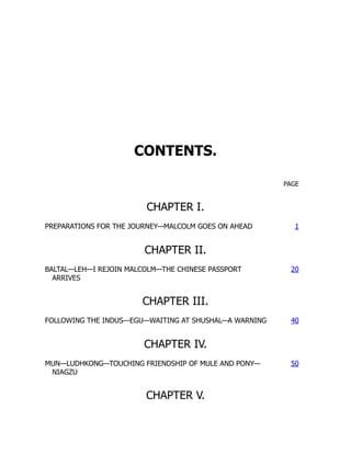
Figure 1 shows three perpendicular cross-sections (coronal, sagittal
and horizontal or axial) of the virtual human brain intersecting in a point
marked by an asterisk in the superior frontal region where the virtual tu-
mour originates. The grey and white matters of the brain domain appear
grey and white, respectively, A contour plot of the tumour cell density is
represented in color with red denoting a high density and blue a low density.
In each image, a single thick black curve defines the edge of the tumour that
the model suggests would be detectable on enhanced CT scan associated
with a threshold of detection of 8000 cells/mm3. The outermost light blue
profile corresponds to an arbitrary threshold of detection 80 times more
sensitive than enhanced CT (that is 100 cells/mm3). The left column of
images in Figure 1 represents the tumour at the time of detection, defined
as an enhanced CT-detectable tumour with average diameter of 3 cm, while
the right column represents the tumour at the time of death, defined by
an enhanced CT-detectable tumour with average diameter of 6 cm. With
our model it is possible to simulate the growth of a tumour starting at any
point we wish.
What is abundantly clear from the figure is how far tumour cells have
diffused beyond any current range of detection. It is also clear why sur-
gical resection is so difficult and ineffectual since the tumour "boundary"
is so diffuse. Even resecting a significant distance outside the detectable
tumour fails to excise all the tumour cells. Previous studies of the motil-
ity of gliomas have demonstrated that diffusion is an accurate estimation
for the method of spread of gliomas [17, 20]. A consequence of modelling
cellular motility by Fickian or gradient-driven diffusion, is the lack of a
definitive interface between malignant and normal tissue. This mathemat-
ical consequence is correlated with the actual biology of human gliomas.
Consider using CT-images, or other visual detection procedures, to delin-
eate the possible interface between cancerous and normal tissue. Radical
excision of the tumour even well beyond these interfaces has been shown
to fail in numerous studies as summarized by [16]. Clearly tumour cells
invade peripheral to the CT or MRI defined boundaries of the tumour.
Even standard histopathological analysis, one of our most sensitive means
of detecting glioma cells, fails in locating all of the tumour cells.
Because of the diffuse nature of gliomas there is no clear boundary
defining the interface of pathological and normal tissue, even though many
attempts have been made to suggest that a boundary exists. Figures 1
shows the spatio-temporal invasion of virtual gliomas at the time of diag-
nosis and death. These simulations clearly reveal the subthreshold invasion
of the tumour well beyond the detectable portion of the tumour. No matter
the extent of resection, the mathematical model indicates that the gross
tumour will ultimately recur and kill (see also [20]).
19.
6 J.D. MURRAY
FIG.1. Sections of the virtual human brain in sagittal, coronal and horizontal
planes that intersect at the site of the glioma originating in the superior frontal region
denoted by an asterisk (*). Red denotes a high density of tumour cells while blue
denotes a low density. A thick black contour defines the edge of the tumour detectable
by enhanced computerized tomography (CT). Cell migration was allowed to occur in a
truly three-dimensional solid representation of the brain.
Unlike real patients with real gliomas, virtual patients with virtual
gliomas can be analyzed by allowing any particular factor to vary while
keeping all the other determining factors constant. Such isolation tech-
niques, of course, require a mathematical model that has sufficient realism
and involves the major variables and parameters. The recent availability
of simulated MRl's, with proportions of grey and white matter accurately
indicated, let us develop this model which is sufficiently complex to allow
20.
BIOLOGICAL PATTERN FORMATION7
different diffusion rates in grey and white matter (for example, a 5-fold
increase in diffusion or migration in white matter) as well as to prevent
spread across certain parts of the brain.
The model is a simple one which focuses on only two key elements,
namely diffusion and growth. Other variables can be introduced into the
model as their relative importance is discovered. Previous studies [18, 20, 5]
showed how to determine estimates for these parameters from patient scans.
With these the present model can be depressingly predictive as to the where
the tumour is likely to grow in real time. Of course many aspects, which
can be included in more complex models, such as swelling and distortion
of tissue should be included. The point of this brief discussion is to show
how even a simple basic model can still be useful'clinically. However, even
without these other effects included what seems clear from these theoretical
studies of virtual gliomas is that current imaging techniques are woefully
inadequate for definitive clinical decisions as to what constitutes the opti-
mal treatment for patients with gliomas.
2. General concluding remarks. Theoretical modelling has been
proven to be useful in the study of a remarkably diverse spectrum of biolog-
ical problems such as wound healing, quantifying disease control strategies,
the effect of introducing genetically engineered organisms in the environ-
ment and suggesting experiments associated with limb development, to
name just a few.
Pattern formation studies are sometimes criticized for their lack of
inclusion of genes in the models. But then criticism can be levelled at any
modelling abstraction of a complex system to a relativley simple one. It
should be remembered that the generation of pattern and form, particularly
in development, is usually a long way from the level of the genome. Of
course genes play crucial roles in development, but they do not actually
create patterns. Many of the evolving patterns could hardly have been
anticipated solely by genetic information.
Why use mathematics to study something as intrinsically complicated
and ill-understood as development, angiogenesis, wound healing, infectious
disease dynamics, regulatory networks and so on? We suggest that math-
ematical modelling must be used if we ever hope to genuinely and real-
istically convert an understanding of the underlying mechanisms into a
predictive science. Mathematics is required to bridge the gap between the
level on which most of our knowledge is accumulating (cellular and below)
and the macroscopic level of the patterns we see. A mathematical approach
lets us explore the logic of pattern formation. Even if the mechanisms were
well understood - and they certainly are far from it at this stage - math-
ematics would be required to explore the consequences of manipulating the
various parameters associated with any particular scenario. In the case of
such things as wound healing, tumour growth and it will be increasingly
so in angiogenesis with the cancer connection, the number of options that
21.
8 J.D. MURRAY
arefast becoming available to wound and cancer managers will become
overwhelming unless we can find a way to simulate particular treatment
protocols before applying them in practice. The latter has already been of
use in understanding the efficacy of various treatment scenarios with brain
tumours [18, 20, 17] and new two step regimes for skin cancer [10].
There is no doubt that we are a long way from being able to reliably
simulate actual developmental scenarios, notwithstanding the multitude of
theories that abound. The active cellular control of key processes is poorly
understood. Despite such limitations, we argue that exploring the logic of
biological processes is worthwhile, in some current situations even essential
in our present state of knowledge. It allows us to take an hypothetical
mechanism and examine its consequences in the form of a mathematical
model, make predictions and suggest experiments that would verify or in-
validate the model; the latter is frequently biologically informative. In fact,
the very process of constructing a mathematical model can be useful in its
own right. Not only must one commit to a particular mechanism, one is
also forced to consider what is truly essential to the process and what the
key players are. We are thus involved in constructing frameworks on which
we can hang our understanding. The equations, the mathematical analysis
and the numerical simulations that follow serve to reveal quantitatively, as
well as qualitatively, the consequences of that logical structure.
The best integrative biology studies have served to highlight where
our knowledge is deficient and to suggest directions in which fruitful exper-
imentation might lead us. A crucial aspect of this research is the interdis-
ciplinary content and, as already mentioned, a crucial test of all theoretical
models should be in their impact on the experimental community. The
field of mathematical or theoretical biology or integrative biology has now
achieved some level of maturity, and we believe that future dialogue be-
tween experimentalists and theoeticians will lead us more rapidly towards
a fuller understanding, if not a complete one, of several biological processes
involving pattern formation.
REFERENCES
[1] T. BOEHM, J. FOLKMAN, T. BROWDER, AND M. O'REILLY. Antiangiogenesis ther-
apy of experimental cancer does not induce acquired drug resistance. Nature,
404-407, 1997.
[2] P.K. BURGESS, P.M. KULESA, J.D. MURRAY, AND E.C. ALVORD, JR. The inter-
action of growth rates and diffusion coefficients in a three-dimensional math-
ematical model of gliomas. J Neuropathol and Exp Neural, 56:704-713, 1997.
[3] D.L. COLLINS, A.P. ZIJDENBOS, V. KOLLOKIAN, J.G. SLED, N.J. KABANI, C.J.
HOLMES, AND A.C. EVANS. Design and construction of a realistic digital brain
phantom. IEEE Transactions on Medical Imaging, 17:463-468, 1998.
[4] V.P. COLLINS, R. K. LOEFFLER, AND H. TIVEY. Observations on growth rates of
human tumors. Am J Roentgenol Radium Ther Nucl Med, 76:988-1000, 1956.
[5] G.C. CRUYWAGEN, D.E. WOODWARD, P. TRACQUI, G.T. BARTOO, J.D. MURRAY,
AND E.C. ALVORD, JR. The modelling of diffusive tumours. J Biological
Systems, 3:937-945, 1995.
22.
BIOLOGICAL PATTERN FORMATION9
[6] J. FOLKMAN. Anti-angiogenesis: New concept for therapy of solid tumors. Annals
of Surgery, 75:409-416, 1971.
[7] J. FOLKMAN. TUmor angiogenesis: therapeutic implications. New England Journal
of Medicine, 285:1182-1186, 1972.
[8] J. FOLKMAN. Angiogenesis in cancer, vascular, rheumatoid and other diseases.
Nature Medicine, 1:27-31, 1995.
[9] A. GIESE, L. KLUWE, B. LAUBE, H. MEISSNER, M. BERENS, AND M. WESTPHAL.
Migration of human glioma cells on myelin. Neurosurg, 38:755-764, 1996.
[10] T. JACKSON, S.R. LUBLIN, N.O. SIEMERS, P.D. SENTER, AND J.D. MURRAY. Math-
ematical and experimental analysis of localization of anti-tumor antibody-
enzyme conjugates. British Journal of Cancer, 80:1747-1753, 1999.
[11] F.W. KRETH, P.C. WARNKE, R. SCHEREMET, AND C.B. OSTERTAG. Surgical resec-
tion and radiation therapy versus biopsy and radiation therapy in the treat-
ment of glioblastoma multiforme. J Neurosurg, 78:762-766, 1993.
[12] B.C. LIANG AND M. WElL. Locoregional approaches to therapy with gliomas as
paradigm. Curro Opinion in Oncol., 10:201-206, 1998.
[13] D. MANOUSSAKI, S.R. LUBKIN, R.B. VERNON, AND J.D. MURRAY. A mechanical
model for the formation of vascular networks in vitro. Acta Biotheretica,
44:271-282, 1996.
[14] J.D. MURRAY, J. COOK, R. TYSON, AND S.R. LUBKIN. Spatial pattern formation in
biology: I dermal wound healing. ii bacterial patterns. Journal of the Franklin
Institute, 335B:303-332, 1998.
[15] J.D. MURRAY, D. MANOUSSAKI, S.R. LUBKIN, AND R.B. VERNON. A mechanical
theory of in vitro vascular network formation. In C. Little, V. Mironov, and
E. Helene Sage, editors, Vascular Morphogenesis in vivo, in vitro, in mente,
pages 173-188. Birkhauser, Boston, 1998.
[16] J .M. NAZZARO AND E.A. NEUWELT. The role of surgery in the management of
supratentorial intermediate and high-grade astrocytomas in adults. J. Neuro-
surg., 73:331-344, 1990.
[17] K.R. SWANSON. Mathematical modeling of the growth and control of tumors. PhD
thesis, University of Washington, 1999.
[18] P. TRACQUI, G.C. CRUYWAGEN, D.E. WOODWARD, G.T. BARTOO, J.D. MURRAY,
AND JR. E.C. ALVORD. A mathematical model of glioma growth: the effect of
chemotherapy on spatial-temporal growth. Cell Poliferation, 28:17-31, 1995.
[19] R. TYSON, S.R. LUBKIN, AND J.D. MURRAY. A minimal mechanism for bacterial
patterns. Proc. Roy. Soc. Lond., pages 299-304, 1998.
[20] D.E. WOODWARD, J. COOK, P. TRACQUI, G.C. CRUYWAGEN, J.D. MURRAY, AND
JR. E.C. ALVORD. A mathematical model of glioma growth: the effect of
extent of surgical resection. Cell Prolif, 29:269-288, 1996.
[21] D.E. WOODWARD, R. TYSON, M.R. MYERSCOUGH, J.D. MURRAY, E.O. Bu-
DRENE, AND H.C. BERG. Spatio-temporal patterns generated by Salmonella
typhimurium. Biophys. J., 68:2181-2189, 1995.
23.
SPATIOTEMPORAL PATTERN FORMATIONIN EARLY
DEVELOPMENT: A REVIEW OF PRIMITIVE STREAK
FORMATION AND SOMITOGENESIS
S. SCHNELL', K.J. PAINTERt, P.K. MAINI' , AND H.G. OTHMERt
Abstract. The basic body plan of a number of vertebrates results from two pro-
cesses that occur early in the development of the blastoderm: large scale rearrangements
of tissue via a process called gastrulation, and axial subdivision of tissue in a process
called somitogenesis. The first step of gastrulation in avians is formation of the prim-
itive streak, which marks the first clear manifestation of the anterior-posterior axis.
Cell movements that occur through the streak ultimately convert the single layered-
blastoderm into a trilaminar blastoderm comprising prospective endodermal, mesoder-
mal and ectodermal tissue. During streak formation a group of cells moves anteriorly as
a coherent column from the posterior end of the blastoderm, and as it proceeds other
cells stream over the lateral edges of the furrow left behind. The anterior end of the
streak is a specialized structure called Hensen's node, which serves as an organizing
center for later axis formation and determination of the left-right asymmetry of the
body. Soon after the primitive streak forms, Hensen's node regresses towards the tail,
leaving the notochord and a pair of segmental plates parallel to the primitive streak in
its wake. The posterior end of the segmental plate moves down the cranio-caudal axis
with the node, as more cells are added to it by cell division within the plate and by cells
entering from the primitive streak. A pair of somites forms from the anterior ends of
the two plates at regular intervals. Despite the fact that much is known about the basic
biological processes, the mechanisms that underlie the formation of the primitive streak
and somitogenesis are still unknown, and elucidating them is one of the major unsolved
problems in developmental biology. Mathematical modelling has been a useful tool in
this process, as it provides a framework in which to study the outcome of proposed
interactions and can make experimentally testable predictions. In this paper we outline
the biological background of these processes and review existing models of them.
Key words. Primitive streak formation, somitogenesis, theoretical models, math-
ematical models, Hox genes, c-hairy-i, Notch-Delta genes.
1. Introduction. Early vertebrate development is a complex process
that involves cell division, cell-cell signaling, cell movement, and cell dif-
ferentiation. Many adult vertebrates exhibit common structures, but the
developmental processes that produce them mayor may not be similar.
For example, formation of a primitive streak is central to avian, reptilian
and mammalian gastrulation, and while it is not present in amphibian blas-
tulae, they contain an analogous structure, called the blastopore. On the
other hand, somitogenesis is common to all vertebrates. This review fo-
cuses on experimental and theoretical aspects of primitive streak formation
and somitogenesis in avian embryogenesis. The chick embryo is a widely-
used model system for experimental studies and, as a result, there is a
'Centre for Mathematical Biology, Mathematical Institute, Oxford University, Ox-
ford, OXI 3LB, UK.
tDepartment of Mathematics, University of Minnesota, Minneapolis, MN 55455,
USA.
11
24.
12 S. SCHNELLET AL.
large amount of experimental data. We begin with a brief description of
the early events: details of these events can be found in [35], [88], and [50].
The chick embryo develops from a small, disk-shaped blastodisc float-
ing on top of the yolk. After the egg is fertilized cells divide repeatedly,
forming a multicellular stratified structure called the blastoderm. The pe-
riod from just prior to laying through several hours afterwards has been
subdivided into 14 stages [31, 50]. Cell division is dominant during stages
I - VI, and morphogenetic movements begin during stages VII-X, when
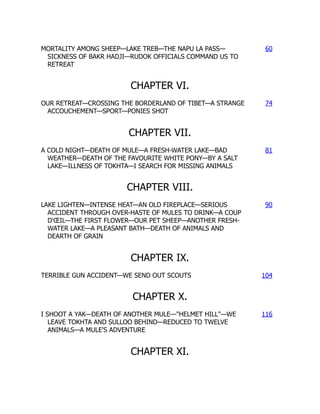
cells of the central blastodisc, called the area pellucida (c/. Figure 1),
separate from the yolk, producing a hollow region beneath the disc called
the subgerminal cavity [75, 99]. Subsequently some cells from the central
blastodisc move into the subgerminal cavity (either actively or passively),
and simultaneously the disc expands radially over the yolk. The opaque
marginal zone of the blastoderm, known as the area opaca, remains in con-
tact with the yolk and may play an active role in the radial movement
(Figure 1 A). The result is that during stages VII-X the central part of
the disc changes from a layer 4-6 cells deep to a translucent layer one cell
thick called the epiblast. The anterior-posterior axis of the embryo is also
determined during these stages [50]. After stage X some cells within the
marginal zone migrate posteriorly, and then leave the marginal zone at
the posterior marginal zone (PMZ)(Figure 1 B). They spread across the
subgerminal cavity beneath the epiblast as a loosely-connected sheet, in-
corporating islands of cells shed from the blastodisc earlier. By stage XIV
this sheet connects with the anterior margin of the disc and forms the hy-
poblast, and at this stage the blastoderm is bi-Iayered with the epiblast and
hypoblast separated by the blastocoel cavity. Fate maps for cell movements
in these stages are available [39].
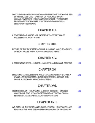
During hypoblast formation the embryonic shield or Koller's sickle de-
velops at the posterior end of the epiblast (cf Figure 2(a)). This consists
of a thickened epiblast [93] comprising primitive streak precursor cells that
have migrated to this area by a series of 'polonnaise movements' [105]. The
first visible sign of gastrulation is formation of the primitive streak, which
arises from Koller's sickle at the posterior midline of the blastodisc [52]
(Figure 1 C and D). The sickle narrows and the primitive streak moves an-
teriorly between the epiblast and the hypoblast. The tip of the ingressing
streak moves"" 60% of the way across the blastoderm before it stops, and
later, regresses. At full primitive streak stage (Hamburger and Hamilton
stage 4, [38]) the organizer of the avian embryo, Hensen's node, develops as
a bulbous structure at the anterior tip of the streak. The period between
the accumulation of cells at the posterior region and full primitive streak
is approximately 12 hours. The structure of the blastoderm at this stage
is illustrated in Figure 2(b). During the advance of the node, epiblast cells
move through the streak and into the interior. Those that migrate through
the node form anterior structures, those that migrate through the lateral
parts of the primitive streak become endodermal and mesodermal cells, and
25.
PRIMITIVE STREAK FORMATIONAND SOMITOGENESIS 13
(A) Anterior
(C)
(E)
node
p<'lIucida
Primitive
groove
Posterior
Anterior
area of
blastoderm
taking shape
Head
process
Hensen's
node
FIG.!' A schematic of the stages in early development of the chick embryo (A)
3-4 hours post-laying, (B) 5-6 hours, (C) 7-8 hours, (D) 10-12 hours, (E) 15-16 hours,
(F) 19-22 hours, . (Reproduced with permission from (35})
the remainder constitute the ectoderm. Simultaneously, the area pellucida
changes from circular to pear-shaped, narrowing in the posterior portion.
The head structure, notochord and somites are laid down during regression
of the node, and when regression is complete the embryo is a flat trilaminar
blastoderm comprising the ectodermal, mesodermal and endodermallayers.
These will form various organs during subsequent morphogenesis, in addi-
tion to the structures formed during regression. The regressing node and
anterior portion of the streak eventually form the tail bud [94]. Regression
proceeds on a slower time scale than progression, taking approximately 24
hours for the node to regress after the streak reaches its maximum length
of approximately 1.9 mm [94].
During regression of the primitive streak the neural folds begin to
gather at the center of the embryo, and the segmental plates, which are
26.
14 S. SCHNELLET AL.
Area Pellucida Marginal
CC======,
===
/
='
=I
======o
==~===o==o=~=o
=o=~
===:>12£::J-
~
t )1V~. 'gJDI5
Primary hypoblast Koller's siCkle ! t ! ro
Seoondary hypoblast ' ,
Deep layer 01 marginal zone
FIG. 2. (a) A schematic cross-section of the blastoderm prior to primitive streak
formation. (b) The blastoderm at the stage of maximal streak ingression (Reproduced
with permission from [35J)
often referred to as paraxial mesoderm or presomitic mesoderm (PSM),
separate into blocks of cells known as somites. They form as paired ep-
ithelial spheres arranged bilaterally along the anterior-posterior axis and
emerge in strict cranio-caudal order [36]. Simultaneously, new cells are in-
corporated into the PSM from the regression of Hensen's node at the same
rate as new somites are formed rostral to the PSM [16, 83]. Figure 3 is a
schematic representation of these early processes. Somites are divided by a
fissure into anterior and posterior halves that differ in their gene expression
and differentiation [104, 36].
The formation and differentiation of somites is the result of three dis-
tinct morphological events progressing in a strict spatio-temporal order: (1)
the prepatterning of the PSM; (2) somite and somitic boundary formation;
and (3) the differentiation of a somite into anterior and posterior halves
[36]. Several experimental observations confirm these events. Scanning
electron microscopy observations [42] and transplantation experiments [49]
show that the PSM displays a prepattern prior to segmentation. In addi-
tion, Hox and Notch-Delta pathway genes are involved in all these events
[104, 25]. These molecular results suggest the existence of a conserved
mechanism for segmentation in protostomes and deuterostomes [61].
The segmental pattern of somites in turn governs the segmental pat-
tern of the peripheral nervous system and determines the shapes and ap-
27.
PRIMITIVE STREAK FORMATIONAND SOMITOGENESIS 15
Head
Somites
Presomitic
Mesoderm
Hensen's --+--t>r
Node
Primitive
Streak
: Anterior
Posterior
FIG. 3. A schematic diagram illustrating the main structures involved in somi-
togenesis. Segmentation of the presomitic mesoderm occurs in an anterior-posterior
sequence and the time taken for the formation of a somite is approximately 90 minutes
in the chick. See text for details. (Redrawn from [10].)
pendage characteristics of the vertebrae. Somites are also the source of
cells for muscles, and influence the metameric distribution of blood ves-
sels. Genetic or/and environmental factors disturbing somitogenesis pro-
duce malformations and abnormal development [117, 27, 36].
Although the sequence of events in early avian development is well
documented, less is known about the mechanisms that give rise to primi-
tive streak formation and somitogenesis. A number of theoretical models
have been proposed to explain somitogenesis, and while these models are
satisfactory in some respects, none can explain the complete set of obser-
vations. In the following subsections we present a brief exposition of the
current experimental facts on primitive streak formation and somitogene-
sis. We then describe the theoretical models developed to explain some of
these observations.
1.1. Formation of the primitive streak and the organizer. The
ability of specific parts of the embryo to induce a primitive streak and
node has been identified by a number of experiments. In particular, two
regions have been tested, the PMZ and Koller's sickle. We should stress
that references below to the PMZ may include Koller's sickle, except where
stated explicitly.
I. Posterior Marginal Zone (PMZ) .
• At stage X, transplants or rotation of the PMZ to lateral or anterior
positions can form an ectopic primitive streak; at stage XI the inner
region in contact with the PMZ also has the potential to form
primitive streak, and at stage XII the PMZ has lost the ability to
28.
16 s. SCHNELLET AL.
induce a primitive streak [53]. At both stages X and XI the size of
the transplanted fraction is also critical in its capacity to initiate
an ectopic axis [30].
• If a fragment of the PMZ is removed and replaced by lateral
marginal zone (LMZ) tissue at stage X, a single primitive streak
always originates in the normal position, but if the fragment of
PMZ is replaced by beads which prevent healing of the wound,
then two primitive streaks form [54].
• If donor PMZ tissue is inserted at 900 to the host PMZ at stage
X, a single primitive streak develops at the site of the host PMZ.
However, if the host PMZ is removed two small primitive streaks
develop, one at the normal site and one at the transplant site.
Khaner and Eyal Giladi [54] have also demonstrated that trans-
plantation of a portion of the PMZ into the LMZ of a host embryo
induces a second primitive streak to grow at 900 to the primitive
streak growing from the PMZ.
• Any part of the blastoderm, provided it contains a portion of the
PMZ and is sufficiently large, has the potential to develop a nor-
mal embryo. The streak is normally initiated along a radius [96].
When the blastodisc is cut in half, perpendicular to the anterior-
posterior axis, the posterior half will form a streak initiated from
the posterior margin. The anterior half can also form a streak,
which is more likely to be initiated from the LMZ, but it may form
from the anterior margin. When the cut is made parallel to the
anterior-posterior axis, two streaks form, one on either side of the
cut.
• Fate map experiments demonstrate that PMZ tissue has the ca-
pacity to induce an ectopic primitive streak without contributing
cells to the streak [6]. This suggests that the PMZ may function
as an avian equivalent of the Nieuwkoop center [66] - a region of
the amphibian blastula that induces an organizer in adjacent cells
without contributing to it. The experiments further demonstrate
that: (i) PMZ does not give rise to hypoblast but remains station-
ary; (ii) transplants of quail PMZ (cut to exclude Koller's sickle)
to the anterior side of a chick anterior region can induce a primi-
tive streak from the anterior pole in a significant number of cases,
and grafts to the posterior side of the anterior region results in a
high frequency of streaks from the posterior end. In neither case,
however, does the graft contribute cells to the streak. These ex-
periments suggest that the PMZ determines the position of the
streak.
II. Koller's sickle.
• It is known that Koller's sickle begins to form in the PMZ at stage
X, and if cell movement in this area is blocked, no primitive streak
is formed [95].
29.
PRIMITIVE STREAK FORMATIONAND SOMITOGENESIS 17
• Transplants of Koller's sickle to lateral portions of host embryos
[13, 41, 14] can induce an ectopic primitive streak. In normal
development, cells of Koller's sickle contribute to the primitive
streak [41].
• Detailed fate mapping of midline cells [6] show that the epiblast
above Koller's sickle and Koller's sickle itself both contribute cells
to the node and primitive streak. The epiblast above and anterior
to Koller's sickle, and cells in the anterior part of Koller's sickle,
contribute cells to the node and anterior streak, whereas those cells
immediately dorsal to the sickle and in the posterior part of the
sickle contribute to the posterior part of the streak. Transplants of
quail PMZ cut in a manner to include Koller's sickle (compare with
previous item) were able to form a primitive streak when grafted
to the anterior-most part of a chick anterior fragment with much
greater frequency than when Koller's sickle was excluded. The
quail cells were found to contribute to the streak when the graft
included Koller's sickle.
• Grafts of PMZ including the sickle retain the competence to induce
a primitive streak at later stages than grafts excluding the sickle
[6]. The ability of Koller's sickle alone to induce an ectopic axis is
lost by stage XIII, but a large fragment of the PMZ together with
Koller's sickle can still induce an ectopic axis [52].
Stimulated in part by the wealth of data unearthed in other model develop-
mental systems, many recent experiments have been directed at discovering
the genes regulating development. For example, the Hox gene goosecoid is
first found in a small population of cells corresponding to Koller's sickle
[41J. Later this gene characterizes cells of the primitive streak, and ex-
pression is highest in cells of Hensen's node and the anterior portion of
the streak. Brachyury (Ch- T) genes are expressed in forming mesoderm in
response to inducing factors and at stage XII in a broad arc in the poste-
rior epiblast. These gene expression patterns suggest that primitive streak
formation can be regulated by gradients of organizer genes [5].
The signals involved in streak formation, particularly the transforming
growth factors, have also been studied recently. A number of members of
the transforming growth factor beta family (TGF-(3) have been shown to
induce primitive streak formation. For example, activin has been shown
to induce development of axial structures [65, 118, 23]' but it does not
have the spatial and temporal distribution expected of an inducer. cVgl
expressed in the PMZ of pre-primitive streak embryos has been shown to
induce development of an ectopic primitive streak [91]. The activation of
the Wnt proto-oncogene pathway potentiates the activity of activin and
cVgl. In contrast, the bone morphogenetic protein-4 (BMP-4) inhibits
primitive streak formation [102]. Furthermore, BMP antagonists such as
chordin can induce both primitive streak formation and organizer genes.
30.
18 S. SCHNELLET AL.
These experiments suggest that areas of the LMZ can form a primitive
streak if they are exposed to fragments of PMZ, but they are inhibited from
doing so by neighboring PMZ. Thus cells in the PMZ are already differen-
tiated from those in other parts of the marginal zone and the remainder of
the blastoderm when ingression of the primitive streak begins.
Traditionally the blastoderm has been considered homogeneous prior
to streak formation, but recent findings suggest earlier cell diversity and
considerable cell movement in the early epiblast [98]. Canning and Stern
[15] identified a subpopulation of cells testing positive for the epitope HNK-
1, which is first expressed on the surface of cells of the PMZ and on those
which later form primary hypoblast. Later it is found in the area of streak
formation, distributed with a distinct anterior-posterior gradient. A prim-
itive streak does not form when these cells are removed. This has led to
the suggestion that HNK-l cells are the source of streak-derived tissue [98].
The precise role of the epitope itself is not clear, but it may have a role in
modulating cell adhesion (see [97] and references therein).
Given the critical role of the organizer in patterning the embryo (for
example, formation of the axial structures and left-right asymmetry), it
is surprising that in embryos where the node and anterior portion of the
streak has been extirpated [37, 113, 112, 84], or replaced in reverse orien-
tation [1], a new organizer can be regenerated and development proceeds
normally (albeit delayed). In fact, a lateral isolate of the embryo, cut such
that both the primitive streak and Hensen's node have been excluded, can
reconstitute a primitive streak and organizer [114, 115].
Using labeling techniques, Joubin and Stern [43] have demonstrated
that the organizer is not a static population of cells, as was tradition-
ally believed, but is a transitory population of cells that have moved into
the node, acquired organizer characteristics (Le. express specific organizer
genes), and then left the node. It appears that the central third of the
primitive streak (axially), characterized by the overlapping expression of
cVg-l and Wnt-Bc, induces the cells anterior to it to acquire organizer
characteristics. The organizer prevents neighboring tissue from acquiring
organizer status by releasing an inhibitory signal. The issue is confused,
however, by the observation of a resident population of cells within the
epiblast which remain part of the node during its regression [89, 90, 83]. It
has been suggested that this population constitutes stem cells which divide
and produce notochord/somite progeny.
1.2. Somitogenesis. During somitogenesis, as in other segmentation
processes, the body axis is divided along the anterior-posterior axis into
similar repetitive structures formed from the embryonic layers. In insects,
such as Drosophila melanogaster, segments are generated by the simulta-
neous division of the syncitial blastoderm. In other invertebrates such as
annelids and crustaceans, and in vertebrates, the mechanism of metameri-
sation is different; the segments are formed at the cranial end of a multi-
cellular embryo and segmentation propagates caudally [110].
31.
PRIMITIVE STREAK FORMATIONAND SOMITOGENESIS 19
During somitogenesis, continuous inductive interactions with Hensen's
node, notochord, neural tube and endoderm are not necessary for somite
formation [7, 11, 100]. For example, explants of PSM are able to form
somites in the absence of all surrounding structures. Further experiments,
in which the PSM is cut into several parts and these parts are rearranged,
show that somites do not form. However, if the disrupted PSM is in contact
with epithelial structures then somites do form, suggesting that some factor
derived from the epithelium may influence somite formation [69].
Scanning electron microscope images show that the PSM is not a ho-
mogeneous tissue. Prior to segmentation, the PSM displays metameric
arrangements of groups of cells, named somitomeres by Meier [62], which
are evidently the predecessors of somites [42, 36]. The existence of this
prepattern is confirmed in microsurgical experiments [70, 18]' where iso-
lated parts of the PSM form somites in strict cranio-caudal order some
time after their isolation, differentiating into anterior and posterior halves
in each somite. The existence of a prepattern is also strongly supported by
the periodic pattern of Hox and Notch-Delta gene expression in the PSM
[104, 57, 25]. Furthermore, the prepattern of anterior and posterior halves
is also established before the formation of a somite [49]. Transplantation
experiments reversing the anterior-posterior axis of the PSM demonstrate
that the anterior-posterior polarity of the resulting pattern of somites is
also reversed, so somite halves develop according to their original orien-
tation [2]. In addition, there is a change in the mechanical properties of
the cells in the PSM before they differentiate into a somite. There is an
increase in cell compaction, and in cell-cell and cell-substratum adhesion,
followed by epithelialization [49, 104] of the ball of cells as they form a
soinite. Several studies suggest that adhesion molecules such as cadherins
playa major role in these processes [26, 85, 59]. It should be noted that
cell labeling experiments indicate that cells of the PSM can contribute to
more than one somite, suggesting that the prepattern of somitomeres does
not preclude mixing between the prospective somites [101].
The total number of somites is regulated in an embryo. The Amputated
mouse mutant, which is shorter than the wild-type mouse, has the same
number of somites, but their somites are considerably smaller than those of
the wild-type embryos [32]. However, the number of somites can be altered
experimentally [49]. For example, heat shock applied to chick embryos can
induce the formation of an extra somite [106, 82], or can result in up to
four repeated somite anomalies, confined to one or to both rows, separated
by relatively constant distances of six to seven normal somites [82]. The
repeated anomalies suggest that heat shock affects an oscillatory process
within the somite precursors [101].
There appears to be some degree of cell cycle synchrony between cells
in the PSM which are destined to segment together to form a somite. The
cell cycle synchrony is observed in the early somite two cell cycles after seg-
mentation [101, 81]. To some extent, cells of the PSM seem to be arranged
32.
20 S. SCHNELLET AL.
in order of developmental age, with cells at a given level having relatively
synchronous cell cycles. The rostral end of the PSM has an increased mi-
totic index, which indicates that this region has a high proportion of cells
in mitosis [82].
Recently, the study of the expression of the transcriptional factor c-
hairy-l in the PSM of chick embryos has provided molecular evidence for
the existence of a segmentation clock [72, 22]. During segmentation, the
cells of the PSM go through 12 cycles of c-hairy-l expression before becom-
ing part of a somite, while more cells are continuously incorporated into the
posterior end of the PSM. This observation suggests that the segmentation
clock controls the time duration of cells in the PSM before they will form
part of a somite. During the time taken for one somite to form, the expres-
sion of c-hairy-l sweeps along the PSM in the posterior-anterior direction,
narrowing as it propagates (see Figure 4). This wavefront-like expression
finally stops and is maintained in a half somite-sized domain which gives
rise to the caudal half of the forming somite. The c-hairy-l expression is
independent of cell movements and does not result from the propagation of
a signal in the plane of the PSMj it is an intrinsic cell autonomous property
of this tissue [61, 79]. More recently, studies by McGrew et al. [60] and
Forsberg et al. [33] have shown that lunatic fringe (i-fng) gene expression
resembles the expression of c-hairy-l in PSM. In fact, they show that both
expressions are coincident and are responding to the same segmentation
clock [80]. In Drosophila, it is known that l-fng plays an important role
in the formation of the wing margin by potentiating Notch activation by
Delta and the inhibition of Notch activation by the alternative ligand Ser-
rate [74, 116]. In l-fng mutant mice, the formation of somites is disrupted
and if a somite forms its anterior-posterior patterning is disturbed [27, 117].
Finally, it is important to mention that the principle differentiation
pattern of all the somites is very similar. However, during morphogenesis
subsequent differentiation forms unique anatomic structures, depending on
the position along the anterior-posterior axis. Experiments in chick em-
bryos demonstrate that the positional specification of somites occurs early
during somitogenesis [55, 20, 21, 19, 107, 17, 12]. When cervical somites
are replaced with somites from the trunk region, rib-like structures develop
in the cervical vertebral column of the embryo. When thoracic somites are
replaced by cervical somites, embryos do not develop ribs [55]. There is
now a large body of experimental work showing that positional specifica-
tion of the PSM requires members of the Hox gene family [57]. Hox gene
activation during development correlates with gene position in the Hox
complex, a property referred to as colinearity. The spatial and temporal
colinearity in the expression of these genes results in unique combinations
of Hox genes in defined groups of somites and their derivatives along the
anterior-posterior axis [34, 40]. This led to the suggestion that a Hox code
specifies the identity of somites [48, 47]. The role of Hox genes in posi-
tional specification has been analyzed by interfering with or altering the
33.
PRIMITIVE STREAK FORMATIONAND SOMITOGENESIS 21
Anterior
Posterior
Time
FIG. 4. Schematic illustration of the wave of c-hairy-l sweeping in the posterior-
anterior direction (bottom to top) along the PSM with time (left to right). The shading
denotes expression of this factor. It begins as a broad wave but narrows as it moves
anteriorly until it finally correlates with the posterior half of the new forming somite.
Then a new wave begins at the posterior margin of the PSM. Similar behavior is observed
for lunatic fringe.
expression of single Hox genes or by simultaneously perturbing the expres-
sion with retinoic acid, which is implicated in the specification of the axes
during development [103].
2. Questions. Early organization of the avian blastoderm clearly in-
volves a carefully controlled sequence of events. At present, very little is
known concerning the mechanisms regulating this development and here
we list some of the major unresolved questions. In the following section we
describe some of the theories postulated to explain these processes.
2.1. Early development.
1. How is the posterior site of the embryo determined? Formation
of the area pellucida involves a gravity induced directional shedding of
cells (posterior to anterior) to form a one-cell thick layer [56]. How is
this translated into the structural differences associated with the posterior
region (e.g. Koller's sickle, secondary hypoblast formation)?
2. Development of the primary hypoblast involves an apparent drop-
ping of cells in the area pellucida to form isolated islands in the subgerminal
cavity [15]. What leads to the early diversification of such cells, and how
do they separate from the area pellucida? One possibility is to link the di-
versification with the cell cycle, such that at the time of primary hypoblast
formation a randomly scattered population in a specific phase of the cycle
experiences a change in its cellular properties, for example adhesion. This
change in adhesion may result in such cells being forced from the area pel-
34.
22 S. SCHNELLET AL.
lucida. To test such a hypothesis, it is necessary to construct a discrete cell
model which incorporates cell adhesion [73).
3. What controls formation of the secondary hypoblast, and does the
hypoblast influence streak formation? The role of the hypoblast in streak
formation is controversial, and earlier experiments in which the hypoblast
has been shown to induce streak formation [108, 3, 4] have been challenged
by recent experiments [51). However it is still not known whether the
hypoblast is able to exert some influence over streak formation.
4. What initiates motion and guides the early migration of cells in
the lateral regions toward the PMZ? Stern [97] observed migration of a
subpopulation of the area pellucida to the posterior marginal zone prior to
streak formation and speculated that a chemoattractant is produced at that
site. Although collagen-gel assays support this theory, no chemoattractant
has been identified.
5. What cues guide elongation and movement of the primitive streak?
A simple anterior-posterior gradient of a diffusible morphogen cannot be
used for positional information along that axis [53), for if it were the 900
transplants of the primitive streak would ingress toward the anterior pole
rather than along a ray through the center of the disk.
6. What is the role of cell division in streak formation? Recent
results by Wei and Mikawa [109) suggest that a subpopulation of cells in the
posterior region may divide in a directional manner to form the primitive
streak. It remains to be understood whether this division is essential for
streak formation, or if it is simply an associated phenomenon.
7. What mechanisms can account for the fact that the primitive streak
maintains its rod-like structure during ingression? Does the primitive
streak ingress by convergent extension [46), whereby cells intercalate at
the posterior marginal zone and push the primitive streak forward? Are
there adhesive differences between cells in the primitive streak and those in
the hypoblast and epiblast, or is the structure maintained by chemotactic
attraction between cells in the primitive streak? Alternatively, is the streak
maintained as a rod by the forces occurring throughout the blastoderm at
these stages.
8. There appears to be a gradient within the marginal zone of poten-
tial to form a streak, with the posterior being the most capable and the
anterior the least. At what stage is this potential determined, and by what
mechanisms?
9. The primitive streak seems to inhibit other streaks from forming.
What is the nature of this inhibition, and is it confined to act along the
marginal zone?
10. The size and age of a blastodisc segment or donor implant are
important in determining the site of streak formation. How do the key
properties involved change with time?
11. How is the organizer defined, and how are the movements of cells
through the organizer to form notochord, head process, paraxial mesoderm,
35.
PRIMITIVE STREAK FORMATIONAND SOMITOGENESIS 23
etc., regulated? Recent experiments have revealed that the organizer is a
transitory population continuously defined by cells in the middle part of the
primitive streak [43]. Previous results, however, suggest that there exists a
resident population of cells within the organizer that moves back with the
node throughout regression. What is the relevance, if any, of this resident
population?
12. What mechanisms control regression of the streak/organizer?
Does regression of the streak simply occur through the disappearance of
anterior cells into axial structures. Does the node regress by being pushed
back by cells that are ingressing through it? Ablation of the node results in
the regeneration of a new node, yet the new node must regenerate before
regression proceeds. Does the static population of cells within the node
control the movements of the node during regression?
13. How is the left-right asymmetry established? The earliest indica-
tion of left-right asymmetry in the avian embryo occurs with the asymmet-
ric expression of sonic hedgehog (shh) in the avian node [58). Studies in the
mouse have revealed the presence of a nodal fluid flow from right to left as
a result of unidirectionally rotating cilia on node cells [67, 68] and this has
been linked with the establishment of the left-right axis. However, no such
cilia have been located in the chick, and the cause of left-right asymmetry
remains unknown.
2.2. Somitogenesis.
1. What regulates the number and size of somites?
2. What determines differentiation into anterior and posterior halves
within a somite?
3. What are the differentiation and mechanical properties involved in
the epithelialization of somites ?
4. What determines the regional specification of somites - that is,
certain somites form certain structures. What is the precise role of the Box
family in this process and how is it controlled?
5. What drives the segmentation clock? Is there a relation between
the cell-cycle and the segmentation clock?
6. What is the precise role of the segmentation clock during somito-
genesis?
7. How is the interplay between the segmentation clock and Notch-
Delta and related components established?
8. What regulates the refinement of the c-hairy-l and l-fng cycles in
the forming somite? How do these cycles interact with the segmentation
clock?
9. How can the heat shock experiments be explained?
3. Models of streak formation and somitogenesis.
3.1. Formation of the primitive streak.
36.
24 S. SCHNELLET AL.
PI. Model of Induction by Gravity: Eyal-Giladi [29] proposed that
substances needed for the initiation of primitive streak formation become
nonuniformly distributed by gravity while the embryo is tilted, moving
from the vegetal pole toward the region that is incorporated into the PMZ.
Alternatively, Eyal-Giladi also suggests that these factors can be located
under the embryo and shifted toward the posterior by the sliding of the
yolk, and could later be found in the PMZ and Koller's sickle. Classic
experiments in chick embryos have established that labile anterior-posterior
polarity is determined 20 hours after fertilization. During this period there
is a critical2-hour time window where the outer albumen layers are rotated
by the uterus while the yolk remains stationary but slightly tilted within
a layer of low friction thin albumen [28]. In these experiments, the side of
the embryo that is tilted upward during the critical window is defined as
posterior. This model is unsatisfactory in some aspects. Little work has
been done on this hypothesis due to the difficulty of obtaining uterine eggs.
In addition, this model does not address the ingression and regression of
the primitive streak.
PII. Model of Induction by the PMZ: In this model, proposed by Bach-
varova [5), the PMZ is considered analogous to the Nieuwkoop Center of
the frog embryo, which is the structure responsible for induction during
the first stages of amphibian development. The PMZ of the chick embryo
acts as an extra-embryonic signaling center promoting formation of the
primitive streak in the adjoining posterior central disc epiblast. According
to the model, factors such as Vgl and Wnt8c produced in the PMZ acti-
vate organizer genes such as goosecoid in Koller's sickle and chordin in the
posterior central-disc epiblast. In turn, chordin suppresses BMP and this
decrease promotes activation of organizer genes in the posterior midline.
Lower concentrations of Vgl or TGF-{3 factors induce Brachyury-like genes
in a broader crescent of posterior central disc epiblast, leading to mesoderm
formation. BMP activity from lateral and anterior marginal zone induces
epidermis in the adjoining central disc. Finally, the activation of the Wnt
pathway in the late uterine and freshly laid egg plays an important role in
the asymmetry observed in cells of Koller's sickle and the hypoblast.
This model incorporates several aspects of primitive streak formation.
However, as in the previous model it does not address the ingression and
regression of the primitive streak. Furthermore, as indicated by Bachvarova
[5), many outstanding problems remain with this model. For example, it
is not clear if factors such as Vgl are required in normal development. In
addition, other factors such as TGF-{3 cannot be present and active in early
embryos; and little is known about the Wnt pathway.
A mathematical formulation of this model could help understand the
outcome of the complex of interactions proposed and make experimentally-
testable predictions.
PIlI. Chemotaxis Model: Chemotaxis (or haptotaxis) is a plausible
37.
PRIMITIVE STREAK FORMATIONAND SOMITOGENESIS 25
mechanism for the observed oriented cell movements both prior to and
during primitive streak formation, and this mechanism has been incorpo-
rated into a model designed for formation and subsequent maintenance of
the streak (though not the determination of the initial site of outgrowth)
[71].
The model assumes that there is a specialized subpopulation of cells
residing at or close to the posterior marginal zone that both respond to
and modulate the level of an attractant. This population serves to mark
the site of the primitive streak and guide the movements of elongation and
regression. Several cell populations have been identified [41, 99, 109] as
having a role in primitive streak formation. The model does not, however,
postulate how other cells ingress through the streak. In Figure 5 we show
the pattern of movements predicted on a two-dimensional domain. To
achieve movement of cells as a rod, rather than a general spreading of cells,
it is necessary to choose conditions such that the chemoattractant initially
has its highest concentration at the center of the domain (corresponding to
the center of the area pellucida) and decreases to zero at the marginal zone.
Plausible mechanisms for generating such conditions are given in [71].
The model makes a number of experimentally-testable predictions
(Figure 6). Firstly, it predicts that any ectopically induced embryonic axis
will develop along radial lines. Secondly, it predicts that disruption of the
center of the area pellucida will have a significant effect on the morphology
of the streak. It also predicts the natural development of an organizer re-
gion at the anterior portion of the streak as a region of higher cell density,
and demonstrates a decrease in the rate of regression as the streak moves
back, in agreement with experimental results [94]. However there is no ex-
perimental evidence for chemotactic motion in streak formation, and it is
unclear whether the same mechanism that drives propagation of the streak
is also responsible for regression. Thus this model simply demonstrates
that chemotaxis can produce the observed behavior.
PIV. Cell Division Model: Wei and Mikawa [109] have proposed a
model for formation of the streak based on directional cell division. In
this model, a specific subpopulation of cells (localized at stage XII to the
epiblast-midline region of the PMZ) undergoes oriented cell division along
the anterior-posterior axis to form the Hamburger and Hamilton stage 3
primitive streak. The model is supported by cell marking experiments
which demonstrate that the Hamburger and Hamilton stage 3 streak com-
prises only cells derived from this region, and not cells which have migrated
in from lateral regions, as has previously been assumed. Furthermore, cells
in the streak were shown to have metaphase chromosome plates (which in-
dicate cleavage direction) perpendicular to the anterior-posterior axis. The
calculation, based on the number of cells in the pre-streak region and Ham-
burger and Hamilton stage 3 streak, of a cell cycle time of approximately
4 hours is consistent with the mitotic index for cells of the chick gastrulae.
38.
26
o
51
o
<D
.!!
a
b
c
S. SCHNELL ETAL.
8
1[
8
!
d
e
8
....
.!!
9
h
FIG. 5. A time sequence showing the cell density for model PIlI on a two-
dimensional rectangular domain. White represents high cell density, black represents
a zero cell density. The results show cell movement across the domain to form a rod
which extends approximately half the way across the domain (e). Subsequent develop-
ment shows a period of reverse movement, which occurs on a slower time scale.
This model is consistent with the observation that the epiblast portion
of the posterior marginal zone contributes to the primitive streak, and with
the idea that a PMZ-derived signal induces primitive streak in the adjoining
epiblast (see model II above). However, it is not yet clear if directional cell
division would be able to induce the streak to form a long straight rod
alone, nor is there any suggestion as to how regression of the streak is
controlled.
PV. Convergent-Extension Model: Schoenwolf [88] has postulated that
primitive streak formation may occur via a convergent-extension mecha-
nism similar to that observed in developing amphibia [44, 45]. In this
model, prospective primitive streak cells from either side of the midline
would converge at the midline, intercalating with those on the opposite
side and thereby producing an elongating primitive streak. This also raises
the possibility that regression may occur through a reverse process.
This model is speculative, yet some evidence for it can be found in the
general cell movements observed to take place in the epiblast during primi-
39.
PRIMITIVE STREAK FORMATIONAND SOMITOGENESIS 27
Simulation Initial Conditions Prediction
Simulation Initial Conditions Prediction
_ I
Simulation Initial Conditions Prediction
FIG. 6. The time course for the development of an ectopic streak following 'trans-
plantation' in model PII!. When a second population of "able" cells is placed at another
point along the marginal zone (top: lateral, middle: anterior), an ectopic streak develops
which moves towards the center of the domain. Fairly small changes in model parame-
ters can result in the fusing of these streaks at the anterior ends. In the bottom figures,
this has been effected by increasing the concentration gradient of the chemoattractant.
tive streak formation [105]. Furthermore, the mechanism could provide an
explanation for the change in morphology of the blastoderm from circular
to pear-shaped during formation of the streak, as intercalation would result
in a streak being driven in both anterior and posterior directions. However
the author does not suggest what determines the posterior marginal zone
as the site of streak formation, nor what the mechanisms are for guiding
cell movement during the convergent-extension process so as to maintain
the rod-like morphology of the streak.
40.
28 S. SCHNELLET AL.
3.2. Somitogenesis. During the last three decades, several models
have been proposed to explain the formation of somites [24, 32, 8, 9, 42,
49, 63, 64, 76, 78, 77, 82, 81, 87]. Some of these incorporate the different
aspects ofsomitogenesis previously mentioned, and are satisfactory in many
respects. It is important to emphasize that these models cannot explain
all the experimental facts presented in section 1.2, but they do lend insight
to many of the observations. The models can be divided into four main
categories:
SI. Induction Models: In these models, somite formation is explained
in terms of inductive interactions with neighboring tissue [8]. These models
are unsatisfactory in many aspects. No single tissue has been shown to in-
duce somite formation. As we previously indicated, somites can be formed
in the absence of Hensen's node, notochord, neural tube and endoderm
[7, 11, 100], but the midline structures are necessary after experimental
disruption of the PSM [69].
SII. Prepattern Models: These models postulate that there is a
spatially-periodic prepattern present in the PSM before formation of the
somites. Bellairs and Veini [11] proposed that somitogenic clusters are gen-
erated during PSM formation. Meier [62] suggested that prior to segmen-
tation the PSM displays metameric arrangements of groups of cells, named
somitomeres. The observation of the prepattern has been confirmed in mi-
crosurgical and transplantation experiments [70, 18, 2]. However, this sort
of model does not address the key problems of how the prepattern is set
up and how it is maintained and regulated.
SIll. Positional Information Models: These assume that a spatial
pattern in chemical morphogen is set up, either via a gradient or a reaction-
diffusion mechanism, and this prepattern determines cell differentiation.
There are two main models:
1. The wave gradient model was proposed by Wilby and Ede [111]
and Flint et al. [32]. This model proposes that regression of Hensen's node
creates two strips of paraxial mesoderm, and that cells recruited into them
start to synthesize a morphogen. The morphogen concentration increases in
the cells until a threshold is reached, at which point an irreversible change
from synthesis to destruction of morphogen occurs. The morphogen con-
centration in these cells falls, establishing a sink relative to cells that are
still producing the morphogen. Neighboring cells maintain a morphogen
concentration below the threshold, as morphogen diffuses from them into
the sink, but cells further back in the paraxial mesoderm exceed the mor-
phogen concentration threshold, and another trough of concentration is
formed. Thus, a pattern of alternating peaks and troughs is created, which
later gives rise to somites and fissures respectively. In this model, the size
of the somites is determined by either the rate of incorporation of cells into
the PSM or the speed of node regression. If the rate of node regression
depends on the size of the embryo, then this model can account for the
41.
PRIMITIVE STREAK FORMATIONAND SOMITOGENESIS 29
observation that there is regulation of somite number so, for example, in
the Amputated mutant mouse embryo, which is only two thirds the nor-
mal size, the number of somites formed is still the same as in the normal
case. This would be consistent with the assumption that the node regresses
more slowly in smaller embryos. However, this model cannot easily explain
the observations after the anterior-posterior axis of the PSM is reversed. It
seems likely that the pattern of morphogen concentration would be severely
disrupted during these experiments and the model would not then predict
somite formation in the normal way.
2. Meinhardt [63, 64] proposed a reaction-diffusion type model, with
two cell states A and P, which locally exclude each other, but stimulate
each other over a long range. Cells switch from one state to the other until
finally reaching a stable state. These can lead to a pattern of stable ...
APAP ... stripes forming from anterior to posterior. If the transition from,
say P to A, allows a change of segmental specification then each AP pair
(or segment) will have a more posterior specification than its predecessor.
Thus a segmental pattern can be generated in which segments have different
regional characteristics.
To set up this pattern, Meinhardt proposed two alternative mecha-
nisms, one involving a morphogen gradient in which threshold concentra-
tions of the morphogen are required for successive P to A transitions, the
other involving outgrowth in which new segments are added as the do-
main grows. Meinhardt's model is in agreement with two observations of
Palmeirim et al. [72]: one full cycle of c-hairy-l oscillation corresponds to
the formation of one somite, and c-hairy-l expression seems to be reminis-
cent of the spatiotemporal dynamics of one of the autocatalytic substances,
because its wavefront expression stops and is maintained in the posterior
half of the somites. It is also the only model, to our knowledge, that ad-
dresses the regional differences ofsomites and the anterior-posterior pattern
of somites.
This model cannot easily explain the results of the experiment in which
an isolated part of the PSM forms normal somites and the experiments that
involve reversing the anterior-posterior axis of the PSM. In the former,
one would expect any diffusion-based structures to be disrupted by the
experiment, while in the latter, the model would predict that somites would
form first in the anterior part of the reversed PSM and somitogenesis would
proceed as normal, but in reality they develop according to their original
location. One would have to assume that rostral-caudal determination
occurs very early and is fixed before isolation or rotation of the PSM.
This possible explanation requires more detailed investigation. It is not
clear that this scenario is consistent with that envisioned for the c-hairy-l
dynamics. Furthermore this model does not explain the cell-autonomous
nature of somite formation which is strongly suggested by the experiments
of Palmeirim et al. [72], McGrew et al. [60] and Forsberg et al. [33]. In
these experiments, a portion of one side of the PSM is removed but the
42.
30 S. SCHNELLET AL.
c-hairy-l waves propagate in synchrony in both sides of the PSM, including
the isolated portion.
As it stands, the model does not appear to explain the heat shock
effects which seem to require a link between cell fate and cell cycle. Such
a link is not apparent in this model.
IV. Clock or Oscillator Models: There are a number of models along
these lines:
1. Cooke and Zeeman [24J were the first to propose he existence of a
cellular oscillator, which they assumed interacts with a progressing wave of
cell determination travelling along the anterior-posterior axis of the PSM.
This model, known as the clock and wavefront model, is able to explain
the control of somite number [92J, but is contradicted by the results of
the experiments which reverse the anterior-posterior polarity of the PSM,
because, as in PIlI, this model would predict that segmentation should
continue in the anterior-posterior direction without disturbance. To be
consistent with the observation of the repetitive anomalies observed after
the single heat shock experiments [82, 81, 101J this model would have to
additionally assume that the cellular oscillator was closely linked to the
cell cycle. The model does not address the formation of the anterior and
posterior halves of a somite.
2. Stern et at. [lOlJ proposed that the cell cycle plays the role of the
oscillator. This cell cycle model relies on an intracellular oscillator that
controls cell division and interacts with a kinematic wave which produces
a signal that recruits other cells in the vicinity shortly before segmentation
[49, 101, 81J. It explains the periodic anomalies of the heat shock experi-
ments, the cell cycle synchrony observed in the PSM, as well as the isolation
and transplantation experiments. This model addresses pattern formation
at the cellular level and therefore does not address molecular issues such
as the oscillations of c-hairy-l and its pattern in the PSM. A direct link
between this model and the c-hairy-l oscillations is not obvious, because in
chick embryos, the period of the cell cycle in the PSM is 9 hours while the
period of the oscillations is only 90 minutes [72J. Furthermore, heat shock
experiments in zebrafish embryos show that the periodic unit of somite
defect (four normally formed somite + one abnormally formed), which cor-
respond to 2.5 hours, does not match the overall cell cycle length (4 hours).
This suggests that the proposed relationship between segmentation clock
and cell cycle in vertebrates should be re-evaluated [86J.
3. In a similar model to the one above, Polezhaev [76, 78, 77J pro-
posed that a wave of cell determination moves along the PSM causing cell
differentiation in a particular phase of the cell cycle, resulting in these cells
secreting an inhibitor which impedes the differentiation of other cells. This
model can explain the results of the heat shock experiments, is consistent
with the observations of the isolation and transplantation experiments,
and the epithelialization observed just before overt segmentation [49, 104J.
43.
PRIMITIVE STREAK FORMATIONAND SOMITOGENESIS 31
However, as in the previous model, this model does not address events at
the molecular level, nor does it address the formation of the anterior and
posterior halves. To explain the regulation of somite number [49] one would
have to assume that the cell determination wave moved at different rates
(as in PIlI 1).
4. Recently, Schnell and Maini [87] have proposed a clock and induc-
tion model in which, as a group of cells destined to form a somite traverses
the PSM, cells undergo a series of l-fng expression pulses, followed by a
longer final pulse which will remain at the posterior half of the newly form-
ing somite. l-fng expression synthesizes a protein associated with the cell
membrane, which increases its membrane levels in a ratchet-like fashion
proportional to the segmentation clock oscillations experienced. The for-
mation of a somite is then assumed to be triggered at a threshold level
of l-fng protein. Elements of the Notch-Delta pathway associated with l-
Ing would allow the formation of a somite boundary and anterior-posterior
pattern, through an induction mechanism. This model is consistent with
the rhythmical expression of c-hairy-l and l-Ing and the expression of the
Notch-Delta pathway genes in PSM. The model can explain the isolation
and transplantation experiments, and the heat shock defects. However, it
cannot explain the cell cycle synchronization or epithelialization.
4. Discussion. Building the early embryo involves an architectural
challenge that higher organisms have addressed through two processes that
occur in early development: large scale rearrangements of tissue via a pro-
cess called gastrulation, and the axial subdivision of tissue in a process
called somitogenesis. Remarkably, somitogenesis has many elements in
common with limb development. In fact both of these phenomena can be
considered as examples of segmentation. For example, in limb development
the anterior-posterior specification of digit elements (see, Dillon, this vol-
ume) is determined by the Box genes, and differentiation and boundary
formation is determined by the Notch-Delta pathway, as in somitogenesis.
In this paper, we have reviewed the theoretical and mathematical
models developed to explain primitive streak formation and somitogene-
sis. Most of these models have been designed to explain particular aspects
of these processes and are successful in doing so. In our critique of pre-
vious models, we have compared the models only with the experimental
results that are widely accepted and which address the gross mechanisms
of primitive streak formation and somitogenesis. As the models stand at
present, none of them can easily explain all of these experimental observa-
tions. It should be noted that the majority of these models were developed
before the discovery of the molecular evidence for the control of primitive
streak formation and somitogenesis and are based on cell and tissue level
observations.
A challenging future problem for theoretical and mathematical mod-
elling will involve linking the pattern formation mechanisms at the cellular
44.
32 S. SCHNELLET AL.
level with the molecular control of cell properties. In Section 2 we listed
22 key questions connected with the problems of primitive streak forma-
tion and somitogenesis. The reader will note that the models presented in
Section 3 addressed only a small fraction of these.
Acknowledgments. This research (SS) has been funded by Jose Gre-
gorio Hernandez and ORS Awards, CONICIT(Venezuela) and a Lord Miles
Senior Scholarship in Science. K. Painter and H. G. Othmer are supported
in part by grant GM29123 from the National Institutes of Health.
We thank Daragh Mcinerney for helping with Figure 3 and Paul Kulesa
for helpful comments on the manuscript.
REFERENCES
[1] M. ABERCROMBIE, The effects of antero-posterior reversal of lengths of the prim-
itive streak in the, Phi!. Trans. Roy. Soc. Lond. B., 234 (1950), pp. 317-338.
[2] H. AOYAMA AND K. ASAMOTO, Determination of somite cells: Independence of
cell differentiation and morphogenesis, Development, 104 (1988), pp. 15-28.
[3] Y. AZAR AND H. EYAL-GILADI, Marginal zone cells, the primitive streak inducing
component of the primary hypoblast in the chick., J. Embryo!. Exp. Morpho!.,
52 (1979), pp. 79-88.
[4] --, Interaction of epiblast and hypoblast in the formation of the primitive
streak and the embryonic axis in the chick, as revealed by hypoblast rotation
experiments., J. Embryo!. Exp. Morpho!., 61 (1981), pp. 133-144.
[5] R. BACHVAROVA, Establishment of anterior-posterior polarity in avian embryos,
Curro Opin. Gen. Dev., 9 (1999), pp. 411-416.
[6] R. BACHVAROVA, I. SKROMME, AND C.D. STERN, Induction of primitive streak
and hensen's node by the posterior marginal zone in the early chick embryo,
Development, 125 (1998), pp. 3521-3534.
[7] R. BELLAIRS, The development of somites in the chick embryo, J. Embryo!. Exp.
Morph., 11 (1963), pp. 697-714.
[8] - - , The segmentation of somites in the chick embryo, Bull. Zool., 47 (1980),
pp. 245-252.
[9] R. BELLAIRS, D.A. EDE, AND J.W. LASH, eds., Somites in Developing Embryos,
Plenum Press, New York, NY, USA; London, UK, 1986.
[10] R. BELLAIRS AND M. OSMOND, The Atlas of Chick Development, Academic Press,
London, 1998.
[I1J R. BELLAIRS AND M. VEINI, An experimental analysis of somite segmentation in
the chick embryo., J. Embryol. Exp. Morph., 55 (1980), pp. 93-108.
[12J J. BUTLER, E. COSMOS, AND P. CAUWENBERGS, Positional signals: Evidence for
a possible role in muscle fibre-type patterning of the embryonic avian limb,
Development, 102 (1988), pp. 763-772.
[13] M. CALLEBAUT AND E.V. NUETEN, Rauber's (koller's) sickle: the early gastrula-
tion organizer of the avian blastoderm., Eur. J. Morph., 32 (1994), pp. 35-48.
[14J M. CALLEBAUT, E. VAN NUETEN, F. HARRISSON, L. VAN NASSAUW,
A. SCHREVENS, AND H. BORTIER, Avian gastrulation and neurulation are
not impaired by the removal of the marginal zone at the unincubated blasto-
derm stage, Eur. J. Morphol., 35 (1997), pp. 69-77.
[15J D. CANNING AND C. STERN, Changes in the expression of the carbohydrate epitope
hnk-l associated with mesoderm induction in the chick embryo., Develop-
ment, 104 (1988), pp. 643-655.
[16] M. CATALA, M.-A. TEILLET, AND N.M.L. DOUARIN, Organization and develop-
ment of the tail bud analyzed with the quail-chick chimera system, Mech.
45.
PRIMITIVE STREAK FORMATIONAND SOMITOGENESIS 33
Dev., 51 (1995), pp. 51-65.
[17] K. CHADA, J. MAGRAM, AND F. CONSTANTINI, An embryonic pattern of expres-
sion of a human fetal globin gene in transgenic mice, Nature, 319 (1986),
pp. 685-689.
[18] E.A.G. CHERNOFF AND S.R. HILFER, Calcium dependence and contraction in
somite formation, Tiss. Cell., 14 (1982), pp. 435-449.
[19] A. CHEVALLIER, Role of the somitic mesoderm in the development of the thorax
in bird embryos. II. Origin of thoracic and appendicular musculature, J.
Embryo!. Exp. Morpho!., 49 (1979), pp. 73-88.
[20] A. CHEVALLIER, M. KIENV, AND A. MAUGER, Limb-somite relationship: origin of
the limb musculature, Journal of Embryology and Experimental Morphology,
41 (1977), pp. 245-258.
[21] ---, Limb-somite relationship: Effects of removal of somitic mesoderm on the
wing musculature, J. Embryo!. Exp. Morph., 43 (1978), pp. 263-278.
[22] J. COOKE, A gene that resuscitates a theory - somitogenesis and a molecular
oscillator, Trends in Genetics (Personal edition), 14 (1998), pp. 85-88.
[23] J. COOKE, S. TAKADA, AND A. McMAHON, Experimental control of axial pattern
in the chick blastoderm by local expression of wnt and activin: the role of
hnk-l positive cells., Dev. Bio!., 164 (1994), pp. 513-527.
[24] J. COOKE AND E.C. ZEEMAN, A clock and wavefront model for control of the
number of repeated structures during animal morphogenesis, Journal of The-
oretical Biology, 58 (1976), pp. 455-476.
[25] I. DEL BARCO BARRANTES, A.J. ELlA, K. WUNSCH, M.H. DE ANGELIS, T.W.
MAK, J. ROSSANT, R.A. CONLON, A. GOSSLER, AND J.L. DE LA POMPA, In-
teraction between notch signalling and lunatic fringe during somite boundary
formation in the mouse, Curro BioI., 9 (1999), pp. 470-480.
[26] J. DUBAND, S. DUFOUR, K. HATTA, M. TAKEICHI, G.M. EDELMAN, AND J.P.
THIERV, Adhesion molecules during somitogenesis in the avian ·embryo, J.
Cell BioI., 104 (1987), pp. 1361-1374.
[27] Y. EVRARD, Y. LUN, A. AULEHLA, L. GAN, AND R.L. JOHNSON, lunatic fringe
is an essential mediator of somite segmentation and patterning, Nature, 394
(1998), pp. 377-381.
[28] H. EVAL-GILADI, Gradual establishment of cell commitments during the early
states of chick development, Cell Differ., 14 (1984), pp. 245-255.
[29] ---, Establishment of the axis in chordates: facts and speculations, Develop-
ment, 124 (1997), pp. 2285-2296.
[30] H. EVAL-GILADI AND O. KHANER, The chick's marginal zone and primitive streak
formation, Dev. Bio!., 134 (1989), pp. 215-221.
[31] H. EVAL-GILADI AND S. KOCHAV, From cleavage to primitive streak formation: a
complementary normal table and a new look at the first stages of the develop-
ment of the chick. i. general morphology, Dev. BioI., 49 (1976), pp. 321-337.
[32] O.P. FLINT, D.A. EDE, O.K. WILBV, AND J. PROCTOR, Control of somite number
in normal and Amputated mutant mouse embryos: an experimental and a
theoretical analysis, Journal of Embryology and Experimental Morphology,
45 (1978), pp. 189-202.
[33] H. FORSBERG, F. CROZET, AND N.A. BROWN, Waves of mouse lunatic fringe ex-
pression, in four-hour cycles at two-hour intervals, precede somite boundary
formation., Curro BioI., 8 (1998), pp. 1027-1030.
[34] S.J. GAUNT, Mouse homeobox gene transcripts occupy different but overlapping
domains in embryonic germ layers and organs: A comparison of Hox-3.1
and Hox-l.5, Development (Cambridge), 103 (1988), pp. 135-144.
[35] S.F. GILBERT, Developmental Biology, Sinauer Associates, fifth ed., 1997.
[36] A. GOSSLER AND M. HRABE DE ANGELIS, Somitogenesis, in Curro Topics in Dev.
BioI., vol. 38, Academic Press, 1998, pp. 225-287. Jackson Laboratory, Bar
Harbor, Maine 04609, USA.
[37] C. GRABOWSKI, The effects of the excision of hensen's node on the early devel-
opment of the chick embryo, J. Exp. Zool., 133 (1956), pp. 301-344.
46.
34 S. SCHNELLET AL.
[38] V. HAMBURGER AND H. HAMILTON, A series of normal stages in the development
of the chick embryo.
[39] Y. HATADA AND C.D. STERN, A fate map of the epiblast of the early chick embryo,
Development, 120 (1994), pp. 2879-2889.
[40] P.W.H. HOLLAND AND B.L.M. HOGAN, Expression of homeo box genes during
development: A review, Genes. Dev., 2 (1988), pp. 773-782.
[41] J. IZPISUA-BELMONTE, E.D. ROBERTIS, K. STOREY, AND C. STERN, The homeobox
gene goosecoid and the origin of organizer cells in the early chick blastoderm.,
Cell., 74 (1993), pp. 645-659.
[42] A. JACOBSON AND S. MEIER, Somites in Developing Embryos, vol. 118 of NATO
ASI series. Series A, Life sciences, New York: Plenum Press., 1986, ch. Somit-
omeres: The primordial body segments, pp. 1-16.
[43] K. JOUBIN AND C. STERN, Molecular interactions continuously define the orga-
nizer during cell movements of gastrulation, Cell, 98 (1999), pp. 559-571.
[44] R. KELLER, Vital dye mapping of the gastrula and neurula of xenopus laevis.
i. prospective areas and morphogenetic movements of the superficial layer,
Dev. BioI., 42 (1975), pp. 222-241.
[45] ---, The cellular basis of epiboly: An sem study of deep cell rearrangement
during gastrulation in xenopus laevis, J. Embryol. Exp. Morphol., 60 (1980),
pp. 201-234.
[46] R. KELLER, J. SHIH, AND P. WILSON, Cell Motility, Control, and Function of
Convergence and Extension During Gastrulation in Xenopus, in Gastrula-
tion: Movements, Patterns, and Molecules, W. C. R. Keller and F. Griffen,
eds., Plenum Press, New York, NY, USA; London, UK, 1991.
[47] M. KESSEL, Respecification of vertebral identities by retinoic acid, Development
(Cambridge), 115 (1992), pp. 487-501.
[48] M. KESSEL AND P. GRUSS, Homeotic transformations of murine prevertebrae and
concomitant alteration ofHox codes induced by retinoic acid, Cell, 67 (1991),
pp.89-104.
[49] R. J. KEYNES AND C.D. STERN, Mechanisms of vertebrate segmentation, Devel-
opment (Cambridge), 103 (1988), pp. 413-429.
[50] O. KHANER, Axis determination in the avian embryo, Curro Topics in Dev. BioI.,
28 (1993), pp. 155-180.
[51] ---, The rotated hypoblast of the chicken embryo does not initiate an ectopic
axis in the epiblast, Proc. Nat. Acad. Sci. USA, 92 (1995), pp. 10733-10737.
[52] ---, The ability to initiate an axis in the avian blastula is concentrated mainly
at a posterior site., Dev BioI, 194 (1998), pp. 257-266. Department of Cell
and Animal Biology, Hebrew University, Jerusalem, Israel.
[53] O. KHANER AND H. EYAL-GILADI, The embryo forming potency of the posterior
marginal zone in stage x through xii of the chick., Dev. BioI., 115 (1986),
pp. 275-281.
[54] ---, The chick's marginal zone and primitive streak formation. I Coordina-
tive effect of induction and inhibition, Developmental Biology, 134 (1989),
pp. 206-214.
[55] M. KIENY, A. MAUGER, AND P. SENGEL, Early regionalization of somite meso-
derm as studied by the development of axil skeleton of the chick embryo,
Dev. BioI., 28 (1972), pp. 142-161.
[56] S. KOCHAV AND H. EYAL-GILADI, Bilateral symmetry in chick embryo, determi-
nation by gravity, Science, 171 (1971), pp. 1027-1029.
[57] R. KRUMLAUF, Hox genes in vertebrate development, Cell, 78 (1994), pp. 191-201.
[58] M. LEVIN, Left-right asymmetry and the chick embryo, Sem. Cell. Dev. BioI.,
(1998).
[59] K. LINASK, C. LUDWIG, M. D. HANG, X. LIU, AND K. K. G. L. RADICE,
N-Cadherin/Catenin-mediated morphoregulation of somite formation, Dev.
BioI., 202 (1998), pp. 85-102.
47.
PRIMITIVE STREAK FORMATIONAND SOMITOGENESIS 35
[60] M. MCGREw, J. DALE, S. FRABOULET, AND O. POURQUiE, The lunatic fringe
gene is a target of the molecular clock linked to somite segmentation in avian
embryos, Curro Bio!., 8 (1998), pp. 979-982.
[61] M.J. MCGREW AND O. POURQUIE, Somitogenesis: segmenting a vertebrate, Cur-
rent opinion in genetics and development, 8 (1998), pp. 487-493.
[62] S. MEIER, Development of the chick embryo mesoblast: Formation of the embry-
onic axis and establishment of the metameric pattern, Dev. Bio!., 73 (1979),
pp.24-45.
[63] H. MEINHARDT, Models of Biological Pattern Formation, Academic Press, New
York, USA, 1982.
[64] ---, Somites in Developing Embryos, vo!. 118 of NATO ASI series. Series A,
Life sciences, New York: Plenum Press., 1986, ch. Models of segmentation,
pp. 179-189.
[65] E. MITRANI AND Y. SHIMONI, Induction by soluble factors of organized axial
structures in chick epiblasts, Science, 247 (1990), pp. 1092-1094.
[66] P. NIEUWKOOP, Origin and establishment of embryonic polar axes in amphibian
development, Curro Topics Dev. Bio!., 11 (1977), pp. 115-132.
[67] S. NONAKA, Y. TANAKA, Y. OKADA, S. TAKEDA, A. HARADA, Y. KANAI,
M. KIDO, AND N. HIROKAWA, Randomization of left-right asymmetry due
to loss of nodal cilia generating leftward flow of extraembryonic fluid lacking
kif3b motor protein, Cell, 95 (1998), pp. 829-837.
[68] Y. OKADA, S. NONAKA, Y. TANAKA, Y. SAIJOH, H. HAMADA, AND N. HIROKAWA,
Abnormal nodal flow precedes situs inversus in iv and inv mice, Molecular
Cell, 4 (1999), pp. 459-468.
[69] D.-J. PACKARD, R. ZHENG, AND D.C. TURNER, Somite pattern regulation in the
avian segmental plate mesoderm, Development, 117 (1993), pp. 779-791.
[70] D.S.-J. PACKARD AND A.G. JACOBSON, The influence of axial structures on chick
somite formation, Dev. Bio!., 53 (1976), pp. 36-48.
[71] K.J. PAINTER, P.K. MAINI, AND H.G. OTHMER, A chemotactic model for the
advance and retreat of the primitive streak in avian development, Bull. Math.
Bio!., 62 (2000). To appear.
[72] 1. PALMEIRIM, D. HENRIQUE, D. ISH-HoROWICZ, AND O. POURQUiE, Avian hairy
gene expression identifies a molecular clock linked to vertbrate segmentation
and somitogenesis, Cell, 91 (1997), pp. 639-648.
[73] E. PALSSON AND H.G. OTHMER, A model for individual and collective cell move-
ment in dictyostelium discoideum, Proc. Nat. Acad. Sci, (2000). Submitted.
[74] V. PANIN, V. PAPAYANNOPOULOS, R. WILSON, AND K.D. IRVINE, Fringe modulates
notch-ligand interactions, Nature, 387 (1997), pp. 908-912.
[75] P. PENNER AND 1. BRICK, Acetylcholinesterase and polyingression in the epiblast
of the primitive streak chick embryo., Roux's Arch. Dev. Bio!., 193 (1984),
pp. 234-241.
[76] A.A. POLEZHAEV, A mathematical model of the mechanism of vertebrate somitic
segmentation, Journal of Theoretical Biology, (1992).
[77] ---, Mathematical model of segmentation in somitogenesis in vertebrates, Bio-
physics, 40 (1995), pp. 583-589.
[78] - - , Mathematical modelling of the mechanism of vertebrate somitic segmen-
tation, J. Bio!. Sys., 3 (1995), pp. 1041-1051.
[79] O. POURQUiE, Clocks regulating developmental processes, Curro Opin. Neurobio!.,
8 (1998), pp. 665-670.
[80] O. POURQUiE, Notch around the clock, Curr. Opin. Gen. Dev., 9 (1999), pp. 559-
565.
[81] D.R.N. PRIMMETT, W.E. NORRIS, G.J. CARLSON, R.J. KEYNES, AND C.D.
STERN, Periodic segmental anomalies induced by heat shock in the chick
embryo are associated with the cell cycle, Development (Cambridge), 105
(1989), pp. 119-130.
48.
36 S. SCHNELLET AL.
[82J D.R.N. PRIMMETT, C.D. STERN, AND R.J. KEYNES, Heat shock causes repeated
segmental anomalies in the chick embryo, Development (Cambridge), 104
(1988), pp. 331-339.
[83J D. PSYCHOYOS AND C.D. STERN, Fates and migratory routes of primitive streak
cells in the chick embryo, Development (Cambridge), 122 (1996), p. 1523.
[84] ---, Restoration of the organizer after radical ablation of hensen's node and
the anterior primitive streak in the chick embryo, Development, 122 (1996),
pp. 3263-3273.
[85J G.L. RADICE, H. RAYBURN, H. MATSUNAMI, K.A. KNUDSEN, M. TAKEICHI, AND
R.O. HYNES, Developmental defects in mouse embryos lacking n-Cadherin,
Dev. Bio!., 181 (1997), pp. 64-78.
[86] M.N. RoY, V.E. PRINCE, AND R.K. Ho, Heat shock produces periodic somitic
disturbances in the zebrafish embryo, Mech. Dev., 85 (1999), pp. 27-34.
[87] S. SCHNELL AND P.K. MAIN!, Clock and induction model for somitogenesis, Dev.
Dyn., 217 (2000), To appear.
[88J G.C. SCHOENWOLF, Cell movements in the epiblast during gastrulation and neu-
ralation in avian embryoes, in Gastrulation, R. Keller, ed., Plenum Press,
New York, NY, USA; London, UK, 1991, pp. 1-28.
[89J M. SELLECK AND C. STERN, Fate mapping and cell lineage analysis of hensen's
node in the chick embryo, Development, 112 (1991), pp. 615-626.
[90J ---, Formation and Differentiation of Early Embryonic Mesoderm, Plenum
Press, New York, 1992, ch. Evidence for stem cells in the mesoderm of
Hensen's node and their role in embryonic pattern formation, pp. 23-3l.
[91J S.B. SHAH, 1. SKROMNE, C.R. HUME, D.S. KESSLER, K.J. LEE, C.D. STERN, AND
J. DODD, Misexpression of chick VgJ in the marginal zone induces primitive
streak formation, Development (Cambridge), 124 (1997), pp. 5127-5138. De-
partment of Physiology and Cellular Biophysics, College of Physicians and
Surgeons of Columbia University, New York, NY 10032, USA.
[92J J.M.W. SLACK, From Egg to Embryo. Regional specification in early develop-
ment, Cambridge: Cambridge University Press, 1991.
[93J N. SPRATT, Location of organ-specific regions and their relationship to the devel-
opment of the primitive streak in the early chick blastoderm, J. Exp. Zoo1.,
89 (1942), pp. 69-10l.
[94J ---, Regression and shortening of the primitive streak in the explanted chick
blastoderm., J. Exp. Zoo1., 104 (1947), pp. 69-100.
[95J - - , Some problems and principles of development, Am. Zoo!., 6 (1966),
pp. 215-254.
[96J N. SPRATT AND H. HAAS, Integrative mechanisms in development of the early
chick blastoderm. I Regulative potentiality of separate parts, J. Exp. Zoo!.,
145 (1960), pp. 97-137.
[97J C. STERN, Gastrulation: Movements, Patterns, and Molecules, Plenum, New
York, 1991, ch. Mesodorm formation in the chick embryo revisited., pp. 29-
4l.
[98] C. STERN AND D. CANNING, Origin of cells giving rise to mesoderm and endoderm
in the chick embryo., Nature, 343 (1990), pp. 273-275. .
[99J C.D. STERN, The marginal zone and its contribution to the hypoblast and prim-
itive streak of the chick embryo, Development (Cambridge), 109 (1990),
p.667.
[100J C.D. STERN AND R. BELLAIRS, Mitotic activity during somite segmentation in
the early chick embryo, Anat. Embryo!. (Berl.), 169 (1984), pp. 97-102.
[101] C.D. STERN, S.E. FRASER, R.J. KEYNES, AND D.R.N. PRIMMETT, A cell lineage
analysis of segmentation in the chick embryo, Development (Cambridge),
104 Supplement (1988), pp. 231-244.
[102] A. STREIT, K. LEE, 1. Woo, C. ROBERTS, T. JESSELL, AND C. STERN, Chordin
regulates primitive streak development and the stability of induced neural
cells, but is not sufficient for neural induction in the chick embryo, Devel-
opment, 125 (1998), pp. 507-519.
49.
PRIMITIVE STREAK FORMATIONAND SOMITOGENESIS 37
[103] D. SUMMERBELL AND M. MADEN, Retinoic acid, a developmental signalling
molecule, Trends in neurosciences (Regular ed.), 13 (1990), pp. 142-147.
[104] P.P.L. TAM AND P.A. TRAINOR, Specification and segmentation of the paraxial
mesoderm, Anat. Embryol. (Berl.), 189 (1994), pp. 275-305.
[105] L. VAKAET, Chimeras in Developmental Biology, Academic Press, London, 1984,
ch. Early development of birds.
[106] M. VEINI AND R. BELLAIRS, Somites in Developing Embryos, vol. 118 of NATO
ASI series. Series A, Life sciences, New York: Plenum Press., 1986, ch. Heat
shock effects in chick embryos, pp. 135-145.
[107] F. WACHTLET, B. CHRIST, AND H.J. JACOB, Grafting experiments on determina-
tion and migratory behaviour of presomitic, somitic and somatopleural cells
in avian embryos, Anat. Embryol. (Berl.), 164 (1982), pp. 369-378.
[108] C. WADDINGTON, Induction by the endodrem in birds., Roux's Arch. Dev. Biol.,
128 (1933), pp. 502-521.
[109] Y. WEI AND T. MIKAWA, Formation of the avian primitive streak from spatially
restricted blastoderm: evidence for polarized cell division in the elongating
streak, Development, 127 (2000), pp. 87-96.
[110] R.G. WEISBLAT, C.J. WEDEEN, AND R.G. KOSTRIKEN, Evolution of develop-
mental mechanisms: Spatial and temporal modes of rostrocaudal patterning,
Curro Top. Dev. Biol., 29 (1994), pp. 101-134.
[111] O.K. WILBY AND D.A. EDE, A model for generating the pattern of cartilage
skeletal elements in the embryonic chick limb, Journal of Theoretical Biology,
52 (1975), pp. 199-217.
[112] S. YUAN, D. DARNELL, AND G. SCHOENWOLF, Identification of inducing, respond-
ing and suppressing regions in an experimental model of notochord formation
in avian embryos, Dev. Biol., 172 (1995), pp. 567-584.
[113] --, Mesodermal patterning during avian gastrulation and neurulation: exper-
imental induction of notochord from non-notochordal precursor cells, Dev.
Genets., 17 (1995), pp. 38-54.
[114] S. YUAN AND G. SCHOENWOLF, De novo induction of the organizer and formation
of the primitive streak in an experimental model of notochord reconstitution
in avian embryos, Development, 125 (1998), pp. 201-213.
[115] ---, Reconstitution of the organizer is both sufficient and required to re-
establish a fully patterned body plan in avian embryos, Development, 126
(1999), pp. 2461-22473.
[116J Y.P. YUAN, J. SCHULTZ, M. MLODZIK, AND P. BORK, Secreted fringe-like sig-
nalling molecules may be glycosyl-transferases, Cell, 88 (1997), pp. 9-11.
[117] N. ZHANG AND T. GRIDLEY, Defect in somite formation in lunatic fringe-deficient
mice, Nature, 394 (1998), pp. 374-377.
[118] T. ZIV, Y. SHIMONI, AND E. MITRANI, Activin can generate ectopic axial struc-
tures in chick blastoderm explants, Development, 115 (1992), pp. 689-694.
50.
MATHEMATICAL MODELING OF
VERTEBRATELIMB DEVELOPMENT'
ROBERT H. DILLONt
Abstract. Vertebrate limb development is a model system in developmental biology
for the study of tissue growth, pattern formation and differentiation. This paper gives an
overview of the development process and experimental results as well as a description
of several modeling approaches. In addition, a new model is described. This model
incorporates both outgrowth due to growth as well as the production and transport
of signaling molecules produced in specialized regions of the limb. Results are shown
from several example simulations. These demonstrate the model's ability to predict
key phenomena described in the experimental literature for normal and experimentally
manipulated embryos.
1. Introduction. The embryonic vertebrate limb is an ideal model
system for the study of growth, differentiation and pattern formation. The
development of the avian, mammal and amphibian limb has been the sub-
ject of extensive experimental and theoretical study for many decades.
Earlier experimental work was motivated in part by the accessibility of
the embryonic chick limb to microsurgical manipuljl.tion. The limb is also
accessible to molecular manipulations and in the past fifteen years there
has been much progress made in understanding the molecular mechanisms
governing limb development. In this paper we give an overview of the de-
veloping limb bud and describe several modeling approaches including a
new model that combines the processes of growth, morphogenesis, and cell
signaling from organizing centers. Finally, we show the results of example
simulations that indicate the ability of the model to predict key phenom-
ena found in normal development as well as in experimentally manipulated
embryos.
2. Overview of limb development. Although the mechanisms gov-
erning limb development are thought to be similar in all tetrapods, much
of the experimental work has been done using chickens and mice as model
systems. We shall discuss limb development in the framework of limb de-
velopment in chicks. The chick develops from fertilized egg to hatchling in
about three weeks via a sequence of developmental events which include
cleavage, gastrulation and the formation of the embryonic axis. At the end
of the third day the limb bud begins to emerge from the embryonic body
and rapidly elongates. The humerus begins to appear during the fourth
day and by the end of the seventh, the cartilage prepattern of the limb's
skeleton is complete.
'This work was supported in part by NSF grant DMS-9805501.
tDepartment of Pure and Applied Mathematics, Washington State University,
Pullman, WA 99164; dillon@math.llsu.edu. This work was supported in part by NSF
grant DMS-9805501.
39
51.
40 ROBERT H.DILLON
Development in chick can be described using the Hamburger-Hamilton
(H-H) system of normal stages [15]. During development the dorsal meso-
derm of the chick segments into blocks known as somites in an anterior-
posterior sequence. Each H-H stage represents the appearance of three
somites. Up to stage 23 each stage represents about four hours; thereafter,
each represents about six hours.
The three orthogonal axes of the limb bud, which are designated
the anterior-posterior (AP), dorsal-ventral (DV), and proximal-distal (PD)
axes are illustrated in Figure 1a. These axes playa key role in the descrip-
tion and modeling of limb bud development. The wing bud's AP polarity
OV
Flank AP
(a)
PO
Wingtip
H~merus
3
(b)
FIG. 1. (a) The orientation of axes used to describe the limb. (b) A schematic of
the adult wing skeleton in chick. From [9J.
is determined prior to stage 8, the time of wing site determination. Its DV
polarity is established during stage 11. The specialized region known as
the apical ectodermal ridge (AER), located along the distal margin of the
limb bud and associated with outgrowth along the PD axis, is induced by
the underlying mesoderm during stages 14-16 [62].
In its early stages, the limb bud consists of a central mass of meso-
dermal cells jacketed by a thin layer of ectoderm. The interior consists
primarily of two morphologically similar cell lines: precartilage cells and
premyogenic cells. The former are derived from the flank region of the
embryo known as the lateral plate and will become the cartilage, bone,
connective tissue, and blood vessels of the fully formed embryo. The lat-
ter, which are derived from the adjacent somites, will develop into the
muscle masses.
When the wing bud emerges from the embryo body it is flat and ap-
proximately elliptical in cross section with its major and minor axes aligned
with the bud's AP and DV axes. By stage 21 the dorsal side of the wing
bud is rounded and the ventral side flattened. During wing bud outgrowth,
the distance from somite to wing tip, known as the PD length, increases
rapidly. Between stages 18, when the AER appears, and stage 25, when
distinct cellular condensations within the humeral region are first observed,
the PD length increases from approximately 0.23 mm to 1.74 mm [27]. As
52.
MATHEMATICAL MODELING OFVERTEBRATE LIMB DEVELOPMENT 41
the bud elongates, the posterior half grows more rapidly than the anterior
[22]. The AP length grows from approximately 0.8mm in width at stage
21 to 2.0 mm at stage 28 [20].
The formation of the limb's bone structure occurs in two stages. Dur-
ing the first, a cartilage prepattern of the bone structure is established.
In the second, the cartilage is replaced with bone through the process of
osteogenesis. The first signs of cartilage differentiation can be observed
at stage 22 with the uptake of radioactively-labeled sulphate eSS-sulfate)
into mucopolysaccharides [51]. The Y-shape of the prospective humerus,
radius, and ulna is first seen in autoradiographs at the end of stage 23
[51]. At about stages 24-25, distinct cellular condensations within the
humeral region are observed [16]. The cartilage elements can be detected
in a proximal-distal and posterior-anterior sequence with alcian green stain-
ing. The humerus and ulna can be seen at stage 24; the radius, at stage
25; posterior wrist parts, at stage 26; anterior wrist parts, at stage 28. The
first digit appears at stage 26 and, when the tip of digit 2 appears at stage
34, the full cartilage pattern is complete [27].
2.1. Specialized regions. Several regions of the developing limb bud
have an important role in determining the final cartilage pattern. A thick-
ened ridge of ectoderm along the distal edge of the limb bud known as
the apical ectodermal ridge (AER) has an essential role in normal limb
development. If the AER is removed, the proximal limb develops normally.
However, the distal limb is truncated, with the level of truncation depen-
dent upon the developmental stage at which the AER was removed [49].
The distal subridge or "progress zone", which consists of the limb bud
mesoderm extending proximally 200-400 j.1m from the AER, is a growth
center [49] of higher mitotic index than the proximal limb tissue [55]. The
subridge fails to develop if the AER is removed and it has been hypothesized
that the AER maintains the tissue of the subridge in an "embryonic state"
preventing differentiation and encouraging a higher rate of cell division
[51]. Premyogenic cells are of somitic origin and migrate into the limb bud
between stages 15 and 18 [50]. As the limb bud elongates, the premyogenic
cells normally migrate distally. Since few premuscle cells are found in the
subridge, the leading edge of premuscle cell colonization is found at the
proximal boundary of the subridge [34].
Normal wings develop a digit pattern of 234 (see Figure Ib). Saunders
and Gasseling [48] showed that grafting a block of tissue from the posterior
margin of the limb bud into the anterior margin of a host could induce
anterior mirror image duplication of the distal limb bud, often with a digital
sequence of 432234 [48]. The region of limb bud capable of eliciting this
mirror image duplication is known as the zone of polarizing activity (ZPA).
The potency of ZPA tissue to stimulate duplication in anterior grafts is
inhomogeneously distributed within the ZPA itself and is stage dependent
[26, 57, 19, 20]. ZPA activity is strongest in stages 19-28, though activity
53.
42 ROBERT H.DILLON
can be detected as early as stage 15 [18]. The nature of the duplication
obtained in ZPA anterior transplants is also dependent upon the location
of the transplant in the host [59]. To be effective, the donor tissue must
be transplanted into a zone of undifferentiated mesenchyme at the tip of
the limb rimmed by the AER, either adjacent to the AER or immediately
under the AER [54].
A intriguing development in understanding the phenomenon of mirror
image limb duplication was the discovery that implants under the anterior
AER of material soaked with retinoic acid (RA) could produce mirror image
limb duplication closely resembling ZPA-induced duplication [60, 56]. In
both cases, the extent of duplication depends on the dose. Depending upon
the RA concentration contained in the implanted bead, extra digits may
develop. With an optimal dosage concentration of RA a complete mirror
image duplication of 432234 often results. At the optimal level and at
lesser levels of RA concentration digit patterns including those of 2234,
3234, 43234, and 4334 may be found.
2.2. Molecular mechanisms. The classical experimental work on
limb development relied extensively on microsurgical intervention. Re-
search in the past two decades has been focused more on the molecular
and cellular mechanisms of limb development. As a result many details
concerning the molecular and cellular basis of limb development are now
known (see [35] for a recent review). These discoveries include the identi-
fication of key signaling molecules produced in the AER and ZPA as well
as transcription factors that are thought to be key determinants of pattern
formation in the limb.
It has long been known that the AER is an essential signaling center
for limb development. As mentioned above, AER removal usually leads
to distal truncations. This fate can be avoided by attaching beads soaked
with growth factors to the limb bud after AER removal. In particular,
several members of the fibroblast growth factor family (FGF) including
FGF-2, FGF-4 can substitute for the AER and result in nearly normal
limb development. Fgf-4 is normally expressed in the AER [36, 24].
A member of the hedgehog family known as Sonic hedgehog (Shh) ,
encodes a protein that may be the primary ZPA signal. Misexpression of
Shh in the limb bud's distal anterior can lead to mirror-image duplication
of the limb's digits in a manner similar to that found in anterior ZPA
transplants and RA-soaked bead implants. The Shh protein is secreted
by cells in the ZPA. Shh expression begins at stage 17 and matches the
location of the ZPA as mapped by Honig and Summerbell [20].
The spatial and temporal distribution of many other proteins or RNA
has also been described (see [61]). Perhaps the most important is the
expression patterns of the vertebrate HOX genes which are thought to
be primary genes involved in the spatial patterning of the embryo and of
the limb as well. The spatial patterns of expression of Hoxa and Hoxd at
54.
MATHEMATICAL MODELING OFVERTEBRATE LIMB DEVELOPMENT 43
various stages of development have been mapped in detail. Five Hoxd genes,
labeled 9-13 from 3' to 5' on the chromosome, are expressed in a nested
pattern that is centered at the ZPA (see Figure 2). Hoxd-9 and Hoxd-lO are
FIG. 2. A schematic of the spatial pattern of Hoxd. After Robertson and Tickle {47}.
expressed throughout the limb at stage 16. Hoxd-11 appears at stage 18 and
Hoxd-12 and Hoxd-13 shortly thereafter [32]. The 5' members of the Hoxa
family, 9-13, are also expressed in a nested pattern centered at the AER
(see Figure 3). There is evidence that Shh can induce Hoxd. However, the
FIG. 3. A schematic of the spatial pattern of Hoxa. After Robertson and Tickle f47}.
details of the relationship are largely unknown. The relationship between
Hox expression and cartilage pattern formation is also unclear.
Sonic Hedgehog is one of the Hedgehog (HH) family of genes found
in vertebrates. The members of this family are homologs to the hedgehog
gene Hh in Drosophila. One of the features of the HH family of proteins is
that they can act as long-range or short-range patterning signals in both
vertebrates and Drosophila. In vertebrates, floor-plate induction depends
on a short-range signal that requires contact between the neural plate and
cells that express Shh. In contrast, motor neuron induction by Shh can
occur over distances of several hundred microns (for a review of Hedge-
hog see [28]). The Sonic Hedgehog protein can be autocleaved into an
amino-terminal product that accounts for all of the signaling activity and
a carboxy-terminal domain that contains the determinants for autopro-
cessing of the Hedgehog protein precursor. The carboxy-terminal domain
diffuses freely but the amino-terminal domain is usually found in tight as-
sociation with the cell surface. Because of this cell-association, it is not
clear how long range Shh signaling is effected. The long range signal may
be due to secondary signaling molecules induced by Shh or low level con-
This ebook isfor the use of anyone anywhere in the United States
and most other parts of the world at no cost and with almost no
restrictions whatsoever. You may copy it, give it away or re-use it
under the terms of the Project Gutenberg License included with this
ebook or online at www.gutenberg.org. If you are not located in the
United States, you will have to check the laws of the country where
you are located before using this eBook.
Title: Through Unknown Tibet
Author: M. S. Wellby
Release date: August 6, 2018 [eBook #57646]
Language: English
Credits: Produced by Melissa McDaniel, Sharon Joiner and the Online
Distributed Proofreading Team at http://www.pgdp.net
(This
file was produced from images generously made available
by The Internet Archive)
*** START OF THE PROJECT GUTENBERG EBOOK THROUGH
UNKNOWN TIBET ***
61.
Transcriber's Note:
Inconsistent hyphenationand spelling in the original
document have been preserved. Obvious typographical
errors have been corrected.
Inconsistent or incorrect accents and spelling have
been left unchanged.
THROUGH UNKNOWN TIBET
PREFACE.
In publishing thefollowing account of a journey across Tibet and
China, it has been my object to describe in a simple manner all that
I did and saw from beginning to end, in the hope that some future
traveller may learn, not so much what he ought to do, as what he
ought not to do.
Those who have experienced the charms of a nomad's life, will, I
trust, be once more reminded of happy days of freedom, will
sympathise with us in our difficulties, and share the pleasures which
they alone can appreciate. Should others, by chance, find some little
interest in perusing these pages, and be tempted to taste for
themselves the sweets of wandering through little known lands, they
will be recompensed for doing so, and I shall have found my reward.
To those who patiently read to the end and close the book with
a feeling of disappointment, I would appeal for leniency. Begun as it
was at Lucknow, amid the distractions of polo, racing, and field-
days, continued at Simla, India's summer capital, and finished in the
wilds of Waziristan, it can lay no claim to literary or scientific merit,
but only to being a plain story plainly told; and as such I give it to
the public.
66.
For the chapteron the Mohammedan rebellion in China, my
thanks are due to my friend Mr. Ridley, of the "China Inland Mission,"
who lived in the very midst of the scene of trouble, and who kindly
allowed me to make every use of his notes. They are likewise due to
Sir Claude and Lady Macdonald, whose kindness and hospitality in
Pekin can never be forgotten, and lastly, to those three faithful ones
who stuck to us through thick and thin.
The names of Duffadar Shahzad Mir, Lassoo, and Esa Tsareng—
known throughout as "Esau"—will always call to my mind three men
without whom this journey could never have been accomplished,
and in saying this I know that I am also expressing the feelings of
my companion, Lieut. Malcolm.
M. S. WELLBY,
Capt. 18th Hussars.
Waziristan,
November, 1897.
67.
CONTENTS.
PAGE
CHAPTER I.
PREPARATIONS FORTHE JOURNEY—MALCOLM GOES ON AHEAD 1
CHAPTER II.
BALTAL—LEH—I REJOIN MALCOLM—THE CHINESE PASSPORT
ARRIVES
20
CHAPTER III.
FOLLOWING THE INDUS—EGU—WAITING AT SHUSHAL—A WARNING 40
CHAPTER IV.
MUN—LUDHKONG—TOUCHING FRIENDSHIP OF MULE AND PONY—
NIAGZU
50
CHAPTER V.
68.
MORTALITY AMONG SHEEP—LAKETREB—THE NAPU LA PASS—
SICKNESS OF BAKR HADJI—RUDOK OFFICIALS COMMAND US TO
RETREAT
60
CHAPTER VI.
OUR RETREAT—CROSSING THE BORDERLAND OF TIBET—A STRANGE
ACCOUCHEMENT—SPORT—PONIES SHOT
74
CHAPTER VII.
A COLD NIGHT—DEATH OF MULE—A FRESH-WATER LAKE—BAD
WEATHER—DEATH OF THE FAVOURITE WHITE PONY—BY A SALT
LAKE—ILLNESS OF TOKHTA—I SEARCH FOR MISSING ANIMALS
81
CHAPTER VIII.
LAKE LIGHTEN—INTENSE HEAT—AN OLD FIREPLACE—SERIOUS
ACCIDENT THROUGH OVER-HASTE OF MULES TO DRINK—A COUP
D'ŒIL—THE FIRST FLOWER—OUR PET SHEEP—ANOTHER FRESH-
WATER LAKE—A PLEASANT BATH—DEATH OF ANIMALS AND
DEARTH OF GRAIN
90
CHAPTER IX.
TERRIBLE GUN ACCIDENT—WE SEND OUT SCOUTS 104
CHAPTER X.
I SHOOT A YAK—DEATH OF ANOTHER MULE—"HELMET HILL"—WE
LEAVE TOKHTA AND SULLOO BEHIND—REDUCED TO TWELVE
ANIMALS—A MULE'S ADVENTURE
116
CHAPTER XI.
69.
SHOOTING AN ANTELOPE—SNOW—AMYSTERIOUS TRACK—THE BED
OF AN ANCIENT LAKE—EMOTION OF MAHOMED RAHIM—
VARIABLE WEATHER—MORE ANTELOPES SHOT—THEODOLITE
BROKEN—EXTRAORDINARILY SUDDEN WIND—HUNGER v.
CEREMONY—NEW FINDS
127
CHAPTER XII.
A FOOTPRINT—SHAHZAD MIR INDISPOSED—DESERTION OF
MULETEERS—A RAINY NIGHT
141
CHAPTER XIII.
RETURN OF THE DESERTERS—SHUKR ALI—LONG MARCHES—DEATH
OF EIGHT MULES AND A PONY—A CHEERING REPAST
152
CHAPTER XIV.
A SERPENTINE RIVER—HUNGER—MARMOTS—A PLEASANT CAMPING 164
CHAPTER XV.
SHOOTING—A TROUBLESOME MULE—A YAK CEMETERY—I CHASE A
KYANG—TENDER HEARTS—INSCRIBED STONES—LASSOO AND
SHUKR ALI SICK—AN ARDUOUS CROSSING
172
CHAPTER XVI.
ANOTHER CHULA—MOUNTAINS—A QUEER ILLUSION—STRANGE
VOICES—WE FIND WE ARE DESCENDING—A TIBETAN CAMP—
ESAU SENT AS AN AMBASSADOR—AN INVITATION
185
CHAPTER XVII.
WE CATCH UP THE MERCHANT'S CAMP—TIBETAN HOSPITALITY—WE
FIND THAT WE HAVE DISCOVERED THE SOURCE OF THE CHU MA
195
70.
—BARGAINING
CHAPTER XVIII.
MANAGEMENT OFTHE TIBETAN CARAVAN—TEA WITH THE
MERCHANT—SHUGATZA RIVER—FRICTION—AN ALARM
204
CHAPTER XIX.
WE LEAVE THE KUSHOK—A USELESS CLIMB—SIGNS OF A
DISASTROUS JOURNEY—A HOUSE OF PRAYER—MALCOLM SHOOTS
A BEAR—ANXIETY FOR FOOD
218
CHAPTER XX.
FOLLOWING THE NAMORAN—WE SPLIT INTO THREE PARTIES—WE
MEET SOME YOUNG MONGOLS—THEIR HOSPITALITY—LOBSAN—
THE BANA TRIBES
227
CHAPTER XXI.
WITH THE MONGOLS—A HOSPITABLE OLD LADY—ON THE WAY TO
TANKAR—A POISONOUS STREAM—BANA TENTS—I ABSTRACT AN
INSCRIBED BONE—OUR COLDEST NIGHT—A WONDERFUL PLACE—
KANJUR RUNGYUM
240
CHAPTER XXII.
ESAU AND I SET OFF—RECEPTION FROM THE BANAS—WE ARRIVE
AT TANKAR—A FRIENDLY GUIDE—AN ABSURD TIP—DR. RIJNHART
—TEA WITH LHASSA OFFICIALS—ARRIVAL OF MALCOLM AND THE
MONGOLS—CHEN-LAO-PAN—CHINESE ETIQUETTE
256
CHAPTER XXIII.
A VISIT TO CHEN-LAO-PAN—COLONEL YANG—THE DAUGHTER OF
PRINCE KOKO NOR—A VISIT TO THE MONASTERY OF KUMBUM
267
71.
CHAPTER XXIV.
THE STORYOF THE FIRST BUDDHA OF THE EMPIRE—THE SACRED
TREE—THE GOLD-TILED TEMPLE—PARTING FROM MINA FU-YEH—
THE GREEN GLAZED-TILE TEMPLE—THE FLOWER TEMPLE—SIGNS
OF THE MOHAMMEDAN REBELLION—AT THE MISSION HOUSE
285
CHAPTER XXV.
THE MOHAMMEDAN REBELLION IN CHINA, 1895–6 298
CHAPTER XXVI.
PARTING FROM LOBSAN—STARTING FOR LANCHEO—A RUINED
SUBURB—GOOD DONE BY MISSIONARIES—WE TAKE LEAVE OF
MR. RIDLEY—OUR FIRST CHINESE INN
315
CHAPTER XXVII.
SHANG TAN—HO TSUI TSI—FIRST VIEW OF THE YELLOW RIVER—
ON A RAFT—AT LANCHEO—A TELEGRAPH TROUBLE
325
CHAPTER XXVIII.
WE PAY OFF SHUKR ALI—LANCHEO TO CHONG WEI—OFFICIAL
INCIVILITY—LOSE RUBY—SHAHZAD MIR MISTAKEN FOR A REBEL
332
CHAPTER XXIX.
MISSIONARY YARNS—CHEAP LIVING—ON THE YELLOW RIVER
AGAIN—CASH
352
CHAPTER XXX.
72.
PAO T'EO—THE SWEDISHMISSION-HOUSE—CHINESE SCHOOLS—
CHINESE INNS—CHINESE BURIAL—KUEI HUA CHENG—FRICTION
WITH CARTERS—WE LEAVE THE MONGOL COUNTRY—THE GREAT
WALL
367
CHAPTER XXXI.
HOW TO MANAGE INNKEEPERS AND CARTERS—SHUEN-HUA-FU
—"SPIRIT'S PAPER"—SHAHZAD MIR LOST AND FOUND—ESAU'S
PRESTIGE
386
CHAPTER XXXII.
A TRUCULENT INNKEEPER—A SEDAN CHAIR—CHINESE WOMEN AND
THEIR FEET—PEKIN—DEPARTURE OF RIJNHART—CARTERS EARN
A BEATING
398
CHAPTER XXXIII.
BACK TO INDIA—DISILLUSIONMENT OF OUR FOLLOWERS WITH
REGARD TO SOME OF THE BLESSINGS OF CIVILIZATION—
MILITARY HOSPITALITY—RETURN TO CALCUTTA
415
APPENDICES 423
INDEX 437
73.
LIST OF ILLUSTRATIONS.
PAGE
CAPTAINWELLBY Frontispiece
THE KUSHOK'S COOK Title
LIEUTENANT MALCOLM Facing 1
CLUB OF NORTHERN INDIA, MURREE, IN SNOW 5
VIEW TOWARDS FIFTH BRIDGE, SRINAGAR 8
A MERCHANT'S HOUSE IN SRINAGAR 9
MOHAMMEDAN MOSQUE, SRINAGAR 11
HINDU TEMPLE, SRINAGAR 12
ON THE RIVER, SRINAGAR 14
DUFFADAR SHAHZAD MIR, 11TH BENGAL
LANCERS
15
SHUKR ALI 18
THE GLACIERS, SONAMERG 21
FIRST SIGNS OF BUDDHISM BEYOND LEH, AT
MULBECK
25
BRIDGE OVER THE SURV RIVER AT KARGIL, ON
THE ROAD TO LEH. BRIDGE ON CANTILEVER
PATTERN
29
SHAHZAD MIR AND OUR TEN MEN 33
74.
CHINESE PASSPORT, ONEAND A HALF FEET
SQUARE
37
MY RED CHINESE VISITING CARD, FIVE INCHES
BROAD AND TEN INCHES LONG
38
ON THE BORDERS OF TIBET; OUR CAMP AT
NIAGZU
57
CROSSING THE NAPU LA (18,500 FEET HIGH) 64
OUR THREE RUNDORE GUIDES 67
KERAMBUTABUK 71
LAKE "LIGHTEN" (WE LIGHTENED OUR LOADS
HERE)
91
17TH JUNE. THREE OF OUR TENTS ARE
ABANDONED AT THIS SPOT
95
WE CAMP IN A GRASSY NULLAH 98
A DAY'S HALT BY A FRESH-WATER LAKE 100
OUR MULES BEING WATERED 109
WE CAMP IN A WILDERNESS AND STEER FOR
"HELMET HILL"
118
MALCOLM AT BREAKFAST WITH ESAU 121
WE CAMP BY TWO FRESH-WATER POOLS 124
TWO ANTELOPES ARE SHOT CLOSE TO CAMP 135
SHAHZAD MIR AT WORK 145
AUTOGRAPH OF THE BUDDHA, page 275 151
THE LAST CAMP OF OUR TWELVE MULES 159
RUBY 165
A DEAD ANTELOPE 180
AN ENORMOUS YAK 183
THE KUSHOK'S TAME YAK 200
SOME MEMBERS OF THE KUSHOK'S CAMP 213
TSOKPO MONGOLS LIVING IN THE BUSH 231
75.
MALCOLM ENGAGES ATTENTIONOF MONGOLS
WITH "CADBURY": THEY THINK IT IS SNUFF
235
MONGOL LADIES ON THE BAYAN GOL 238
MONGOL CAMP: ONE OF OUR HALTS 249
THREE REMAINING MULES AT KOKO NOR 253
BUDDHIST PRAYER-WHEEL, WITH SCARF OF
BLESSING
268
DR. RIJNHART. DAUGHTER OF THE PRINCE OF
KOKO NOR
269
BRIDGE IN CHINA, FIVE MILES FROM TANKAR.
MISSIONARY AND MULE ON BRIDGE
271
BRIDGE OF SHANG-HO-RI (SOUNDING RIVER)
ON THE ROAD TO TANKAR
273
AUDIENCE ROOM OF THE KAMBO (ABBOT) OF
KUMBUM
275
MINA FU-YEH (BUDDHA) 277
CHORTENS OF KUMBUM 286
KUMBUM 287
TWO SMALL SHRINES, WITH NUMBERS OF
PRAYER-WHEELS
291
PRAYER-WHEELS, BOARDS, ROSARY FROM
LUSAR
295
RUINS OF THE E. SUBURB OF SINING 318
A SEDAN CHAIR 321
THE ONLY WAY TO DRIVE IN NORTH CHINA 333
ON THE ROAD FROM LANCHEO, CHINA 339
STREET IN THE TONG KUAN (E. SUBURB),
CHONG WEI, YELLOW RIVER
343
OUR INN AT CHONG WEI 349
OUR WOOL BOAT ON THE YELLOW RIVER 355
HALTING FOR THE NIGHT ON THE YELLOW
RIVER
357
76.
A FERRY ACROSSTHE HUANG HO 359
ALL THE "CASH" ISSUED FROM THE FIRST OF
THE EMPERORS OF THE TS'ING OR MANCHU
DYNASTY
361
ON THE ROAD IN CHINA 369
ON THE ROAD FROM PAO T'EO TO PEKIN 373
BUDDHIST TEMPLE, OUTSIDE KUEI HUA CHENG,
CHINA
377
MONGOL ENCAMPMENT 382
TEMPLE OUTSIDE SI-YANG HO 384
BRIDAL CHAIR 387
A GORGE WITH GREAT WALL IN DISTANCE 390
THE GREAT WALL AT CHA-TAO 395
BUDDHIST ARCHWAY BY NAN KOU 399
CLOSE TO THE CELESTIAL CAPITAL 403
A PORTION OF THE WALLS OF THE CAPITAL 407
STREET WHEREIN IS THE ENTRANCE TO THE
BRITISH LEGATION
409
PEKIN 413
LASSOO AND ESAU 420
Welcome to ourwebsite – the perfect destination for book lovers and
knowledge seekers. We believe that every book holds a new world,
offering opportunities for learning, discovery, and personal growth.
That’s why we are dedicated to bringing you a diverse collection of
books, ranging from classic literature and specialized publications to
self-development guides and children's books.
More than just a book-buying platform, we strive to be a bridge
connecting you with timeless cultural and intellectual values. With an
elegant, user-friendly interface and a smart search system, you can
quickly find the books that best suit your interests. Additionally,
our special promotions and home delivery services help you save time
and fully enjoy the joy of reading.
Join us on a journey of knowledge exploration, passion nurturing, and
personal growth every day!
ebookbell.com


![Philip K. Maini
Mathematical Institute
University of Oxford
Oxford, OXl 3LB
UK
maini@maths.ox.ac.uk
Series Editor:
Willard Miller, Ir.
Institute for Mathematics and its
Applications
University of Minnesota
Minneapolis, MN 55455, USA
Hans G. Othmer
School of Mathematics
University of Minnesota
Minneapolis, MN 55455
USA
othmer@math.umn.edu
Mathematics Subject Classification (2000): 92CIO, 92Cl5, 92C17, 92EIO
Library of Congress Cataloging-in-Publication Data
Mathematical models for biological pattern formation : frontiers in biological
mathematics / [edited by] Philip K. Maini, Hans G. Otbmer.
p. cm. - (The IMA volumes in mathematics and its applications; v. 121)
Includes bibliographical references (p. ).
ISBN 978-1-4612-6524-5 ISBN 978-1-4613-0133-2 (eBook)
DOI 10.1007/978-1-4613-0133-2
1. Pattern formation (Biology)-Mathematical models. I. Maini, Philip K. II. Othmer,
H.G. (Hans G.), 1943- III. Series.
QH491 .M29 2000
570'.5'118-dc21 00-044018
Printed on acid-free paper.
© 2001 Springer Science+Business Media New York
Originally published by Springer-Verlag New York, Inc. in 2001
Softcover reprint ofthe hardcover Ist edition 2001
All rights reserved. This work may not be translated or copied in whole or in part without the written
permission of the publisher (Springer Science+Business Media, LLC), except for brief excerpts in
connection with reviews or scholarly analysis. Use in connection with any form of information storage
and retrieval, electronic adaptation, computer software, or by similar or dissimilar methodology now
known or hereafter developed is forbidden. The use of general descriptive names, trade names,
trademarks, etc., in this publication, even if the former are not especially identified, is not to be taken as
a sign that such names, as understood by the Trade Marks and Merchandise Marks Act, may
accordingly be used freely by anyone. Authorization to photocopy items for internal or personal use, or
the internal or personal use of specific clients, is granted by Springer Science+Business Media, LLC,
provided that the appropriate fee is paid directly to Copyright Clearance Center, 222 Rosewood Drive,
Danvers, MA 01923, USA (Telephone: (508) 750-8400), stating the ISBN number, the title ofthe book,
and the first and last page numbers of each article copied. The copyright owner's consent does not
include copying for general distribution, promotion, new works, or resale. In these cases, specific
written permission must first be obtained from the publisher.
Production managed by A. Orrantia; manufacturing supervised by Joe Quatela.
Camera-ready copy prepared by the IMA.
9 8 7 6 5 4 3 2 1
ISBN 978-1-4612-6524-5 SPIN 10774025](https://image.slidesharecdn.com/27962819-250523222021-fae36494/85/Mathematical-Models-For-Biological-Pattern-Formation-2001th-Edition-Philip-Maini-9-320.jpg)

![BIOLOGICAL PATTERN FORMATION - A MARRIAGE
OF THEORY AND EXPERIMENT
J.D. MURRAY'
Abstract. The interdisciplinary challenges to discover the underlying mechanisms
in the generation of biological pattern and form are central issues in development. Here
I briefly discuss a philosophy of such an integrative biology approach. I then describe, by
way of example, the successful use of a very simple model-even linear - for the growth
of brain tumours in an anatomically accurate brain. All of the model parameters are
estimated from experiment and patient data. Even with such a basic model the results
highlight the inadequacies of current medical intervention treatment of brain tumours.
I conclude with some brief general views on the use of models in biology.
Although the biomedical world is in the throes of the genetic revolu-
tion the basic question which genes do not address is the development of
spatio-temporal pattern and form, whether it is the growth of a tumour
or the development of stripes on a fish. During the past 20 some years a
large amount of research in mathematical biology, or biomathematics or
whatever name is given to the application of mathematics to the biomed-
ical sciences, has been devoted to trying to increase our understanding of
the underlying biological processes involved in pattern formation processes.
The relatively few people working in the field in the 1970's has blossomed
into the several thousand who are now actively involved in modelling a vast
and ever widening spectrum of biomedical problems. The collection of pa-
pers in this volume demonstrate not only how powerful such mathematical
models can be, but how far the field has come in even just the past 10 years.
Although we still do not know the complete detailed mechanism involved
in any specific situation I am optimistic that we are approaching the situa-
tion when we shall. Several of the theoretical studies of pattern formation
paradigms, such as the organisation of social amoebae like Dictyostelium
discoideum and bacterial patterns [19, 21], have resulted in major advances
in our understanding and guided the direction of illuminating experimental
programmes. What is, I feel, indisputable, is that major progress has come
about by genuine interaction between the theoreticians and the experimen-
talists. Gone are the days when papers in which functions describing blood
cell density as being "imbedded in some appropriate Banach space" with
statements such as "this will be of great interest to cardiologists" tagged
at the end of a paper replete with theorems and lemmas with as much rel-
evance to biological pattern formation as the length of the the latest pop
singer's earing. The articles in this collection deal with real problems and,
irrespective of the mathematical sophistication involved in the model anal-
yses, relate directly to real biological problems and importantly increase
our basic understanding of the biological processes.
"Department of Applied Mathematics, Box 352420, University of Washingon, Seattle,
Washington 98195-2420.
1](https://image.slidesharecdn.com/27962819-250523222021-fae36494/85/Mathematical-Models-For-Biological-Pattern-Formation-2001th-Edition-Philip-Maini-14-320.jpg)
![2 J.D. MURRAY
Although it is generally accepted, it should perhaps be stressed again
that mathematical descriptions of patterning phenomena are not expla-
nations. One of the principal uses of any theory is in its biological pre-
dictions. From a theoretical point of view, the art of good modelling in
biology relies not only on sufficient mathematical expertise (often not at
all sophisticated), but also on: (i) a sound understanding and appreciation
of the biological problem; (ii) a realistic mathematical representation of the
important biological phenomena; and (iii) a biological interpretation of the
mathematical analysis and results in terms of insights and predictions. Sci-
entifically relevant mathematical or theoretical biology is unquestionably
an interdisciplinary science par excellence.
An important point arising from theoretical models is that any pattern
contains its own history. Consider a simple engineering analogy of our role
in trying to understand a biological process [14]. It is one thing to suggest
that a bridge requires a thousand tons of steel, that any less will result
in too weak a structure, and any more will result in excessive rigidity. It
is quite another matter to instruct the workers on how best to put the
pieces together. In morphogenesis, for example, it is conceivable that the
cells involved in tissue formation and deformation have enough expertise
that given the right set of ingredients and initial instructions they could be
persuaded to construct whatever element one wants. This is the hope of
many who are searching for a full and predictive understanding. However,
it seems very likely that the global effect of all this sophisticated cellular
activity would be critically sensitive to the sequence of events occurring
during development. As scientists we should concern ourselves with how
to take advantage of the limited opportunities we have for communicating
with the workforce so as to direct experiment towards an acceptable end-
product. This is perhaps a little philosophical, but even a cursory look at
many theories in the literature reveal a fixation on simplistic explanations.
On the other hand, in situations which frequently arise especially in medical
problems, the complexity is such that if we wish to be useful we often have
to start with what is clearly an oversimplistic scenario and build into the
models progressively more realism as we discover more about the problem.
There are certainly no ground rules as to how complex or simple a model
has to be to be useful.
None of the individual models that have been suggested for any bio-
logical patterning process, and not even all of them put together, could be
considered a complete model. In the case of some of the widely studied
problems (such as Dictyiostelium discoideum) , each model has shed light
on different aspects of the process and we can now say what the most im-
portant conceptual elements have to be in a complete model. These studies
have served to highlight where our knowledge is deficient and to suggest di-
rections in which fruitful experimentation might lead us. Indeed, a critical
test of these theoretical constructs is in their impact on the experimental
community.](https://image.slidesharecdn.com/27962819-250523222021-fae36494/85/Mathematical-Models-For-Biological-Pattern-Formation-2001th-Edition-Philip-Maini-15-320.jpg)
![BIOLOGICAL PATTERN FORMATION 3
Since the articles in this volume are primarily concerned with biological
as opposed to medical spatial problems, it is perhaps appropriate to briefly
describe a particularly simple model for the highly complex and poorly
understood problem of the growth of human brain tumours (glioblastomas).
The fast pace of medical discoveries, real and spurious, is a fruitful field
for genuine integrative interdisciplinary research. Some of these discoveries
bring new uses for extant theories. For example, the recent experimental
work on the importance of anti-angiogenetic drug [8, 1] for the control of
tumours first suggested by Judah Folkman in the 1970's [6, 7] has brought
the developmental problem of the mechanisms that could be involved in
angiogenesis to the fore [13, 15]: without angiogenesis the tumour cannot
grow.
1. A simple mathematical model for virtual brain tumours
(gliomas) - enhancing medical imaging. Gliomas are particularly
nasty brain tumours that diffuse aggressively, thereby invading the sur-
rounding normal tissue. That the spatial spread involves diffusion is now
fairly generally accepted. Although other processes are probably involved,
diffusion and cell mitosis play major (arguably the major) roles in the
spread of cancer cells. Being a diffusion process there is a long tail where
the cell density is extremely low. There is clearly a threshold level be-
low which even the most sophisticated scans cannot detect in spite of the
continuing development of medical imaging such as enhanced computer-
ized tomography (CT) and magnetic resonance imaging (MRI). At least
one inadequacy of current medical imaging is that even extensive surgi-
cal resection or local irradiation of gliomas, based on where the tumour
"boundary" is as defined by the scans, is followed by tumour recurrence at
or near the edge of the excised tumour [12].
In an attempt to try and get some understanding of the growth of
such tumours, Dr. Elsworth Alvord MD (Pathology, Health Science, Uni-
versity of Washington), myself and several of my graduate students and
post-doctorals over the past six years have looked at some very simple
diffusion models to try and obtain some quantitative estimates of brain
tumour growth, both with and without medical intervention [18, 5, 20, 2].
Perhaps the most damning demonstration of the inadequacies of current
medical treatment has been given by the work [17] with Dr. Alvord and
a former student, Dr. Kristin Swanson. We started with a basic diffusion
model for the cancer cells involving exponential cell growth (justified by
the data on such tumours). The major difference to previous work along
these lines is that the diffusion was simulated within anatomically accurate
heterogeneous brain tissue in three spatial dimensions. The work will be
reported in detail elsewhere. Here I give only a brief sketch of the model
and results, since it highlights the above point that even simple models can
be clinically useful.](https://image.slidesharecdn.com/27962819-250523222021-fae36494/85/Mathematical-Models-For-Biological-Pattern-Formation-2001th-Edition-Philip-Maini-16-320.jpg)
![4 J.D. MURRAY
The availability of the BrainWeb [3] brain atlas database let us de-
fine the gross anatomical boundaries and to vary the degree of motility
of glioma cells in grey or white matter in heterogeneous, anatomically ac-
curate brain tissue. Glioma cells are reported to migrate more rapidly in
white matter than in grey matter [9] so we allow the motility coefficient to
differ depending on the local tissue composition.
Our mathematical model for glioma growth and invasion, including the
differential motility of gliomas in grey and white matter, can be written as
(1)
Be
8t =7 . (D(x)7c) + pc ,
where c(x, t) is the concentration of tumour cells at position x and time
t. D(x), a function of position x in the brain, is the diffusion coefficient
defining the random motility of the glioma cells with D(x) = Dg , Dw,
constants for x in grey and white matter, respectively. p represents the
net proliferation rate of the glioma cells. The diffusion coefficient in white
matter is larger than that in grey, so Dw > Dg • The difference in the
diffusion coefficients has been estimated to range from 2 to 100 fold [17],
but we chose 5 as an arbitrary first approximation to illustrate the model's
potential. To complete the model formulation, we required zero flux of cells
across the brain boundaries and assumed that the tumour had grown to
about 4,000 cells as a local mass before it began to diffuse and the model
equation (1) applies.
The BrainWeb lets us simulate the growth of a virtual glioma in any of
the 3 standard planes (coronal, sagittal and axial or horizontal) to demon-
strate a pseudo-3-dimensional tumour. (The numerical simulation was a
challenging problem.)
For every current medical imaging technique there is a threshold of
detection below which gliomas cells are not detectable. Even microscopy
has a limit beyond which individual cells cannot be detected.
Survival time. Previous models assumed that diagnosis is made when
the volume of an enhanced CT-detectable tumour has reached a size equiv-
alent to a sphere of an average 3 cm diameter, and that death occurs when
the volume reaches an average 6 cm diameter. The difference between
these two times can be defined as the survival time of the hypothetical or
virtual patient. With earlier models, and even simpler brain structure, the
comparison of calculated survival times [20] with extant data [11] was very
good.
Crucial to all successful modelling, particularly those which give rise
to simple models which have fewer parameters, is the ability to determine
reasonable estimates of the critical parameters, here the growth rate p and
the diffusion coefficient D. For high-grade gliomas (glioblastomas) previous
estimates, based on extant data, have suggested a net proliferation rate of
p ::::; 0.012/day [20, 2, 17, 4], corresponding to a volume-doubling time of 60](https://image.slidesharecdn.com/27962819-250523222021-fae36494/85/Mathematical-Models-For-Biological-Pattern-Formation-2001th-Edition-Philip-Maini-17-320.jpg)
![BIOLOGICAL PATTERN FORMATION 5
days, and a diffusion coefficient of D ~ .0013 cm2/ day [2, 17]. The actual
ranges of these values are quite extreme but real values for any actual
patient could be substituted if they could be measured.
Figure 1 shows three perpendicular cross-sections (coronal, sagittal
and horizontal or axial) of the virtual human brain intersecting in a point
marked by an asterisk in the superior frontal region where the virtual tu-
mour originates. The grey and white matters of the brain domain appear
grey and white, respectively, A contour plot of the tumour cell density is
represented in color with red denoting a high density and blue a low density.
In each image, a single thick black curve defines the edge of the tumour that
the model suggests would be detectable on enhanced CT scan associated
with a threshold of detection of 8000 cells/mm3. The outermost light blue
profile corresponds to an arbitrary threshold of detection 80 times more
sensitive than enhanced CT (that is 100 cells/mm3). The left column of
images in Figure 1 represents the tumour at the time of detection, defined
as an enhanced CT-detectable tumour with average diameter of 3 cm, while
the right column represents the tumour at the time of death, defined by
an enhanced CT-detectable tumour with average diameter of 6 cm. With
our model it is possible to simulate the growth of a tumour starting at any
point we wish.
What is abundantly clear from the figure is how far tumour cells have
diffused beyond any current range of detection. It is also clear why sur-
gical resection is so difficult and ineffectual since the tumour "boundary"
is so diffuse. Even resecting a significant distance outside the detectable
tumour fails to excise all the tumour cells. Previous studies of the motil-
ity of gliomas have demonstrated that diffusion is an accurate estimation
for the method of spread of gliomas [17, 20]. A consequence of modelling
cellular motility by Fickian or gradient-driven diffusion, is the lack of a
definitive interface between malignant and normal tissue. This mathemat-
ical consequence is correlated with the actual biology of human gliomas.
Consider using CT-images, or other visual detection procedures, to delin-
eate the possible interface between cancerous and normal tissue. Radical
excision of the tumour even well beyond these interfaces has been shown
to fail in numerous studies as summarized by [16]. Clearly tumour cells
invade peripheral to the CT or MRI defined boundaries of the tumour.
Even standard histopathological analysis, one of our most sensitive means
of detecting glioma cells, fails in locating all of the tumour cells.
Because of the diffuse nature of gliomas there is no clear boundary
defining the interface of pathological and normal tissue, even though many
attempts have been made to suggest that a boundary exists. Figures 1
shows the spatio-temporal invasion of virtual gliomas at the time of diag-
nosis and death. These simulations clearly reveal the subthreshold invasion
of the tumour well beyond the detectable portion of the tumour. No matter
the extent of resection, the mathematical model indicates that the gross
tumour will ultimately recur and kill (see also [20]).](https://image.slidesharecdn.com/27962819-250523222021-fae36494/85/Mathematical-Models-For-Biological-Pattern-Formation-2001th-Edition-Philip-Maini-18-320.jpg)
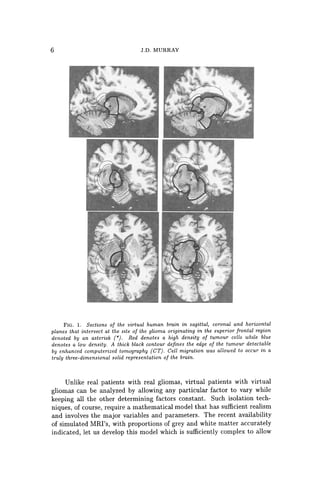
![BIOLOGICAL PATTERN FORMATION 7
different diffusion rates in grey and white matter (for example, a 5-fold
increase in diffusion or migration in white matter) as well as to prevent
spread across certain parts of the brain.
The model is a simple one which focuses on only two key elements,
namely diffusion and growth. Other variables can be introduced into the
model as their relative importance is discovered. Previous studies [18, 20, 5]
showed how to determine estimates for these parameters from patient scans.
With these the present model can be depressingly predictive as to the where
the tumour is likely to grow in real time. Of course many aspects, which
can be included in more complex models, such as swelling and distortion
of tissue should be included. The point of this brief discussion is to show
how even a simple basic model can still be useful'clinically. However, even
without these other effects included what seems clear from these theoretical
studies of virtual gliomas is that current imaging techniques are woefully
inadequate for definitive clinical decisions as to what constitutes the opti-
mal treatment for patients with gliomas.
2. General concluding remarks. Theoretical modelling has been
proven to be useful in the study of a remarkably diverse spectrum of biolog-
ical problems such as wound healing, quantifying disease control strategies,
the effect of introducing genetically engineered organisms in the environ-
ment and suggesting experiments associated with limb development, to
name just a few.
Pattern formation studies are sometimes criticized for their lack of
inclusion of genes in the models. But then criticism can be levelled at any
modelling abstraction of a complex system to a relativley simple one. It
should be remembered that the generation of pattern and form, particularly
in development, is usually a long way from the level of the genome. Of
course genes play crucial roles in development, but they do not actually
create patterns. Many of the evolving patterns could hardly have been
anticipated solely by genetic information.
Why use mathematics to study something as intrinsically complicated
and ill-understood as development, angiogenesis, wound healing, infectious
disease dynamics, regulatory networks and so on? We suggest that math-
ematical modelling must be used if we ever hope to genuinely and real-
istically convert an understanding of the underlying mechanisms into a
predictive science. Mathematics is required to bridge the gap between the
level on which most of our knowledge is accumulating (cellular and below)
and the macroscopic level of the patterns we see. A mathematical approach
lets us explore the logic of pattern formation. Even if the mechanisms were
well understood - and they certainly are far from it at this stage - math-
ematics would be required to explore the consequences of manipulating the
various parameters associated with any particular scenario. In the case of
such things as wound healing, tumour growth and it will be increasingly
so in angiogenesis with the cancer connection, the number of options that](https://image.slidesharecdn.com/27962819-250523222021-fae36494/85/Mathematical-Models-For-Biological-Pattern-Formation-2001th-Edition-Philip-Maini-20-320.jpg)
![8 J.D. MURRAY
are fast becoming available to wound and cancer managers will become
overwhelming unless we can find a way to simulate particular treatment
protocols before applying them in practice. The latter has already been of
use in understanding the efficacy of various treatment scenarios with brain
tumours [18, 20, 17] and new two step regimes for skin cancer [10].
There is no doubt that we are a long way from being able to reliably
simulate actual developmental scenarios, notwithstanding the multitude of
theories that abound. The active cellular control of key processes is poorly
understood. Despite such limitations, we argue that exploring the logic of
biological processes is worthwhile, in some current situations even essential
in our present state of knowledge. It allows us to take an hypothetical
mechanism and examine its consequences in the form of a mathematical
model, make predictions and suggest experiments that would verify or in-
validate the model; the latter is frequently biologically informative. In fact,
the very process of constructing a mathematical model can be useful in its
own right. Not only must one commit to a particular mechanism, one is
also forced to consider what is truly essential to the process and what the
key players are. We are thus involved in constructing frameworks on which
we can hang our understanding. The equations, the mathematical analysis
and the numerical simulations that follow serve to reveal quantitatively, as
well as qualitatively, the consequences of that logical structure.
The best integrative biology studies have served to highlight where
our knowledge is deficient and to suggest directions in which fruitful exper-
imentation might lead us. A crucial aspect of this research is the interdis-
ciplinary content and, as already mentioned, a crucial test of all theoretical
models should be in their impact on the experimental community. The
field of mathematical or theoretical biology or integrative biology has now
achieved some level of maturity, and we believe that future dialogue be-
tween experimentalists and theoeticians will lead us more rapidly towards
a fuller understanding, if not a complete one, of several biological processes
involving pattern formation.
REFERENCES
[1] T. BOEHM, J. FOLKMAN, T. BROWDER, AND M. O'REILLY. Antiangiogenesis ther-
apy of experimental cancer does not induce acquired drug resistance. Nature,
404-407, 1997.
[2] P.K. BURGESS, P.M. KULESA, J.D. MURRAY, AND E.C. ALVORD, JR. The inter-
action of growth rates and diffusion coefficients in a three-dimensional math-
ematical model of gliomas. J Neuropathol and Exp Neural, 56:704-713, 1997.
[3] D.L. COLLINS, A.P. ZIJDENBOS, V. KOLLOKIAN, J.G. SLED, N.J. KABANI, C.J.
HOLMES, AND A.C. EVANS. Design and construction of a realistic digital brain
phantom. IEEE Transactions on Medical Imaging, 17:463-468, 1998.
[4] V.P. COLLINS, R. K. LOEFFLER, AND H. TIVEY. Observations on growth rates of
human tumors. Am J Roentgenol Radium Ther Nucl Med, 76:988-1000, 1956.
[5] G.C. CRUYWAGEN, D.E. WOODWARD, P. TRACQUI, G.T. BARTOO, J.D. MURRAY,
AND E.C. ALVORD, JR. The modelling of diffusive tumours. J Biological
Systems, 3:937-945, 1995.](https://image.slidesharecdn.com/27962819-250523222021-fae36494/85/Mathematical-Models-For-Biological-Pattern-Formation-2001th-Edition-Philip-Maini-21-320.jpg)
![BIOLOGICAL PATTERN FORMATION 9
[6] J. FOLKMAN. Anti-angiogenesis: New concept for therapy of solid tumors. Annals
of Surgery, 75:409-416, 1971.
[7] J. FOLKMAN. TUmor angiogenesis: therapeutic implications. New England Journal
of Medicine, 285:1182-1186, 1972.
[8] J. FOLKMAN. Angiogenesis in cancer, vascular, rheumatoid and other diseases.
Nature Medicine, 1:27-31, 1995.
[9] A. GIESE, L. KLUWE, B. LAUBE, H. MEISSNER, M. BERENS, AND M. WESTPHAL.
Migration of human glioma cells on myelin. Neurosurg, 38:755-764, 1996.
[10] T. JACKSON, S.R. LUBLIN, N.O. SIEMERS, P.D. SENTER, AND J.D. MURRAY. Math-
ematical and experimental analysis of localization of anti-tumor antibody-
enzyme conjugates. British Journal of Cancer, 80:1747-1753, 1999.
[11] F.W. KRETH, P.C. WARNKE, R. SCHEREMET, AND C.B. OSTERTAG. Surgical resec-
tion and radiation therapy versus biopsy and radiation therapy in the treat-
ment of glioblastoma multiforme. J Neurosurg, 78:762-766, 1993.
[12] B.C. LIANG AND M. WElL. Locoregional approaches to therapy with gliomas as
paradigm. Curro Opinion in Oncol., 10:201-206, 1998.
[13] D. MANOUSSAKI, S.R. LUBKIN, R.B. VERNON, AND J.D. MURRAY. A mechanical
model for the formation of vascular networks in vitro. Acta Biotheretica,
44:271-282, 1996.
[14] J.D. MURRAY, J. COOK, R. TYSON, AND S.R. LUBKIN. Spatial pattern formation in
biology: I dermal wound healing. ii bacterial patterns. Journal of the Franklin
Institute, 335B:303-332, 1998.
[15] J.D. MURRAY, D. MANOUSSAKI, S.R. LUBKIN, AND R.B. VERNON. A mechanical
theory of in vitro vascular network formation. In C. Little, V. Mironov, and
E. Helene Sage, editors, Vascular Morphogenesis in vivo, in vitro, in mente,
pages 173-188. Birkhauser, Boston, 1998.
[16] J .M. NAZZARO AND E.A. NEUWELT. The role of surgery in the management of
supratentorial intermediate and high-grade astrocytomas in adults. J. Neuro-
surg., 73:331-344, 1990.
[17] K.R. SWANSON. Mathematical modeling of the growth and control of tumors. PhD
thesis, University of Washington, 1999.
[18] P. TRACQUI, G.C. CRUYWAGEN, D.E. WOODWARD, G.T. BARTOO, J.D. MURRAY,
AND JR. E.C. ALVORD. A mathematical model of glioma growth: the effect of
chemotherapy on spatial-temporal growth. Cell Poliferation, 28:17-31, 1995.
[19] R. TYSON, S.R. LUBKIN, AND J.D. MURRAY. A minimal mechanism for bacterial
patterns. Proc. Roy. Soc. Lond., pages 299-304, 1998.
[20] D.E. WOODWARD, J. COOK, P. TRACQUI, G.C. CRUYWAGEN, J.D. MURRAY, AND
JR. E.C. ALVORD. A mathematical model of glioma growth: the effect of
extent of surgical resection. Cell Prolif, 29:269-288, 1996.
[21] D.E. WOODWARD, R. TYSON, M.R. MYERSCOUGH, J.D. MURRAY, E.O. Bu-
DRENE, AND H.C. BERG. Spatio-temporal patterns generated by Salmonella
typhimurium. Biophys. J., 68:2181-2189, 1995.](https://image.slidesharecdn.com/27962819-250523222021-fae36494/85/Mathematical-Models-For-Biological-Pattern-Formation-2001th-Edition-Philip-Maini-22-320.jpg)
![12 S. SCHNELL ET AL.
large amount of experimental data. We begin with a brief description of
the early events: details of these events can be found in [35], [88], and [50].
The chick embryo develops from a small, disk-shaped blastodisc float-
ing on top of the yolk. After the egg is fertilized cells divide repeatedly,
forming a multicellular stratified structure called the blastoderm. The pe-
riod from just prior to laying through several hours afterwards has been
subdivided into 14 stages [31, 50]. Cell division is dominant during stages
I - VI, and morphogenetic movements begin during stages VII-X, when
cells of the central blastodisc, called the area pellucida (c/. Figure 1),
separate from the yolk, producing a hollow region beneath the disc called
the subgerminal cavity [75, 99]. Subsequently some cells from the central
blastodisc move into the subgerminal cavity (either actively or passively),
and simultaneously the disc expands radially over the yolk. The opaque
marginal zone of the blastoderm, known as the area opaca, remains in con-
tact with the yolk and may play an active role in the radial movement
(Figure 1 A). The result is that during stages VII-X the central part of
the disc changes from a layer 4-6 cells deep to a translucent layer one cell
thick called the epiblast. The anterior-posterior axis of the embryo is also
determined during these stages [50]. After stage X some cells within the
marginal zone migrate posteriorly, and then leave the marginal zone at
the posterior marginal zone (PMZ)(Figure 1 B). They spread across the
subgerminal cavity beneath the epiblast as a loosely-connected sheet, in-
corporating islands of cells shed from the blastodisc earlier. By stage XIV
this sheet connects with the anterior margin of the disc and forms the hy-
poblast, and at this stage the blastoderm is bi-Iayered with the epiblast and
hypoblast separated by the blastocoel cavity. Fate maps for cell movements
in these stages are available [39].
During hypoblast formation the embryonic shield or Koller's sickle de-
velops at the posterior end of the epiblast (cf Figure 2(a)). This consists
of a thickened epiblast [93] comprising primitive streak precursor cells that
have migrated to this area by a series of 'polonnaise movements' [105]. The
first visible sign of gastrulation is formation of the primitive streak, which
arises from Koller's sickle at the posterior midline of the blastodisc [52]
(Figure 1 C and D). The sickle narrows and the primitive streak moves an-
teriorly between the epiblast and the hypoblast. The tip of the ingressing
streak moves"" 60% of the way across the blastoderm before it stops, and
later, regresses. At full primitive streak stage (Hamburger and Hamilton
stage 4, [38]) the organizer of the avian embryo, Hensen's node, develops as
a bulbous structure at the anterior tip of the streak. The period between
the accumulation of cells at the posterior region and full primitive streak
is approximately 12 hours. The structure of the blastoderm at this stage
is illustrated in Figure 2(b). During the advance of the node, epiblast cells
move through the streak and into the interior. Those that migrate through
the node form anterior structures, those that migrate through the lateral
parts of the primitive streak become endodermal and mesodermal cells, and](https://image.slidesharecdn.com/27962819-250523222021-fae36494/85/Mathematical-Models-For-Biological-Pattern-Formation-2001th-Edition-Philip-Maini-24-320.jpg)
![PRIMITIVE STREAK FORMATION AND SOMITOGENESIS 13
(A) Anterior
(C)
(E)
node
p<'lIucida
Primitive
groove
Posterior
Anterior
area of
blastoderm
taking shape
Head
process
Hensen's
node
FIG.!' A schematic of the stages in early development of the chick embryo (A)
3-4 hours post-laying, (B) 5-6 hours, (C) 7-8 hours, (D) 10-12 hours, (E) 15-16 hours,
(F) 19-22 hours, . (Reproduced with permission from (35})
the remainder constitute the ectoderm. Simultaneously, the area pellucida
changes from circular to pear-shaped, narrowing in the posterior portion.
The head structure, notochord and somites are laid down during regression
of the node, and when regression is complete the embryo is a flat trilaminar
blastoderm comprising the ectodermal, mesodermal and endodermallayers.
These will form various organs during subsequent morphogenesis, in addi-
tion to the structures formed during regression. The regressing node and
anterior portion of the streak eventually form the tail bud [94]. Regression
proceeds on a slower time scale than progression, taking approximately 24
hours for the node to regress after the streak reaches its maximum length
of approximately 1.9 mm [94].
During regression of the primitive streak the neural folds begin to
gather at the center of the embryo, and the segmental plates, which are](https://image.slidesharecdn.com/27962819-250523222021-fae36494/85/Mathematical-Models-For-Biological-Pattern-Formation-2001th-Edition-Philip-Maini-25-320.jpg)
![14 S. SCHNELL ET AL.
Area Pellucida Marginal
CC======,
===
/
='
=I
======o
==~===o==o=~=o
=o=~
===:>12£::J-
~
t )1V~. 'gJDI5
Primary hypoblast Koller's siCkle ! t ! ro
Seoondary hypoblast ' ,
Deep layer 01 marginal zone
FIG. 2. (a) A schematic cross-section of the blastoderm prior to primitive streak
formation. (b) The blastoderm at the stage of maximal streak ingression (Reproduced
with permission from [35J)
often referred to as paraxial mesoderm or presomitic mesoderm (PSM),
separate into blocks of cells known as somites. They form as paired ep-
ithelial spheres arranged bilaterally along the anterior-posterior axis and
emerge in strict cranio-caudal order [36]. Simultaneously, new cells are in-
corporated into the PSM from the regression of Hensen's node at the same
rate as new somites are formed rostral to the PSM [16, 83]. Figure 3 is a
schematic representation of these early processes. Somites are divided by a
fissure into anterior and posterior halves that differ in their gene expression
and differentiation [104, 36].
The formation and differentiation of somites is the result of three dis-
tinct morphological events progressing in a strict spatio-temporal order: (1)
the prepatterning of the PSM; (2) somite and somitic boundary formation;
and (3) the differentiation of a somite into anterior and posterior halves
[36]. Several experimental observations confirm these events. Scanning
electron microscopy observations [42] and transplantation experiments [49]
show that the PSM displays a prepattern prior to segmentation. In addi-
tion, Hox and Notch-Delta pathway genes are involved in all these events
[104, 25]. These molecular results suggest the existence of a conserved
mechanism for segmentation in protostomes and deuterostomes [61].
The segmental pattern of somites in turn governs the segmental pat-
tern of the peripheral nervous system and determines the shapes and ap-](https://image.slidesharecdn.com/27962819-250523222021-fae36494/85/Mathematical-Models-For-Biological-Pattern-Formation-2001th-Edition-Philip-Maini-26-320.jpg)
![PRIMITIVE STREAK FORMATION AND SOMITOGENESIS 15
Head
Somites
Presomitic
Mesoderm
Hensen's --+--t>r
Node
Primitive
Streak
: Anterior
Posterior
FIG. 3. A schematic diagram illustrating the main structures involved in somi-
togenesis. Segmentation of the presomitic mesoderm occurs in an anterior-posterior
sequence and the time taken for the formation of a somite is approximately 90 minutes
in the chick. See text for details. (Redrawn from [10].)
pendage characteristics of the vertebrae. Somites are also the source of
cells for muscles, and influence the metameric distribution of blood ves-
sels. Genetic or/and environmental factors disturbing somitogenesis pro-
duce malformations and abnormal development [117, 27, 36].
Although the sequence of events in early avian development is well
documented, less is known about the mechanisms that give rise to primi-
tive streak formation and somitogenesis. A number of theoretical models
have been proposed to explain somitogenesis, and while these models are
satisfactory in some respects, none can explain the complete set of obser-
vations. In the following subsections we present a brief exposition of the
current experimental facts on primitive streak formation and somitogene-
sis. We then describe the theoretical models developed to explain some of
these observations.
1.1. Formation of the primitive streak and the organizer. The
ability of specific parts of the embryo to induce a primitive streak and
node has been identified by a number of experiments. In particular, two
regions have been tested, the PMZ and Koller's sickle. We should stress
that references below to the PMZ may include Koller's sickle, except where
stated explicitly.
I. Posterior Marginal Zone (PMZ) .
• At stage X, transplants or rotation of the PMZ to lateral or anterior
positions can form an ectopic primitive streak; at stage XI the inner
region in contact with the PMZ also has the potential to form
primitive streak, and at stage XII the PMZ has lost the ability to](https://image.slidesharecdn.com/27962819-250523222021-fae36494/85/Mathematical-Models-For-Biological-Pattern-Formation-2001th-Edition-Philip-Maini-27-320.jpg)
![16 s. SCHNELL ET AL.
induce a primitive streak [53]. At both stages X and XI the size of
the transplanted fraction is also critical in its capacity to initiate
an ectopic axis [30].
• If a fragment of the PMZ is removed and replaced by lateral
marginal zone (LMZ) tissue at stage X, a single primitive streak
always originates in the normal position, but if the fragment of
PMZ is replaced by beads which prevent healing of the wound,
then two primitive streaks form [54].
• If donor PMZ tissue is inserted at 900 to the host PMZ at stage
X, a single primitive streak develops at the site of the host PMZ.
However, if the host PMZ is removed two small primitive streaks
develop, one at the normal site and one at the transplant site.
Khaner and Eyal Giladi [54] have also demonstrated that trans-
plantation of a portion of the PMZ into the LMZ of a host embryo
induces a second primitive streak to grow at 900 to the primitive
streak growing from the PMZ.
• Any part of the blastoderm, provided it contains a portion of the
PMZ and is sufficiently large, has the potential to develop a nor-
mal embryo. The streak is normally initiated along a radius [96].
When the blastodisc is cut in half, perpendicular to the anterior-
posterior axis, the posterior half will form a streak initiated from
the posterior margin. The anterior half can also form a streak,
which is more likely to be initiated from the LMZ, but it may form
from the anterior margin. When the cut is made parallel to the
anterior-posterior axis, two streaks form, one on either side of the
cut.
• Fate map experiments demonstrate that PMZ tissue has the ca-
pacity to induce an ectopic primitive streak without contributing
cells to the streak [6]. This suggests that the PMZ may function
as an avian equivalent of the Nieuwkoop center [66] - a region of
the amphibian blastula that induces an organizer in adjacent cells
without contributing to it. The experiments further demonstrate
that: (i) PMZ does not give rise to hypoblast but remains station-
ary; (ii) transplants of quail PMZ (cut to exclude Koller's sickle)
to the anterior side of a chick anterior region can induce a primi-
tive streak from the anterior pole in a significant number of cases,
and grafts to the posterior side of the anterior region results in a
high frequency of streaks from the posterior end. In neither case,
however, does the graft contribute cells to the streak. These ex-
periments suggest that the PMZ determines the position of the
streak.
II. Koller's sickle.
• It is known that Koller's sickle begins to form in the PMZ at stage
X, and if cell movement in this area is blocked, no primitive streak
is formed [95].](https://image.slidesharecdn.com/27962819-250523222021-fae36494/85/Mathematical-Models-For-Biological-Pattern-Formation-2001th-Edition-Philip-Maini-28-320.jpg)
![PRIMITIVE STREAK FORMATION AND SOMITOGENESIS 17
• Transplants of Koller's sickle to lateral portions of host embryos
[13, 41, 14] can induce an ectopic primitive streak. In normal
development, cells of Koller's sickle contribute to the primitive
streak [41].
• Detailed fate mapping of midline cells [6] show that the epiblast
above Koller's sickle and Koller's sickle itself both contribute cells
to the node and primitive streak. The epiblast above and anterior
to Koller's sickle, and cells in the anterior part of Koller's sickle,
contribute cells to the node and anterior streak, whereas those cells
immediately dorsal to the sickle and in the posterior part of the
sickle contribute to the posterior part of the streak. Transplants of
quail PMZ cut in a manner to include Koller's sickle (compare with
previous item) were able to form a primitive streak when grafted
to the anterior-most part of a chick anterior fragment with much
greater frequency than when Koller's sickle was excluded. The
quail cells were found to contribute to the streak when the graft
included Koller's sickle.
• Grafts of PMZ including the sickle retain the competence to induce
a primitive streak at later stages than grafts excluding the sickle
[6]. The ability of Koller's sickle alone to induce an ectopic axis is
lost by stage XIII, but a large fragment of the PMZ together with
Koller's sickle can still induce an ectopic axis [52].
Stimulated in part by the wealth of data unearthed in other model develop-
mental systems, many recent experiments have been directed at discovering
the genes regulating development. For example, the Hox gene goosecoid is
first found in a small population of cells corresponding to Koller's sickle
[41J. Later this gene characterizes cells of the primitive streak, and ex-
pression is highest in cells of Hensen's node and the anterior portion of
the streak. Brachyury (Ch- T) genes are expressed in forming mesoderm in
response to inducing factors and at stage XII in a broad arc in the poste-
rior epiblast. These gene expression patterns suggest that primitive streak
formation can be regulated by gradients of organizer genes [5].
The signals involved in streak formation, particularly the transforming
growth factors, have also been studied recently. A number of members of
the transforming growth factor beta family (TGF-(3) have been shown to
induce primitive streak formation. For example, activin has been shown
to induce development of axial structures [65, 118, 23]' but it does not
have the spatial and temporal distribution expected of an inducer. cVgl
expressed in the PMZ of pre-primitive streak embryos has been shown to
induce development of an ectopic primitive streak [91]. The activation of
the Wnt proto-oncogene pathway potentiates the activity of activin and
cVgl. In contrast, the bone morphogenetic protein-4 (BMP-4) inhibits
primitive streak formation [102]. Furthermore, BMP antagonists such as
chordin can induce both primitive streak formation and organizer genes.](https://image.slidesharecdn.com/27962819-250523222021-fae36494/85/Mathematical-Models-For-Biological-Pattern-Formation-2001th-Edition-Philip-Maini-29-320.jpg)
![18 S. SCHNELL ET AL.
These experiments suggest that areas of the LMZ can form a primitive
streak if they are exposed to fragments of PMZ, but they are inhibited from
doing so by neighboring PMZ. Thus cells in the PMZ are already differen-
tiated from those in other parts of the marginal zone and the remainder of
the blastoderm when ingression of the primitive streak begins.
Traditionally the blastoderm has been considered homogeneous prior
to streak formation, but recent findings suggest earlier cell diversity and
considerable cell movement in the early epiblast [98]. Canning and Stern
[15] identified a subpopulation of cells testing positive for the epitope HNK-
1, which is first expressed on the surface of cells of the PMZ and on those
which later form primary hypoblast. Later it is found in the area of streak
formation, distributed with a distinct anterior-posterior gradient. A prim-
itive streak does not form when these cells are removed. This has led to
the suggestion that HNK-l cells are the source of streak-derived tissue [98].
The precise role of the epitope itself is not clear, but it may have a role in
modulating cell adhesion (see [97] and references therein).
Given the critical role of the organizer in patterning the embryo (for
example, formation of the axial structures and left-right asymmetry), it
is surprising that in embryos where the node and anterior portion of the
streak has been extirpated [37, 113, 112, 84], or replaced in reverse orien-
tation [1], a new organizer can be regenerated and development proceeds
normally (albeit delayed). In fact, a lateral isolate of the embryo, cut such
that both the primitive streak and Hensen's node have been excluded, can
reconstitute a primitive streak and organizer [114, 115].
Using labeling techniques, Joubin and Stern [43] have demonstrated
that the organizer is not a static population of cells, as was tradition-
ally believed, but is a transitory population of cells that have moved into
the node, acquired organizer characteristics (Le. express specific organizer
genes), and then left the node. It appears that the central third of the
primitive streak (axially), characterized by the overlapping expression of
cVg-l and Wnt-Bc, induces the cells anterior to it to acquire organizer
characteristics. The organizer prevents neighboring tissue from acquiring
organizer status by releasing an inhibitory signal. The issue is confused,
however, by the observation of a resident population of cells within the
epiblast which remain part of the node during its regression [89, 90, 83]. It
has been suggested that this population constitutes stem cells which divide
and produce notochord/somite progeny.
1.2. Somitogenesis. During somitogenesis, as in other segmentation
processes, the body axis is divided along the anterior-posterior axis into
similar repetitive structures formed from the embryonic layers. In insects,
such as Drosophila melanogaster, segments are generated by the simulta-
neous division of the syncitial blastoderm. In other invertebrates such as
annelids and crustaceans, and in vertebrates, the mechanism of metameri-
sation is different; the segments are formed at the cranial end of a multi-
cellular embryo and segmentation propagates caudally [110].](https://image.slidesharecdn.com/27962819-250523222021-fae36494/85/Mathematical-Models-For-Biological-Pattern-Formation-2001th-Edition-Philip-Maini-30-320.jpg)
![PRIMITIVE STREAK FORMATION AND SOMITOGENESIS 19
During somitogenesis, continuous inductive interactions with Hensen's
node, notochord, neural tube and endoderm are not necessary for somite
formation [7, 11, 100]. For example, explants of PSM are able to form
somites in the absence of all surrounding structures. Further experiments,
in which the PSM is cut into several parts and these parts are rearranged,
show that somites do not form. However, if the disrupted PSM is in contact
with epithelial structures then somites do form, suggesting that some factor
derived from the epithelium may influence somite formation [69].
Scanning electron microscope images show that the PSM is not a ho-
mogeneous tissue. Prior to segmentation, the PSM displays metameric
arrangements of groups of cells, named somitomeres by Meier [62], which
are evidently the predecessors of somites [42, 36]. The existence of this
prepattern is confirmed in microsurgical experiments [70, 18]' where iso-
lated parts of the PSM form somites in strict cranio-caudal order some
time after their isolation, differentiating into anterior and posterior halves
in each somite. The existence of a prepattern is also strongly supported by
the periodic pattern of Hox and Notch-Delta gene expression in the PSM
[104, 57, 25]. Furthermore, the prepattern of anterior and posterior halves
is also established before the formation of a somite [49]. Transplantation
experiments reversing the anterior-posterior axis of the PSM demonstrate
that the anterior-posterior polarity of the resulting pattern of somites is
also reversed, so somite halves develop according to their original orien-
tation [2]. In addition, there is a change in the mechanical properties of
the cells in the PSM before they differentiate into a somite. There is an
increase in cell compaction, and in cell-cell and cell-substratum adhesion,
followed by epithelialization [49, 104] of the ball of cells as they form a
soinite. Several studies suggest that adhesion molecules such as cadherins
playa major role in these processes [26, 85, 59]. It should be noted that
cell labeling experiments indicate that cells of the PSM can contribute to
more than one somite, suggesting that the prepattern of somitomeres does
not preclude mixing between the prospective somites [101].
The total number of somites is regulated in an embryo. The Amputated
mouse mutant, which is shorter than the wild-type mouse, has the same
number of somites, but their somites are considerably smaller than those of
the wild-type embryos [32]. However, the number of somites can be altered
experimentally [49]. For example, heat shock applied to chick embryos can
induce the formation of an extra somite [106, 82], or can result in up to
four repeated somite anomalies, confined to one or to both rows, separated
by relatively constant distances of six to seven normal somites [82]. The
repeated anomalies suggest that heat shock affects an oscillatory process
within the somite precursors [101].
There appears to be some degree of cell cycle synchrony between cells
in the PSM which are destined to segment together to form a somite. The
cell cycle synchrony is observed in the early somite two cell cycles after seg-
mentation [101, 81]. To some extent, cells of the PSM seem to be arranged](https://image.slidesharecdn.com/27962819-250523222021-fae36494/85/Mathematical-Models-For-Biological-Pattern-Formation-2001th-Edition-Philip-Maini-31-320.jpg)
![20 S. SCHNELL ET AL.
in order of developmental age, with cells at a given level having relatively
synchronous cell cycles. The rostral end of the PSM has an increased mi-
totic index, which indicates that this region has a high proportion of cells
in mitosis [82].
Recently, the study of the expression of the transcriptional factor c-
hairy-l in the PSM of chick embryos has provided molecular evidence for
the existence of a segmentation clock [72, 22]. During segmentation, the
cells of the PSM go through 12 cycles of c-hairy-l expression before becom-
ing part of a somite, while more cells are continuously incorporated into the
posterior end of the PSM. This observation suggests that the segmentation
clock controls the time duration of cells in the PSM before they will form
part of a somite. During the time taken for one somite to form, the expres-
sion of c-hairy-l sweeps along the PSM in the posterior-anterior direction,
narrowing as it propagates (see Figure 4). This wavefront-like expression
finally stops and is maintained in a half somite-sized domain which gives
rise to the caudal half of the forming somite. The c-hairy-l expression is
independent of cell movements and does not result from the propagation of
a signal in the plane of the PSMj it is an intrinsic cell autonomous property
of this tissue [61, 79]. More recently, studies by McGrew et al. [60] and
Forsberg et al. [33] have shown that lunatic fringe (i-fng) gene expression
resembles the expression of c-hairy-l in PSM. In fact, they show that both
expressions are coincident and are responding to the same segmentation
clock [80]. In Drosophila, it is known that l-fng plays an important role
in the formation of the wing margin by potentiating Notch activation by
Delta and the inhibition of Notch activation by the alternative ligand Ser-
rate [74, 116]. In l-fng mutant mice, the formation of somites is disrupted
and if a somite forms its anterior-posterior patterning is disturbed [27, 117].
Finally, it is important to mention that the principle differentiation
pattern of all the somites is very similar. However, during morphogenesis
subsequent differentiation forms unique anatomic structures, depending on
the position along the anterior-posterior axis. Experiments in chick em-
bryos demonstrate that the positional specification of somites occurs early
during somitogenesis [55, 20, 21, 19, 107, 17, 12]. When cervical somites
are replaced with somites from the trunk region, rib-like structures develop
in the cervical vertebral column of the embryo. When thoracic somites are
replaced by cervical somites, embryos do not develop ribs [55]. There is
now a large body of experimental work showing that positional specifica-
tion of the PSM requires members of the Hox gene family [57]. Hox gene
activation during development correlates with gene position in the Hox
complex, a property referred to as colinearity. The spatial and temporal
colinearity in the expression of these genes results in unique combinations
of Hox genes in defined groups of somites and their derivatives along the
anterior-posterior axis [34, 40]. This led to the suggestion that a Hox code
specifies the identity of somites [48, 47]. The role of Hox genes in posi-
tional specification has been analyzed by interfering with or altering the](https://image.slidesharecdn.com/27962819-250523222021-fae36494/85/Mathematical-Models-For-Biological-Pattern-Formation-2001th-Edition-Philip-Maini-32-320.jpg)
![PRIMITIVE STREAK FORMATION AND SOMITOGENESIS 21
Anterior
Posterior
Time
FIG. 4. Schematic illustration of the wave of c-hairy-l sweeping in the posterior-
anterior direction (bottom to top) along the PSM with time (left to right). The shading
denotes expression of this factor. It begins as a broad wave but narrows as it moves
anteriorly until it finally correlates with the posterior half of the new forming somite.
Then a new wave begins at the posterior margin of the PSM. Similar behavior is observed
for lunatic fringe.
expression of single Hox genes or by simultaneously perturbing the expres-
sion with retinoic acid, which is implicated in the specification of the axes
during development [103].
2. Questions. Early organization of the avian blastoderm clearly in-
volves a carefully controlled sequence of events. At present, very little is
known concerning the mechanisms regulating this development and here
we list some of the major unresolved questions. In the following section we
describe some of the theories postulated to explain these processes.
2.1. Early development.
1. How is the posterior site of the embryo determined? Formation
of the area pellucida involves a gravity induced directional shedding of
cells (posterior to anterior) to form a one-cell thick layer [56]. How is
this translated into the structural differences associated with the posterior
region (e.g. Koller's sickle, secondary hypoblast formation)?
2. Development of the primary hypoblast involves an apparent drop-
ping of cells in the area pellucida to form isolated islands in the subgerminal
cavity [15]. What leads to the early diversification of such cells, and how
do they separate from the area pellucida? One possibility is to link the di-
versification with the cell cycle, such that at the time of primary hypoblast
formation a randomly scattered population in a specific phase of the cycle
experiences a change in its cellular properties, for example adhesion. This
change in adhesion may result in such cells being forced from the area pel-](https://image.slidesharecdn.com/27962819-250523222021-fae36494/85/Mathematical-Models-For-Biological-Pattern-Formation-2001th-Edition-Philip-Maini-33-320.jpg)
![22 S. SCHNELL ET AL.
lucida. To test such a hypothesis, it is necessary to construct a discrete cell
model which incorporates cell adhesion [73).
3. What controls formation of the secondary hypoblast, and does the
hypoblast influence streak formation? The role of the hypoblast in streak
formation is controversial, and earlier experiments in which the hypoblast
has been shown to induce streak formation [108, 3, 4] have been challenged
by recent experiments [51). However it is still not known whether the
hypoblast is able to exert some influence over streak formation.
4. What initiates motion and guides the early migration of cells in
the lateral regions toward the PMZ? Stern [97] observed migration of a
subpopulation of the area pellucida to the posterior marginal zone prior to
streak formation and speculated that a chemoattractant is produced at that
site. Although collagen-gel assays support this theory, no chemoattractant
has been identified.
5. What cues guide elongation and movement of the primitive streak?
A simple anterior-posterior gradient of a diffusible morphogen cannot be
used for positional information along that axis [53), for if it were the 900
transplants of the primitive streak would ingress toward the anterior pole
rather than along a ray through the center of the disk.
6. What is the role of cell division in streak formation? Recent
results by Wei and Mikawa [109) suggest that a subpopulation of cells in the
posterior region may divide in a directional manner to form the primitive
streak. It remains to be understood whether this division is essential for
streak formation, or if it is simply an associated phenomenon.
7. What mechanisms can account for the fact that the primitive streak
maintains its rod-like structure during ingression? Does the primitive
streak ingress by convergent extension [46), whereby cells intercalate at
the posterior marginal zone and push the primitive streak forward? Are
there adhesive differences between cells in the primitive streak and those in
the hypoblast and epiblast, or is the structure maintained by chemotactic
attraction between cells in the primitive streak? Alternatively, is the streak
maintained as a rod by the forces occurring throughout the blastoderm at
these stages.
8. There appears to be a gradient within the marginal zone of poten-
tial to form a streak, with the posterior being the most capable and the
anterior the least. At what stage is this potential determined, and by what
mechanisms?
9. The primitive streak seems to inhibit other streaks from forming.
What is the nature of this inhibition, and is it confined to act along the
marginal zone?
10. The size and age of a blastodisc segment or donor implant are
important in determining the site of streak formation. How do the key
properties involved change with time?
11. How is the organizer defined, and how are the movements of cells
through the organizer to form notochord, head process, paraxial mesoderm,](https://image.slidesharecdn.com/27962819-250523222021-fae36494/85/Mathematical-Models-For-Biological-Pattern-Formation-2001th-Edition-Philip-Maini-34-320.jpg)
![PRIMITIVE STREAK FORMATION AND SOMITOGENESIS 23
etc., regulated? Recent experiments have revealed that the organizer is a
transitory population continuously defined by cells in the middle part of the
primitive streak [43]. Previous results, however, suggest that there exists a
resident population of cells within the organizer that moves back with the
node throughout regression. What is the relevance, if any, of this resident
population?
12. What mechanisms control regression of the streak/organizer?
Does regression of the streak simply occur through the disappearance of
anterior cells into axial structures. Does the node regress by being pushed
back by cells that are ingressing through it? Ablation of the node results in
the regeneration of a new node, yet the new node must regenerate before
regression proceeds. Does the static population of cells within the node
control the movements of the node during regression?
13. How is the left-right asymmetry established? The earliest indica-
tion of left-right asymmetry in the avian embryo occurs with the asymmet-
ric expression of sonic hedgehog (shh) in the avian node [58). Studies in the
mouse have revealed the presence of a nodal fluid flow from right to left as
a result of unidirectionally rotating cilia on node cells [67, 68] and this has
been linked with the establishment of the left-right axis. However, no such
cilia have been located in the chick, and the cause of left-right asymmetry
remains unknown.
2.2. Somitogenesis.
1. What regulates the number and size of somites?
2. What determines differentiation into anterior and posterior halves
within a somite?
3. What are the differentiation and mechanical properties involved in
the epithelialization of somites ?
4. What determines the regional specification of somites - that is,
certain somites form certain structures. What is the precise role of the Box
family in this process and how is it controlled?
5. What drives the segmentation clock? Is there a relation between
the cell-cycle and the segmentation clock?
6. What is the precise role of the segmentation clock during somito-
genesis?
7. How is the interplay between the segmentation clock and Notch-
Delta and related components established?
8. What regulates the refinement of the c-hairy-l and l-fng cycles in
the forming somite? How do these cycles interact with the segmentation
clock?
9. How can the heat shock experiments be explained?
3. Models of streak formation and somitogenesis.
3.1. Formation of the primitive streak.](https://image.slidesharecdn.com/27962819-250523222021-fae36494/85/Mathematical-Models-For-Biological-Pattern-Formation-2001th-Edition-Philip-Maini-35-320.jpg)
![24 S. SCHNELL ET AL.
PI. Model of Induction by Gravity: Eyal-Giladi [29] proposed that
substances needed for the initiation of primitive streak formation become
nonuniformly distributed by gravity while the embryo is tilted, moving
from the vegetal pole toward the region that is incorporated into the PMZ.
Alternatively, Eyal-Giladi also suggests that these factors can be located
under the embryo and shifted toward the posterior by the sliding of the
yolk, and could later be found in the PMZ and Koller's sickle. Classic
experiments in chick embryos have established that labile anterior-posterior
polarity is determined 20 hours after fertilization. During this period there
is a critical2-hour time window where the outer albumen layers are rotated
by the uterus while the yolk remains stationary but slightly tilted within
a layer of low friction thin albumen [28]. In these experiments, the side of
the embryo that is tilted upward during the critical window is defined as
posterior. This model is unsatisfactory in some aspects. Little work has
been done on this hypothesis due to the difficulty of obtaining uterine eggs.
In addition, this model does not address the ingression and regression of
the primitive streak.
PII. Model of Induction by the PMZ: In this model, proposed by Bach-
varova [5), the PMZ is considered analogous to the Nieuwkoop Center of
the frog embryo, which is the structure responsible for induction during
the first stages of amphibian development. The PMZ of the chick embryo
acts as an extra-embryonic signaling center promoting formation of the
primitive streak in the adjoining posterior central disc epiblast. According
to the model, factors such as Vgl and Wnt8c produced in the PMZ acti-
vate organizer genes such as goosecoid in Koller's sickle and chordin in the
posterior central-disc epiblast. In turn, chordin suppresses BMP and this
decrease promotes activation of organizer genes in the posterior midline.
Lower concentrations of Vgl or TGF-{3 factors induce Brachyury-like genes
in a broader crescent of posterior central disc epiblast, leading to mesoderm
formation. BMP activity from lateral and anterior marginal zone induces
epidermis in the adjoining central disc. Finally, the activation of the Wnt
pathway in the late uterine and freshly laid egg plays an important role in
the asymmetry observed in cells of Koller's sickle and the hypoblast.
This model incorporates several aspects of primitive streak formation.
However, as in the previous model it does not address the ingression and
regression of the primitive streak. Furthermore, as indicated by Bachvarova
[5), many outstanding problems remain with this model. For example, it
is not clear if factors such as Vgl are required in normal development. In
addition, other factors such as TGF-{3 cannot be present and active in early
embryos; and little is known about the Wnt pathway.
A mathematical formulation of this model could help understand the
outcome of the complex of interactions proposed and make experimentally-
testable predictions.
PIlI. Chemotaxis Model: Chemotaxis (or haptotaxis) is a plausible](https://image.slidesharecdn.com/27962819-250523222021-fae36494/85/Mathematical-Models-For-Biological-Pattern-Formation-2001th-Edition-Philip-Maini-36-320.jpg)
![PRIMITIVE STREAK FORMATION AND SOMITOGENESIS 25
mechanism for the observed oriented cell movements both prior to and
during primitive streak formation, and this mechanism has been incorpo-
rated into a model designed for formation and subsequent maintenance of
the streak (though not the determination of the initial site of outgrowth)
[71].
The model assumes that there is a specialized subpopulation of cells
residing at or close to the posterior marginal zone that both respond to
and modulate the level of an attractant. This population serves to mark
the site of the primitive streak and guide the movements of elongation and
regression. Several cell populations have been identified [41, 99, 109] as
having a role in primitive streak formation. The model does not, however,
postulate how other cells ingress through the streak. In Figure 5 we show
the pattern of movements predicted on a two-dimensional domain. To
achieve movement of cells as a rod, rather than a general spreading of cells,
it is necessary to choose conditions such that the chemoattractant initially
has its highest concentration at the center of the domain (corresponding to
the center of the area pellucida) and decreases to zero at the marginal zone.
Plausible mechanisms for generating such conditions are given in [71].
The model makes a number of experimentally-testable predictions
(Figure 6). Firstly, it predicts that any ectopically induced embryonic axis
will develop along radial lines. Secondly, it predicts that disruption of the
center of the area pellucida will have a significant effect on the morphology
of the streak. It also predicts the natural development of an organizer re-
gion at the anterior portion of the streak as a region of higher cell density,
and demonstrates a decrease in the rate of regression as the streak moves
back, in agreement with experimental results [94]. However there is no ex-
perimental evidence for chemotactic motion in streak formation, and it is
unclear whether the same mechanism that drives propagation of the streak
is also responsible for regression. Thus this model simply demonstrates
that chemotaxis can produce the observed behavior.
PIV. Cell Division Model: Wei and Mikawa [109] have proposed a
model for formation of the streak based on directional cell division. In
this model, a specific subpopulation of cells (localized at stage XII to the
epiblast-midline region of the PMZ) undergoes oriented cell division along
the anterior-posterior axis to form the Hamburger and Hamilton stage 3
primitive streak. The model is supported by cell marking experiments
which demonstrate that the Hamburger and Hamilton stage 3 streak com-
prises only cells derived from this region, and not cells which have migrated
in from lateral regions, as has previously been assumed. Furthermore, cells
in the streak were shown to have metaphase chromosome plates (which in-
dicate cleavage direction) perpendicular to the anterior-posterior axis. The
calculation, based on the number of cells in the pre-streak region and Ham-
burger and Hamilton stage 3 streak, of a cell cycle time of approximately
4 hours is consistent with the mitotic index for cells of the chick gastrulae.](https://image.slidesharecdn.com/27962819-250523222021-fae36494/85/Mathematical-Models-For-Biological-Pattern-Formation-2001th-Edition-Philip-Maini-37-320.jpg)
![26
o
51
o
<D
.!!
a
b
c
S. SCHNELL ET AL.
8
1[
8
!
d
e
8
....
.!!
9
h
FIG. 5. A time sequence showing the cell density for model PIlI on a two-
dimensional rectangular domain. White represents high cell density, black represents
a zero cell density. The results show cell movement across the domain to form a rod
which extends approximately half the way across the domain (e). Subsequent develop-
ment shows a period of reverse movement, which occurs on a slower time scale.
This model is consistent with the observation that the epiblast portion
of the posterior marginal zone contributes to the primitive streak, and with
the idea that a PMZ-derived signal induces primitive streak in the adjoining
epiblast (see model II above). However, it is not yet clear if directional cell
division would be able to induce the streak to form a long straight rod
alone, nor is there any suggestion as to how regression of the streak is
controlled.
PV. Convergent-Extension Model: Schoenwolf [88] has postulated that
primitive streak formation may occur via a convergent-extension mecha-
nism similar to that observed in developing amphibia [44, 45]. In this
model, prospective primitive streak cells from either side of the midline
would converge at the midline, intercalating with those on the opposite
side and thereby producing an elongating primitive streak. This also raises
the possibility that regression may occur through a reverse process.
This model is speculative, yet some evidence for it can be found in the
general cell movements observed to take place in the epiblast during primi-](https://image.slidesharecdn.com/27962819-250523222021-fae36494/85/Mathematical-Models-For-Biological-Pattern-Formation-2001th-Edition-Philip-Maini-38-320.jpg)
![PRIMITIVE STREAK FORMATION AND SOMITOGENESIS 27
Simulation Initial Conditions Prediction
Simulation Initial Conditions Prediction
_ I
Simulation Initial Conditions Prediction
FIG. 6. The time course for the development of an ectopic streak following 'trans-
plantation' in model PII!. When a second population of "able" cells is placed at another
point along the marginal zone (top: lateral, middle: anterior), an ectopic streak develops
which moves towards the center of the domain. Fairly small changes in model parame-
ters can result in the fusing of these streaks at the anterior ends. In the bottom figures,
this has been effected by increasing the concentration gradient of the chemoattractant.
tive streak formation [105]. Furthermore, the mechanism could provide an
explanation for the change in morphology of the blastoderm from circular
to pear-shaped during formation of the streak, as intercalation would result
in a streak being driven in both anterior and posterior directions. However
the author does not suggest what determines the posterior marginal zone
as the site of streak formation, nor what the mechanisms are for guiding
cell movement during the convergent-extension process so as to maintain
the rod-like morphology of the streak.](https://image.slidesharecdn.com/27962819-250523222021-fae36494/85/Mathematical-Models-For-Biological-Pattern-Formation-2001th-Edition-Philip-Maini-39-320.jpg)
![28 S. SCHNELL ET AL.
3.2. Somitogenesis. During the last three decades, several models
have been proposed to explain the formation of somites [24, 32, 8, 9, 42,
49, 63, 64, 76, 78, 77, 82, 81, 87]. Some of these incorporate the different
aspects ofsomitogenesis previously mentioned, and are satisfactory in many
respects. It is important to emphasize that these models cannot explain
all the experimental facts presented in section 1.2, but they do lend insight
to many of the observations. The models can be divided into four main
categories:
SI. Induction Models: In these models, somite formation is explained
in terms of inductive interactions with neighboring tissue [8]. These models
are unsatisfactory in many aspects. No single tissue has been shown to in-
duce somite formation. As we previously indicated, somites can be formed
in the absence of Hensen's node, notochord, neural tube and endoderm
[7, 11, 100], but the midline structures are necessary after experimental
disruption of the PSM [69].
SII. Prepattern Models: These models postulate that there is a
spatially-periodic prepattern present in the PSM before formation of the
somites. Bellairs and Veini [11] proposed that somitogenic clusters are gen-
erated during PSM formation. Meier [62] suggested that prior to segmen-
tation the PSM displays metameric arrangements of groups of cells, named
somitomeres. The observation of the prepattern has been confirmed in mi-
crosurgical and transplantation experiments [70, 18, 2]. However, this sort
of model does not address the key problems of how the prepattern is set
up and how it is maintained and regulated.
SIll. Positional Information Models: These assume that a spatial
pattern in chemical morphogen is set up, either via a gradient or a reaction-
diffusion mechanism, and this prepattern determines cell differentiation.
There are two main models:
1. The wave gradient model was proposed by Wilby and Ede [111]
and Flint et al. [32]. This model proposes that regression of Hensen's node
creates two strips of paraxial mesoderm, and that cells recruited into them
start to synthesize a morphogen. The morphogen concentration increases in
the cells until a threshold is reached, at which point an irreversible change
from synthesis to destruction of morphogen occurs. The morphogen con-
centration in these cells falls, establishing a sink relative to cells that are
still producing the morphogen. Neighboring cells maintain a morphogen
concentration below the threshold, as morphogen diffuses from them into
the sink, but cells further back in the paraxial mesoderm exceed the mor-
phogen concentration threshold, and another trough of concentration is
formed. Thus, a pattern of alternating peaks and troughs is created, which
later gives rise to somites and fissures respectively. In this model, the size
of the somites is determined by either the rate of incorporation of cells into
the PSM or the speed of node regression. If the rate of node regression
depends on the size of the embryo, then this model can account for the](https://image.slidesharecdn.com/27962819-250523222021-fae36494/85/Mathematical-Models-For-Biological-Pattern-Formation-2001th-Edition-Philip-Maini-40-320.jpg)
![PRIMITIVE STREAK FORMATION AND SOMITOGENESIS 29
observation that there is regulation of somite number so, for example, in
the Amputated mutant mouse embryo, which is only two thirds the nor-
mal size, the number of somites formed is still the same as in the normal
case. This would be consistent with the assumption that the node regresses
more slowly in smaller embryos. However, this model cannot easily explain
the observations after the anterior-posterior axis of the PSM is reversed. It
seems likely that the pattern of morphogen concentration would be severely
disrupted during these experiments and the model would not then predict
somite formation in the normal way.
2. Meinhardt [63, 64] proposed a reaction-diffusion type model, with
two cell states A and P, which locally exclude each other, but stimulate
each other over a long range. Cells switch from one state to the other until
finally reaching a stable state. These can lead to a pattern of stable ...
APAP ... stripes forming from anterior to posterior. If the transition from,
say P to A, allows a change of segmental specification then each AP pair
(or segment) will have a more posterior specification than its predecessor.
Thus a segmental pattern can be generated in which segments have different
regional characteristics.
To set up this pattern, Meinhardt proposed two alternative mecha-
nisms, one involving a morphogen gradient in which threshold concentra-
tions of the morphogen are required for successive P to A transitions, the
other involving outgrowth in which new segments are added as the do-
main grows. Meinhardt's model is in agreement with two observations of
Palmeirim et al. [72]: one full cycle of c-hairy-l oscillation corresponds to
the formation of one somite, and c-hairy-l expression seems to be reminis-
cent of the spatiotemporal dynamics of one of the autocatalytic substances,
because its wavefront expression stops and is maintained in the posterior
half of the somites. It is also the only model, to our knowledge, that ad-
dresses the regional differences ofsomites and the anterior-posterior pattern
of somites.
This model cannot easily explain the results of the experiment in which
an isolated part of the PSM forms normal somites and the experiments that
involve reversing the anterior-posterior axis of the PSM. In the former,
one would expect any diffusion-based structures to be disrupted by the
experiment, while in the latter, the model would predict that somites would
form first in the anterior part of the reversed PSM and somitogenesis would
proceed as normal, but in reality they develop according to their original
location. One would have to assume that rostral-caudal determination
occurs very early and is fixed before isolation or rotation of the PSM.
This possible explanation requires more detailed investigation. It is not
clear that this scenario is consistent with that envisioned for the c-hairy-l
dynamics. Furthermore this model does not explain the cell-autonomous
nature of somite formation which is strongly suggested by the experiments
of Palmeirim et al. [72], McGrew et al. [60] and Forsberg et al. [33]. In
these experiments, a portion of one side of the PSM is removed but the](https://image.slidesharecdn.com/27962819-250523222021-fae36494/85/Mathematical-Models-For-Biological-Pattern-Formation-2001th-Edition-Philip-Maini-41-320.jpg)
![PRIMITIVE STREAK FORMATION AND SOMITOGENESIS 31
However, as in the previous model, this model does not address events at
the molecular level, nor does it address the formation of the anterior and
posterior halves. To explain the regulation of somite number [49] one would
have to assume that the cell determination wave moved at different rates
(as in PIlI 1).
4. Recently, Schnell and Maini [87] have proposed a clock and induc-
tion model in which, as a group of cells destined to form a somite traverses
the PSM, cells undergo a series of l-fng expression pulses, followed by a
longer final pulse which will remain at the posterior half of the newly form-
ing somite. l-fng expression synthesizes a protein associated with the cell
membrane, which increases its membrane levels in a ratchet-like fashion
proportional to the segmentation clock oscillations experienced. The for-
mation of a somite is then assumed to be triggered at a threshold level
of l-fng protein. Elements of the Notch-Delta pathway associated with l-
Ing would allow the formation of a somite boundary and anterior-posterior
pattern, through an induction mechanism. This model is consistent with
the rhythmical expression of c-hairy-l and l-Ing and the expression of the
Notch-Delta pathway genes in PSM. The model can explain the isolation
and transplantation experiments, and the heat shock defects. However, it
cannot explain the cell cycle synchronization or epithelialization.
4. Discussion. Building the early embryo involves an architectural
challenge that higher organisms have addressed through two processes that
occur in early development: large scale rearrangements of tissue via a pro-
cess called gastrulation, and the axial subdivision of tissue in a process
called somitogenesis. Remarkably, somitogenesis has many elements in
common with limb development. In fact both of these phenomena can be
considered as examples of segmentation. For example, in limb development
the anterior-posterior specification of digit elements (see, Dillon, this vol-
ume) is determined by the Box genes, and differentiation and boundary
formation is determined by the Notch-Delta pathway, as in somitogenesis.
In this paper, we have reviewed the theoretical and mathematical
models developed to explain primitive streak formation and somitogene-
sis. Most of these models have been designed to explain particular aspects
of these processes and are successful in doing so. In our critique of pre-
vious models, we have compared the models only with the experimental
results that are widely accepted and which address the gross mechanisms
of primitive streak formation and somitogenesis. As the models stand at
present, none of them can easily explain all of these experimental observa-
tions. It should be noted that the majority of these models were developed
before the discovery of the molecular evidence for the control of primitive
streak formation and somitogenesis and are based on cell and tissue level
observations.
A challenging future problem for theoretical and mathematical mod-
elling will involve linking the pattern formation mechanisms at the cellular](https://image.slidesharecdn.com/27962819-250523222021-fae36494/85/Mathematical-Models-For-Biological-Pattern-Formation-2001th-Edition-Philip-Maini-43-320.jpg)
![32 S. SCHNELL ET AL.
level with the molecular control of cell properties. In Section 2 we listed
22 key questions connected with the problems of primitive streak forma-
tion and somitogenesis. The reader will note that the models presented in
Section 3 addressed only a small fraction of these.
Acknowledgments. This research (SS) has been funded by Jose Gre-
gorio Hernandez and ORS Awards, CONICIT(Venezuela) and a Lord Miles
Senior Scholarship in Science. K. Painter and H. G. Othmer are supported
in part by grant GM29123 from the National Institutes of Health.
We thank Daragh Mcinerney for helping with Figure 3 and Paul Kulesa
for helpful comments on the manuscript.
REFERENCES
[1] M. ABERCROMBIE, The effects of antero-posterior reversal of lengths of the prim-
itive streak in the, Phi!. Trans. Roy. Soc. Lond. B., 234 (1950), pp. 317-338.
[2] H. AOYAMA AND K. ASAMOTO, Determination of somite cells: Independence of
cell differentiation and morphogenesis, Development, 104 (1988), pp. 15-28.
[3] Y. AZAR AND H. EYAL-GILADI, Marginal zone cells, the primitive streak inducing
component of the primary hypoblast in the chick., J. Embryo!. Exp. Morpho!.,
52 (1979), pp. 79-88.
[4] --, Interaction of epiblast and hypoblast in the formation of the primitive
streak and the embryonic axis in the chick, as revealed by hypoblast rotation
experiments., J. Embryo!. Exp. Morpho!., 61 (1981), pp. 133-144.
[5] R. BACHVAROVA, Establishment of anterior-posterior polarity in avian embryos,
Curro Opin. Gen. Dev., 9 (1999), pp. 411-416.
[6] R. BACHVAROVA, I. SKROMME, AND C.D. STERN, Induction of primitive streak
and hensen's node by the posterior marginal zone in the early chick embryo,
Development, 125 (1998), pp. 3521-3534.
[7] R. BELLAIRS, The development of somites in the chick embryo, J. Embryo!. Exp.
Morph., 11 (1963), pp. 697-714.
[8] - - , The segmentation of somites in the chick embryo, Bull. Zool., 47 (1980),
pp. 245-252.
[9] R. BELLAIRS, D.A. EDE, AND J.W. LASH, eds., Somites in Developing Embryos,
Plenum Press, New York, NY, USA; London, UK, 1986.
[10] R. BELLAIRS AND M. OSMOND, The Atlas of Chick Development, Academic Press,
London, 1998.
[I1J R. BELLAIRS AND M. VEINI, An experimental analysis of somite segmentation in
the chick embryo., J. Embryol. Exp. Morph., 55 (1980), pp. 93-108.
[12J J. BUTLER, E. COSMOS, AND P. CAUWENBERGS, Positional signals: Evidence for
a possible role in muscle fibre-type patterning of the embryonic avian limb,
Development, 102 (1988), pp. 763-772.
[13] M. CALLEBAUT AND E.V. NUETEN, Rauber's (koller's) sickle: the early gastrula-
tion organizer of the avian blastoderm., Eur. J. Morph., 32 (1994), pp. 35-48.
[14J M. CALLEBAUT, E. VAN NUETEN, F. HARRISSON, L. VAN NASSAUW,
A. SCHREVENS, AND H. BORTIER, Avian gastrulation and neurulation are
not impaired by the removal of the marginal zone at the unincubated blasto-
derm stage, Eur. J. Morphol., 35 (1997), pp. 69-77.
[15J D. CANNING AND C. STERN, Changes in the expression of the carbohydrate epitope
hnk-l associated with mesoderm induction in the chick embryo., Develop-
ment, 104 (1988), pp. 643-655.
[16] M. CATALA, M.-A. TEILLET, AND N.M.L. DOUARIN, Organization and develop-
ment of the tail bud analyzed with the quail-chick chimera system, Mech.](https://image.slidesharecdn.com/27962819-250523222021-fae36494/85/Mathematical-Models-For-Biological-Pattern-Formation-2001th-Edition-Philip-Maini-44-320.jpg)
![PRIMITIVE STREAK FORMATION AND SOMITOGENESIS 33
Dev., 51 (1995), pp. 51-65.
[17] K. CHADA, J. MAGRAM, AND F. CONSTANTINI, An embryonic pattern of expres-
sion of a human fetal globin gene in transgenic mice, Nature, 319 (1986),
pp. 685-689.
[18] E.A.G. CHERNOFF AND S.R. HILFER, Calcium dependence and contraction in
somite formation, Tiss. Cell., 14 (1982), pp. 435-449.
[19] A. CHEVALLIER, Role of the somitic mesoderm in the development of the thorax
in bird embryos. II. Origin of thoracic and appendicular musculature, J.
Embryo!. Exp. Morpho!., 49 (1979), pp. 73-88.
[20] A. CHEVALLIER, M. KIENV, AND A. MAUGER, Limb-somite relationship: origin of
the limb musculature, Journal of Embryology and Experimental Morphology,
41 (1977), pp. 245-258.
[21] ---, Limb-somite relationship: Effects of removal of somitic mesoderm on the
wing musculature, J. Embryo!. Exp. Morph., 43 (1978), pp. 263-278.
[22] J. COOKE, A gene that resuscitates a theory - somitogenesis and a molecular
oscillator, Trends in Genetics (Personal edition), 14 (1998), pp. 85-88.
[23] J. COOKE, S. TAKADA, AND A. McMAHON, Experimental control of axial pattern
in the chick blastoderm by local expression of wnt and activin: the role of
hnk-l positive cells., Dev. Bio!., 164 (1994), pp. 513-527.
[24] J. COOKE AND E.C. ZEEMAN, A clock and wavefront model for control of the
number of repeated structures during animal morphogenesis, Journal of The-
oretical Biology, 58 (1976), pp. 455-476.
[25] I. DEL BARCO BARRANTES, A.J. ELlA, K. WUNSCH, M.H. DE ANGELIS, T.W.
MAK, J. ROSSANT, R.A. CONLON, A. GOSSLER, AND J.L. DE LA POMPA, In-
teraction between notch signalling and lunatic fringe during somite boundary
formation in the mouse, Curro BioI., 9 (1999), pp. 470-480.
[26] J. DUBAND, S. DUFOUR, K. HATTA, M. TAKEICHI, G.M. EDELMAN, AND J.P.
THIERV, Adhesion molecules during somitogenesis in the avian ·embryo, J.
Cell BioI., 104 (1987), pp. 1361-1374.
[27] Y. EVRARD, Y. LUN, A. AULEHLA, L. GAN, AND R.L. JOHNSON, lunatic fringe
is an essential mediator of somite segmentation and patterning, Nature, 394
(1998), pp. 377-381.
[28] H. EVAL-GILADI, Gradual establishment of cell commitments during the early
states of chick development, Cell Differ., 14 (1984), pp. 245-255.
[29] ---, Establishment of the axis in chordates: facts and speculations, Develop-
ment, 124 (1997), pp. 2285-2296.
[30] H. EVAL-GILADI AND O. KHANER, The chick's marginal zone and primitive streak
formation, Dev. Bio!., 134 (1989), pp. 215-221.
[31] H. EVAL-GILADI AND S. KOCHAV, From cleavage to primitive streak formation: a
complementary normal table and a new look at the first stages of the develop-
ment of the chick. i. general morphology, Dev. BioI., 49 (1976), pp. 321-337.
[32] O.P. FLINT, D.A. EDE, O.K. WILBV, AND J. PROCTOR, Control of somite number
in normal and Amputated mutant mouse embryos: an experimental and a
theoretical analysis, Journal of Embryology and Experimental Morphology,
45 (1978), pp. 189-202.
[33] H. FORSBERG, F. CROZET, AND N.A. BROWN, Waves of mouse lunatic fringe ex-
pression, in four-hour cycles at two-hour intervals, precede somite boundary
formation., Curro BioI., 8 (1998), pp. 1027-1030.
[34] S.J. GAUNT, Mouse homeobox gene transcripts occupy different but overlapping
domains in embryonic germ layers and organs: A comparison of Hox-3.1
and Hox-l.5, Development (Cambridge), 103 (1988), pp. 135-144.
[35] S.F. GILBERT, Developmental Biology, Sinauer Associates, fifth ed., 1997.
[36] A. GOSSLER AND M. HRABE DE ANGELIS, Somitogenesis, in Curro Topics in Dev.
BioI., vol. 38, Academic Press, 1998, pp. 225-287. Jackson Laboratory, Bar
Harbor, Maine 04609, USA.
[37] C. GRABOWSKI, The effects of the excision of hensen's node on the early devel-
opment of the chick embryo, J. Exp. Zool., 133 (1956), pp. 301-344.](https://image.slidesharecdn.com/27962819-250523222021-fae36494/85/Mathematical-Models-For-Biological-Pattern-Formation-2001th-Edition-Philip-Maini-45-320.jpg)
![34 S. SCHNELL ET AL.
[38] V. HAMBURGER AND H. HAMILTON, A series of normal stages in the development
of the chick embryo.
[39] Y. HATADA AND C.D. STERN, A fate map of the epiblast of the early chick embryo,
Development, 120 (1994), pp. 2879-2889.
[40] P.W.H. HOLLAND AND B.L.M. HOGAN, Expression of homeo box genes during
development: A review, Genes. Dev., 2 (1988), pp. 773-782.
[41] J. IZPISUA-BELMONTE, E.D. ROBERTIS, K. STOREY, AND C. STERN, The homeobox
gene goosecoid and the origin of organizer cells in the early chick blastoderm.,
Cell., 74 (1993), pp. 645-659.
[42] A. JACOBSON AND S. MEIER, Somites in Developing Embryos, vol. 118 of NATO
ASI series. Series A, Life sciences, New York: Plenum Press., 1986, ch. Somit-
omeres: The primordial body segments, pp. 1-16.
[43] K. JOUBIN AND C. STERN, Molecular interactions continuously define the orga-
nizer during cell movements of gastrulation, Cell, 98 (1999), pp. 559-571.
[44] R. KELLER, Vital dye mapping of the gastrula and neurula of xenopus laevis.
i. prospective areas and morphogenetic movements of the superficial layer,
Dev. BioI., 42 (1975), pp. 222-241.
[45] ---, The cellular basis of epiboly: An sem study of deep cell rearrangement
during gastrulation in xenopus laevis, J. Embryol. Exp. Morphol., 60 (1980),
pp. 201-234.
[46] R. KELLER, J. SHIH, AND P. WILSON, Cell Motility, Control, and Function of
Convergence and Extension During Gastrulation in Xenopus, in Gastrula-
tion: Movements, Patterns, and Molecules, W. C. R. Keller and F. Griffen,
eds., Plenum Press, New York, NY, USA; London, UK, 1991.
[47] M. KESSEL, Respecification of vertebral identities by retinoic acid, Development
(Cambridge), 115 (1992), pp. 487-501.
[48] M. KESSEL AND P. GRUSS, Homeotic transformations of murine prevertebrae and
concomitant alteration ofHox codes induced by retinoic acid, Cell, 67 (1991),
pp.89-104.
[49] R. J. KEYNES AND C.D. STERN, Mechanisms of vertebrate segmentation, Devel-
opment (Cambridge), 103 (1988), pp. 413-429.
[50] O. KHANER, Axis determination in the avian embryo, Curro Topics in Dev. BioI.,
28 (1993), pp. 155-180.
[51] ---, The rotated hypoblast of the chicken embryo does not initiate an ectopic
axis in the epiblast, Proc. Nat. Acad. Sci. USA, 92 (1995), pp. 10733-10737.
[52] ---, The ability to initiate an axis in the avian blastula is concentrated mainly
at a posterior site., Dev BioI, 194 (1998), pp. 257-266. Department of Cell
and Animal Biology, Hebrew University, Jerusalem, Israel.
[53] O. KHANER AND H. EYAL-GILADI, The embryo forming potency of the posterior
marginal zone in stage x through xii of the chick., Dev. BioI., 115 (1986),
pp. 275-281.
[54] ---, The chick's marginal zone and primitive streak formation. I Coordina-
tive effect of induction and inhibition, Developmental Biology, 134 (1989),
pp. 206-214.
[55] M. KIENY, A. MAUGER, AND P. SENGEL, Early regionalization of somite meso-
derm as studied by the development of axil skeleton of the chick embryo,
Dev. BioI., 28 (1972), pp. 142-161.
[56] S. KOCHAV AND H. EYAL-GILADI, Bilateral symmetry in chick embryo, determi-
nation by gravity, Science, 171 (1971), pp. 1027-1029.
[57] R. KRUMLAUF, Hox genes in vertebrate development, Cell, 78 (1994), pp. 191-201.
[58] M. LEVIN, Left-right asymmetry and the chick embryo, Sem. Cell. Dev. BioI.,
(1998).
[59] K. LINASK, C. LUDWIG, M. D. HANG, X. LIU, AND K. K. G. L. RADICE,
N-Cadherin/Catenin-mediated morphoregulation of somite formation, Dev.
BioI., 202 (1998), pp. 85-102.](https://image.slidesharecdn.com/27962819-250523222021-fae36494/85/Mathematical-Models-For-Biological-Pattern-Formation-2001th-Edition-Philip-Maini-46-320.jpg)
![PRIMITIVE STREAK FORMATION AND SOMITOGENESIS 35
[60] M. MCGREw, J. DALE, S. FRABOULET, AND O. POURQUiE, The lunatic fringe
gene is a target of the molecular clock linked to somite segmentation in avian
embryos, Curro Bio!., 8 (1998), pp. 979-982.
[61] M.J. MCGREW AND O. POURQUIE, Somitogenesis: segmenting a vertebrate, Cur-
rent opinion in genetics and development, 8 (1998), pp. 487-493.
[62] S. MEIER, Development of the chick embryo mesoblast: Formation of the embry-
onic axis and establishment of the metameric pattern, Dev. Bio!., 73 (1979),
pp.24-45.
[63] H. MEINHARDT, Models of Biological Pattern Formation, Academic Press, New
York, USA, 1982.
[64] ---, Somites in Developing Embryos, vo!. 118 of NATO ASI series. Series A,
Life sciences, New York: Plenum Press., 1986, ch. Models of segmentation,
pp. 179-189.
[65] E. MITRANI AND Y. SHIMONI, Induction by soluble factors of organized axial
structures in chick epiblasts, Science, 247 (1990), pp. 1092-1094.
[66] P. NIEUWKOOP, Origin and establishment of embryonic polar axes in amphibian
development, Curro Topics Dev. Bio!., 11 (1977), pp. 115-132.
[67] S. NONAKA, Y. TANAKA, Y. OKADA, S. TAKEDA, A. HARADA, Y. KANAI,
M. KIDO, AND N. HIROKAWA, Randomization of left-right asymmetry due
to loss of nodal cilia generating leftward flow of extraembryonic fluid lacking
kif3b motor protein, Cell, 95 (1998), pp. 829-837.
[68] Y. OKADA, S. NONAKA, Y. TANAKA, Y. SAIJOH, H. HAMADA, AND N. HIROKAWA,
Abnormal nodal flow precedes situs inversus in iv and inv mice, Molecular
Cell, 4 (1999), pp. 459-468.
[69] D.-J. PACKARD, R. ZHENG, AND D.C. TURNER, Somite pattern regulation in the
avian segmental plate mesoderm, Development, 117 (1993), pp. 779-791.
[70] D.S.-J. PACKARD AND A.G. JACOBSON, The influence of axial structures on chick
somite formation, Dev. Bio!., 53 (1976), pp. 36-48.
[71] K.J. PAINTER, P.K. MAINI, AND H.G. OTHMER, A chemotactic model for the
advance and retreat of the primitive streak in avian development, Bull. Math.
Bio!., 62 (2000). To appear.
[72] 1. PALMEIRIM, D. HENRIQUE, D. ISH-HoROWICZ, AND O. POURQUiE, Avian hairy
gene expression identifies a molecular clock linked to vertbrate segmentation
and somitogenesis, Cell, 91 (1997), pp. 639-648.
[73] E. PALSSON AND H.G. OTHMER, A model for individual and collective cell move-
ment in dictyostelium discoideum, Proc. Nat. Acad. Sci, (2000). Submitted.
[74] V. PANIN, V. PAPAYANNOPOULOS, R. WILSON, AND K.D. IRVINE, Fringe modulates
notch-ligand interactions, Nature, 387 (1997), pp. 908-912.
[75] P. PENNER AND 1. BRICK, Acetylcholinesterase and polyingression in the epiblast
of the primitive streak chick embryo., Roux's Arch. Dev. Bio!., 193 (1984),
pp. 234-241.
[76] A.A. POLEZHAEV, A mathematical model of the mechanism of vertebrate somitic
segmentation, Journal of Theoretical Biology, (1992).
[77] ---, Mathematical model of segmentation in somitogenesis in vertebrates, Bio-
physics, 40 (1995), pp. 583-589.
[78] - - , Mathematical modelling of the mechanism of vertebrate somitic segmen-
tation, J. Bio!. Sys., 3 (1995), pp. 1041-1051.
[79] O. POURQUiE, Clocks regulating developmental processes, Curro Opin. Neurobio!.,
8 (1998), pp. 665-670.
[80] O. POURQUiE, Notch around the clock, Curr. Opin. Gen. Dev., 9 (1999), pp. 559-
565.
[81] D.R.N. PRIMMETT, W.E. NORRIS, G.J. CARLSON, R.J. KEYNES, AND C.D.
STERN, Periodic segmental anomalies induced by heat shock in the chick
embryo are associated with the cell cycle, Development (Cambridge), 105
(1989), pp. 119-130.](https://image.slidesharecdn.com/27962819-250523222021-fae36494/85/Mathematical-Models-For-Biological-Pattern-Formation-2001th-Edition-Philip-Maini-47-320.jpg)
![36 S. SCHNELL ET AL.
[82J D.R.N. PRIMMETT, C.D. STERN, AND R.J. KEYNES, Heat shock causes repeated
segmental anomalies in the chick embryo, Development (Cambridge), 104
(1988), pp. 331-339.
[83J D. PSYCHOYOS AND C.D. STERN, Fates and migratory routes of primitive streak
cells in the chick embryo, Development (Cambridge), 122 (1996), p. 1523.
[84] ---, Restoration of the organizer after radical ablation of hensen's node and
the anterior primitive streak in the chick embryo, Development, 122 (1996),
pp. 3263-3273.
[85J G.L. RADICE, H. RAYBURN, H. MATSUNAMI, K.A. KNUDSEN, M. TAKEICHI, AND
R.O. HYNES, Developmental defects in mouse embryos lacking n-Cadherin,
Dev. Bio!., 181 (1997), pp. 64-78.
[86] M.N. RoY, V.E. PRINCE, AND R.K. Ho, Heat shock produces periodic somitic
disturbances in the zebrafish embryo, Mech. Dev., 85 (1999), pp. 27-34.
[87] S. SCHNELL AND P.K. MAIN!, Clock and induction model for somitogenesis, Dev.
Dyn., 217 (2000), To appear.
[88J G.C. SCHOENWOLF, Cell movements in the epiblast during gastrulation and neu-
ralation in avian embryoes, in Gastrulation, R. Keller, ed., Plenum Press,
New York, NY, USA; London, UK, 1991, pp. 1-28.
[89J M. SELLECK AND C. STERN, Fate mapping and cell lineage analysis of hensen's
node in the chick embryo, Development, 112 (1991), pp. 615-626.
[90J ---, Formation and Differentiation of Early Embryonic Mesoderm, Plenum
Press, New York, 1992, ch. Evidence for stem cells in the mesoderm of
Hensen's node and their role in embryonic pattern formation, pp. 23-3l.
[91J S.B. SHAH, 1. SKROMNE, C.R. HUME, D.S. KESSLER, K.J. LEE, C.D. STERN, AND
J. DODD, Misexpression of chick VgJ in the marginal zone induces primitive
streak formation, Development (Cambridge), 124 (1997), pp. 5127-5138. De-
partment of Physiology and Cellular Biophysics, College of Physicians and
Surgeons of Columbia University, New York, NY 10032, USA.
[92J J.M.W. SLACK, From Egg to Embryo. Regional specification in early develop-
ment, Cambridge: Cambridge University Press, 1991.
[93J N. SPRATT, Location of organ-specific regions and their relationship to the devel-
opment of the primitive streak in the early chick blastoderm, J. Exp. Zoo1.,
89 (1942), pp. 69-10l.
[94J ---, Regression and shortening of the primitive streak in the explanted chick
blastoderm., J. Exp. Zoo1., 104 (1947), pp. 69-100.
[95J - - , Some problems and principles of development, Am. Zoo!., 6 (1966),
pp. 215-254.
[96J N. SPRATT AND H. HAAS, Integrative mechanisms in development of the early
chick blastoderm. I Regulative potentiality of separate parts, J. Exp. Zoo!.,
145 (1960), pp. 97-137.
[97J C. STERN, Gastrulation: Movements, Patterns, and Molecules, Plenum, New
York, 1991, ch. Mesodorm formation in the chick embryo revisited., pp. 29-
4l.
[98] C. STERN AND D. CANNING, Origin of cells giving rise to mesoderm and endoderm
in the chick embryo., Nature, 343 (1990), pp. 273-275. .
[99J C.D. STERN, The marginal zone and its contribution to the hypoblast and prim-
itive streak of the chick embryo, Development (Cambridge), 109 (1990),
p.667.
[100J C.D. STERN AND R. BELLAIRS, Mitotic activity during somite segmentation in
the early chick embryo, Anat. Embryo!. (Berl.), 169 (1984), pp. 97-102.
[101] C.D. STERN, S.E. FRASER, R.J. KEYNES, AND D.R.N. PRIMMETT, A cell lineage
analysis of segmentation in the chick embryo, Development (Cambridge),
104 Supplement (1988), pp. 231-244.
[102] A. STREIT, K. LEE, 1. Woo, C. ROBERTS, T. JESSELL, AND C. STERN, Chordin
regulates primitive streak development and the stability of induced neural
cells, but is not sufficient for neural induction in the chick embryo, Devel-
opment, 125 (1998), pp. 507-519.](https://image.slidesharecdn.com/27962819-250523222021-fae36494/85/Mathematical-Models-For-Biological-Pattern-Formation-2001th-Edition-Philip-Maini-48-320.jpg)
![PRIMITIVE STREAK FORMATION AND SOMITOGENESIS 37
[103] D. SUMMERBELL AND M. MADEN, Retinoic acid, a developmental signalling
molecule, Trends in neurosciences (Regular ed.), 13 (1990), pp. 142-147.
[104] P.P.L. TAM AND P.A. TRAINOR, Specification and segmentation of the paraxial
mesoderm, Anat. Embryol. (Berl.), 189 (1994), pp. 275-305.
[105] L. VAKAET, Chimeras in Developmental Biology, Academic Press, London, 1984,
ch. Early development of birds.
[106] M. VEINI AND R. BELLAIRS, Somites in Developing Embryos, vol. 118 of NATO
ASI series. Series A, Life sciences, New York: Plenum Press., 1986, ch. Heat
shock effects in chick embryos, pp. 135-145.
[107] F. WACHTLET, B. CHRIST, AND H.J. JACOB, Grafting experiments on determina-
tion and migratory behaviour of presomitic, somitic and somatopleural cells
in avian embryos, Anat. Embryol. (Berl.), 164 (1982), pp. 369-378.
[108] C. WADDINGTON, Induction by the endodrem in birds., Roux's Arch. Dev. Biol.,
128 (1933), pp. 502-521.
[109] Y. WEI AND T. MIKAWA, Formation of the avian primitive streak from spatially
restricted blastoderm: evidence for polarized cell division in the elongating
streak, Development, 127 (2000), pp. 87-96.
[110] R.G. WEISBLAT, C.J. WEDEEN, AND R.G. KOSTRIKEN, Evolution of develop-
mental mechanisms: Spatial and temporal modes of rostrocaudal patterning,
Curro Top. Dev. Biol., 29 (1994), pp. 101-134.
[111] O.K. WILBY AND D.A. EDE, A model for generating the pattern of cartilage
skeletal elements in the embryonic chick limb, Journal of Theoretical Biology,
52 (1975), pp. 199-217.
[112] S. YUAN, D. DARNELL, AND G. SCHOENWOLF, Identification of inducing, respond-
ing and suppressing regions in an experimental model of notochord formation
in avian embryos, Dev. Biol., 172 (1995), pp. 567-584.
[113] --, Mesodermal patterning during avian gastrulation and neurulation: exper-
imental induction of notochord from non-notochordal precursor cells, Dev.
Genets., 17 (1995), pp. 38-54.
[114] S. YUAN AND G. SCHOENWOLF, De novo induction of the organizer and formation
of the primitive streak in an experimental model of notochord reconstitution
in avian embryos, Development, 125 (1998), pp. 201-213.
[115] ---, Reconstitution of the organizer is both sufficient and required to re-
establish a fully patterned body plan in avian embryos, Development, 126
(1999), pp. 2461-22473.
[116J Y.P. YUAN, J. SCHULTZ, M. MLODZIK, AND P. BORK, Secreted fringe-like sig-
nalling molecules may be glycosyl-transferases, Cell, 88 (1997), pp. 9-11.
[117] N. ZHANG AND T. GRIDLEY, Defect in somite formation in lunatic fringe-deficient
mice, Nature, 394 (1998), pp. 374-377.
[118] T. ZIV, Y. SHIMONI, AND E. MITRANI, Activin can generate ectopic axial struc-
tures in chick blastoderm explants, Development, 115 (1992), pp. 689-694.](https://image.slidesharecdn.com/27962819-250523222021-fae36494/85/Mathematical-Models-For-Biological-Pattern-Formation-2001th-Edition-Philip-Maini-49-320.jpg)
![40 ROBERT H. DILLON
Development in chick can be described using the Hamburger-Hamilton
(H-H) system of normal stages [15]. During development the dorsal meso-
derm of the chick segments into blocks known as somites in an anterior-
posterior sequence. Each H-H stage represents the appearance of three
somites. Up to stage 23 each stage represents about four hours; thereafter,
each represents about six hours.
The three orthogonal axes of the limb bud, which are designated
the anterior-posterior (AP), dorsal-ventral (DV), and proximal-distal (PD)
axes are illustrated in Figure 1a. These axes playa key role in the descrip-
tion and modeling of limb bud development. The wing bud's AP polarity
OV
Flank AP
(a)
PO
Wingtip
H~merus
3
(b)
FIG. 1. (a) The orientation of axes used to describe the limb. (b) A schematic of
the adult wing skeleton in chick. From [9J.
is determined prior to stage 8, the time of wing site determination. Its DV
polarity is established during stage 11. The specialized region known as
the apical ectodermal ridge (AER), located along the distal margin of the
limb bud and associated with outgrowth along the PD axis, is induced by
the underlying mesoderm during stages 14-16 [62].
In its early stages, the limb bud consists of a central mass of meso-
dermal cells jacketed by a thin layer of ectoderm. The interior consists
primarily of two morphologically similar cell lines: precartilage cells and
premyogenic cells. The former are derived from the flank region of the
embryo known as the lateral plate and will become the cartilage, bone,
connective tissue, and blood vessels of the fully formed embryo. The lat-
ter, which are derived from the adjacent somites, will develop into the
muscle masses.
When the wing bud emerges from the embryo body it is flat and ap-
proximately elliptical in cross section with its major and minor axes aligned
with the bud's AP and DV axes. By stage 21 the dorsal side of the wing
bud is rounded and the ventral side flattened. During wing bud outgrowth,
the distance from somite to wing tip, known as the PD length, increases
rapidly. Between stages 18, when the AER appears, and stage 25, when
distinct cellular condensations within the humeral region are first observed,
the PD length increases from approximately 0.23 mm to 1.74 mm [27]. As](https://image.slidesharecdn.com/27962819-250523222021-fae36494/85/Mathematical-Models-For-Biological-Pattern-Formation-2001th-Edition-Philip-Maini-51-320.jpg)
![MATHEMATICAL MODELING OF VERTEBRATE LIMB DEVELOPMENT 41
the bud elongates, the posterior half grows more rapidly than the anterior
[22]. The AP length grows from approximately 0.8mm in width at stage
21 to 2.0 mm at stage 28 [20].
The formation of the limb's bone structure occurs in two stages. Dur-
ing the first, a cartilage prepattern of the bone structure is established.
In the second, the cartilage is replaced with bone through the process of
osteogenesis. The first signs of cartilage differentiation can be observed
at stage 22 with the uptake of radioactively-labeled sulphate eSS-sulfate)
into mucopolysaccharides [51]. The Y-shape of the prospective humerus,
radius, and ulna is first seen in autoradiographs at the end of stage 23
[51]. At about stages 24-25, distinct cellular condensations within the
humeral region are observed [16]. The cartilage elements can be detected
in a proximal-distal and posterior-anterior sequence with alcian green stain-
ing. The humerus and ulna can be seen at stage 24; the radius, at stage
25; posterior wrist parts, at stage 26; anterior wrist parts, at stage 28. The
first digit appears at stage 26 and, when the tip of digit 2 appears at stage
34, the full cartilage pattern is complete [27].
2.1. Specialized regions. Several regions of the developing limb bud
have an important role in determining the final cartilage pattern. A thick-
ened ridge of ectoderm along the distal edge of the limb bud known as
the apical ectodermal ridge (AER) has an essential role in normal limb
development. If the AER is removed, the proximal limb develops normally.
However, the distal limb is truncated, with the level of truncation depen-
dent upon the developmental stage at which the AER was removed [49].
The distal subridge or "progress zone", which consists of the limb bud
mesoderm extending proximally 200-400 j.1m from the AER, is a growth
center [49] of higher mitotic index than the proximal limb tissue [55]. The
subridge fails to develop if the AER is removed and it has been hypothesized
that the AER maintains the tissue of the subridge in an "embryonic state"
preventing differentiation and encouraging a higher rate of cell division
[51]. Premyogenic cells are of somitic origin and migrate into the limb bud
between stages 15 and 18 [50]. As the limb bud elongates, the premyogenic
cells normally migrate distally. Since few premuscle cells are found in the
subridge, the leading edge of premuscle cell colonization is found at the
proximal boundary of the subridge [34].
Normal wings develop a digit pattern of 234 (see Figure Ib). Saunders
and Gasseling [48] showed that grafting a block of tissue from the posterior
margin of the limb bud into the anterior margin of a host could induce
anterior mirror image duplication of the distal limb bud, often with a digital
sequence of 432234 [48]. The region of limb bud capable of eliciting this
mirror image duplication is known as the zone of polarizing activity (ZPA).
The potency of ZPA tissue to stimulate duplication in anterior grafts is
inhomogeneously distributed within the ZPA itself and is stage dependent
[26, 57, 19, 20]. ZPA activity is strongest in stages 19-28, though activity](https://image.slidesharecdn.com/27962819-250523222021-fae36494/85/Mathematical-Models-For-Biological-Pattern-Formation-2001th-Edition-Philip-Maini-52-320.jpg)
![42 ROBERT H. DILLON
can be detected as early as stage 15 [18]. The nature of the duplication
obtained in ZPA anterior transplants is also dependent upon the location
of the transplant in the host [59]. To be effective, the donor tissue must
be transplanted into a zone of undifferentiated mesenchyme at the tip of
the limb rimmed by the AER, either adjacent to the AER or immediately
under the AER [54].
A intriguing development in understanding the phenomenon of mirror
image limb duplication was the discovery that implants under the anterior
AER of material soaked with retinoic acid (RA) could produce mirror image
limb duplication closely resembling ZPA-induced duplication [60, 56]. In
both cases, the extent of duplication depends on the dose. Depending upon
the RA concentration contained in the implanted bead, extra digits may
develop. With an optimal dosage concentration of RA a complete mirror
image duplication of 432234 often results. At the optimal level and at
lesser levels of RA concentration digit patterns including those of 2234,
3234, 43234, and 4334 may be found.
2.2. Molecular mechanisms. The classical experimental work on
limb development relied extensively on microsurgical intervention. Re-
search in the past two decades has been focused more on the molecular
and cellular mechanisms of limb development. As a result many details
concerning the molecular and cellular basis of limb development are now
known (see [35] for a recent review). These discoveries include the identi-
fication of key signaling molecules produced in the AER and ZPA as well
as transcription factors that are thought to be key determinants of pattern
formation in the limb.
It has long been known that the AER is an essential signaling center
for limb development. As mentioned above, AER removal usually leads
to distal truncations. This fate can be avoided by attaching beads soaked
with growth factors to the limb bud after AER removal. In particular,
several members of the fibroblast growth factor family (FGF) including
FGF-2, FGF-4 can substitute for the AER and result in nearly normal
limb development. Fgf-4 is normally expressed in the AER [36, 24].
A member of the hedgehog family known as Sonic hedgehog (Shh) ,
encodes a protein that may be the primary ZPA signal. Misexpression of
Shh in the limb bud's distal anterior can lead to mirror-image duplication
of the limb's digits in a manner similar to that found in anterior ZPA
transplants and RA-soaked bead implants. The Shh protein is secreted
by cells in the ZPA. Shh expression begins at stage 17 and matches the
location of the ZPA as mapped by Honig and Summerbell [20].
The spatial and temporal distribution of many other proteins or RNA
has also been described (see [61]). Perhaps the most important is the
expression patterns of the vertebrate HOX genes which are thought to
be primary genes involved in the spatial patterning of the embryo and of
the limb as well. The spatial patterns of expression of Hoxa and Hoxd at](https://image.slidesharecdn.com/27962819-250523222021-fae36494/85/Mathematical-Models-For-Biological-Pattern-Formation-2001th-Edition-Philip-Maini-53-320.jpg)
![MATHEMATICAL MODELING OF VERTEBRATE LIMB DEVELOPMENT 43
various stages of development have been mapped in detail. Five Hoxd genes,
labeled 9-13 from 3' to 5' on the chromosome, are expressed in a nested
pattern that is centered at the ZPA (see Figure 2). Hoxd-9 and Hoxd-lO are
FIG. 2. A schematic of the spatial pattern of Hoxd. After Robertson and Tickle {47}.
expressed throughout the limb at stage 16. Hoxd-11 appears at stage 18 and
Hoxd-12 and Hoxd-13 shortly thereafter [32]. The 5' members of the Hoxa
family, 9-13, are also expressed in a nested pattern centered at the AER
(see Figure 3). There is evidence that Shh can induce Hoxd. However, the
FIG. 3. A schematic of the spatial pattern of Hoxa. After Robertson and Tickle f47}.
details of the relationship are largely unknown. The relationship between
Hox expression and cartilage pattern formation is also unclear.
Sonic Hedgehog is one of the Hedgehog (HH) family of genes found
in vertebrates. The members of this family are homologs to the hedgehog
gene Hh in Drosophila. One of the features of the HH family of proteins is
that they can act as long-range or short-range patterning signals in both
vertebrates and Drosophila. In vertebrates, floor-plate induction depends
on a short-range signal that requires contact between the neural plate and
cells that express Shh. In contrast, motor neuron induction by Shh can
occur over distances of several hundred microns (for a review of Hedge-
hog see [28]). The Sonic Hedgehog protein can be autocleaved into an
amino-terminal product that accounts for all of the signaling activity and
a carboxy-terminal domain that contains the determinants for autopro-
cessing of the Hedgehog protein precursor. The carboxy-terminal domain
diffuses freely but the amino-terminal domain is usually found in tight as-
sociation with the cell surface. Because of this cell-association, it is not
clear how long range Shh signaling is effected. The long range signal may
be due to secondary signaling molecules induced by Shh or low level con-](https://image.slidesharecdn.com/27962819-250523222021-fae36494/85/Mathematical-Models-For-Biological-Pattern-Formation-2001th-Edition-Philip-Maini-54-320.jpg)


![This ebook is for the use of anyone anywhere in the United States
and most other parts of the world at no cost and with almost no
restrictions whatsoever. You may copy it, give it away or re-use it
under the terms of the Project Gutenberg License included with this
ebook or online at www.gutenberg.org. If you are not located in the
United States, you will have to check the laws of the country where
you are located before using this eBook.
Title: Through Unknown Tibet
Author: M. S. Wellby
Release date: August 6, 2018 [eBook #57646]
Language: English
Credits: Produced by Melissa McDaniel, Sharon Joiner and the Online
Distributed Proofreading Team at http://www.pgdp.net
(This
file was produced from images generously made available
by The Internet Archive)
*** START OF THE PROJECT GUTENBERG EBOOK THROUGH
UNKNOWN TIBET ***](https://image.slidesharecdn.com/27962819-250523222021-fae36494/85/Mathematical-Models-For-Biological-Pattern-Formation-2001th-Edition-Philip-Maini-60-320.jpg)

![[All rights reserved]
TO
OUR BROTHER OFFICERS](https://image.slidesharecdn.com/27962819-250523222021-fae36494/85/Mathematical-Models-For-Biological-Pattern-Formation-2001th-Edition-Philip-Maini-64-320.jpg)