This document discusses open fractures, including their definition, classification, epidemiology, microbiology, treatment principles, and management. Some key points:
- Open fractures involve a break in the skin and soft tissue leading directly to the fracture site. They were historically associated with high infection and mortality rates.
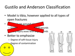
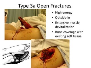
- The Gustilo-Anderson classification system grades open fractures based on the degree of soft tissue injury from I to III (A, B, C). Higher grades correlate with increased risk of infection and impaired fracture healing.
- Most open fracture infections are caused by bacteria acquired in the hospital rather than the initial trauma. Appropriate antibiotic treatment is crucial.
- Goals of management include preserving life and limb,
This document provides a history and overview of open fractures. It defines open fractures as fractures where there is a breach in the soft tissue envelope exposing the fracture. It discusses classifications of open fractures including the Gustilo-Anderson classification. It notes that open fractures often have high rates of infection, delayed healing, and amputation. The document traces historical approaches to treatment and how understanding of microbiology and use of antibiotics has improved outcomes over time.
Dr. Suman Paul provides a historical overview of the treatment of open fractures. Early civilizations like Egyptians recognized the need to cover open fracture wounds to minimize morbidity. Through the 19th century, amputation was often used for treatment after open fractures. Advances in the 20th century included debridement and stabilization principles from World War I, and the introduction of local antibiotics in World War II which reduced wound sepsis rates. The Gustilo classification system, introduced in 1976, provides guidelines for prognosis and treatment of open fractures based on the degree of soft tissue injury. Later classifications like the Bowen system in 2005 incorporated host risk factors to better predict infection risk. Common bacteria associated with open fractures include staph, strep,
This document provides an overview of the management of open fractures. It defines an open fracture as a soft tissue injury complicated by a broken bone with communication to the external environment. The history of open fracture treatment is discussed, from ancient practices like debridement to modern advances with antibiotics and fixation methods. Classification systems for open fractures are presented, including the Gustilo-Anderson classification which correlates the degree of soft tissue injury with infection risk. Key steps in managing open fractures are described, including thorough debridement and irrigation, antibiotic administration, fracture stabilization options like external or internal fixation depending on the injury, and wound management. Overall infection rates and healing times are correlated with the classification of the soft tissue injury.
1) The initial management of open fractures, including timing of debridement, choice of antibiotics, and timing of wound coverage, involves several controversies with little consensus in the literature.
2) While early antibiotic administration and debridement are agreed upon, there is no evidence that debridement must occur within 6 hours as was once believed; many surgeons now find urgent rather than emergency debridement acceptable.
3) Timing of wound coverage is also debated, but most evidence suggests covering Type III wounds within 7 days is appropriate once tissues have stabilized and debridement is complete.
1) The initial management of open fractures, including the timing of debridement, choice of antibiotic regimen, and wound coverage, remains controversial with debate around several key issues and a lack of strong evidence to guide certain practices.
2) While early debridement and antibiotics are generally recommended, the literature does not provide clear guidance on the optimal timing of debridement or whether adding gram-negative coverage improves outcomes for Type III fractures.
3) Antibiotic regimens of short-course cephalosporins begun promptly are supported, but prolonged courses lack evidence. Routine wound cultures also do not predict infection.
management priorities in high energy trauma
Define the terms of fracture, dislocation and Subluxation
Identify the clinical and radiological pictures of fractures
Classify the different types of fractures
general principles of fracture management
Principles of open fracture management
This document discusses open fractures and mangled extremities. It covers the goals of treatment which are to preserve life, limb and function while preventing infection and restoring stability and soft tissue coverage. Open fractures require urgent assessment and debridement to remove non-viable tissue which can be aided by lavage and the use of tourniquets. Skeletal stabilization is also important. Scores can help determine if limb salvage is possible or if amputation is required based on the extent of soft tissue and bone damage. Proper antibiotic use, wound coverage and further reconstruction are also outlined.
Open tibia fractures can range from minor soft tissue wounds to severe injuries with extensive soft tissue damage and bone loss. The goals of treatment are to prevent infection, achieve bone union, and restore function. Management involves thorough debridement and irrigation, temporary stabilization, soft tissue coverage within 7-10 days if possible, and definitive stabilization once the soft tissues have healed sufficiently. Complications include infection, malunion, nonunion, and compartment syndrome, which require additional treatment such as antibiotics, bone grafting, or surgery.
Open fractures are unique, complex, and emergently presenting injuries that expose sterile bone to the contaminated environment.
Because a fracture disrupts the intramedullary blood supply, the additionally stripped soft tissue envelope further devitalizes the bone.
The more severe the soft tissue injury or open wound, the more severe the osseous injury.
Historically, open fractures were associated with infection, delayed union, nonunion, amputation, or death.
This document discusses the impact of infection on fracture fixation. It notes that trauma and surgery disrupt the body's natural barriers against infection. Recent studies found infection rates of 2.5-25% in open fractures, and 17.6-23.6% in bicondylar tibial plateau fractures. Factors that affect infection risk include surgical technique, time, antibiotic timing, host characteristics, and implant colonization by antibiotic-resistant bacterial biofilms. Proper diagnosis and treatment of surgical site infections aims to eliminate infection while healing the fracture and maintaining patient function, though infections increase 30-day and 1-year mortality rates.
This document discusses the evaluation and management of infected nonunions. It begins by defining infected nonunion and describing the associated patient presentation. Evaluation involves investigating potential contributing factors, imaging to identify the extent of infection, and assessing patient goals. Management includes thorough debridement, stabilization with external fixation, culture-directed antibiotics, and dead space management. Once infection clears, methods to achieve bony union include internal fixation, bone grafting, bone transport, or free flaps. The document outlines various surgical strategies and complications to consider in treating these challenging cases.
This document discusses the principles of damage control orthopedics (DCO) for treatment of polytrauma patients. DCO involves a staged approach, with initial temporary stabilization of fractures using external fixation, followed by resuscitation and optimization of the patient. Definitive stabilization is then performed after 4 days once the acute inflammatory response has subsided to minimize additional surgical stress. Femur fractures stabilized with external fixation can be converted to intramedullary nailing within 2-3 weeks once the risk of infection is low. Pelvic and acetabular fractures may also be definitively treated 7-10 days after injury. The goal of DCO is to balance life-saving care of injuries with avoiding lethal complications through additional
The document discusses open fractures, providing details on epidemiology, classification, management principles, antibiotic treatment, wound irrigation and debridement. Open fractures commonly involve the fingers, tibia and distal radius, and result from high-energy trauma like vehicle accidents or falls. Immediate evaluation and treatment is needed, including antibiotics, wound cleaning, and splinting or stabilization to prevent infection while facilitating healing.
Open fractures involve a break in the bone that communicates with the external environment through a break in the skin and soft tissue. They are often caused by high-energy trauma like traffic accidents or falls. The initial management involves thorough debridement to remove all non-viable tissue, irrigation to clean the wound, fracture stabilization, and antibiotic treatment. Further debridement may be needed over subsequent days to fully clean the wound. The goal is to prevent infection while stabilizing the fracture and achieving soft tissue coverage. Outcomes depend on adequate initial management and reconstruction as needed.
This includes the basics of open fracture including classifications and treatment.
References are from the latest edition of rockwood and greens fracture in adults.
This document discusses open fractures of the tibial diaphysis (shaft). It notes that open tibia fractures account for about 1/4 of all tibial fractures and occur more commonly in the tibia than any other long bone. Treatment involves thorough debridement and irrigation of the soft tissue wound, antibiotics, and stabilization of the bone fracture, which may be via internal or external fixation depending on the fracture pattern and soft tissue injury severity. Complications include infection, nonunion, malunion, and compartment syndrome. Outcomes depend highly on the severity of soft tissue and neurovascular damage based on the Gustilo-Anderson classification.
Open fractures of the tibial diaphysis are common injuries that require urgent treatment to prevent infection and achieve bone union. The treatment priorities include addressing life threats, administering antibiotics and tetanus prophylaxis, debriding wounds, stabilizing fractures, and covering soft tissue defects. Surgical management often involves external or internal fixation along with soft tissue reconstruction using flaps or grafts. Despite treatment, complications like infection, nonunion, and malunion are common, especially with higher grade open fractures and bone/soft tissue loss. The goals of treatment are to prevent sepsis, achieve bone union, and restore function.
This document summarizes a journal article discussing the use of noncontact locking plates as an internal fixator for open fractures. Some key points:
- Open fractures involve communication between the external environment and fracture site, complicating treatment. Traditional fixation methods like plates and screws risk high infection rates.
- The study evaluated outcomes of using noncontact locking plates in 42 patients with open tibia or femur fractures. All fractures united within 19.7 weeks on average with minimal complications.
- Advantages of this technique include minimizing contact between implant and bone to reduce risks of infection, while still providing stable fixation comparable to traditional plating. The results were satisfactory compared to other studies.
A 13-year-old boy presented with 4 months of right knee pain. Radiographs, bone scan, CT scan, and biopsy were performed. The most likely diagnosis based on the imaging and biopsy is Ewing's sarcoma. Ewing's sarcoma commonly presents in long bones of adolescents with pain and is an aggressive round cell tumor that typically affects the metaphysis of long bones. The imaging and biopsy are consistent with this diagnosis.
This document provides information on fractures of the tibia. It begins with definitions of fractures and their various classifications. The causes of tibial fractures include direct forces, indirect forces, twisting, bending, and pathological fractures. Fracture patterns include transverse, oblique, spiral, impacted, comminuted, and compression fractures. Treatment options for tibial fractures depend on the fracture type and include casting, intramedullary nailing, plating, and external fixation. Complications can include nonunion, malunion, infection, and hardware failure. Open fractures require urgent debridement and antibiotics to prevent infection.
This document discusses fracture classification systems. It provides an overview of why fractures are classified (as a treatment guide, to assist with prognosis, and to facilitate communication between physicians) and issues with existing systems like poor reliability. It then reviews the Tscherne classification system for closed fractures and the Gustilo classification for open fractures. The document concludes by introducing the OTA comprehensive fracture classification system as a universal system to classify fractures of any bone based on location, type, group, and subgroup.
This document discusses the management of vascular injuries associated with extremity trauma. It begins by outlining the importance of rapid diagnosis and treatment, as irreversible tissue damage can occur after only 6 hours. Both non-operative and operative management approaches are then described. Non-operative options include observation of some low-risk injuries and endovascular occlusion. Operative treatment involves open surgical repair or ligation of injured vessels. Special considerations like fasciotomy, wound coverage, and the need for multi-specialty involvement are also noted. The document concludes by discussing guidelines for amputation in cases of severe "mangled extremity" injuries.
Damage control orthopaedics emphasizes the stabilization and control of the injury, often with use of spanning external fixation, rather than immediate fracture repair. The concept of damage control orthopaedics is not new; it has evolved out of the rich history of fracture care and abdominal surgery.
The document discusses ventral hernias, including:
- Incidence and risk factors for ventral hernias
- Options for mesh placement during hernia repair surgery
- Types of prosthetic meshes used, including benefits and disadvantages of polypropylene, ePTFE, polyester, and absorbable barrier-coated meshes
- Studies comparing surgical outcomes and complications between different mesh types
1. Open fractures occur when a broken bone pierces the overlying soft tissue, exposing the bone. The most common causes are motor vehicle accidents, motorcycle accidents, falls, and pedestrian injuries.
2. Treatment goals are to preserve life, limb, and function. This involves assessing for other injuries, stabilizing the patient, cleaning and debriding the wound, administering antibiotics and tetanus prophylaxis, and stabilizing the fracture—often initially with external fixation.
3. Further debridement and irrigation is done in the operating room, followed by temporary stabilization. Definitive reconstruction and internal or intramedullary fixation is done later, once the risk of infection decreases. Close monitoring is
Physeal injuries occur in the growth plate of children's long bones. The growth plate is made up of zones of cartilage that allow for bone growth. Injuries can disrupt these zones and cause growth abnormalities.
The Salter-Harris classification system categorizes physeal injuries into 5 types based on the location and extent of the fracture line. Type 1 injuries involve just the growth plate while Type 2-4 injuries also involve the epiphysis or metaphysis. Type 5 is a crush injury.
Evaluation involves x-rays and sometimes CT or MRI to identify the fracture pattern. Treatment aims to restore the normal anatomy through closed or open reduction and stabilize the bone to allow healing and prevent growth disturbances. Comp
Physeal injuries occur in the growth plates of children and can disrupt bone growth. There are several classifications of physeal injuries, most notably the Salter-Harris classification which categorizes injuries based on the location of the fracture line. Type I and II injuries have a low risk of growth disturbance while types III and IV are more likely to affect growth. Treatment aims to reduce fractures while avoiding further damage to the physis. Younger age, open injuries, and delays in treatment can worsen prognosis. Complications may include growth abnormalities, malunion, infection, and avascular necrosis.
The presentation discusses evidence based medicine in the stream of Orthopaedics. Here I have discussed a case of Ipsilateral Intertronchanteric and Femoral shaft Fracture and its various treatment modalities. The presentation was done at J.N. Medical College Belagavi, India. Lets share, discuss and keep learning.
Rasamanikya is a excellent preparation in the field of Rasashastra, it is used in various Kushtha Roga, Shwasa, Vicharchika, Bhagandara, Vatarakta, and Phiranga Roga. In this article Preparation& Comparative analytical profile for both Formulationon i.e Rasamanikya prepared by Kushmanda swarasa & Churnodhaka Shodita Haratala. The study aims to provide insights into the comparative efficacy and analytical aspects of these formulations for enhanced therapeutic outcomes.
Histololgy of Female Reproductive System.pptxAyeshaZaid1
Dive into an in-depth exploration of the histological structure of female reproductive system with this comprehensive lecture. Presented by Dr. Ayesha Irfan, Assistant Professor of Anatomy, this presentation covers the Gross anatomy and functional histology of the female reproductive organs. Ideal for students, educators, and anyone interested in medical science, this lecture provides clear explanations, detailed diagrams, and valuable insights into female reproductive system. Enhance your knowledge and understanding of this essential aspect of human biology.
Adhd Medication Shortage Uk - trinexpharmacy.comreignlana06
The UK is currently facing a Adhd Medication Shortage Uk, which has left many patients and their families grappling with uncertainty and frustration. ADHD, or Attention Deficit Hyperactivity Disorder, is a chronic condition that requires consistent medication to manage effectively. This shortage has highlighted the critical role these medications play in the daily lives of those affected by ADHD. Contact : +1 (747) 209 – 3649 E-mail : sales@trinexpharmacy.com
Does Over-Masturbation Contribute to Chronic Prostatitis.pptxwalterHu5
In some case, your chronic prostatitis may be related to over-masturbation. Generally, natural medicine Diuretic and Anti-inflammatory Pill can help mee get a cure.
Muktapishti is a traditional Ayurvedic preparation made from Shoditha Mukta (Purified Pearl), is believed to help regulate thyroid function and reduce symptoms of hyperthyroidism due to its cooling and balancing properties. Clinical evidence on its efficacy remains limited, necessitating further research to validate its therapeutic benefits.
Cell Therapy Expansion and Challenges in Autoimmune DiseaseHealth Advances
There is increasing confidence that cell therapies will soon play a role in the treatment of autoimmune disorders, but the extent of this impact remains to be seen. Early readouts on autologous CAR-Ts in lupus are encouraging, but manufacturing and cost limitations are likely to restrict access to highly refractory patients. Allogeneic CAR-Ts have the potential to broaden access to earlier lines of treatment due to their inherent cost benefits, however they will need to demonstrate comparable or improved efficacy to established modalities.
In addition to infrastructure and capacity constraints, CAR-Ts face a very different risk-benefit dynamic in autoimmune compared to oncology, highlighting the need for tolerable therapies with low adverse event risk. CAR-NK and Treg-based therapies are also being developed in certain autoimmune disorders and may demonstrate favorable safety profiles. Several novel non-cell therapies such as bispecific antibodies, nanobodies, and RNAi drugs, may also offer future alternative competitive solutions with variable value propositions.
Widespread adoption of cell therapies will not only require strong efficacy and safety data, but also adapted pricing and access strategies. At oncology-based price points, CAR-Ts are unlikely to achieve broad market access in autoimmune disorders, with eligible patient populations that are potentially orders of magnitude greater than the number of currently addressable cancer patients. Developers have made strides towards reducing cell therapy COGS while improving manufacturing efficiency, but payors will inevitably restrict access until more sustainable pricing is achieved.
Despite these headwinds, industry leaders and investors remain confident that cell therapies are poised to address significant unmet need in patients suffering from autoimmune disorders. However, the extent of this impact on the treatment landscape remains to be seen, as the industry rapidly approaches an inflection point.
These lecture slides, by Dr Sidra Arshad, offer a quick overview of the physiological basis of a normal electrocardiogram.
Learning objectives:
1. Define an electrocardiogram (ECG) and electrocardiography
2. Describe how dipoles generated by the heart produce the waveforms of the ECG
3. Describe the components of a normal electrocardiogram of a typical bipolar lead (limb II)
4. Differentiate between intervals and segments
5. Enlist some common indications for obtaining an ECG
6. Describe the flow of current around the heart during the cardiac cycle
7. Discuss the placement and polarity of the leads of electrocardiograph
8. Describe the normal electrocardiograms recorded from the limb leads and explain the physiological basis of the different records that are obtained
9. Define mean electrical vector (axis) of the heart and give the normal range
10. Define the mean QRS vector
11. Describe the axes of leads (hexagonal reference system)
12. Comprehend the vectorial analysis of the normal ECG
13. Determine the mean electrical axis of the ventricular QRS and appreciate the mean axis deviation
14. Explain the concepts of current of injury, J point, and their significance
Study Resources:
1. Chapter 11, Guyton and Hall Textbook of Medical Physiology, 14th edition
2. Chapter 9, Human Physiology - From Cells to Systems, Lauralee Sherwood, 9th edition
3. Chapter 29, Ganong’s Review of Medical Physiology, 26th edition
4. Electrocardiogram, StatPearls - https://www.ncbi.nlm.nih.gov/books/NBK549803/
5. ECG in Medical Practice by ABM Abdullah, 4th edition
6. Chapter 3, Cardiology Explained, https://www.ncbi.nlm.nih.gov/books/NBK2214/
7. ECG Basics, http://www.nataliescasebook.com/tag/e-c-g-basics
Osteoporosis - Definition , Evaluation and Management .pdfJim Jacob Roy
Osteoporosis is an increasing cause of morbidity among the elderly.
In this document , a brief outline of osteoporosis is given , including the risk factors of osteoporosis fractures , the indications for testing bone mineral density and the management of osteoporosis
2. Definition
• Break in the skin and underlying soft tissue
leading directly into or communicating with
the fracture and its hematoma
3. • Last century, high mortality with open fractures
of long bones
• Early amputation in order to prevent death
• WWI, mortality of open femur fractures > 70%
• 1939 Trueta “closed treatment of war fractures”
– Included open wound treatment and then enclosure
of the extremity in a cast
– “Greatest danger of infection lay in muscle, not bone”
Trueta J: "Closed" treatment of war fractures, Lacet 1939;1:1452-1455
History
4. • 1943 PCN on the battlefield quickly reduced
rate of wound sepsis
• Delayed closure of wounds
• Hampton: closure btwn 4th and 7th day
• Larger defects continued to be left open to
heal by secondary intention
Hampton OP Jr: Basic principles in management of open fractures; JAMA 1955; 159:417-419
History
5. • Advances shifted the focus
– Preservation of life and limb preservation of
function and prevention of complications
• However, amputation rates still exceed 50% in
the most severe open tibial fractures assoc
with vascular injury*
Lange RH, Bach AW, Hansen ST et al: Open tibial fractures with associated vascular injuries: prognosis for limb salvage. J Trauma; 25(3):203-
208
History
9. Open fracture classification
• Allows comparison of results
• Provides guidelines on prognosis and
treatment
– Fracture healing, infection and amputation rate
correlate with the degree of soft tissue injury
• Gustilo upgraded to Gustilo and Anderson
• AO open fracture classification
• Host classification of open fractures
10. Gustilo and Anderson Classification
• Model is tibia, however applied to all types of
open fractures
• Emphasis on wound size
– Crush injury assoc with small wounds
– Sharp injury assoc with large wounds
• Better to emphasize
– Degree of soft tissue injury
– Degree of contamination
12. Type 1 Open Fractures
• Wound less than 1 cm,
• Inside-out injury
• Clean wound
• Minimal soft tissue damage
• No significant periosteal
stripping
http://www.unboundedmedicine.com/2005/11/08/open-fractures-classification-and-its-clinical-manifestations-3/
18. Type 3b Open Fractures
• High energy
• Outside in
• Extensive muscle
devitalization
• Requires a flap for
bone coverage
and soft tissue
closure
• Periosteal stripping
20. Type 3c Open Fractures
• High energy
• Increased risk of
amputation and
infection
• Any grade 3 with
major vascular injury
requiring repair
21. Why use this classification?
• Grades of soft tissue injury correlates with infection
and fracture healing
Grade 1 2 3A 3B 3C
Infection
Rates
0-2% 2-7% 10-25% 10-50% 25-50%
Fracture
Healing
(weeks)
21-28 28-28 30-35 30-35
Amputation
Rate
50%
22. Gustilo and Anderson
Bowen and Widmaier*
• 2005 Host classification predicts infection
after open fracture
– Gustilo and Anderson classification and the
number of comorbidities predict infection risk
– 174 patients with open fractures of long bones
– Sorted into three classes based on 14
immunocompromising factors
• Age>80, current nicotine use, DM, malignancy,
pulmonary insufficiency, systemic immunodeficiency,
etc
Bowen TR, Widmaier JC. Host classification predicts infection after open fracture. Clin Orthop Relat Res. 2005;433:205-11.
23. What they found…
• Patients with any compromising risk factor has
increased risk of infection
• May benefit from additional therapies that
decrease the risk of infection.
Bowen TR, Widmaier JC. Host classification predicts infection after open fracture. Clin Orthop Relat Res. 2005;433:205-11.
Class Compromising factors Infection rates
A 0 4%
B 1-2 15%
C 3 or more 31%
24. Gustilo Classification:
a simple and useful tool, but is it accurate?
• 1994 Brumback et al.
• 125 randomized open fractures
• 245 surgeons of various levels of training
• 12 cases of open tibia fractures, videos used
• Interobserver agreement poor
– Range 42-94% for each fracture
• Ortho attendings - 59% agreement
• Ortho Trauma Fellowship trained attendings - 66% agreement
Brumback RJ, Jones AL (1994) Interobserver agreement in the classification of open fractures of the tibia. The results of a survey of two
hundred and forty-five orthopaedic surgeons. J Bone and Joint Am; 76(8):1162–1166.
25.
26. So……….
• Fracture type should not be classified in the
ER
• Most reliably done in the OR at the
completion of primary wound care and
debridement
28. Radiological Examination
• Usually, only AP and lateral radiographs are
required
– They should include adjacent joints and any
associated injuries.
• There are a number of features that the
surgeon should look for when examining the
radiographs
http://www.lww.com/static/docs/product/samplechapters/978-0-7817-5096-7_Chapter%204.pdf
30. Radiological Examination
• MRI and CT scans are rarely required in the
acute situation but may be helpful in open
pelvic, intra-articular, carpal, and tarsal
fractures.
• Angiography may be required in Gustilo IIIb or
IIIc fractures.
• In the polytraumatized patient, the surgeon
must decide if a delay for further imaging is
appropriate.
http://www.lww.com/static/docs/product/samplechapters/978-0-7817-5096-7_Chapter%204.pdf
31. Microbiology
• Most acute infections are caused by pathogens
acquired in the hospital
• 1976 Gustilo and Anderson
– most infections in their study of 326 open fxs
developed secondarily
• When left open for >2wks, wounds were prone to
nocosomial contaminants such as Pseudomonas
and other GN bacteria
• Currently most open fracture infections are
caused by GNR and GP staph
Gustilo RB, Anderson JT: Prevention of Infection in the Treatment of One Thousand and Twenty-five Open Fractures of Long Bones; JBJS,
58(4):453-458, June 1976
32. Nocosomial infection?!!!!
• Only 18% of infections were caused by the
same organism initially isolated in the
perioperative cultures*
• Carsenti-Etesse et al. 1999
– 92% of open fracture infections were caused by
bacteria acquired while the patient was in the
hospital**
*Patzakis MJ, Wilkins J, Moore TM: Considerations in reducing the infection rate in open tibial fractures. Clin Orthop Relat Res. 1983
Sep;(178):36-41.
*Patzakis MJ, Bains RS, Lee J, Shepherd L, Singer G, Ressler R, Harvey F, Holtom P: Prospective, randomized, double-blind study comparing
single antibiotic therapy, ciprofloxacin, to combo antibiotic therapy in open fracture wounds. J Orthop Trauma. 2000 Nov;14(8):529-33.
**Carsenti-Etesse H, Doyon F, Desplaces N, Gagey O, Tancrede C, Pradier C, Dunais B, Dellamonica P. Epidemiology of bacterial infection
during management of open leg fractures. Eur J Clin Microbiol Infect Dis. 1999;18:315-23.
Cover the
wounds
quickly
33. Common bacteria encountered with
open fractures
Blunt Trauma, Low Energy GSW Staph, Strept
Farm Wounds Clostridia
Fresh Water Pseudomonas, Aeromonas
Sea Water Aeromonas, Vibrios
War Wounds, High Energy GSW Gram Negative
35. Antibiotic comparisons
• No difference btwn clindamycin and cefazolin*
• Patzakis et al. **
– For type 1&2, cipro = cefamandole+gentamicin
– For type 3, cipro worse (31% vs 7.7% infection)
• Cipro and other fluoroquinolones inhibit
osteoblast activity and fracture healing***
*Benson DR, Riggins RS, Lawrence RM, Hoeprich PD, Huston AC, Harrison JA. Treatment of open fractures: a prospective study. J Trauma.
1983;23:25-30.
**Patzakis MJ, Bains RS, Lee J, Shepherd L, Singer G, Ressler R, Harvey F, Holtom P. Prospective, randomized, double-blind study comparing
single-agent antibiotic therapy, ciprofloxacin, to combination antibiotic therapy in open fracture wounds. J Orthop Trauma. 2000;14:529-33.
***Holtom PD, Pavkovic SA, Bravos PD, Patzakis MJ, Shepherd LE, Frenkel B. Inhibitory effects of the quinolone antibiotics trovafloxacin,
ciprofloxacin, and levofloxacin on osteoblastic cells in vitro. J Orthop Res. 2000;18:721-7.
***Huddleston PM, Steckelberg JM, Hanssen AD, Rouse MS, Bolander ME, Patel R. Ciprofloxacin inhibition of experimental fracture healing.
J Bone Joint Surg Am. 2000;82:161-73.
36. When and for how long?
• Start abx as soon as possible*
– Less than 3 hours 4.7 % infection rate
– Greater than 3 hours 7.4%
• No difference btwn 1 and 5 days of post op
abx treatment**
• Mass Gen recommended treatment:***
– Cefazolin Q 8 until 24 hours after wound closed
– Gentamicin or levofloxacin added for type 3
*Patzakis MJ, Wilkins J. Factors influencing infection rate in open fracture wounds. Clin Orthop Relat Res. 1989;243:36-40.
**Dellinger EP, Caplan ES, Weaver LD, Wertz MJ, Brumback R, Burgess A, Poka A, Benirschke SK, Lennard S, Lou MA. Duration of preventive
antibiotic administration for open extremity fractures. Arch Surg. 1988;123:333-9.
***Okike K, Bhattacharyya T: Trends in the management of open fractures. A critical analysis. J Bone Joint Surg. 2006 Dec;88(12):2739-48.
37. Local antibiotic therapy
• High abx conc within the wound and low
systemic conc
– Reduces risk of systemic side effect
• Vancomycin or aminoglycosides
– Heat stable
– Available in powder form
– Active against suspected pathogens
Eckman JB Jr, Henry SL, Mangino PD, Seligson D. Wound and serum levels of tobramycin with the prophylactic use of tobramycin-
impregnated polymethylmethacrylate beads in compound fractures. Clin Orthop Relat Res. 1988; 237:213-5.
38. Antibiotics - locally
• Prevents secondary contamination by
nocosomial pathogens
• Useful adjunct to systemic abx
• Potential for abx impregnated bone graft,
bone graft substitute, and abx coated IMN
Ostermann PA, Seligson D, Henry SL: Local antibiotic therapy for severe open fractures. A review of 1085 consecutive cases; J Bone Joint
Surg Br. 1995 Jan;77(1):93-7.
Antibiotic Infection Rate
IV Abx 12%
IV Abx + local aminoglycoside
impregnated PMMA beads
3.7%
39. Antibiotic Beads
• Pros
– Very high levels of
antibiotics locally
– Dead space
management
• Cons
– Requires removal
– Limited to heat
stable antibiotics
– Increased drainage
from wound
40. Goals of treatment
• 1. preserve life
• 2. preserve limb
• 3. preserve function
• Also….
– Prevent infection
– Fracture stabilization
– Soft tissue coverage
43. Initial assessment & management
• ABC’s
• Assess entire patient
• Careful PE, neurovasc
• Abx and tetanus
• Local irrigation 1-2 liters
Lee J. Efficacy of cultures in the management of open fractures. Clin Orthop Relat Res. 1997;339:71-5.
44. Initial assessment & management
• Sterile compressive dressings
• Realign fracture and splint
• Do not culture wound in the
ED*
– 8% of bugs grown caused deep
infection
– cultures were of no value and not
to be done
• Recheck pulse, motor and
sensation
Lee J. Efficacy of cultures in the management of open fractures. Clin Orthop Relat Res. 1997;339:71-5.
45. Primary surgery
• Objectives of initial surgical
management
– Preservation of life and limb
– Wound debridement
– Definitive injury assessment
– Fracture stabilization
Stages of open fracture management in the OR
46. Surgical emergency!
• 1898 Friedrich guinea pigs
– Take to the OR within 6-8 hours*
• 1973 Robson:
bacteria multiply in contaminated
wounds **
– 105 organisms/gram of tissue is the
infection threshold
– Reached at 5.17 hours
• 1995 Kindsfater et al:
– 47 G2/3 fxs at 4.8 months out….
• Less than 5 hrs 7% infection
• Greater than 5 hrs 38% infection
– However G3 fxs were treated later
*Friedrich PL. Die aseptische Versorgung frischer Wundern. Arch Klin Chir. 1898;57:288-310.
**Robson MC, Duke WF, Krizek TJ. Rapid bacterial screening in the treatment of civilian wounds. J Surg Res. 1973;14:426-30.
47. Or not?....
Calling the “6 hour rule” into question
• 1993 Bednar and Parikh…. No significant difference *
– 3.4% vs 9%; 82 open femoral/tibial fxs
• 2004 Ashford et al…. No significant difference **
– 11% vs 17%; pts from the austrailian outback
• 2004 Spencer et al.... No significant difference ***
– 10.1% vs 10.9%; 142 open long bone fxs from UK
• 2003 Pollack and the LEAP investigators…. No correlation****
– 315 open long bone fxs
• 2005 Skaggs et al….No significant difference *****
– children with all types of open fractures; 554 open fractures
*Bednar DA, Parikh J. Effect of time delay from injury to primary management on the incidence of deep infection after open fractures of the lower extremities
caused by blunt trauma in adults. J Orthop Trauma. 1993;7:532-5.
**Ashford RU, Mehta JA, Cripps R. Delayed presentation is no barrier to satisfactory outcome in the management of open tibial fractures. Injury. 2004;35:411-6.
***Spencer J, Smith A, Woods D. The effect of time delay on infection in open long-bone fractures: a 5-year prospective audit from a district general hospital. Ann R
Coll Surg Engl. 2004;86:108-12.
****Pollack AN, Castillo RC, Jones AL, Bosse MJ, MacKenzie EJ, and the LEAP Study Group. Time to definitive treatment significantly influences incidence of infection
after open high-energy lower-extremity trauma. Read at the Annual Meeting of the Orthopaedic Trauma Association; 2003 Oct 9-11; Salt Lake City, UT.
*****Skaggs DL, Friend L, Alman B, Chambers HG, Schmitz M, Leake B, Kay RM, Flynn JM. “The Effect of Surgical Delay on Acute Infection Following 554 Open
Fractures in Children.” JBJS-A 2005. 87:8-12
No significant
difference
before or after 6
hours!!!
48. Do we even need to do operative
debridement?
• Orcutt et al... No significant difference, BUT…*
– 50 type 1 &2 open fractures
– less infection in nonoperative group (3% vs 6%)
– Less delayed union in nonop group (10% vs 16%)
• Yang et al….0% infections **
– 91 type 1 open fractures treated without I&D
*Orcutt S, Kilgus D, Ziner D. The treatment of low-grade open fractures without operative debridement. Read at the Annual Meeting of the
Orthopaedic Trauma Association; 1988 Oct 28; Dallas, TX.
**Yang EC, Eisler J. “Treatment of Isolated Type 1 Open Fractures: Is Emergent Operative Debridement Necessary?” Clin Orthop Relat Res
2003. 410: 289-294.
Do we even
need to debride
low grade open
fractures?
49. However, after review of all literature….….
• Okike et al. states….
• “Thorough operative debridement is the standard
of care for all open fractures.”
• “Even if the benefits of formal I&D were
insignificant for low grade fractures, operative
debridement is still required for proper wound
classification.”
• “Open fractures graded on the basis of superficial
characteristics are often misclassified.”
• Huge risk not to explore and debride!
Okike K, Bhattacharyya T: Trends in the management of open fractures. A critical analysis. J Bone Joint Surg Am. 2006 Dec;88(12):2739-48.
51. URGENTLY debride, not EMERGENTLY
• Time to OR is probably less important than:*
– Adequacy of debridement
– Time to soft tissue coverage
• Timing depends on….**
– Is patient stable?
– Is the OR prepared?
– Is appropriate assistance available?
• Ortho trained scrub techs, assistant surgeons, xray
techs, and other OR staff
• 2005 Skaggs et al:***
– If after 10pm, keep until the morning! Or at least
within 24 hours.
– Unless….
• neurovasc compromise
• horrible soft tissue contamination
• compartment syndrome
*Okike K, Bhattacharyya T: Trends in the management of open fractures. A critical analysis. J Bone Joint Surg. 2006 Dec;88(12):2739-48.
**Werner CM, Pierpont Y, Pollak AN: The urgency of surgical débridement in the management of open fractures. J Am Acad Orthop Surg.
2008 Jul;16(7):369-75.
***Stewart DJ, Kay RM, Skaggs DL: Open Fractures in Children. Principles of Evaluation and Management. JBJS-A. 2005;87:2784-2798.
Within
24
hours
Within
6 hours
52. I&D in the OR
• Trauma scrub
– Soap and saline to remove gross debris
• “Zone of injury”
– Skin wound is the window through which
the true wound communicates with the
exterior
• Extend the traumatic wound
– Excise margins
– Resect muscle and skin to healthy tissue
• color, consistency, capacity to bleed and
contractility
53. I&D in the OR
• Bone ends are exposed and debrided
• Irrigate
• Serial debridements?
– If needed, 2nd or 3rd debridement after 24-
48 hours should be planned
54. The Irrigation
• Amount
– No good data, copious is better
– Animal studies show improved
removal of particulate matter
and bacteria but effect plateaus
– Irrigation bags typically contain
3 L of fluid
– Anglen recommends:*
• 3L (one bag) for type 1
• 6L (two bags) for type 2
• 9L (three bags) for type 3
*Anglen JO. “Wound Irrigation in Musculoskeletal Injury.” JAAOS 2001. 9: 219-226.
55. How to deliver the irrigation?
(what animal studies show)
• Bulb Syringe vs Pulsatile Lavage
– Pulsatile lavage
• Detrimental for early bone healing
– this is no longer present at 2 wks*
• More soft tissue destruction**
• More effective in removing
particulate matter and bacteria***
*Dirschl DR, Duff GP, Dahners LE, Edin M, Rahn BA, Miclau T. “High Pressure Pulsatile Lavage Irrigation of Intraarticular Fractures: Effects on
Fracture Healing.” JOT 1998. 12(7): 460-463.
**Boyd JI, Wongworawat MD. “High-Pressure Pulsatile Lavage Causes Soft Tissue Damage.” CORR 2004. 427: 13-17
***Bhandari M, Schemitsch EH, Adili A, Lachowski RJ, Shaughnessy SG. “High and Low Pressure Pulsatile Lavage of Contaminated Tibial
Fractures: An in vitro Study of Bacterial Adherence and Bone Damage.” JOT 1999. 13: 526-533.
56. How to deliver the irrigation?
(what animal studies show)
• High or low pressure?
– Higher pressure
• Better bone cleaning
• Worse soft tissue cleaning
• Slows bone healing
*Dirschl DR, Duff GP, Dahners LE, Edin M, Rahn BA, Miclau T. “High Pressure Pulsatile Lavage Irrigation of Intraarticular Fractures: Effects on
Fracture Healing.” JOT 1998. 12(7): 460-463.
**Boyd JI, Wongworawat MD. “High-Pressure Pulsatile Lavage Causes Soft Tissue Damage.” CORR 2004. 427: 13-17
***Bhandari M, Schemitsch EH, Adili A, Lachowski RJ, Shaughnessy SG. “High and Low Pressure Pulsatile Lavage of Contaminated Tibial
Fractures: An in vitro Study of Bacterial Adherence and Bone Damage.” JOT 1999. 13: 526-533.
57. Antibiotics in the irrigation?
• Antibiotics (bacitracin and/or neomycin)
– Mixed results, controversial
– Costly
• bacitracin alone around $500/washout
– ?? Causing resistance
– Wound healing problems?
– Few reported cases of anaphylaxis
– Anglen: “No proven value in the care of open
fracture wounds…some risk, albeit small.”
No proven
benefit!
*Anglen JO. “Wound Irrigation in Musculoskeletal Injury.” JAAOS 2001. 9: 219-226.
58. Soaps in the irrigation?
• Surfactants (i.e. Soaps)
– Less bacteria adhesion
– Emulsify and remove debris
– No significant difference in
infection or bone healing
compared to bacitracin
solution, but more wound
healing problems in
bacitracin group
Anglen JO. “Comparison of Soap and Antibiotic Solutions for Irrigation of Lower-Limb Open Fracture Wounds: A Prospective, Randomized
Study.” JBJS-A 2005. 87(7):1415-1422.
59. Level 4 evidence based recommendations
• 1st washout, highly contaminated
Soap solution
• Repeat washout of clean wounds
Saline
• Infected wounds
Soap, then antibiotic
*Anglen JO. “Wound Irrigation in Musculoskeletal Injury.” JAAOS 2001. 9: 219-226.
60. Wound closure after contaminated fracture
• Timing and technique is
controversial
OPEN WOUND should be left OPEN!
– Prevents anaerobic conditions in
wound: Clostridium
– Facilitates drainage
– Allows repeat debridement
Zalavras CG, Patzakis MJ:Open fractures: evaluation and management. J Am Acad Orthop Surg. 2003 May-Jun;11(3):212-9.
Dubunked!
61. To close or not to close?
• Recently, renewed interest
in primary closure
• Collinge, OTA 2004
• Moola, OTA 2005
• Russell, OTA 2005
• DeLong, J Trauma 2004/
• Bosse, JAAOS 2002
– Improved abx management
– Better stabilization
– Less morbidity
– Shorter hospital stay, lower
cost
– NO increase in wound
infection
• These wounds are at
higher risk of clostridia
perfringens if they do get
infected.
• 1999 Delong et al: 119 open fxs
– No significant difference
• delayed/nonunion and infection rates btwn
immediate and delayed closure
– Immediate closure is a “viable option”
DeLong WG Jr, Born CT, Wei SY, Petrik ME, Ponzio R, Schwab CW: Aggressive treatment of 119 open fracture wounds. J Trauma. 1999
Jun;46(6):1049-54.
infection rate 7%
Overall delayed/nonunion rate 16%
Grade Percent of primary closures
1 88%
2 86%
3a 75%
3b 33%
3c 0%
62. Contraindications to primary closure
• Inadequate debridement
• Gross contamination
• Farm related or freshwater immersion injuries
• Delay in treatment >12 hours
• Delay in giving abx
• Compromised host or tissue viability
63. When to cover the wound?
• ASAP after wound adequately debrided
– Only 18% of infections are caused by the same organism
isolated in initial perioperative culture*
• Suggests hospital acquired etiology of infection
• “Fix and Flap”**
– For Type IIIB & IIIC open tibia fractures
– Early if not immediate flap coverage
•Patzakis MJ, Bains RS, Lee J, et al. “Prospective, randomized, double-blind study comparing single-agent antibiotic therapy, ciprofloxacin, to
combination antibiotic therapy in open fracture wounds.” JOT 2000. 14: 529-533.
**Gopal S, Majumder S, Batchelor A, Knight S, De Boer P, Smith RM. “Fix and flap: the radical orthopaedic and plastic treatment of severe
open fractures of the tibia.” JBJS-B 2000. 82(7): 959 – 966.
Timing of flap placement Infection rate
< 72 hours 6%
> 72 hours 30%
65. VAC
• Vacuum assisted wound closure
– Recommended for temporary management
– Mechanically induced negative pressure in a closed
system
– Removes fluid from extravascular space
– Reduced edema
– Improves microcirculation
– Enhances proliferation of reparative granulation tissue
• Open cell polyurethane foam dressing ensures an
even distribution of negative pressure
-Webb LX: New techniques in wound management: vacuum-assisted wound closure. J Am Acad Orthop Surg. 2002 Sep-Oct;10(5):303-11.
-Dedmond BT, Kortesis B, Punger K, Simpson J, Argenta A, Kulp B, Morykwas M, Webb L. “The use of Negative Pressure Wound Therapy in
the Temporary Treatment of Soft Tissue Injuries associated with High Energy Open Tibial Shaft Fractures.” JOT. 2007
66. Types of fracture stabilization
• Splint
– Good option if operative
fixation not required
• Internal fixation
– Wound is clean and soft tissue
coverage available
• External fixation
– Dirty wounds or extensive soft
tissue injury
67. Fracture stabilization
• Gustilo type 1 injury can be treated the same
way as a comparable closed fracture
• Most cases involve surgical fixation
• Outcome is similar to closed counterparts
68. Fracture stabilization
• Gustilo type 2&3 usually displaced and unstable
– dictate surgical fixation
• Restore length, alignment, rotation and provide
stability
– ideal environment for soft tissue healing and reduces
wound infection
– reduces dead space and hematoma volume
• Inflammatory response dampened
• Exudates and edema is reduced
• Tissue revascularization is encouraged
69. When to use plates?
• Open diaphyseal fractures of arm & forearm
• Open diaphyseal fractures lower extremity
– NOT recommended
– Open tibial shaft plating assoc high infection rate*
• Open periarticular fractures
– Treatment of choice in both upper and lower
extremities
Bach AW, Hansen ST Jr.: Plates versus external fixation in severe open tibial shaft fractures. A randomized trial. Clin Orthop Relat Res.
1989 Apr;(241):89-94.
70. When to use IM nails?
• Treatment of choice for most
diaphyseal fractures of the
lower extremity
• Inserted without disrupting
the already injured soft tissue
envelope
• Preserves the remaining extra
osseous blood supply to
cortical bone
• Malunion is uncommon
71. To ream or not to ream?
• Does reaming cause additional damage to the
endosteal blood supply?
• Solid IM nails without reaming has a lower risk of
infection that tubular nails with a large dead space*
• However reamed IM nails are biomechanically stronger
and can reliably maintain fracture reduction if statically
locked
• 2000 Finkemeier et al.
– reamed vs unreamed interlocked nails of open tibias
– NO statistical difference in outcome and risk of
complication**
*Melcher GA, Claudi B, Schlegel U, Perren SM, Printzen G, Munzinger J.Influence of type of medullary nail on the development of local
infection. An experimental study of solid and slotted nails in rabbits; .J Bone Joint Surg Br. 1994 Nov;76(6):955-9.
**Keating JF, O'Brien PJ, Blachut PA, Meek RN, Broekhuyse HM: Locking intramedullary nailing with and without reaming for open fractures
of the tibial shaft. A prospective, randomized study. J Bone Joint Surg Am. 1997 Mar;79(3):334-41.
**Finkemeier CG, Schmidt AH, Kyle RF, Templeman DC, Varecka TF: A prospective, randomized study of intramedullary nails inserted with
and without reaming for the treatment of open and closed fractures of the tibial shaft. J Orthop Trauma. 2000 Mar-Apr;14(3):187-93.
72. When to use external fixation?
• Diaphyseal fractures
not amenable to IM
nails
• Ring fixators for
periarticular
fractures
• Temporary joint
spanning ex fix is
popular for knee,
ankle, elbow and
wrist
• If temporary, plan
for conversion to IM
nail within 3 weeks
73. Ex-fix: Weigh the pros and cons!
• Historically was definitive treatment
• Now, more commonly as temporary fixation
• Can be applied almost always and everywhere
• Severe soft tissue damage and contamination
74. Advantages
• Easy and quick
• Relatively stable fixation
• No further damage done
• Avoids hardware in the open wound
76. Skin cover and soft tissue reconstruction
• Do these early!
• 1994 Osterman et al.*
– Retrospective 1085 fractures, 115 G2 and 239 G3
• All treated with appropriate IV Abx and I&D
– No infection if wounds closed at 7.6 days
– Yes infection if wounds closed at 17.9 days
*Ostermann PA, Seligson D, Henry SL: Local antibiotic therapy for severe open fractures: A review of 1085 consecutive cases. J Bone Joint
Surg Br 1995;77:93–97.
Infection risk
increases if wound
open > 7 days
77. Reconstructive ladder: options for wound
coverage
Primary closure
Secondary intention
Skin graft
Local flap
Regional flap
Distant flap
Free flap
Tissue
expansion
Type 1 open fx
Type 3B open
fx
Type 2/3A open fx
80. Type 3c, a bad injury!
• Devastating damage to
bone and soft tissue
• Major arterial injuries
that require repair
• Poor functional outcome
• Consensus btwn ortho,
vascular and plastics
• Salvage is technically
possible in most cases
• However it is not always
the correct choice esp
type 3c tibia fractures
81. We can do both, salvage & amputate.
• Vascular surgery can revascularize
with bypass graft
– Generally before fracture stabilization
• Plastics can provide soft tissue
coverage
• However, in the tibia, the severity
to soft tissue envelope and bone
may result in infected nonunion
• If salvage…. long course of
repeated surgical procedures
– Painful and psychologically distressing
– Functional outcome may be poor and
no better than amputation
82. How to decide, salvage or amputate?
• Important factors in decision making:*
– General condition of the patient (shock)
– Warm ischemia time (>6hours)
– Age (>30 years)
– Cut to crush ratio (blunt injuries has a large zone
of crush)
Howe HR Jr, Poole GV Jr, Hansen KJ, Clark T, Plonk GW, Koman LA, Pennell TC: Salvage of lower extremities following combined orthopedic
and vascular trauma. A predictive salvage index. Am Surg. 1987 Apr;53(4):205-8.
83. Gunshot injuries
• Energy dissipated at impact = damage
severity
• High velocity rifles and close range
shotguns
– Worst, high energy of impact
– Huge secondary cavitation
– Secondary effects of shattered bone
fragments
• Bullets lodged in joints should be removed
– avoid lead arthropathy and systemic lead
poisoning
84. Low velocity GSW <2000 ft/sec
• Low velocity handguns
– Less severe, not treated like open fractures
– Cavitation is not significant
– Secondary missile effects are minimal
– Bone fragments rarely stripped of soft tissue
attachments and blood supply
– Soft tissue injuries not severe and skin wounds are
small
85. Low velocity GSW open fractures
• Geisslar et al. *
• If neurovascular status
normal, do local
debridement
• NO formal I&D needed
• IV Abx
• Approach fx fixation as
if closed
• Dickey et al.**
– No abx vs IV Ancef x 3d
– 67 low velocity GSW fxs
– Not requiring operative
fixation
– No difference in
infection rates
*Geisslar ett al, J Ortho Trauma, 4;39-41,1990
**Dickey et al, J Ortho Trauma, 3;6-10,1989
Treat open
fractures from low
velocity GSW as
closed fractures
without Abx
86. Pitfalls and complications
• Infection delayed union, nonunion, malunion
and loss of function
• Plan ahead to avoid delayed union and nonunion
• Predict nonunion in severe injuries with bone loss
– Bone grafting usually delayed 6 weeks when soft
tissues have soundly healed
– Autogenous bone grafting is usual strategy
– Fibular transfer, free composite graft or distraction
osteogenesis for complex defects
– Recombinant human BMP in open tibia fracture
reduces risk of delayed union
87. Advances…
• BMPs
– 40% decreased infection rate with BMP in type 3
open tibia fractures*
• Antibiotic Laden Bone Graft**
– Tobramycin-impregnated calcium sulfate pellets
with demineralized bone matrix
– Animal study: successful in preventing infection
*BESTT Study Group, Govender S, Csimma C, Genant H, Valentin-Opran A. “Recombinant Human Bone Morphogenetic Protein-2 for
Treatment of Open Tibial Fractures: A prospective, controlled, randomized study of four hundred and fifty patients.” JBJS-A 2002.
84(12): 2123-2134.
**Beardmore AA, Brooks DE, Wenke JC, Thomas DB. “Effectiveness of local antibiotic delivery with an osteoinductive and
osteoconductive bone-graft substitute.” JBJS-A 2005. 87(1): 107-112.
88. Summary
A = good evidence (level 1 studies)
B = fair evidence (level 2/3 studies)
C = poor quality evidence (level 4/5 studies)
I = insufficient or conflicting evidence
Okike K, Bhattacharyya T: Trends in the management of open fractures. A critical analysis. J Bone Joint Surg. 2006 Dec;88(12):2739-48.