1. Special Topic
Liposuction of the Legs and Ankles: A Review
of the Literature
Frederick G. Weniger, M.D., M.B.A., Jay W. Calvert, M.D., and E. Douglas Newton, M.D.
Pittsburgh, Pa.; and Orange, Calif.
A TABOO PROCEDURE
Lipodystrophy of the legs and ankles pre-
sents a particularly challenging problem in aes-
thetic surgery. This is true both historically and
presently. Anatomic peculiarities and technical
challenges delayed the development of treat-
ment for this condition. Today, attempts to
overcome these same obstacles continue to
rapidly change the face of liposuction in this
anatomical area.
Lipodystrophy of the legs and ankles is dis-
tressing to patients for several reasons. This
condition, which is mostly limited to female
patients and usually appears during the emo-
tionally vulnerable time of adolescence,1,2
tends to make patients look heavier than they
are. The problem is seen in all ethnic groups.3
The appearance of the leg may be so ill defined
as to cause the patient severe psychological
pain and loss of self-esteem. Legs and ankles
are frequently not camouflaged by clothing
and are therefore among the more conspicu-
ous areas of lipodystrophy.3–5
These factors
combine to make this an emotionally frustrat-
ing cosmetic deformity. Frustration is exacer-
bated by the fact that lipodystrophy in this area
appears to be largely genetically predeter-
mined and is especially resistant to exercise
and diet.1,2,6
Despite a need for therapy, liposuction of
the legs and ankles was slow to develop. The
earliest attempt at treatment of these areas in
the literature dates to the 1920s, when Dujar-
rier in France attempted to remove fat by cu-
rettage in the lower legs of a well-known balle-
rina. He transected a major artery, and this
eventually resulted in amputation.7
Thus be-
gan the long-lived stigma surrounding lipec-
tomy of the leg. In 1964, another surgeon,
Schrudde of Cologne, attempted to remove
such fat in his patients by curettage. Despite
several good results, he reported skin necrosis
in four of 15 patients in his series.4,1
In 1977,
Fischer and Fischer presented the new lipec-
tomy technique of suction curettage.8
This
technique was attempted in the trochanteric
areas but was abandoned because it left lym-
phorrhea, large cavities filled with lymph and
serum, and terrible secondary sagging deformi-
ties over the affected areas.9
After disappointing experiences with closed
curettage and closed suction curettage in the
early 1970s, the modern era of lipectomy be-
gan in 1977 with Illouz in Europe.7,9,10
The first
U.S. experience was in 1981, when American
physicians of various specialties observed the
new technique of suction lipectomy in France.2
Liposuction is now one of the most common
cosmetic procedures,2
but the growth of this
modality in the specific areas of the legs and
ankles lagged behind. By the late 1980s, the
legs and ankles were being treated more fre-
quently, but large numbers of successful cases
were not reported until 1989 and 1990.4
The ignition of the lipectomy wildfire by
Illouz’s development of blunt liposuction tech-
niques in 1978 resurrected the stigmata associ-
ated with circumferential lipectomy of the legs
and ankles.7
The procedure had been por-
trayed as “fraught with complications and of
limited benefit.”2
Others have said that lipo-
plasty of these areas frequently combines high
expectations with, at best, modest results.4,11
Thus, early on, it became clear that liposuction
From the Division of Plastic Surgery, University of Pittsburgh School of Medicine; Aesthetic and Plastic Surgery Institute, University of California
Irvine; and Division of Plastic Surgery, The Western Pennsylvania Hospital. Received for publication July 12, 2002; revised August 4, 2003.
DOI: 10.1097/01.PRS.0000117299.55812.2E
1771
2. of the legs and ankles represented an entity far
different from liposuction in other anatomic
areas, with unique anatomical and technical
challenges to be overcome. Feared complica-
tions then and now include poor patient satis-
faction, persistent pigmentation, contour irreg-
ularity, ulceration, and especially persistent
edema.1,2,7
For these reasons, liposuction of the
legs and ankles was slow to develop.
As early as 1983, Illouz was finding much of
this stigma of liposuction of the calves and
ankles to be well-founded. In his report of 3000
cases of liposuction that year, he included 159
patients who had liposuction of the ankles.12
Although he described good early results in
the ankle region, he recognized and warned
that the postoperative edema in this area was
much greater than in any other anatomical
zone, lasting up to 6 months. As is discussed in
more detail, efforts to overcome this seemingly
inevitable edema have been a major front in
the battle to treat lipodystrophy in the legs and
ankles. Even cases of eventual good outcomes
are tainted by the fact that the edema, which
persists in the legs longer than in other body
areas, often makes patients prematurely feel
that they had a poor result.13
Illouz also found other major complications
to include long-term postoperative pigmenta-
tion and contour irregularities.14
Such predis-
position to hyperpigmentation is attributable
to the dependent location.4
Irregularities,
which have been claimed to be the most com-
mon complication, are more common in this
area because the fat is relatively thin. These
irregularities are more commonly palpable
than visible.4
In 1990, Watanabe’s study of 166 cases of
liposuction of the legs and ankles showed that
patient dissatisfaction was secondary to these
and multiple other factors. In addition to the
aforementioned edema, irregularities, and
prolonged postoperative pigmentation, these
complaints included legs that appeared less
slender than anticipated by the patient, asym-
metries in shape, hypesthesia, and unexpect-
edly severe postoperative pain.13
In fact, lipo-
suction of these areas may be one of the most
painful cosmetic procedures, and this discom-
fort often remains for many weeks.7
When the
legs and ankles are treated with liposuction,
the recovery will also be longer than for most
other areas because of factors such as edema.
The reality of these complications in the con-
text of a recovery period that can be challeng-
ing to both the patient and the physician ex-
plains the continued hesitancy of many
surgeons to attempt this procedure.
Still, with advances in technique over the last
15 years, complication rates seem to have dra-
matically improved. Newer literature suggests
that good results usually can be expected. De-
spite prolonged postoperative edema, after 6
months these patients are some of the most
grateful.2
Watanabe showed 84 percent satisfac-
tion in this patient population, although this is
compared with 98 percent satisfaction with li-
posuction in other areas of the body.13
None-
theless, cautious attitudes toward this proce-
dure have been slow to change. As of 1997,
there were still virtually no published articles
or textbooks emphasizing the significant ben-
efit, high patient satisfaction, and progressively
lower complication rates of liposuction in
these areas.2
Recently, more investigators have
discussed new techniques that have contrib-
uted to overcoming some of the aforemen-
tioned obstacles. This review presents the ana-
tomic basis for the challenges of liposuction of
the legs and ankles and examines recent au-
thors’ strategies for successful liposuction in
these difficult areas. It is intended to provide
clinicians with an overview of the developing
state of the art, and to assist in sorting through
the multiple techniques that have been pre-
sented. Table I summarizes the individual au-
thors’ differing strategies.
ANATOMIC CONSIDERATIONS OF THE LEGS AND
ANKLES
Illouz felt that the legs and ankles required
special attention because of their unique ana-
tomic features.7
He described this area as rela-
tively taboo for liposuction because of these
characteristics.13–15
Understanding of these
unique anatomic considerations elucidates the
basis for most of the unique challenges of lipo-
suction in the legs and ankles. First, many au-
thors describe this area as having only one
layer of fat, with dense fibrous tissue and many
lymphatics.7,13–15
This is thought to be largely
responsible for easily caused postoperative skin
irregularities.7
Also, because of this thin subcu-
taneous fat layer, postoperative hematomas or
ecchymosis may result in pigmentation of the
skin itself.13
In addition, the dependent nature
of the lower legs, the fibrous nature of the
fat,13–15
and possibly the disruption of the
dense lymphatics from certain techniques16
en-
gender a constant tendency toward postopera-
1772 PLASTIC AND RECONSTRUCTIVE SURGERY, May 2004
3. tive edema. A delay in the absorption of this
swelling may cause a tendency for more fibrous
tissue to form, thereby threatening permanent
“woody” induration of the subcutaneous
layer.13
Anatomically, the larger, less sculptured leg
is rarely the result of undesirable fat distribu-
tion alone, and usually is also the product of
bone and muscle configuration.11
This impor-
tant anatomical consideration is addressed
later in the discussion of preoperative evalua-
tion. The heterogeneic character of the fat
throughout the lower leg also presents a chal-
lenge. For example, the subcutaneous fat in
the medial lower calf is softer and easier to
suction than that more laterally.13
This tends to
lead to a greater reduction and resultant over-
all concavity medially if this anatomic factor is
not taken into account.
Because of the neurovascular structures, li-
posuction at the popliteal fossa should never
be performed.6
Although the motor nerves and
deep vascular structures of the leg lie deep to
the investing fascia, the greater and lesser sa-
phenous veins and the anatomically associated
saphenous and sural nerves, respectively, live
within the fat. Although one must take care not
to injure these structures with stab wounds for
cannulas, Grazer claimed no serious hemato-
mas, superficial vein thromboses, or other
complications from suctioning around the sa-
phenous system.5
Within this anatomic setting, the pattern of
lipodystrophy of the calves and ankles can be
considered. Women tend to present with a
common pattern of lipodystrophy of the legs
and ankles. Chamosa presents a thorough re-
view of the aesthetic ideals in the legs, and
explains the typical adiposities that deviate
from this form.17
In brief, the knee should
normally be bony, with some medial convexity
and mild lateral concavity. More inferiorly, a
concavity should also occur between the knee
and the medial protrusion of the gastrocne-
mius. This area is commonly deformed by ad-
iposities that exaggerate the medial convexity
of the knee and that fill in the aesthetic con-
cavities of the lateral knee and the area be-
tween the medial knee and the calf. In lipodys-
trophy of the leg, there is also proportionally
more fat accumulation over the lower half of
the leg than the upper areas.13
Again, these
aesthetic ideals and their common dystrophic
deviations are well presented graphically by
Chamosa.17
In 1997, Chamosa presented a uniquely dif-
ferent conceptualization of the lipodystrophies
of this area by representing the cross-sectional
anatomy of the leg and ankle as a rhomboid,
the points of which were the anterior edge of
the tibia, the Achilles tendon, and the two
malleoli.18
In this model, fat tends to accumu-
late along the lines between these points and
thus obscures these prominences. He found
the anterolateral and posterolateral aspects to
be the most frequent areas of lipodystrophy.
Interestingly, Chamosa suggested using lipo-
suction to visually camouflage malalignment of
the knee, claiming that the aesthetic impact of
a varus or valgus knee can be decreased by
concentrating liposuction toward the lipodys-
trophic areas on the convex side of the leg.18
Most patients’ concerns regard areas of ex-
cessive fat distally over the medial and lateral
malleoli.1
In addition, excess fat often blunts
the definition of the inferior bellies of the
gastrocnemius muscles, especially medially.1,2
Such an obscure transition between the gas-
trocnemius muscles and the more inferior leg
contributes to a columnar appearance. Special
attention toward the contouring of this “tran-
sition zone” is a focus of Mladick’s recent
work,19
and is further discussed.
Another localized excess of fat commonly
exists distally on either side of the Achilles
tendon, creating medial and lateral fullness at
the ankle.4
These normally concave areas
present a unique technical challenge.
Despite these common foci of lipodystrophy
in the legs and ankles, specific fat distributions
in this area of the body may be generally less
well defined than in other commonly lipodys-
trophic areas.14
Lipodystrophy of the legs and
ankles is often more circumferential than in
other locations, which presents special techni-
cal problems secondary to the unique and
complex contours of these zones. There are
two important exemptions to the commonly
circumferential nature of this lipodystrophy.
First, excess fat rarely becomes confluent be-
tween the perimalleolar regions across the
pretibial region.1,5
This paucity of fat extends
superiorly over the anterior tibia.4
Second, sig-
nificant fat is never present over the Achilles
tendon distally.1,4,5
The consequence is that li-
posuction rarely changes the anteroposterior
dimension of the lower leg or ankle, which is
instead determined by the distance between
the anterior tibia and the posterior surface of
Vol. 113, No. 6 / LIPOSUCTION OF THE LEGS AND ANKLES 1773
6. the Achilles tendon.4
This is an important pre-
operative consideration.
Whether “circumferential” or more local-
ized, lipodystrophy in the legs and ankles must
be considered in the context of the whole
lower extremity. As Chamosa stressed, “beauty
is based on the different body regions being in
proportion to one another. . .correcting a sin-
gle area usually leads to an imbalance in the
contour of the lower limbs.”17
The aesthetic
goal is the achievement of normal leg and
ankle contour, but this must be in proportion
to neighboring body regions. Watanabe noted
that the medial knee should be addressed at
the same time as the leg, if needed, to give an
overall balanced shape.13
Although liposuction
of the knees is not further discussed here,
much literature exists on this topic and such
treatment should be considered when it ap-
plies to the achievement of well-proportioned
leg contour in a given patient.
PREOPERATIVE EVALUATION
Understanding the patterns of lipodystrophy
and the unique relevant anatomy of the leg,
one can assess individual candidates for surgi-
cal treatment. When evaluating patients for
liposuction of the legs and ankles, certain con-
traindications should be respected. Because of
the high risk of embolism from this procedure,
patients with a history of deep venous throm-
bosis, phlebitis, hypercoagulability, or any his-
tory of a previous thrombotic event should be
excluded.7,11
In addition, the procedure should
not be performed on patients with circulatory
insufficiency.1,2
The procedure should also be
avoided in patients with chronic edema, tro-
phic anomalies, and severe varicosities.14
Mild
varicosities do not preclude liposuction, and
these patients are not subject to increased post-
operative morbidity.4
Active phlebitis, though,
is an absolute contraindication to this proce-
dure. If vein stripping is planned for severe
varicosities, this should precede liposuction by
3 months.4
The problem of skin laxity and draping after
liposuction of the legs and ankles is not as
great a concern as in other areas.3
Originally, it
was felt that patients with good skin elasticity
(younger patients) should be the best candi-
dates, but experience has shown that the great
elasticity and the limited amount of skin in this
area allows for very large volumes of fat to be
removed in almost all patients without concern
for postoperative skin laxity.1,3,10
Still, Te-
imourian claims his best results to be in pa-
tients under the age of 40, although most pa-
tients requesting this procedure are in their
20s and 30s.20
Patients who may be candidates must be exam-
ined to determine whether their complaints can
in fact be addressed appropriately by this proce-
dure. Visual inspection should not be considered
sufficient to assess fat deposits. In general, pa-
tients with lipodystrophy of the calves and ankles
will show blunting of definition of the gastrocne-
mius muscles and the ankles as previously de-
scribed. If the legs have a large circumference
but the gastrocnemius muscles are actually well
defined, the “problem” is likely that of increased
muscle tissue, often in the soleus.1,2
Older litera-
ture endorsed the use of xerograms to evaluate
fat thickening.14
Most authors, though, use only
the pinch test to determine whether the excess
tissue is actually removable fat. Recommenda-
tions for surgical treatment vary between authors,
but pinch tests of 1.5 to 2 cm of fat at the calf and
1 to 1.5 cm at the ankle are felt by many to
represent an indication for surgery.4,10,13
Despite
the fact that fat can usually be distinguished from
muscle in this way, at times patients with firm
pinch tests suggestive of increased muscle tissue
over both calf and ankle areas have had a surpris-
ing amount of fat at surgery.1
Most authors perform the pinch test while
the patients stand on a stool. Usually, it is
performed with the patient standing flat-
footed, and then standing on the toes.1,3,4,10,11,14
The thought is that standing on the toes will
flex and therefore help to define the edges of
the gastrocnemius muscle bellies for marking.
These authors perform the final preoperative
evaluation and marking in this manner. Re-
cently, Mladick proposed the benefits of mark-
ing the patient in the sitting position with the
legs dangling, claiming that “dangling legs are
more supple, which makes it easier to differen-
tiate the thickness of the fat bulges from the
underlying muscles and tendons.”19
Klein sug-
gests using the pinch test while the patient is
resting her leg in the horizontal position on a
chair or stool with the ipsilateral knee bent at
approximately 90 degrees while standing on
the contralateral leg.6
Rohrich espoused a com-
bination of dangling, standing, and sitting, cit-
ing that the relaxed tone of the muscles allows
better differentiation of tissues in the sitting
position.21
We also feel that the combination of
all three positions is useful, with the benefit of
the dangling position or Klein’s resting posi-
1776 PLASTIC AND RECONSTRUCTIVE SURGERY, May 2004
7. tion coming not so much from the decreased
tone of the muscles, but from the decreased
tension on all of the soft tissues—especially
skin and subcutaneous tissue—as the foot is
allowed to naturally plantar flex, thus facilitat-
ing distinction between relaxed tissue types
during the pinch test.
Klein recommends that contour lines should
be drawn in the preoperative evaluation posi-
tion because distinct concentrations of fat can
be subtle and can be lost when tumid.6
Most
authors mark areas of relatively greater or
lesser fat deposits as would be done in other
areas of the body to intraoperatively demon-
strate areas of fat requiring intensive resection.
Mladick’s markings emphasize the contours of
the transition zone, where the leg narrows pos-
teriorly at the inferior edge of the gastrocne-
mius muscles and then tapers down to the
ankle, approximately midway between the pop-
liteal crease and the maleoli.19
Areas of fat are
often difficult to document with photographs,
and this fact should be discussed with the pa-
tient and the discussion documented in the
chart, as it may be hard to show postoperative
changes in the photographs.6
Toledo notes
that results are usually subtle; therefore, some
authors recommend measuring preoperative
and postoperative leg circumferences for this
reason.5,22
He does mention, though, that pa-
tients are often very pleased with even the most
subtle objective changes, which may be imper-
ceptible to others.22
As will be discussed further, many surgeons
put their patients in compressive garments for
extended periods postoperatively. Some sur-
geons require fitted garments. Authors suggest-
ing the use of these garments recommend that
the patient be fitted preoperatively for a prop-
erly fitting garment.4,10,11
It is unclear what ef-
fect the operative change in size has on the
accuracy of such planning.
The final preoperative concern after evalu-
ating the patient is that of patient selection and
counseling. Because the outcome of this pro-
cedure eventually relies heavily on postopera-
tive management, this operation should be re-
served for patients who are responsible enough
to follow postoperative instructions closely.1,2
TECHNIQUE
Positioning and Anesthesia
Over the past decade, the progression of the
literature concerning liposuction of the calves
and ankles has demonstrated changes in tech-
niques, but there are still many disagreements.
In 1990, Watanabe and others described lipo-
suction in the prone position.3,13
Others have
championed the idea of an awake patient able
to change positions between both sides and
prone, to better visualize and assess the circum-
ferential fat.1,2,23
In contrast, Mladick in 1994
described the technique using a supine posi-
tion, which he still recommends.10,19
Klein
places most patients in the lateral decubitus
position and turns the patient to the other side
during the procedure.6
The literature has shown no real trend over
time among these alternatives toward a pre-
ferred type of positioning. Most authors tend
to endorse their technique on the basis of its
convenient marriage with a certain type of an-
esthesia, as some combinations tend to work
better together.
Opinions also differ on the type of anesthe-
sia used. In 1985, Teimourian described using
only general anesthesia.24
Klein used straight
local tumescent infiltration of anesthetic into
the tissue with a spinal needle.6
Watanabe used
epidurals in his patients and infiltrated 0.5%
lidocaine and 1:200,000 epinephrine but did
not describe actual tumescing.13
Many others
have used sedation with “tumescent-type” infil-
tration of a solution usually consisting of dilute
local anesthetic (lidocaine), epinephrine, and
sometimes bicarbonate in saline or lactated
Ringer’s solution.1,7
Lillis described liposuc-
tion, using only lidocaine as a tumescent fluid,
on an awake patient who was therefore able to
change positions.2
This was also reported by
Mladick, who then used only local anesthetic
for touchups.2,10
In 1996, Chamosa claimed to
perform liposuction in a two-stage procedure.
In the first stage, liposuction of only half of the
circumference on each leg and ankle was per-
formed under general anesthesia. The second
stage was performed at 3 weeks, which involved
completion of the procedure under local anes-
thesia.17
From these diverse possibilities, one
can understand that the best anesthetic may
depend on such factors as the extent of the
procedure and whether it is to be combined
with other procedures.
The Role of Tumescence and Tourniquets
Most authors use some form of tumescent
liposuction of the legs and ankles. There have
been multiple techniques described that use
tumescent infiltration of local anesthetic. The
Vol. 113, No. 6 / LIPOSUCTION OF THE LEGS AND ANKLES 1777
8. various compositions of the actual tumescent
fluids are presented in Table I. Here, some of
the arguments that have been presented in the
literature for and against tumescent infiltration
will be reviewed to provide the reader with a
context in which to balance these suggestions
and make his or her own decisions.
In 1995, Hunstad discussed the advantages
of the tumescent technique for liposuction in
general as described by Klein,25
adding that
tumescence magnifies areas to be treated and
that it not only improves safety but decreases
cost.26
Hunstad suggested the use of warm so-
lutions to avoid shivering or heat loss in the
patient. He found that his tumescent tech-
nique resulted in less ecchymosis and appar-
ently less pain, citing a survey showing that
patients felt overwhelmingly that general anes-
thesia was unnecessary for their procedures.
He also endorsed the use of lactated Ringer’s
solution over normal saline because lactated
Ringer’s solution gave a lower acidity without
requiring the addition of bicarbonate. Finally,
Hunstad suggested that lactated Ringer’s solu-
tion, being more physiologic, may be less trau-
matic to adipocytes. He recognized that this
may prove important in the context of grafting
the aspirated fat.26
In 1989, Illouz and de Vil-
lers espoused many of these same advantages
of tumescence, claiming benefits of the “wet
technique” to be hydrodissection and magnifi-
cation of the fat layer in which to work, thus
facilitating staying in the proper plane. They
stated that the dry technique is rougher, being
merely “curettage.”14
In 1993, Klein showed
that the tumescent technique improved safety
in large-volume liposuction by eliminating the
need for general anesthesia, intravenous seda-
tion, and narcotic analgesia, and by also virtu-
ally eliminating surgical blood loss.25
Neither
of these authors discussed the application of
these techniques specifically in the area of the
legs and ankles.
With these benefits in mind, Ersek and Salis-
bury7
in 1995 described a superwet technique
in the legs and ankles. They too claimed that
this helped to provide adequate analgesia and
to reduce bleeding and subsequent bruising.
Mladick also championed a superwet tech-
nique in this anatomical area, claiming the
advantages of making the fat more turgid, mag-
nifying fat accumulations, and decreasing
blood loss through the use of tumescence flu-
id.19
Lillis specified that the lower legs require
a reduced infiltration rate compared with
other anatomical locations, citing that the tis-
sue is naturally more taut in this area and
therefore becomes more uncomfortable with
distention compared with other areas. There-
fore, less volume should be used to maximally
tumesce this area, which he notes causes severe
but expected blanching and tautness of the
skin.1,2
Mladick indeed clarifies that the volume
of fluid used for strictly defined tumescence
(specifically, the infusion of twice the volume
of expected fat aspirate) is “unnecessary and
excessive and makes the leg excessively tight.”
Instead, he uses approximately 1 cc of tumes-
cent fluid per 1 cc of expected fat to be re-
moved. In his technique, approximately 1000
cc is injected per leg through multiholed fine
injecting needles.19
Lillis added another goal to tumescing, add-
ing triamcinolone to his tumescent fluid.1
Chamosa also reported this technique and
claimed less edema since his addition of triam-
cinolone to the tumescent solution.18
Although most authors champion some ver-
sion of tumescing, there are those who have
described liposuction of the legs and ankles
without the use of tumescent fluid infiltration.
The dry technique was described by Fournier
and Otteni in 1983 for use in general liposuc-
tion.9
It was championed because it saved time,
left the tissues less distorted for evaluation, and
left the aspirated tissue mostly unchanged his-
tologically. Because of the lack of local anes-
thetic, they used mostly general anesthesia with
some regional blocks. They admitted that this
technique had the disadvantage of requiring
volume resuscitation, sometimes with colloid
use in large-volume cases.
The variation of dry liposuction of the lower
leg with a tourniquet inflated at the midthigh
was first reported by Teimourian in 1985 and
in 1987.20,24
Although he did not describe his
cases or technique in much detail, he claimed
to have experienced no intraoperative blood
loss and minimal bleeding afterward, having
placed drains overnight through the incisions.
The legs were wrapped with Ace wrap before
the tourniquet was released to minimize
edema formation.24
In 1990, Stallings sug-
gested that a dry technique under tourniquet
could give very precise sculpting. He also ex-
plained that the technique causes almost no
bleeding and therefore no hemosiderin tissue
staining, and that this technique resulted in
less postoperative swelling. The longevity of
these benefits was ensured after the tourniquet
1778 PLASTIC AND RECONSTRUCTIVE SURGERY, May 2004
9. was released by placing a large bulky pressure
dressing of Dacron fluffs and an Ace wrap be-
fore the tourniquet was released. No drains
were used.27
In that same year, Watanabe ar-
gued against such use of a tourniquet, reason-
ing that although the lower leg is more subject
to persistent edema than other areas of the
body, it is also one of the least vascularized
areas.3
The basis for this argument, which was
not explained in more detail, remains unclear.
In 1998, Karacalar and Ozcan described li-
posuction of the kneecap area with a “super-
dry” technique under tourniquet.28
Then, in
2000, they reported the use of this technique
for liposuction of the leg.29
They too felt that
this technique should give the best intraoper-
ative prediction of a final result because the
end result on the table is not distorted by
swelling from trauma or by injection of fluid.
They reported first exsanguinating the leg with
an Esmarch bandage from toes to upper
thighs. Although they did not tumesce, they
did pretunnel with a 3-mm, single-hole, blunt
rod. These authors also firmly wrapped the legs
from the toes to above the knee with an elastic
bandage before the tourniquet was let down, as
was done by Teimourian.24
They point out that
such dressing application under tourniquet is a
common, safe technique in hand surgery. Very
little bruising was noted in these patients,
which was conjectured to be secondary to hav-
ing applied pressure to the tiny injured vessels
before the tourniquet was released. They noted
that the only patient in their series who devel-
oped significant bruising was the single one in
whom the Esmarch bandage had been prema-
turely released during the procedure.28
Thus, it
appeared that liposuction and compressive
dressing application under tourniquet control
may be helpful in preventing both bruising
and edema from ever developing.
Of interest, Karacalar and Ozcan histologically
studied the fat aspirated using this dry technique.
Using as controls the contralateral legs with wet
technique and no tourniquet, they found the fat
from the dry, tourniquet side to be almost blood-
less and far more intact than the control tis-
sue.28,29
This may have great significance as the
practice of lipoaugmentation becomes more
commonplace and the need for harvesting reli-
able autograft material is realized.
Intraoperative Techniques
Authors differ slightly in their incision loca-
tion, but fat removal always takes precedence
over attempts to limit the number of scars from
incisions.1,2,22
Ersek and Salisbury used only in-
cisions in the posterior knee crease and in the
crease above the Achilles tendon both medially
and laterally.7
Pitman4
and Lillis1,2
both de-
scribed starting with three incisions both me-
dially and laterally on each leg—one just below
the knee, one midleg, and one just above the
ankle. Lillis stressed the point that additional
incisions are often required, especially on the
posterior calf.2
Grazer described access to the
ankle and posterior calf through stab incisions
on either side of the Achilles tendon.5
Most
authors use one or a combination of these
groups of incisions. Most importantly, the con-
sensus stands that incisions should be added as
needed to achieve a good contour.
Watanabe claimed that, before beginning
suctioning, the use of a 5-mm feathering rod to
create an artificial double layer of fat is neces-
sary to prevent the suctioning of the most su-
perficial fat.13
Mladick also endorsed pretun-
neling without suction.10,19,30
Rohrich21
reported the use of internal ultrasound to assist
suctioning, whereas Klein6
felt that this is con-
traindicated in the legs and ankles.
One of the most significant differences in
techniques between liposuction in the legs and
ankles and liposuction in other areas of the
body is the small cannulas used in this area.
This trend has developed among authors over
the past dozen years, and many authors discuss
how they came to realize this necessity. Table I
shows the choices of various authors and the
year in which their work was published. In
general, most authors currently use cannulas
in the range of 2 to 5 mm, often using the
smallest for refinements around the ankles.
The type of cannula also differs between
authors, but with no consensus. Lillis, for ex-
ample, favors smaller cannulas in the range of
2 to 3 mm with openings recessed from the tip
on the underside to avoid traumatizing the
underside of the dermis and to leave 2 to 3 mm
of subdermal fat.1,2
In contrast, Mladick favors
the use of triple-holed cannulas.10,19,30
Some authors endorse the use of bent and
straight cannulas to deal with the anatomic
complexities of this area. In his earlier work,
Mladick recognized that the delicate curves of
the legs make liposuction with straight cannu-
las difficult.13
Mladick described the use of 2-,
3-, and 4-mm triple-holed cannulas, adding the
use of both convexly and concavely curved can-
nulas.10,19,30
He claimed these curved cannulas
Vol. 113, No. 6 / LIPOSUCTION OF THE LEGS AND ANKLES 1779
10. to be requisite to following the contours in
these tight areas. Chamosa also uses slight
bends to the cannulas as needed to allow easier
contouring of the complex curves of the leg
and ankle.19
In 1995, Hunstad promoted the use of sy-
ringes in liposuction in general. He cited that
the use of syringes was quieter, less expensive,
easier, faster, more lightweight, gave more con-
trol and precision, avoided the awkward tub-
ing, and caused less surgeon fatigue. It also
allowed for multiple surgeons and made the
suction more immediately adjustable. In addi-
tion, he claimed less trauma, bleeding, and
bruising with this technique, and felt that sy-
ringes allowed for better quantitative assess-
ment of the fat removed.26
Toledo preferred syringes also, and used
60-cc Toomey-tip syringes with 2- and 3-mm
cannulas.22
Chamosa18
also preferred the use of
syringes, whereas Mladick31
disagreed and uses
machine-generated suction because it is hard
to maintain a seal around the cannulas when
working in these small areas near the incisions.
Most authors have not specified which type of
suction they prefer.
Many surgeons differ on the endpoint of
liposuction in the calves and ankles. Most sur-
geons believe that some thickness of fat should
be left behind. Authors such as Klein feel that
attempting to remove all the fat is a mistake
because an incongruously muscular, masculine
leg on a woman is generally undesirable. In
addition, if skin is adherent to muscle in all
areas except where small bits of fat were left
behind, future weight gain will accentuate
these areas.6
Ersek and Salisbury felt that it was
very important to ensure that some subcutane-
ous fat be left to provide shape to the calves
and ankles.7
Aiche suggested thinning to a pinchable 1
cm.3
In 1989, Illouz also suggested leaving 0.5
cm of fat or a pinchable 1 cm.14
Watanabe first
pretunneled to artificially create a double layer
of fat, and attempted to leave 3 to 5 mm of fat
in this top layer, for a final postoperative pinch
test of 5 to 7 mm (except at the Achilles ar-
ea).13
He felt that a thinner layer of fat would
make irregularities more noticeable. Lillis fa-
vors small cannulas with openings recessed
from the tip on the underside to avoid trauma-
tizing the underside of the dermis and to leave
2 to 3 mm of subdermal fat.1,2
Lillis also
pointed out that the aggressive use of the op-
posite hand through downward pressure or
grasping of the cannula is contraindicated.
Some mild downward pressure may sometimes
be necessary, and in fibrous pockets of fat
around the malleoli, grasping and lifting fat
and more aggressive cannula use is warranted.
Other authors strive to leave little or no fat.
Mladick espoused very superficial work with 2-
and 3-mm cannulas around the ankle, origi-
nally specifying a single-holed cannula for this
area,10
with no attempt to preserve any subder-
mal fat in the ankles to approximately 5 cm
above the malleoli. He noted that the most
frequent postoperative problem was, in fact,
persistent ankle thickness in 10 percent of his
patients, which he attributed partly to persis-
tent edema and partly to underresection.19
He
noted that “The degree of success is usually
determined by the improvement of the ankle
region.”10
Pioneers of liposuction of the calves and an-
kles suggested that hypertrophy is often asym-
metrical—often being more medial or later-
al—and that isolated treatment was all that was
required.14
This preference for local treat-
ments corresponded with an old belief that
there are colliding zones in the dorsal calves
and ankles that should not be crossed to avoid
irregularities.22
It was felt that the circumferen-
tial technique should be avoided so that ve-
nous and lymphatic return would not be se-
verely altered.14
Mladick presented the concept
of circumferential liposuction in 1994.19
Over
the past several years, an understanding of the
need to blend the appearance of the entire leg
has developed. Aiche stressed the importance
of feathering between the junctions of defatted
and nondefatted portions of the leg with a
4-mm cannula.3
Chamosa pointed out that one
single area must never be done alone,17
and
Lillis encouraged an organized approach to
avoid skipped areas.2
Klein felt that there are
some individuals who can benefit from circum-
ferential work in all areas, but that other patients
should have work limited to discrete areas.6
Most
recent authors, though, have accepted Mladick’s
concept of circumferential liposuction. Still, To-
ledo feels many if not most patients only need
localized treatments.22
Although Mladick was one of the first to
encourage circumferential liposuction, he
more recently stresses the importance of
sculpting the transition zone, previously de-
scribed as the area where the leg narrows pos-
teriorly at the inferior edge of the gastrocne-
mius muscles and then tapers down to the
1780 PLASTIC AND RECONSTRUCTIVE SURGERY, May 2004
11. ankle, approximately midway between the pop-
liteal crease and the malleoli. This transition
zone had already been alluded to by Reed in
1989.11
Mladick claimed, “The importance of
the Transition zone cannot be understated; it
is a major aesthetic improvement.” He stressed
medial transition zone suctioning more aggres-
sively than lateral on the posterior leg. This
area is suctioned transversely and obliquely
through stab wounds placed posteriorly and 3
cm medial and lateral to the posterior
midline.19
In contrast, Lillis claims that this concern
over defining the inferior gastrocnemius mus-
cle so that it can be sculpted is no longer
warranted. He claims that with his technique of
removing all but a thin layer of subdermal fat
circumferentially in all areas, the shape of the
gastrocnemius will naturally define itself.1,2
In addition to the transition zone conceptual
modification to the circumferential model of
liposuction of the leg and ankle, other varia-
tions have been suggested. Chamosa feels that
reproducing the shape of the ankle by concen-
trating on the reduction of fat in the four facets
of this aforementioned rhomboid produces a
better result than the circumferential liposuc-
tion approach championed by Ersek, Mladick,
and Watanabe.18
Earlier in the literature, be-
cause of what he saw as a tendency to aspirate
more of the softer medial fat, Watanabe rec-
ommended focusing on removing more fat lat-
erally.5
This concept has not been revisited by
other authors.
Recently, the optimal direction to suck with
the cannula has also come into question.
Chamosa specified working in most areas longi-
tudinally.17,18
In 1999, Frick studied the effect of
liposuction of the lower legs on epifascial lym-
phatics.16
He examined the effect of dry liposuc-
tion with 4-mm blunt cannulas both in parallel
and perpendicular to the long axis of the leg. He
found severe or moderate lymphatic injury in
approximately half of all transversely suctioned
areas, whereas no severe injury and only a few
moderate injuries were seen in areas suctioned
parallel to the long axis. Importantly, Frick
claimed that “the flexibility and quality of the
lymph vessels visualized and tested after lipec-
tomy seem to be comparable to those of the
lymph vessels harvested during lymph vessel
transplantation.”16,31
Klein works mostly in the longitudinal direc-
tion for exactly this reason.6
Mladick recog-
nized the value of this study but claimed to not
have seen an increase in postoperative edema
from his transverse suctioning of the transition
zone. He suggests that this may be because of
his use of 2- and 3-mm cannulas or because
these lymphatics may regenerate in the normal
leg.32
Lillis also felt that cross-tunneling is
desirable.2
The studies by Frick are difficult to interpret
in the setting of tissue that has been infiltrated
with tumescent fluid, especially with the super-
wet and tumescent techniques. The results of
previous authors do not show any significant
prolonged edema with aggressive transverse li-
posuction at the transition zone from the gas-
trocnemius and soleus muscle bellies to the
tendon of Achilles. One must postulate that
the increased volume of interstitial fluid pro-
vides a protective effect on the lymphatics.
Also, the cannula size used by the majority of
authors is less than 4 mm. Of course, there is
also the unknown importance of injury re-
sponse of live tissue versus cadaveric tissue. In
addition, it is difficult to provide any data re-
garding the strength, mobility, and elasticity of
live versus cadaveric tissue in these operative
situations. These factors, which have not been
studied, and the clearly documented favorable
results of authors that do use transverse strokes
during liposuction make it difficult to draw any
conclusions from Frick’s study.
One of the more recent techniques de-
scribed is the use of ultrasound assistance with
liposuction of the leg and ankle. Mladick feels
that the use of external ultrasonic assisted lipo-
suction may help to disperse the tumescence
fluid, soften the fat, and possibly increase post-
operative skin contracture.19
Rohrich com-
mented that although he does not find exter-
nal ultrasonic assisted liposuction helpful, he
has begun using internal ultrasound to im-
prove results, especially in the transition
zone.21
No consensus has developed in the literature
regarding the management of the stab wounds
for the cannulas. Many authors mention clos-
ing the wounds with nonabsorbable sutures,
whereas some authors leave the wounds open
to drain.
DRESSINGS AND POSTOPERATIVE CARE
As postoperative edema is one of the most
daunting hurdles of liposuction of the legs and
ankles, postoperative care to prevent this prob-
lem has proven to be fundamental to improve-
ments in this treatment modality. The specific
Vol. 113, No. 6 / LIPOSUCTION OF THE LEGS AND ANKLES 1781
12. modalities used by the authors is presented in
Table I. Most authors use some form of com-
pressive therapy and attention to postoperative
leg elevation. Despite agreement on the need
for attention to postoperative care, no clear
preferred regimen has developed, and the sug-
gested postoperative treatments continue to
vary from the simple to the most complex pro-
tocols. The various interventions described at
progressive stages of the procedure to combat
edema are presented.
Aiche3
felt that the fluid and blood from
surgery would collect in dependent areas, lead-
ing to continued tender swelling. He thought
that this could be addressed in two ways. First,
one can flush the wounds with saline to remove
debris that could produce an oncotic load and
leave pigments. Second, drains can be placed
dependently for 2 days, after which the wounds
can be closed with delayed sutures. Te-
imourian placed one Jackson-Pratt and three
Penrose drains in each leg for 24 hours.20,24
Mladick described milking excess fluid out
through incisions after suctioning each leg.10
Another concept described to battle the for-
mation of edema is the prevention of its for-
mation even before the patient leaves the op-
erating room. Chamosa wraps the first leg in an
Esmarch strap elastic tourniquet while working
on the second leg. This is not removed until
the definitive postoperative dressing is applied,
a strategy that he feels also prevents postoper-
ative hematomas.18
Similarly, after finishing the
first leg, Mladick tightly wrapped it with a
6-inch Ace wrap to above the knee while the
contralateral leg was contoured and likewise
bandaged. When both legs were done, the first
was unwrapped and the incisions closed, fol-
lowed by the second leg. Mladick claimed that
this “maintains continuous firm compression
on the legs to avoid accumulation of blood.
The compression has also been successful in
minimizing bruising.”19
Techniques involving pneumatic tourniquet
use focus specifically on this goal of minimiz-
ing edema formation in the operative and im-
mediate postoperative periods. Application of
a compressive dressing before the release of
the tourniquet prevents edema and bleeding
before it has the chance to develop because the
traumatized tissues are never exposed to blood
flow without a compressive dressing in place.
After placing his drains, Teimourian applied
stockinette and then an elastic bandage from
midfoot to above the knee before the tourni-
quet was released. The procedure was then
performed on the other leg.20
Karacalar and
Ozcan described a similar technique.28,29
Even without the use of a tourniquet, the
cornerstone of postoperative care is compres-
sive therapy. The simplest form of compressive
therapy is the use of criss-crossed elastic tape
placed posteriorly on the legs.14
More com-
monly, postoperative care includes compres-
sive stockings, and some authors feel the use of
medical grade graduated compression stock-
ings in the range of 35 to 40 mmHg to be
imperative. Such proponents suggest that these
support hose be fitted and purchased preoper-
atively.4,11
Other authors use an Ace wrap in-
stead of, or in addition to, compressive
stockings.5,7,10,19,20,24,27,31
Foam padding has been placed beneath the
elastic dressing to provide more compression.
Watanabe13
placed a 1-cm-thick urethane
sponge on the fossa created lateral to the Achil-
les tendon to prevent blood accumulation.
Over this, he wrapped the leg with an elastic
bandage. Postoperatively, tight stockings were
initiated during the first week after the pres-
sure bandages were removed. Grazer described
placing a Reston sponge more circumferen-
tially under thromboembolic disease stockings
and occasionally applying an Ace bandage over
top in addition. Reexamination after removing
the foam at 24 hours showed reduction in ec-
chymosis where the Reston foam was.5
Mladick
applies tight surgical hose to above the knee,
and then circumferentially covers the areas
from the ankles to the calves with soft nonad-
hesive foam wrapped with a 6-inch Ace wrap
over top.10,19,31
These authors suggest leaving
compressive dressings in place for varying
lengths of time, as shown in Table I.
Several authors endorse the use of massage
in the postoperative period to decrease edema
while the body repairs its ability to remove this
excess fluid. Chamosa considered the “use of
body support therapy wear and massage for
lymph drainage to be fundamental.”17
Mladick modernized the concept of postop-
erative massage to reduce edema through the
application of sequential compression devices
in the postoperative period. He claimed that
the use of sequential compression devices
markedly decreased the period of postopera-
tive edema compared with his early experi-
ence, with most patients using sequential com-
pression devices being almost edema-free by 2
months.19
He begins the use of sequential com-
1782 PLASTIC AND RECONSTRUCTIVE SURGERY, May 2004
13. pression devices in the recovery room, and the
original dressings are not changed until the
fourth postoperative day. Mladick requires that
his patients use the sequential compression de-
vices as much as possible, at least at night, with
firm stockings during the day for 2 months.
Varying authors differ in their opinions on
the need for postoperative leg elevation and
rest. Lillis felt strongly that “strict attention to
leg elevation especially in the first three days
will greatly decrease the amount and duration
of peripheral edema.” Although he admits to
one patient who went mountain climbing 1
week postoperatively and still had an excellent
result, he enumerates complications that can
be expected with noncompliance, such as huge
recurrent bilateral seromas.2
Other authors
suggest widely varying activity restrictions,
which are listed in Table I.
Several other modalities have been used to
combat postoperative edema. Chamosa added
triamcinolone to his tumescent fluid,18
as did
Lillis.1
Watanabe claimed that local injection of
1 to 2 cc of double-diluted dexamethasone
acetate every 2 weeks was very effective in com-
bating edema.13
Rohrich reported the use of
external ultrasound two to three times per
week for 3 or 4 weeks combined with manual
massage in the office to facilitate edema
resolution.21
Despite this shift toward aggressive means of
edema prophylaxis, some surgeons remain
minimalists. Klein wraps the legs with the inci-
sions left open with absorbent pads and then
elastic bandages that are kept on until the
drainage stops. He considers further compres-
sion to be optional, and feels that bed rest and
elevation are unnecessary. His patients are en-
couraged to ambulate as of the first postoper-
ative day.6
OUR PREFERRED TECHNIQUE
We prefer a combination of the aforemen-
tioned techniques to achieve the best possible
result with the fastest recovery and the least
postoperative edema. The use of a tourniquet
and compression dressings is thought to be
essential to the success of this technique. Ag-
gressive manual massage and thromboembolic
disease hose are also emphasized.
Preoperative Evaluation and Markings
The patient is marked in a seated position.
The transition zone from calf to ankle is
marked for aggressive suctioning and easy
identification intraoperatively. Other areas
that will be reduced by suctioning are also
marked, and care is taken to identify the area
overlying the tibia, as this will not be violated
by suction cannulas.
Operative Technique
The patient is placed in the supine position
and either general anesthesia or spinal anes-
thesia with sedation is used. Prophylactic anti-
biotics are given 1 hour before coming to the
operating room. The lower extremities are pre-
pared and draped in the usual sterile fashion,
and sterile tourniquets are applied to the
thighs. One of the legs is elevated for 5 minutes
and the tourniquet is inflated to 300 mmHg.
The areas to be suctioned are infiltrated with a
solution containing 1 mg of epinephrine and
20 cc of 0.25% bupivacaine per liter of lactated
Ringer’s solution. This will provide pain reduc-
tion long after the operative anesthetic has
worn off. A wet technique is used.
Incisions are made laterally and medially to
address the lower leg fat along the Achilles
tendon and up to the transition zone. Incisions
are made medial and lateral to the palpable
tibia at the middle third of the lower leg to
address the transition zone. At the proximal
third of the leg, less liposuction is performed,
to create the appearance of a muscular calf
transitioning to a thinner ankle. A combina-
tion of 2- and 3-mm suction cannulas are used,
and the tip configuration is generally varied
according to the thickness of the fat layer being
suctioned. After completion of the first leg
liposuction, attention is turned to the other
leg, where the same procedure is performed.
The tourniquet is not deflated on either limb
until the conclusion of the procedure.
Postoperative Dressings
With the tourniquets still inflated bilaterally,
the dressings are applied. Wounds are dressed
with 2 ϫ 2-inch gauze pads, and the legs are
wrapped with Reston foam and 6-inch Ace ban-
dages over the foam. Care is taken to make the
foam circumferential in nature up to the level
of the knees. The adhesive side is placed defin-
itively away from the patient’s skin. The tour-
niquets are then deflated and the patient taken
to recovery.
Postoperative Care and Management
The patient is to minimize the activity of
standing and walking for 1 week. Preferably,
Vol. 113, No. 6 / LIPOSUCTION OF THE LEGS AND ANKLES 1783
14. they should take 1 week off from work or travel
and remain supine with their legs elevated the
majority of the first week postoperatively. Full
bed rest is not recommended. The dressings
are removed at 1 week and the patient is placed
in thromboembolic disease hose for the next 3
to 6 months. Leg elevation is strongly empha-
sized, with two pillows every evening, and man-
ual massage is started as soon as the patient is
able to tolerate this maneuver. The patient is
able to remove the thromboembolic disease
hose completely when all residual edema is
eradicated from the lower extremities (Fig. 1).
CONCLUSIONS
Liposuction of the calves and ankles is cer-
tainly an important operation to offer the ap-
propriate patients. Many authors have ap-
proached the problem in many different ways,
which indicates that there is no one best way to
handle the problem of lipodystrophy of this
difficult region. The most important point is to
develop a technique that is safe and reliable
and produces excellent results. The extensive
review presented here will help one formulate
an approach that is customized to the surgeon
and to the patient. When the “best” technique
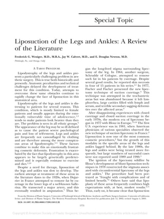
FIG. 1. Preoperative views (left), preoperative markings (center), and postoperative views (right) of a patient with lipodystrophy.
Notice the poor definition of the gastrocnemius muscles and the thickness around the malleoli preoperatively. The transition
zone is marked and a total of six incisions are planned: one medial and one lateral at the knees, one medial and lateral at the
central portion of the transition zone, and one medial and one lateral just above the malleoli. Anterior and posterior views of
postoperative result at 1 year show definition retention and no edema.
1784 PLASTIC AND RECONSTRUCTIVE SURGERY, May 2004
15. is not immediately obvious, it is important to
keep in mind the principles of excellent surgi-
cal patient care and synthesize the best ap-
proach that generates the best results in your
hands.33
Frederick G. Weniger, M.D., M.B.A.
Division of Plastic Surgery
6B Scaife Hall
3550 Terrace Street
Pittsburgh, Pa. 15261
wenigerf@msx.upmc.edu
REFERENCES
1. Lillis, P. J. Liposuction of the knees, calves, and ankles.
Dermatol. Clin. 17: 865, 1999.
2. Lillis, P. J. Liposuction of the arms, calves, and ankles.
Dermatol. Surg. 23: 1161, 1997.
3. Aiche, A. E. Lipoplasty of the calves and ankles. In G. P.
Hetter (Ed.), Lipoplasty: The Theory and Practice of Blunt
Suction Lipectomy, 2nd Ed. Boston: Little, Brown, 1990.
Pp. 347-353.
4. Pitman, G. H. Liposuction & Aesthetic Surgery. St. Louis:
Quality Medical Publishing, 1993. Pp. 413-444.
5. Grazer, F. M. Knees, calves, and ankles. In F. M. Grazer
(Ed.), Atlas of Suction Assisted Lipectomy. New York:
Churchill Livingstone, 1992. Pp. 297-300.
6. Klein, J. A. Tumescent Technique: Tumescent Anesthesia and
Microcannular Liposuction. Philadelphia: Mosby, 2000.
Pp. 440-443.
7. Ersek, R. A., and Salisbury, A. V. Circumferential lipo-
suction of knees, calves, and ankles. Plast. Reconstr.
Surg. 98: 880, 1996.
8. Fischer, A., and Fischer, G. M. Revised technique for
cellulitic fat reduction in riding breeches deformity.
Bull. Int. Acad. Cosmet. Surg. 2: 40, 1977.
9. Fournier, P. F., and Otteni, F. M. Lipodissection in body
sculpting: The dry procedure. Plast. Reconstr. Surg. 72:
598, 1983.
10. Mladick, R. A. Circumferential “intermediate” lipo-
plasty of the legs. Aesthetic Plast. Surg. 18: 165, 1994.
11. Reed, L. S. Lipoplasty of the calves and ankles. Clin.
Plast. Surg. 16: 365, 1989.
12. Illouz, Y.-G. Body contouring by lipolysis: A 5-year ex-
perience with over 3000 cases. Plast. Reconstr. Surg. 72:
591, 1983.
13. Watanabe, K. Circumferential liposuction of calves and
ankles. Aesthetic Plast. Surg. 14: 259, 1990.
14. Illouz, Y.-G., and de Villers, Y. T. Body Sculpturing by
Lipoplasty. New York: Churchill Livingstone, 1989. Pp.
124-126, 275-280.
15. Illouz, Y.-G. Surgical remodeling of the silhouette by
aspiration lipolysis or selective lipectomy. Aesthetic
Plast. Surg. 9: 7, 1985.
16. Frick, A., Hoffmann, J. N., Baumeister, R. G. H., and Putz,
R. Liposuction technique and lymphatic lesions in
lower legs: Anatomic study to reduce risks. Plast. Re-
constr. Surg. 103: 1868, 1999.
17. Chamosa, M. Comprehensive liposuction of lower
limbs: Basic concepts. Aesthetic Plast. Surg. 20: 49, 1996.
18. Chamosa, M. Suction lipectomy of the ankle area. Plast.
Reconstr. Surg. 100: 1047, 1997.
19. Mladick, R. A. Advances in liposuction contouring of
calves and ankles. Plast. Reconstr. Surg. 104: 823, 1999.
20. Teimourian, B. Suction Lipectomy & Body Sculpting. St.
Louis: Mosby, 1987. Pp. 495-510.
21. Rohrich, R. J. Advances in liposuction contouring of
calves and ankles (Discussion). Plast. Reconstr. Surg.
104: 832, 1999.
22. Toledo, L. S. Refinements in Facial and Body Contouring.
Philadelphia: Lippincott-Raven, 1999. Pp. 165-167.
23. Gasparotti, M. Superficial liposuction: A new applica-
tion for the technique for aged and flaccid skin. Aes-
thetic Plast. Surg. 16: 141, 1992.
24. Teimourian, B. Tourniquet after suction lipectomy of
the lower extremity. Plast. Reconstr. Surg. 75: 442, 1985.
25. Klein, J. A. Tumescent technique for local anesthesia
improves safety in large-volume liposuction. Plast. Re-
constr. Surg. 92: 1085, 1993.
26. Hunstad, J. P. Tumescent and syringe liposculpture: A
logical partnership. Aesthetic Plast. Surg. 19: 321, 1995.
27. Stallings, J. O. The use of the pneumatic tourniquet. In
G. P. Hetter (Ed.), Lipoplasty: The Theory and Practice of
Blunt Suction Lipectomy, 2nd Ed. Boston: Little, Brown,
1990. P. 354.
28. Karacalar, A., and Ozcan, M. Liposuction of the knee-
cap area under tourniquet: A superdry procedure.
Aesthetic Plast. Surg. 22: 408, 1998.
29. Karacalar, A., and Ozcan, M. The superdry technique
for lipoplasty of the leg and the no-touch technique
for autologous fat transplantation. Plast. Reconstr. Surg.
106: 738, 2000.
30. Mladick, R. A. Lipoplasty of the calves and ankles. Plast.
Reconstr. Surg. 86: 84, 1990.
31. Mladick, R. A. Suction lipectomy of the ankle area (Dis-
cussion). Plast. Reconstr. Surg. 100: 1053, 1997.
32. Mladick R. A. Liposuction techniques and lymphatic
lesions on lower legs: Anatomic study to reduce
risks (Discussion). Plast. Reconstr. Surg. 103: 1874,
1999.
33. Asken, S. Liposuction Surgery and Autologous Fat Trans-
plantation. Norwalk, Conn.: Appleton & Lange, 1988.
Pp. 106-107.
Vol. 113, No. 6 / LIPOSUCTION OF THE LEGS AND ANKLES 1785