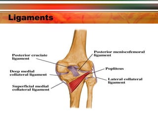
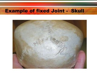
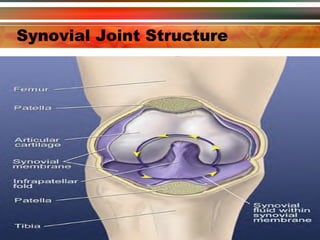
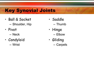
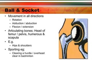
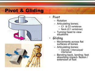
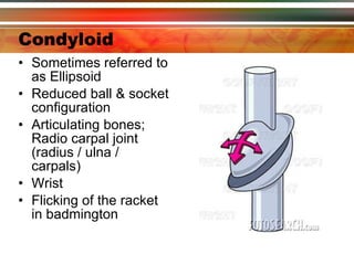
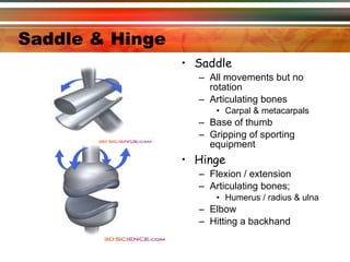
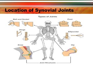
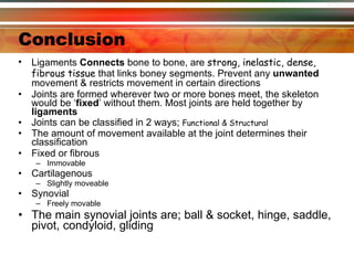
Exercise and Fitness Lvl 2 Lesson 2 – Joints in Action
Lesson 1 Re-cap Progress Test Complete Work sheet 3 on your own
Classification of Bones There are 206 bones in the body, these can be classified as: Long Short Flat Irregular Sesamoid
Long Bone Such as Femur, tibia, fibula, radius ulna, humerous,  Blood Production occurs in the red bone marrow of long bones Long bones provide the body with leverage e.g throwing a cricket ball.
Short bones Short bones are found in the wrist and ankle. Short bones are useful for delicate movement where precision is paramount  e.g. writing or a surgical operations. Small bones also help dissipate large compressive forces generated during walking and running.
Flat Bones Flat can be found in the scapulae, rib cage, sternum and cranium Flat bones offer protection and muscle attachment e.g. the rib cage protects the heart and lungs
Irregular Bones Irregular bones can be found in the vertebral column Irregular bones provide muscle attachment points and allow varying movement patterns to occur e.g. The vertebral column
Sesamoid Bones Sesamoid bones are small bones formed in tendons e.g. patella ( knee cap). Sesamoid bones provide protection for delicate areas.
Joints – 3 types Fused – Immovable e.g. skull Cartiliginous – Slightly movable e.g. between vertebral column Synovial – Freely movable e.g hip & knee
Key Joint Facts   Formed wherever two or more bones meet The skeleton would be ‘ fixed ’ without them Bones that form a joint ‘fit well’, if irregularities exist they are evened out by extra layers of  cartilage The above are called ‘ menisci ’ if they only partly divide a joint cavity If they fully divide the joint cavity this cartilage is called a  disc Most joints are held together by  ligaments
Ligaments
Fixed or Fibrous Joints Has no movement Tough fibrous  tissue lies between the ends of the bones The ends are ‘ dovetailed ’ together There is  no joint cavity  (space between the bones’ E.g.  sutures  in the skull
Example of fixed Joint -  Skull
Cartilagenous Joints Allows some  slight movement Ends of the bones are covered in  articular cartilage Ends are separated by pads of white  fibro cartilage Compression of these pads permits  slight movement These pads also act as ‘ shock absorbers ’ Eg:  intervertebral  discs of the spinal column
Synovial Joints Distinguishing features; Ends are covered in  articular  cartilage Inners surface of joint capsules is lined with a thin synovial membrane that produces  synovial fluid Freely movable joints Characterised by the presence of a joint capsule and cavity (space between bones) Range of movement can vary due to the shape of the bony surfaces forming the joint The synovial fluid acts as a  lubricant  for the joint, provides  nutrients   E.G. knee
Synovial Joint Structure
Key Synovial Joints Ball & Socket Shoulder, Hip Pivot Neck Condyloid Wrist Saddle Thumb Hinge Elbow Gliding Carpels
Ball & Socket   Movement in all directions  Rotation Adduction / abduction Flexion / extension Articulating bones; Head of femur / pelvis, humerous & scapula E.g.  Hips & shoulders Sporting eg; Clearing a hurdle / overhead clear in badminton
Pivot & Gliding   Pivot Rotation Articulating bones; C1  & C2 vertebrae  Neck (C1 vertebrae) Turning head to view situations Gliding Movements across flat surfaces of bones Articulating bones; Clavicle / intercarpal/ intertarsal Bending back, landing; feet absorbing impact- flexion / extension of foot
Condyloid Sometimes referred to as Ellipsoid Reduced ball & socket configuration Articulating bones; Radio carpal joint (radius / ulna / carpals) Wrist Flicking of the racket in badmington
Saddle & Hinge Saddle All movements but no rotation Articulating bones Carpal & metacarpals Base of thumb Gripping of sporting equipment Hinge Flexion / extension Articulating bones; Humerus / radius & ulna Elbow Hitting a backhand
Location of Synovial Joints
Ligaments
Conclusion Ligaments  Connects  bone to bone, are  strong, inelastic, dense, fibrous tissue  that links boney segments. Prevent any  unwanted  movement & restricts movement in certain directions Joints are formed wherever two or more bones meet, the skeleton would be ‘ fixed ’ without them. Most joints are held together by  ligaments Joints can be classified in 2 ways;  Functional & Structural  The amount of movement available at the joint determines their classification   Fixed or fibrous Immovable Cartilagenous Slightly moveable Synovial Freely movable The main synovial joints are; ball & socket, hinge, saddle, pivot, condyloid, gliding
Now complete…. Now complete worksheet 4

Lesson 2 joints

  • 1.
    Exercise and FitnessLvl 2 Lesson 2 – Joints in Action
  • 2.
    Lesson 1 Re-capProgress Test Complete Work sheet 3 on your own
  • 3.
    Classification of BonesThere are 206 bones in the body, these can be classified as: Long Short Flat Irregular Sesamoid
  • 4.
    Long Bone Suchas Femur, tibia, fibula, radius ulna, humerous, Blood Production occurs in the red bone marrow of long bones Long bones provide the body with leverage e.g throwing a cricket ball.
  • 5.
    Short bones Shortbones are found in the wrist and ankle. Short bones are useful for delicate movement where precision is paramount e.g. writing or a surgical operations. Small bones also help dissipate large compressive forces generated during walking and running.
  • 6.
    Flat Bones Flatcan be found in the scapulae, rib cage, sternum and cranium Flat bones offer protection and muscle attachment e.g. the rib cage protects the heart and lungs
  • 7.
    Irregular Bones Irregularbones can be found in the vertebral column Irregular bones provide muscle attachment points and allow varying movement patterns to occur e.g. The vertebral column
  • 8.
    Sesamoid Bones Sesamoidbones are small bones formed in tendons e.g. patella ( knee cap). Sesamoid bones provide protection for delicate areas.
  • 9.
    Joints – 3types Fused – Immovable e.g. skull Cartiliginous – Slightly movable e.g. between vertebral column Synovial – Freely movable e.g hip & knee
  • 10.
    Key Joint Facts Formed wherever two or more bones meet The skeleton would be ‘ fixed ’ without them Bones that form a joint ‘fit well’, if irregularities exist they are evened out by extra layers of cartilage The above are called ‘ menisci ’ if they only partly divide a joint cavity If they fully divide the joint cavity this cartilage is called a disc Most joints are held together by ligaments
  • 11.
  • 12.
    Fixed or FibrousJoints Has no movement Tough fibrous tissue lies between the ends of the bones The ends are ‘ dovetailed ’ together There is no joint cavity (space between the bones’ E.g. sutures in the skull
  • 13.
    Example of fixedJoint - Skull
  • 14.
    Cartilagenous Joints Allowssome slight movement Ends of the bones are covered in articular cartilage Ends are separated by pads of white fibro cartilage Compression of these pads permits slight movement These pads also act as ‘ shock absorbers ’ Eg: intervertebral discs of the spinal column
  • 15.
    Synovial Joints Distinguishingfeatures; Ends are covered in articular cartilage Inners surface of joint capsules is lined with a thin synovial membrane that produces synovial fluid Freely movable joints Characterised by the presence of a joint capsule and cavity (space between bones) Range of movement can vary due to the shape of the bony surfaces forming the joint The synovial fluid acts as a lubricant for the joint, provides nutrients E.G. knee
  • 16.
  • 17.
    Key Synovial JointsBall & Socket Shoulder, Hip Pivot Neck Condyloid Wrist Saddle Thumb Hinge Elbow Gliding Carpels
  • 18.
    Ball & Socket Movement in all directions Rotation Adduction / abduction Flexion / extension Articulating bones; Head of femur / pelvis, humerous & scapula E.g. Hips & shoulders Sporting eg; Clearing a hurdle / overhead clear in badminton
  • 19.
    Pivot & Gliding Pivot Rotation Articulating bones; C1 & C2 vertebrae Neck (C1 vertebrae) Turning head to view situations Gliding Movements across flat surfaces of bones Articulating bones; Clavicle / intercarpal/ intertarsal Bending back, landing; feet absorbing impact- flexion / extension of foot
  • 20.
    Condyloid Sometimes referredto as Ellipsoid Reduced ball & socket configuration Articulating bones; Radio carpal joint (radius / ulna / carpals) Wrist Flicking of the racket in badmington
  • 21.
    Saddle & HingeSaddle All movements but no rotation Articulating bones Carpal & metacarpals Base of thumb Gripping of sporting equipment Hinge Flexion / extension Articulating bones; Humerus / radius & ulna Elbow Hitting a backhand
  • 22.
  • 23.
  • 24.
    Conclusion Ligaments Connects bone to bone, are strong, inelastic, dense, fibrous tissue that links boney segments. Prevent any unwanted movement & restricts movement in certain directions Joints are formed wherever two or more bones meet, the skeleton would be ‘ fixed ’ without them. Most joints are held together by ligaments Joints can be classified in 2 ways; Functional & Structural The amount of movement available at the joint determines their classification Fixed or fibrous Immovable Cartilagenous Slightly moveable Synovial Freely movable The main synovial joints are; ball & socket, hinge, saddle, pivot, condyloid, gliding
  • 25.
    Now complete…. Nowcomplete worksheet 4