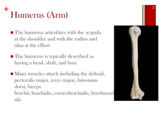
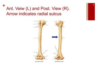
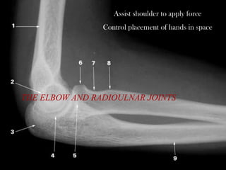
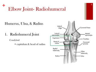
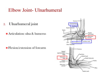
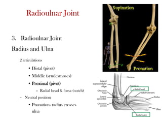
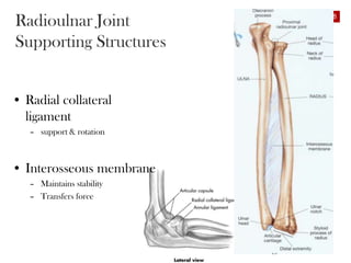
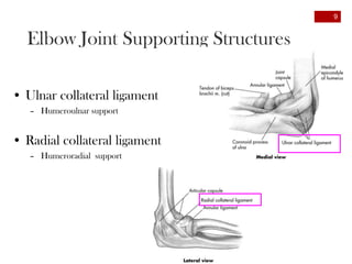
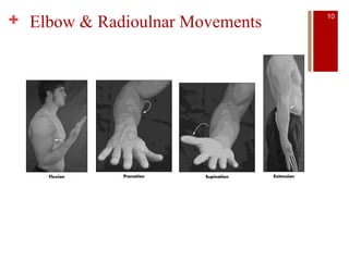
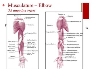
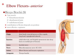
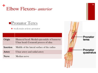
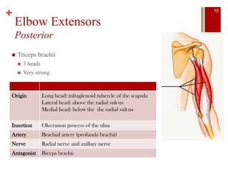
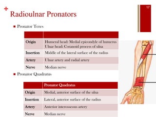
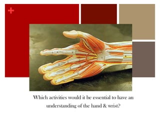
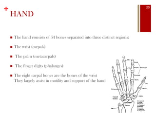
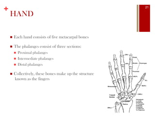
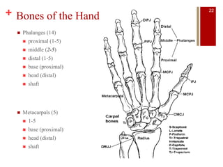
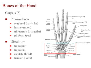
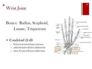
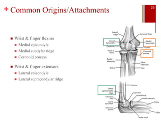
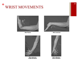
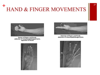
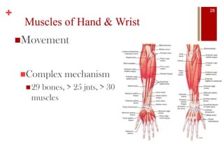
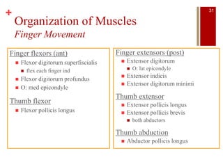
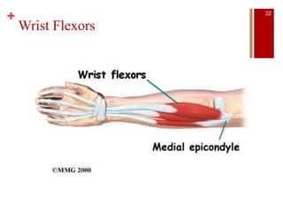
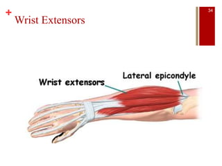
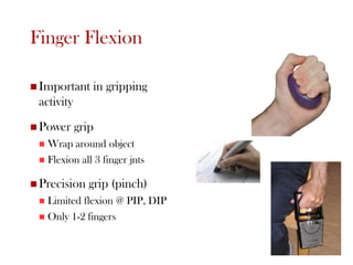
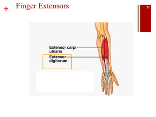
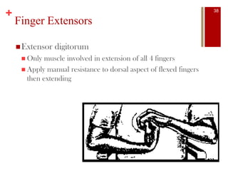
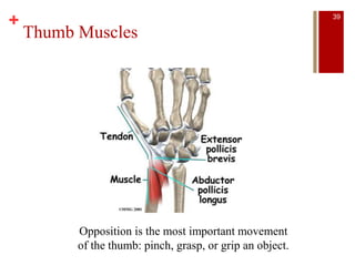
+ Upper Limb
1
+
Humerus (Arm)
 The humerus articulates with the scapula
at the shoulder and with the radius and
ulna at the elbow
 The humerus is typically described as
having a head, shaft, and base
 Many muscles attach including the deltoid,
pectoralis major, teres major, latissimus
dorsi, biceps
brachii, brachialis, coracobrachialis, brachioradi
alis
2
+
Ant. Veiw (L) and Post. View (R).
Arrow indicates radial sulcus
3
+
THE ELBOW AND RADIOULNAR JOINTS
Assist shoulder to apply force
Control placement of hands in space
+
Humerus, Ulna, & Radius
1. Radiohumeral Joint
Condyloid
• capitulum & head of radius
Elbow Joint- Radiohumeral
5
Elbow Joint- Ulnarhumeral
2. Ulnarhumeral joint
 Articulation: ulna & humerus
 Flexion/extension of forearm
6
Radioulnar Joint
3. Radioulnar Joint
Radius and Ulna
2 articulations
• Distal (pivot)
• Middle (syndesmoses)
• Proximal (pivot)
– Radial head & fossa (notch)
– Neutral position
• Pronation- radius crosses
ulna
7
Radioulnar Joint
Supporting Structures
• Radial collateral
ligament
– support & rotation
• Interosseous membrane
– Maintains stability
– Transfers force
8
Elbow Joint Supporting Structures
• Ulnar collateral ligament
– Humeroulnar support
• Radial collateral ligament
– Humeroradial support
9
+ Elbow & Radioulnar Movements 10
+ Musculature – Elbow
24 muscles cross
P
A
11
+
Elbow Flexors
Anterior
 Biceps brachii (S)
 Brachialis
 Brachioradialis (P & S)
 Pronator teres (weak)
 Elbow flexion: effectiveness of sup/pron ↑ as elbow flexion ↑
Origin Anterior surface of the humerus, specifically the distal portion
Insertion Coronoid process and the tuberosity of the ulna
Artery Radial recurrent artery
Nerve Musculocutaneous nerve (C5,C6)
12
+ Elbow Flexors- anterior
Biceps Brachii (S)
 3 jnt muscle
1= Glenohumeral joint
2= ulnarhumeral joint
3= radiohumeral joint
 contribution to flexion depends on position
 most effective- supinator
Origin short head: coracoid process of the scapula.
long head: supraglenoid tubercle
Insertion radial tuberosity and bicipital aponeurosis into deep
fascia on medial part of forearm
Artery brachial artery
Nerve Musculocutaneous nerve (C5–C6)
Action Elbow flexion and forearm supination
Antagonist Triceps brachii
13
+
Elbow Flexors- anterior
Pronator Teres
 weak-main action: pronator
Origin Humeral head: Medial epicondyle of humerus
Ulnar head: Coranoid process of ulna
Insertion Middle of the lateral surface of the radius
Artery Ulnar artery and radial artery
Nerve Median nerve
14
+
Elbow Extensors
Posterior
 Triceps brachii
 3 heads
 Very strong
Origin Long head: infraglenoid tubercle of the scapula
Lateral head: above the radial sulcus
Medial head: below the the radial sulcus
Insertion Olecranon process of the ulna
Artery Brachial artery (profunda brachii)
Nerve Radial nerve and axillary nerve
Antagonist Biceps brachii
15
+ Flexor Strength
 Twice as strong as extensors
 Are we better pullers or pushers?
 Elbow flexion:
 ADLs = more neutral
 Strongest in semi-prone positions
16
+
Radioulnar Pronators
 Pronator Teres
 Pronator Quadratus
Origin Humeral head: Medial epicondyle of humerus
Ulnar head: Coranoid process of ulna
Insertion Middle of the lateral surface of the radius
Artery Ulnar artery and radial artery
Nerve Median nerve
Pronator Quadratus
Origin Medial, anterior surface of the ulna
Insertion Lateral, anterior surface of the radius
Artery Anterior interosseous artery
Nerve Median nerve
17
+
Conditioning Pronators & Supinators
Pronators
 Loosening a screw, turning a door
Supinators
 Tightening a screw
 Throwing a curveball (extension, before release)
18
+THE HAND AND WRIST
Which activities would it be essential to have an
understanding of the hand & wrist?
+
HAND
 The hand consists of 54 bones separated into three distinct regions:
 The wrist (carpals)
 The palm (metacarpals)
 The finger digits (phalanges)
 The eight carpal bones are the bones of the wrist
They largely assist in motility and support of the hand
20
+
HAND
 Each hand consists of five metacarpal bones
 The phalanges consist of three sections:
 Proximal phalanges
 Intermediate phalanges
 Distal phalanges
 Collectively, these bones make up the structure
known as the fingers
21
+ Bones of the Hand
 Metacarpals (5)
 1-5
 base (proximal)
 head (distal)
 shaft
 Phalanges (14)
 proximal (1-5)
 middle (2-5)
 distal (1-5)
 base (proximal)
 head (distal)
 shaft
22
Bones of the Hand
Carpals (8)
 Proximal row
 scaphoid (navicular)
 lunate (moon)
 triquetrum (triangular)
 pisiform (pea)
 Distal row
 trapezium
 trapezoid
 capitate (head)
 hamate (hook)
23
+
Bones: Radius, Scaphoid,
Lunate, Triquetrum
• Condyloid (2 df)
– flexion/extension/hyperextension
– radial deviation/flexion (abduction)
– ulnar deviation/flexion (adduction)
Wrist Joint
24
+ Common Origins/Attachments
 Wrist & finger flexors
 Medial epicondyle
 Medial condylar ridge
 Coronoid process
 Wrist & finger extensors
 Lateral epicondyle
 Lateral supracondylar ridge
25
+
WRIST MOVEMENTS
26
+
HAND & FINGER MOVEMENTS
27
+
Muscles of Hand & Wrist
Movement
Complex mechanism
29 bones, > 25 jnts, > 30
muscles
28
+
Intrinsic vs. Extrinsic
Muscles acting at wrist & fingers
Originate outside hand (extrinsic)
 Enter hand as tendons
Intrinsic (originate in hand)
29
+
Names Identify Function/Location
 Flexor or extensor
 Carpi – carpals
 Radialis – radial side
 Ulnaris – ulnar side
 Digitorum- digits/fingers
 Superfiscialis – superfiscial aspect
 Profunda – deep
 Indicis – index finger
 Pollicis – thumb
 Digiti minimi – pinky/little finger
30
+
Organization of Muscles
Finger Movement
Finger flexors (ant)
 Flexor digitorum superfiscialis
 flex each finger ind
 Flexor digitorum profundus
 O: med epicondyle
Thumb flexor
 Flexor pollicis longus
Finger extensors (post)
 Extensor digitorum
 O: lat epicondyle
 Extensor indicis
 Extensor digitorum minimi
Thumb extensor
 Extensor pollicis longus
 Extensor pollicis brevis
 both abductors
Thumb abduction
 Abductor pollicis longus
31
+
Wrist Flexors
32
+
Wrist Flexors
 Curling the wrist
 Stabilize the wrist against resistance
 Snaps hand fwd
 spiking VB, throwing BB
33
+
Wrist Extensors
34
+
Wrist Extensors
 Curling the wrist
 Stabilize the wrist against resistance when forearm is
_____________
 Important in backhand (racquet sports)
 Draws the hand back
 dribbling BK, throwing BB
35
Finger Flexion
 Important in gripping
activity
 Power grip
 Wrap around object
 Flexion all 3 finger jnts
 Precision grip (pinch)
 Limited flexion @ PIP, DIP
 Only 1-2 fingers
36
+ Finger Extensors 37
+
Finger Extensors
Extensor digitorum
 Only muscle involved in extension of all 4 fingers
 Apply manual resistance to dorsal aspect of flexed fingers
then extending
38
+
Thumb Muscles
Opposition is the most important movement
of the thumb: pinch, grasp, or grip an object.
39

Upper limb (week 2 3)

  • 1.
  • 2.
    + Humerus (Arm)  Thehumerus articulates with the scapula at the shoulder and with the radius and ulna at the elbow  The humerus is typically described as having a head, shaft, and base  Many muscles attach including the deltoid, pectoralis major, teres major, latissimus dorsi, biceps brachii, brachialis, coracobrachialis, brachioradi alis 2
  • 3.
    + Ant. Veiw (L)and Post. View (R). Arrow indicates radial sulcus 3
  • 4.
    + THE ELBOW ANDRADIOULNAR JOINTS Assist shoulder to apply force Control placement of hands in space
  • 5.
    + Humerus, Ulna, &Radius 1. Radiohumeral Joint Condyloid • capitulum & head of radius Elbow Joint- Radiohumeral 5
  • 6.
    Elbow Joint- Ulnarhumeral 2.Ulnarhumeral joint  Articulation: ulna & humerus  Flexion/extension of forearm 6
  • 7.
    Radioulnar Joint 3. RadioulnarJoint Radius and Ulna 2 articulations • Distal (pivot) • Middle (syndesmoses) • Proximal (pivot) – Radial head & fossa (notch) – Neutral position • Pronation- radius crosses ulna 7
  • 8.
    Radioulnar Joint Supporting Structures •Radial collateral ligament – support & rotation • Interosseous membrane – Maintains stability – Transfers force 8
  • 9.
    Elbow Joint SupportingStructures • Ulnar collateral ligament – Humeroulnar support • Radial collateral ligament – Humeroradial support 9
  • 10.
    + Elbow &Radioulnar Movements 10
  • 11.
    + Musculature –Elbow 24 muscles cross P A 11
  • 12.
    + Elbow Flexors Anterior  Bicepsbrachii (S)  Brachialis  Brachioradialis (P & S)  Pronator teres (weak)  Elbow flexion: effectiveness of sup/pron ↑ as elbow flexion ↑ Origin Anterior surface of the humerus, specifically the distal portion Insertion Coronoid process and the tuberosity of the ulna Artery Radial recurrent artery Nerve Musculocutaneous nerve (C5,C6) 12
  • 13.
    + Elbow Flexors-anterior Biceps Brachii (S)  3 jnt muscle 1= Glenohumeral joint 2= ulnarhumeral joint 3= radiohumeral joint  contribution to flexion depends on position  most effective- supinator Origin short head: coracoid process of the scapula. long head: supraglenoid tubercle Insertion radial tuberosity and bicipital aponeurosis into deep fascia on medial part of forearm Artery brachial artery Nerve Musculocutaneous nerve (C5–C6) Action Elbow flexion and forearm supination Antagonist Triceps brachii 13
  • 14.
    + Elbow Flexors- anterior PronatorTeres  weak-main action: pronator Origin Humeral head: Medial epicondyle of humerus Ulnar head: Coranoid process of ulna Insertion Middle of the lateral surface of the radius Artery Ulnar artery and radial artery Nerve Median nerve 14
  • 15.
    + Elbow Extensors Posterior  Tricepsbrachii  3 heads  Very strong Origin Long head: infraglenoid tubercle of the scapula Lateral head: above the radial sulcus Medial head: below the the radial sulcus Insertion Olecranon process of the ulna Artery Brachial artery (profunda brachii) Nerve Radial nerve and axillary nerve Antagonist Biceps brachii 15
  • 16.
    + Flexor Strength Twice as strong as extensors  Are we better pullers or pushers?  Elbow flexion:  ADLs = more neutral  Strongest in semi-prone positions 16
  • 17.
    + Radioulnar Pronators  PronatorTeres  Pronator Quadratus Origin Humeral head: Medial epicondyle of humerus Ulnar head: Coranoid process of ulna Insertion Middle of the lateral surface of the radius Artery Ulnar artery and radial artery Nerve Median nerve Pronator Quadratus Origin Medial, anterior surface of the ulna Insertion Lateral, anterior surface of the radius Artery Anterior interosseous artery Nerve Median nerve 17
  • 18.
    + Conditioning Pronators &Supinators Pronators  Loosening a screw, turning a door Supinators  Tightening a screw  Throwing a curveball (extension, before release) 18
  • 19.
    +THE HAND ANDWRIST Which activities would it be essential to have an understanding of the hand & wrist?
  • 20.
    + HAND  The handconsists of 54 bones separated into three distinct regions:  The wrist (carpals)  The palm (metacarpals)  The finger digits (phalanges)  The eight carpal bones are the bones of the wrist They largely assist in motility and support of the hand 20
  • 21.
    + HAND  Each handconsists of five metacarpal bones  The phalanges consist of three sections:  Proximal phalanges  Intermediate phalanges  Distal phalanges  Collectively, these bones make up the structure known as the fingers 21
  • 22.
    + Bones ofthe Hand  Metacarpals (5)  1-5  base (proximal)  head (distal)  shaft  Phalanges (14)  proximal (1-5)  middle (2-5)  distal (1-5)  base (proximal)  head (distal)  shaft 22
  • 23.
    Bones of theHand Carpals (8)  Proximal row  scaphoid (navicular)  lunate (moon)  triquetrum (triangular)  pisiform (pea)  Distal row  trapezium  trapezoid  capitate (head)  hamate (hook) 23
  • 24.
    + Bones: Radius, Scaphoid, Lunate,Triquetrum • Condyloid (2 df) – flexion/extension/hyperextension – radial deviation/flexion (abduction) – ulnar deviation/flexion (adduction) Wrist Joint 24
  • 25.
    + Common Origins/Attachments Wrist & finger flexors  Medial epicondyle  Medial condylar ridge  Coronoid process  Wrist & finger extensors  Lateral epicondyle  Lateral supracondylar ridge 25
  • 26.
  • 27.
    + HAND & FINGERMOVEMENTS 27
  • 28.
    + Muscles of Hand& Wrist Movement Complex mechanism 29 bones, > 25 jnts, > 30 muscles 28
  • 29.
    + Intrinsic vs. Extrinsic Musclesacting at wrist & fingers Originate outside hand (extrinsic)  Enter hand as tendons Intrinsic (originate in hand) 29
  • 30.
    + Names Identify Function/Location Flexor or extensor  Carpi – carpals  Radialis – radial side  Ulnaris – ulnar side  Digitorum- digits/fingers  Superfiscialis – superfiscial aspect  Profunda – deep  Indicis – index finger  Pollicis – thumb  Digiti minimi – pinky/little finger 30
  • 31.
    + Organization of Muscles FingerMovement Finger flexors (ant)  Flexor digitorum superfiscialis  flex each finger ind  Flexor digitorum profundus  O: med epicondyle Thumb flexor  Flexor pollicis longus Finger extensors (post)  Extensor digitorum  O: lat epicondyle  Extensor indicis  Extensor digitorum minimi Thumb extensor  Extensor pollicis longus  Extensor pollicis brevis  both abductors Thumb abduction  Abductor pollicis longus 31
  • 32.
  • 33.
    + Wrist Flexors  Curlingthe wrist  Stabilize the wrist against resistance  Snaps hand fwd  spiking VB, throwing BB 33
  • 34.
  • 35.
    + Wrist Extensors  Curlingthe wrist  Stabilize the wrist against resistance when forearm is _____________  Important in backhand (racquet sports)  Draws the hand back  dribbling BK, throwing BB 35
  • 36.
    Finger Flexion  Importantin gripping activity  Power grip  Wrap around object  Flexion all 3 finger jnts  Precision grip (pinch)  Limited flexion @ PIP, DIP  Only 1-2 fingers 36
  • 37.
  • 38.
    + Finger Extensors Extensor digitorum Only muscle involved in extension of all 4 fingers  Apply manual resistance to dorsal aspect of flexed fingers then extending 38
  • 39.
    + Thumb Muscles Opposition isthe most important movement of the thumb: pinch, grasp, or grip an object. 39