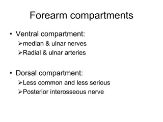
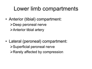
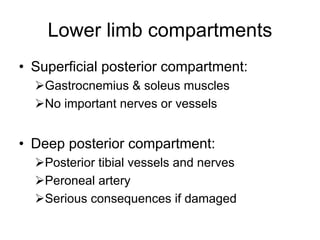
This document provides an overview of orthopedic anatomy and traumatology, including:
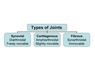
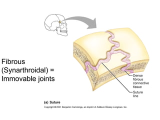
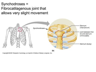
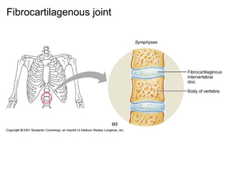
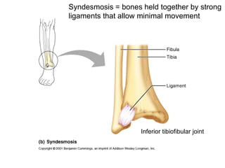
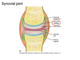
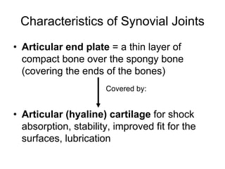
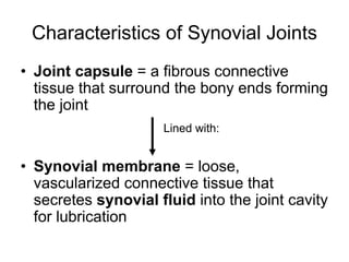
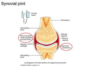
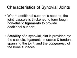
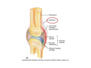
1. The three types of joints - synovial, cartilaginous, and fibrous - and characteristics of synovial joints.
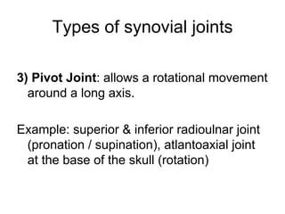
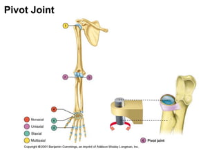
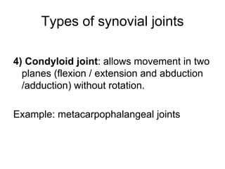
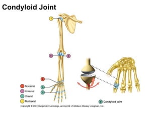
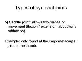
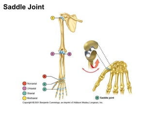
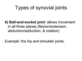
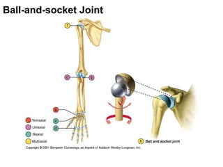
2. The six types of synovial joints - plane, hinge, pivot, condyloid, saddle, and ball-and-socket.
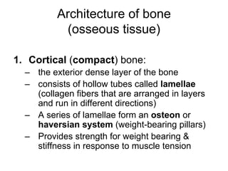
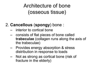
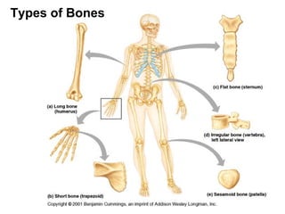
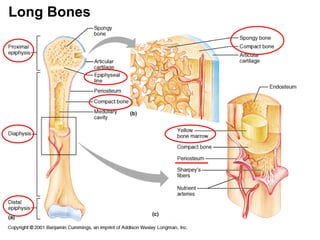
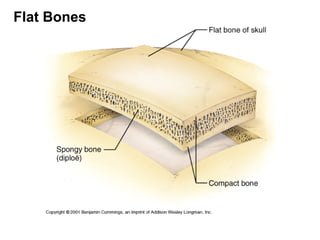
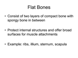
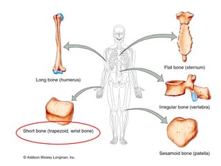
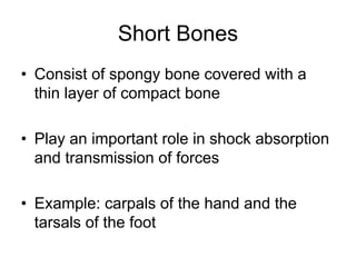
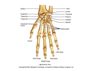
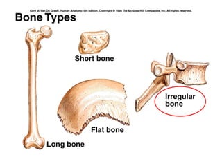
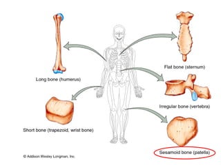
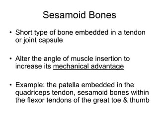
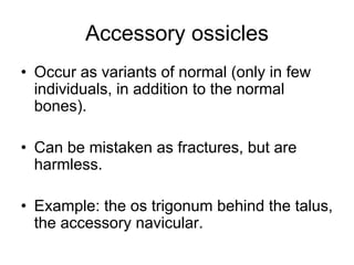
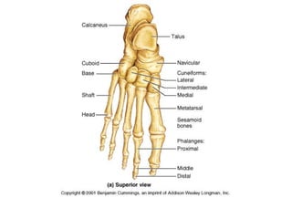
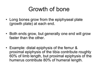
3. The architecture and types of bones, including long, flat, short, irregular, and sesamoid bones.


![PERI-PROSTHETIC FRACTURE NAIL-PLATE CONSTRUCT [NPC].pptx](https://cdn.slidesharecdn.com/ss_thumbnails/drarunkumardrmohamedashrafperiprostheticfrasturenail-plateconstructnpc-260209164459-7e9d15a1-thumbnail.jpg?width=640&height=640&fit=bounds)

