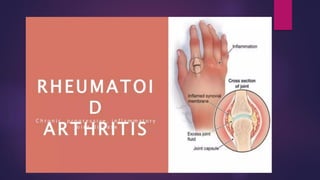
17. BLOOD TEST
Rheumatoid Factor (RF)
A rheumatoid factor test measures the amount of
rheumatoid factor in your blood.
Rheumatoid factors are proteins made by your
immune system that can attack healthy tissue in
the body.
High levels of rheumatoid factor in the blood are
most often related to autoimmune diseases, such
as rheumatoid arthritis.
18. BLOOD TEST
Anti- citrullinated Protein Antibodies (
ACPAS)
Like RF, this testing is only positive in a
proportion of all RA cases.
Unlike RF, this test is rarely round
positive if RA is not present,giving s
specificity of about 95%.
19.
20. CLINICAL MANIFESTATIONS
Fatigue , anorexia, malaise, weight loss, slight temperature
elevation
Painful, warm, swollen joints with limited motion ( stiff in the
morning and after period of inactivity ( non-use of joints).
Crippling deformity in a long standing disease.
Muscle weakness secondary to inactivity
Some patients have other manifestations: subcutaneous
nodules, eye, vascular, lung or cardiac problems.
Sjoren’s Syndrome is characterized by:
• Excessive dryness of the eyes, mouth and vagina.
21. Felty’s Syndrome is characterized by:
• Leukopenia (causes low resistance to
infection).
• Splenomegaly causes hemolytic anemia
because trapped rbc’s in the spleen
undergo hemolysis.
22. COLLABORATIVE MANAGEMENT
Bed rest during acute pain.
Passive ROM exercises of joints, To prevent
contractures.
Splint painful joints.
Heat and cold application
o Cold application during acute pain; 20 minutes
at a time. Then followed by heat application.
Warm bath in the morning
Protect the client from infection.
Physical therapy as prescribed
23. Surgery
Osteotomy
o Surgical removal of a wedge from the
joint.
Synovectomy
o Removal of synovia.
Arthoplasty
o Replacement of joints with prostheses.
24. Pharmacotherapy
Aspirin – mainstay of treatment, has both analgesic and anti-inflammatory
effects.
Nonsteroidal anti-inflammatory drugs (NSAIDS):
Indocin ( Indomethacin)
Butazolidin (Phenylbutazone)
Motrin (Ibuprofen)
Nalfon (Fenoprofen)
Naprosyn (Naproxen)
Clinoril (Sulindac)
Gold compounds (chrysotherapy)
o Injectable forms: Myochrysine; Solganal; given IM once a week ( takes 3-6
months to become effective)
o Oral form: Ridaura; smaller doses are effective; diarrhea is a common side
effect.
Corticosteroids – if pain becomes intolerable.
25. OSTEOARTHRITIS
Chronic, nonsystemic disorder of joints
characterized by degeneration of joint
cartilage.
Women and men affected equally;
incidence increases with age. Also related
to obesity and joint trauma.
Weight bearing joints – spine, knees, hips
and ends of fingers are most commonly
affected.
26. The clinical manifestations of
osteoarthritis are as follows:
Pain aggravated by use and relieved by rest.
Stiffness of joints.
Heberden’s nodes – bony overgrowth at terminal
interphalangeal joints.
Bouchard’s nodes – bony overgrowth at the
proximal interphalangeal joints.
Decreased ROM, crepitus.
27. COLLABORATIVE MANAGEMENT:
Assess joints for pain and ROM.
Relieve strain & prevent further trauma to joints:
o Use can or walker when indicated
o Maintain good posture & body mechanics,
avoid excessive weight – bearing & continuous
standing.
o Physical therapy to maintain joint mobility &
muscle strength
o Promote comfort/relief of pain (analgesics and
NSAIDS).
o Joint replacement as needed.
28. GOUT
It is a disorder of purine metabolism.
It is characterized by high levels of
uric acid in the blood & in the urine.
There is precipitation of urate crystals
(tophi) in the joints. This causes
inflammation & pain.
Occurs most often in males: and it is
familial.
29. CLINICAL MANIFESTATIONS:
Joint pain, redness, heat, swelling;
great/big toe ankle are most
commonly affected.
Headache, malaise, anorexia.
Tachycardia, fever, tophi in the great
toe, outer ear, hands & feet.
30. COLLABORATIVE MANAGEMENT:
Drug therapy
Acute attack – Colchicine (discontinue if
diarrhea or nausea & vomiting occur); or NSAIDS
– Indocin, Butazolidin.
Prevention – uricosuric agents. These are agents
that increase excretion of uric acid in the urine.
o Benemid (Probenecid), Anturane
(sulfinpyrazone).
o Zyloprim (allopurinol) inhibits uric acid formation.
31. NURSING INTERVENTIONS:
1. Antigout medications should be used cautiously in
clients with gastrointestinal, renal, cardiac or hepatic
disease.
2. Maintain a fluid intake of atleast 2000 to 3000 ml a day
to avoid kidney stones.
3. Instruct client to avoid alcohol and caffeine, These
products can increase uric acid level.
4. Avoid purine – rich foods
5. Instruct client to take medications with food.
6. Instruct client to avoid large doses of Vit.C while taking
allopurinol to prevent kidney stones.
7. Advise client to have yearly eye examination.
8. Do not take A.S.A with antigout medications.
32. 10. Observe for the ff. side effects of antigout
medications:
o Headache
o Nausea, vomiting, diarrhea
o Bone marrow depression
o Flushed skin & skin rash
o Uric acid kidney stone
o Sore gums
o Metallic taste
33. Low purine diet
Foods to avoid
o Organ meats
o Shellfish
o Legumes
o Sardines
o Salted anchovies
o Mushrooms
o Herring
o Sweetbreads
o Consome
o Beer/Wine
34. SYSTEMIC LUPUS ERYTHEMATOSUS
(SLE)
It is a chronic, multisystem, collagen disorder.
The causes are as ff:
Unknown
Autoimmune
Drugs
Viral Infections
Genetics
Higher incidence in females especially those
who are 15 to 40 years of age.
35. SLE is precipitated by the ff. medications:
Pronestyl
Phenergan
Apresoline
Dilantin
INH
Quinidine
Diagnostic Tests that support diagnosis of SLE are as ff:
CBC – pancytopenia
Increased ESR
ANA
Anti – DNA (most specific)
LE factor
40. OSTEOMYELITIS
Acute or chronic infection of the bone &
surrounding soft tissues, most commonly
caused by Staphylococcus Aureus.
Infection may reach bone through open
wound; through the blood stream or by
direct extension from infected adjacent
structures.
41. CLINICAL MANIFESTATIONS:
malaise, fever
Pain & tenderness of bone, redness & swelling of
bone
Difficulty with weight-bearing
Drainage from wound site may be present ;
necrosis of bone tissues (sequestrum)
Diagnostic tests
CBC – WBC may be elevated (indicates
presence of infection)
ESR – may be elevated (indicates inflammatory
process)
42. HERNIATED NUCLEUS PULPOSUS
(HNP)
It is rupture of intervertebral disk.
It involves protrusion of nucleus polposus into spine
causing compression of spine nerve roots.
Occurs more often in men.
The 4th & 5th intervertebral disks in the lumbar region are
most commonly affected.
Predisposing factors of HNP are as ff.
1. Heavy lifting or pulling & trauma.
2. Degeneration of the intervertebral disks.
3. Congenital predisposition.
44. CLINICAL MANIFESTATIONS:
Lumbosacral disk
Back pain radiating across the buttock &
down the leg
Weakness of leg & foot on affected side.
Numbness & tingling in toes & foot.
Positive straight leg raise test; pain on leg
below the knee when leg raised from a
supine position (Lasegue’s sign).
Depressed or absent Achilles reflex.
Muscle spasm in lumbar region.
45. CONT.
Cervical disk
Shoulder pain radiating down the arm to
hand, weakness of upper extremity,
paresthesia & sensory disturbances.
Diagnostic tests
Myelogram
CT scan or MRI; MRI has greater sensitivity.
46. COLLABORATIVE MANAGEMENT:
Conservative management
Bed rest on a firm mattress with bed board.
Traction (pelvic traction).
Drug therapy.
o Anti-inflammatory agents (ASA, NSAIDS, steroids)
o Muscle relaxants
Lioresal (baclofen)
Soma (carisoprodol)
Maolate (chlorphenesin carbanate)
Flexeril (Cyclobenzaprine)
48. CONT.
Local application of heat & diathermy.
Use of corset for lumbosacral disk; cervical
collar for cervical disk.
Epidural injections of corticosteroids if pain
becomes intolerable.
Prevent complications of immobility.
49. SURGERY
Chemonucleolysis (less common invasive treatment for
lumbar disk herniation).
o Chymopapain (Chymodiactin) into disc to reduce
size & pressure on affected nerve foot.
o Used as alternative to laminectomy in selected cases.
May cause severe complications such as transverse
myelitis, allergic reactions, persistent muscle spasm.
o Pre-op care for patient receiving chemonucleolysis
Cimetidine (Tagamet)
Diaphenhydramine HCI (Benadryl)
Corticosteroids before procedure.
50. CONT.
Post – op care for patient receiving
chemonucleolysis
Observe for anaphylaxis.
Observe for less serious allergic
reaction.
Monitor for neurologic deficits
(numbness or tingling in extremities or
inability to void).
51. LAMINECTOMY
Surgical excision of a part of posterior arch of vertebra &
removal of protruded disc.
Nursing interventions – preoperative period
Routine pre – op care.
Teach patient log rolling & use of bedpan.
Nursing interventions – postoperative period
Routine post-op care
Position as ordered
o Lower spinal surgery (lumbar) : flat position
o Cervical spinal surgery : slight elevation of head of
bed.
52. CONT.
o Maintain proper body alignment.
o For cervical spinal surgery: avoid flexion of neck & apply
cervical collar.
o Turn patient every 2 hrs.
Use log rolling technique or stryker frame.
Put small pillow under the head & 2 pillows between legs
while on side.
o Assess for complications
• Monitor sensory & motor status every 2-4 hrs. Report any
new deficit.
• With cervical spinal surgery; assess swallowing, coughing;
check for respiratory distress; have suction &
tracheostomy set at bedside.
53. CONT.
Check dressing for hemorrhage, CSF leakage,
signs of infection.
Promote comfort.
o Analgesics as ordered.
o Provide additional comfort measures &
positioning.
Assess for adequate bowel & bladder function;
check every 2-4hrs for bladder distention.
Assess bowel sounds.
Prevent complications of immobility.
Assist with ambulation.
54. PATIENT TEACHING & DISCHARGE
PLANNING
AVOID THE FF FACTORS:
• Acute hip flexion
• Prolonged sitting/standing
• Running, jogging, horseback riding.
• Heavy lifting of more than 20 lbs.
• Back – strengthening exercises.
• Lie in side.
• Wound care.
• Good posture & proper body mechanics
• Activity level as ordered.
• Recognition & reporting of complications
55. SPINAL FUSION
Fusion of spinous processes with bone graft from iliac crest to
provide stabilization of spine.
Nursing interventions
o Pre – op care (same as laminectomy)
o Post – op care
Position: lower spinal fusion – keep bed flat for first 12 hrs., then may
elevate HOB 20 – 30 degrees; keep off back for the 1st 48 hrs.
Cervical spinal fusion – elevate HOB slightly; assist with ambulation.
Usually OOB 3-4 post – op days; apply brace before OOB; apply
special cervical collar for cervical fusion.
Promote comfort – client may have considerable pain from graft
site.
56. CONT.
Advise client that brace will be needed for 4
mos. and lighter corset for 1 year after surgery.
It takes 1 year for the graft to become stable.
No bending, stooping, lifting, or sitting for
prolonged periods for 4 months.
Walking without excessive tiring is good; diet
modification will help prevent weight gain from
decreased activity.
57. CARPAL TUNNEL SYNDROME (CTS)
It occurs when the median nerve at the
wrist is compressed by thickened flexor
sheath, skeletal encroachment, edema or
soft tissue mass. It is commonly due to
repetitive hand activities.
58. CLINICAL MANIFESTATIONS:
Pain from the wrist to the shoulders.
Numbness, paresthesia.
Thumb, index, and middle fingers are affected.
(+) tinels sign (tingling sensation on percussion of
inner wrist)
(+) phalen’s sign (tingling sensation on holding
the wrist in flexion for few minutes).
Weak grip of hands.
59. COLLABORATIVE MANAGEMENT:
Rest & splint the affected wrist.
Avoid repetitive flexion of the wrist.
NSAIDS are prescribed.
Carpal canal cortisone injections.
Surgical release of transverse carpal
ligament.
60. DUPUYTREN’S CONTRACTURE
A flexion deformity of the 4th & 5th
fingers, sometimes the middle finger.
This is due to progressive contracture
of the palmar fascia. It is caused by
an inherited autosomal dominant
trait. It requires surgery.
61. AMPUTATION OF THE LIMB
It is a surgical procedure done for peripheral vascular
disease if medical management is ineffective. It may
also be done in trauma.
Types of amputation
Guillotine
Closed/flap
Nursing interventions – preoperative
Establish open & honest communication.
Offer support & encouragement & accept patient’s
response of anger / grief.
Discuss treatment during the postop period:
62. CONT.
Rehabilitation program & use of prosthesis
Upper extremity exercises such as push ups in bed
Crutch walking
Amputation dressing / Cast
Phantom limb sensation as a normal occurrence
Nursing interventions – postoperative
Observe stump dressing for signs of hemorrhage & mark
outside of dressing so, rate of bleeding can be assessed
Prevent edema
o Raise extremity with pillow support for first 24 hrs.
63. CONT.
Prevent hip/knee contractures.
Avoid letting patient sit in chair with hips flexed for long
periods of time.
Have patient assume prone position several times a day
& position hip in extension.
Avoid elevation of stump after 24 hrs; keep stump
adducted with the unaffected leg
Pain medication as ordered
Ensure that stump bandages fit snugly & are applied
properly to enhance prosthesis fitting.
Initiate active ROM of all joints, crutch walking &
arm/shoulder exercises.
64. CONT.
Provide stump care.
Inspect daily signs of skin irritation
Wash thoroughly daily with warm water & bacteriostatic
soap; rinse & dry thoroughly, avoid use of irritating
substances such as lotions, alcohol, powders.
Wear cotton or woolen stump socks, avoid nylon socks.
Do not use torn socks.
Put on prosthesis upon arising & keep it on all day.
Continue prescribed exercises; push stump against hard
surface, e.g. foot stool with pillow to toughen it for better
prosthesis fitting.
65. DON’T’S ON THE STUMP
Hang stump over the bed
Sit in wheelchair or edge of the bed with
stump flexed
Place pillow under hip or knee
Place pillow between thighs
Rest above knee stump on crutch handle
Abduct above knee stump
66. PAGET’S DISEASE (OSTEITIS
DEFROMANS)
Increased rate of bone tissue breakdown
by osteoclasts followed by excessive
abnormal bone formation by the
osteoblasts.
The diseased bone is weak & prone to
fracture.
Usually affects femur, tibia, lower spine,
pelvis & cranium.
69. LEG – CALVE – PERTHE’S DISEASE
(LCPD)
Coxa planca or osteochondritis deformans
juvenilis.
It is characterized by aseptic necrosis of the
head of the femur. It may lead to fracture.
The cause is unknown, however it is associated
with decreased circulation to the head of the
femur,
The disease is self – limiting.
Children who are 3 to 12 years of age are most
commonly affected.
71. COLLABORATIVE MANAGEMENT:
Relief of pain in the joint
Limitation of activities, bed rest to relieve
synovitis, muscle spasm, & pain in the joint.
Encourage activities to maintain ROM
Provide mobility equipment.
Use of brace.
Cast application.
Surgery (osteotomy)
Prevent trauma to the hips
73. CLINICAL MANIFESTATIONS:
Diffuse pain & tender points on the skin which is
symmetrical.
Sleep disturbance.
Headache.
Restless leg syndrome.
Excess response to cold.
Periods of depression.
Memory loss.
Sleep apnea.
74. COLLABORATIVE MANAGEMENT:
Massage back, neck, or shoulders as desired.
Use heat via ultrasound for tender points.
Group therapy
Passive stretching exercises by physical therapist
Active exercises, aerobic conditioning guided by
physical therapist; regular aerobic exercises, a minimum
of 30 minutes, 3 times weekly.
Aqua therapy.
Continuous positive airway pressure (CPAP) machine is
used for sleep apnea during naps or nighttime sleep.
75. PHARMACOTHERAPY
1. NSAIDs (e.g. Motrin/Ibuprofen). These are the
most commonly used drugs for fibromyalgia.
2. Narcotic – analgesic (e.g.
Percodan/Oxycodone). For short – term
treatment of headaches. It is used infrequently.
3. Antidepressant agents (e.g. Elavil/Amitriptyline,
Prozac/Fluoxetine).
4. Anti-parkinsonism agents (Sinemet/Carbidopa;
Klonipin/Clonazepam). For treatment of restless
leg.
76. OSTEOPOROSIS
Is a systemic skeletal disease
characterized by low bone mass,
leading to enhanced bone fragility &
a consequent increase in fracture risk.
77. RISK FACTORS:
Age – 50 years old & above
Low body weight ( less than 70kg.)
Family history
Low physical activity
White race
Medications (especially glucocorticoids)
Female
Tobacco use
Previous fracture
Estrogen deficiency
Low calcium intake
78. CLINICAL MANIFESTATIONS:
Decreasing height (10 to 15 cm) due to
collapsing vertebrae
Back pain (t5 to l5)
Dowager’s hump (curved upper back)
Fracture with minimal trauma
The most commonly used bone mineral density
(BMD) screening is Dual – energy X – ray
Absorptiometry (DXA)
79. COLLABORATIVE MANAGEMENT:
Diet & supplementation: calcium, vit.
D, phytoestrogen
Exercise at least 30 minutes. 5 days a
week, then work up to 60 minutes.
Avoid use of tobacco products &
alcohol.
80. PHARMACOTHERAPY:
Evista (Raloxifene)
Fosamax (Alendronate sodium)
Calcitonin
Forteo (Teriparatide)
Bonefos (Clodronate)
Didronel (Etidronate)
Aredia (Pamidronate)
Skelid (Tiludronate)
The client should not eat or drink anything for 30 mins. Ff
administration of the medication to increase absorption of
the drug.
81. MUSCULAR DYSTROPHY
Is a muscular disease characterized by
progressive muscle weakness & deformity.
The child will be confined to wheelchair by
age 8 to 19 years (Duchenne type).
Death occurs by age 20 years in 75% of
clients (Duchenne Type)
82. CLINICAL MANIFESTATIONS:
Pelvic girdle weakness ( the child waddles
& falls)
Gower’s sign ( uses hands to push up from
the floor )
Scoliosis ( weakness of shoulder girdle )
Contractures & hypertrophy of muscles.
83. DIAGNOSTIC TESTS:
1. Muscle biopsy shows degeneration of
muscle fibers & replacement of fibers with
fat.
2. EMG show decrease in amplitude &
duration of potentials.
3. Serum enzymes are elevated, especially
CK – MM.
84. COLLABORATIVE MANAGEMENT:
Maintain function at optimal level; keep
the child as active & independent as
possible.
Plan diet to avoid obesity.
Support child & parents; provide
information on availability of community
agencies & support groups.