ISCHEMIC OPTIC NEUROPATHY-NATURAL REMEDIES
•
0 likes•37 views

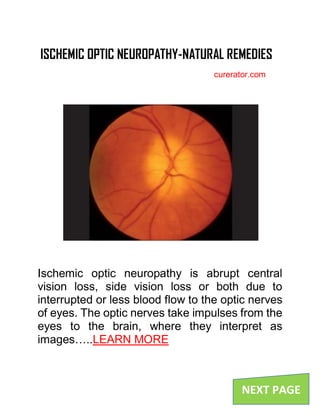
Ischemic optic neuropathy is abrupt central vision loss, side vision loss or both due to interrupted or less blood flow to the optic nerves of eyes. The optic nerves take impulses from the eyes to the brain, where they interpret as images.
Report
Share
Report
Share
Download to read offline

Recommended
General Medicine for the Optometrist
The document discusses several medical conditions and their ocular manifestations. It covers anaemia, sickle cell disease, demyelinating diseases, tumours of the nervous system, cerebrovascular disease, and seronegative arthropathies. For sickle cell disease, it describes ocular features such as vascular segments on the conjunctiva and retinal findings including venous tortuosity and peripheral scars. For demyelinating diseases like multiple sclerosis, it lists ocular features such as optic neuritis, eye movement disorders, and nerve palsies.
NEUROPATHOLOGY LECTURE 2009*
THIS LECTURE WAS PRESENTED TO THE OSTEOPATHIC STUDENTS AT THE PACIFIC NORTHWEST UNIVERSITY OF HEALTH SCIENCES IN YAKIMA WASHINGTON.
Optic nerve 1
Optic neuritis can be caused by viral or infectious etiologies. Signs include a pale optic disc, edema, and hyperfluorescence. Visual field defects are typically altitudinal and color vision is diminished. Management includes serologic tests, fasting lipid profile, and blood tests. Treatment focuses on underlying diseases, stopping smoking, and low-dose aspirin. Arteritic anterior ischemic optic neuropathy presents with scalp tenderness and jaw pain and requires high-dose steroids to prevent vision loss. Papilledema is caused by increased intracranial pressure and presents as optic disc swelling, with risks of long-term vision damage if not addressed.
Optic atrophy (b)
This document provides an overview of optic atrophy, including:
1. It defines optic atrophy as degeneration of the optic nerve due to damage to the visual pathways from the retina to the lateral geniculate body.
2. It classifies optic atrophy based on whether damage originates in the retina or more centrally, and by cause. Primary optic atrophy occurs without swelling, while secondary involves prior swelling.
3. Causes of primary optic atrophy include optic neuritis, compression, hereditary conditions, toxins, trauma, and multiple sclerosis. Secondary optic atrophy follows conditions like papilledema.
4. Treatment focuses on the underlying cause, with vitamins sometimes used
Struge weber syndrome case presentation
This document describes a case of Struge Weber syndrome in a 9-month-old female child presenting with fever and right-sided focal seizures for 7 days. Struge Weber syndrome is a rare neurocutaneous disorder characterized by a port-wine stain on the face along with leptomeningeal angiomas. Key findings included a port-wine stain on the left side of the face and right arm, CT scans showing left-sided cerebral calcifications and atrophy, and raised intraocular pressure in the left eye. The child was diagnosed with Struge Weber syndrome and treated with antipyretics, anti-epileptics, antibiotics, and management of glaucoma.
Acute Central Nervous System Demyelination
This document summarizes and compares several acute and chronic demyelinating conditions of the central nervous system. Acute disseminated encephalomyelitis is an acute multifocal condition following infection or vaccination, treated with steroids or IVIg. Multiple sclerosis is a chronic condition with recurrent episodes over space and time, potentially showing oligoclonal bands and treated with immunomodulators. Optic neuritis and transverse myelitis can occur in isolation or with other conditions and are treated with high dose steroids, while other conditions like SLE, sarcoidosis and cancer should also be considered.
Complications of systemic lupus erythematosus
This document summarizes various complications that can occur as a result of systemic lupus erythematosus (SLE). Complications can affect the blood, heart and circulation, lungs, kidneys, central nervous system, gastrointestinal system, joints/muscles/bones, and eyes. Common issues include anemia, blood clots, Raynaud's phenomenon, pericarditis, myocarditis, pleuritis, pleural effusion, lupus nephritis, CNS inflammation, nausea, weight loss, osteoporosis, and retinal damage. Left untreated, some complications like gangrene, coma, and retinal hemorrhages can become serious.
Aneurysm early signs and symptoms
A brain aneurysm is a bulge or ballooning in a blood vessel in the brain that is caused by a weakness in the vessel walls. Risk factors include older age, smoking, high blood pressure, drug/alcohol abuse, and inherited connective tissue disorders. Symptoms of a ruptured aneurysm are sudden and severe headache, nausea, stiff neck, blurred vision, sensitivity to light, seizures, drooping eyelid, loss of consciousness and confusion. Unruptured aneurysms may cause pain above or behind the eye, dilated pupil, changes in vision, or numbness of the face. The best treatment depends on each individual case.
Alzheimer's disease
This document discusses Alzheimer's disease, including what it is, how it affects the brain, stages of the disease, and treatment options. Specifically:
- Alzheimer's disease is a progressive brain disorder that causes brain cells to waste away and die, leading to cognitive decline.
- In Alzheimer's disease, the brain shrinks due to death of nerve cells caused by amyloid plaques and neurofibrillary tangles forming. This damages neurons responsible for memories, feelings, and thoughts.
- Treatment options aim to block excess glutamate toxicity and prevent breakdown of acetylcholine in the brain, such as the drugs memantine and donepezil. Other treatments target the cholinergic hypothesis of Alzheimer's disease pathogenesis
Sturge weber syndrome
This document summarizes Encephalotrigeminal Angiomatosis, also known as Sturge-Weber Syndrome (SWS), a rare congenital neurological disorder characterized by a facial birthmark and abnormalities of the brain and eyes. SWS is caused by a somatic mutation in the GNAQ gene and results in errors in skin and nervous system development. Clinical manifestations include seizures, glaucoma, hemiparesis, and developmental delays. Diagnosis involves imaging tests like MRI, CT, and angiography showing abnormal blood vessels and lesions. Treatment focuses on controlling seizures and reducing pressure in the eyes. Prognosis is often poor if seizures begin before age 2, with increased risk of developmental delays.
Anterior ischemic optic neuropathy
This document provides information on anterior ischemic optic neuropathy (AION), which is the most common cause of acute optic neuropathy in older age groups. It can be divided into two types: arteritic AION, which is due to giant cell arteritis; and non-arteritic AION, which makes up most cases. Both types present with sudden painless vision loss and optic disc swelling. Arteritic AION carries a worse prognosis and requires high-dose steroid treatment to prevent loss of vision in the fellow eye. Non-arteritic AION has a variable course but generally a poor rate of recovery without any proven effective treatments.
Spinal cord injury
1) Spinal cord injury can occur from trauma such as motor vehicle accidents, falls, or violence. It results in loss of movement and sensation below the site of injury.
2) Injuries can be complete or incomplete. Complete injuries result in total loss of function below the injury while incomplete injuries cause mixed losses.
3) Common complications include respiratory issues, pressure ulcers, blood clots, and autonomic dysreflexia. Management involves steroids, surgery, and preventative care measures.
Kearns Sayre Syndrome
Kearns-sayre syndrome is a rare genetic disorder caused by abnormalities in the mitochondria's DNA. It affects people under age 20 and can cause paralysis of eye muscles, retinal degeneration, and weakness/damage of muscles, brain, heart and other organs over time. Doctors diagnose it through tests of protein/lactate levels or muscle biopsy to examine mitochondrial DNA. There is no cure currently, and symptoms worsen over time.
Paj 5103 clinical neuropahtophys ii hn10
The document discusses several topics related to brain injury and disorders. It begins by explaining the mechanisms of primary and secondary brain injury, including ischemia, cellular energy failure, excitatory amino acids, and reperfusion injury. It then discusses types of traumatic brain injury like concussions and hematomas. It also covers cerebrovascular disease and strokes, central nervous system infections like meningitis and abscesses, and chronic neurological disorders such as seizures, dementia, Parkinson's disease, and hydrocephalus.
Optic atrophy and neuroretinitis
Optic atrophy refers to changes in the optic nerve resulting from axonal degeneration between the retina and lateral geniculate body, causing visual disturbance and changes in the optic nerve head appearance. It can be classified as primary, secondary, or consecutive. Primary optic atrophy occurs without prior nerve swelling and may result from lesions along the visual pathway. Secondary optic atrophy is preceded by long-term nerve swelling and includes causes like chronic papilledema. Consecutive optic atrophy is caused by diseases of the inner retina or its blood supply. Neuroretinitis refers to optic neuritis with retinal inflammation, most commonly caused by cat scratch fever, and presents with papillitis, macular edema, and sometimes a macular
Ischaemic Optic Neuropathy
This document discusses various types of ischemic optic neuropathy (ION). It begins by introducing ION as a major cause of vision loss and outlines its classification into anterior and posterior forms. It then details the anatomy and vascular supply of the optic nerve, risk factors for ION such as nocturnal blood pressure changes, and the pathogenesis involving hypoperfusion and axonal swelling. Non-arteritic anterior ION is described as the most common type, while posterior ION and arteritic forms are less prevalent but can involve vascular inflammation. The document outlines signs, investigations, management approaches including steroids, and variable prognoses for the different ION types.
Optic disc swelling
This document discusses optic disc swelling (papilledema) caused by increased intracranial pressure. It presents a case of a 35-year-old woman with severe headaches and vision issues. Examination found bilateral disc edema. CT scan revealed a brain tumor causing pressure. The patient was diagnosed with papilledema from the tumor and underwent surgery. The document then discusses the causes, presentation, stages, histopathology, and treatment of papilledema, emphasizing the importance of an eye exam for patients with headaches to identify potential intracranial issues.
Diesase Project 20.4.09
This document provides an overview of various diseases that affect different body systems, including the integumentary, skeletal, muscular, nervous, and circulatory systems. For each system, it describes two representative diseases, their causes, symptoms, and potential treatments. The integumentary diseases covered are albinism and herpes, the skeletal diseases are osteochondroma and osteoarthritis, the muscular diseases are tendonitis and PCL injury, the nervous diseases are Alzheimer's disease and Parkinson's disease, and the circulatory diseases are peripheral vascular disease and congenital heart defect.
Cva
This document discusses cerebrovascular disorders and stroke. It defines cerebrovascular disorders as any abnormality that disrupts the normal blood supply to the brain. Stroke is described as a sudden neurological event caused by disrupted blood flow. The two main types of stroke discussed are ischemic stroke and hemorrhagic stroke. Assessment, diagnostics, medical management, surgical options, and nursing management are summarized for patients experiencing a cerebrovascular event or stroke.
SLE & APS for undergraduates: diagnosis & treatment.
The document provides an overview of systemic lupus erythematosus (SLE) for medical students. It defines SLE, discusses its epidemiology and pathophysiology. It then describes the clinical presentation of SLE including cutaneous, musculoskeletal, serosal, renal, neurological, and hematological manifestations. It also covers investigations such as autoantibody tests and renal biopsy. Finally, it discusses lupus nephritis as a serious complication of SLE. The document aims to ensure students understand the definition, clinical picture, classification criteria, investigations, prognosis, complications and treatment approaches for SLE.
Ischemic optic neuropathies
INTRODUCTION
Optic nerve ischemia most frequently occurs at the optic nerve head, where structural crowding of nerve fibers and reduction of the vascular supply may combine to impair perfusion to a critical degree and produce optic disc edema. The most common such syndrome is termed anterior ischemic optic neuropathy(AION).
Generally, AION is categorized as either arteritic (associated with temporal arteritis) or nonarteritic .
Classic sign crbral aneurysm pbl
This document classifies cerebral aneurysms into three cases: ruptured, leaking, and unruptured. [1] A ruptured aneurysm causes a sudden, extremely severe headache described as the "worst headache ever" along with nausea, vomiting, neck stiffness, vision changes, sensitivity to light, seizures, drooping eyelid, loss of consciousness, or confusion. [2] A leaking aneurysm may cause a mild headache and will likely rupture fully. [3] An unruptured aneurysm may be asymptomatic, but a large one could press on brain tissue and cause eye or facial pain, vision changes, numbness, or weakness. The defining symptom of an aneurysm is the extreme rapid onset of
Leukodystrophies
This document summarizes several genetic leukodystrophies, including their inheritance pattern, affected enzyme or gene, clinical features, and MRI appearance. It describes adrenoleukodystrophy as an X-linked disorder caused by ABCD1 gene defects that leads to failure of peroxisomal fatty acid oxidation and can present as childhood cerebral adrenoleukodystrophy, adrenomyeloneuropathy, or Addison's disease. On MRI, it shows a characteristic pattern of different intensities in the central white matter. Zellweger syndrome is an autosomal recessive peroxisomal disorder caused by PEX1 defects that results in facial features, hepatomegaly, renal cysts, and diffuse
Paraplegia
Paraplegia is defined as impairment of motor function in the lower extremities, with or without sensory involvement, and is usually caused by involvement of the spinal cord, nerves supplying the lower limbs, or muscles directly. It is classified as spastic or flaccid depending on the affected part of the nervous system and resulting muscle tone. Common causes include spinal cord injuries, infections, tumors, and vascular disorders. A thorough history, neurological examination, and imaging tests are used to diagnose the condition and determine the specific cause and level of spinal involvement.
Ueda2015 vascular optic neuropathies dr.sherif kamel
This document discusses a new classification system for vascular optic neuropathies. It proposes several stages: prodromal, incipient, subacute, chronic (slowly progressive), acute, and post ischemic. For each stage it provides details on characteristics such as optic disc appearance and visual symptoms. It also notes that diabetes can cause several presentations of vascular optic nerve disease including acute, papillopathy, and chronic stages with or without neovascularization. The classification aims to improve early detection and management of vascular optic neuropathies.
Ocular Manifestations of Systemic Disease in Dogs
This document outlines various ocular manifestations of systemic diseases in dogs. It discusses cardiovascular issues like hypertension and how they can cause retinal hemorrhages. Hematologic disorders like anemia, thrombocytopenia, and hypertriglyceridemia are covered. Neurologic conditions such as granulomatous meningoencephalitis can lead to blindness. Dermatologic diseases including autoimmune disorders and demodex mites are addressed. Infectious diseases from ticks, parasites, fungi and more are outlined. Endocrine conditions like diabetes and hypothyroidism are discussed. Finally, oncologic topics like lymphoma and other metastatic cancers to the eyes are summarized.
Brain tumor
Stroke occurs when a blood vessel in the brain bursts or is blocked by a clot, depriving brain cells of oxygen. Without treatment, brain cells quickly begin to die, which can lead to serious disability or death. Signs of a stroke include sudden numbness or weakness, vision changes, and severe headache. It is critical to seek emergency treatment immediately if someone is experiencing stroke symptoms, as clot-busting drugs must be given within three hours to minimize brain damage. The most common type of stroke is ischemic stroke, caused by a blood clot blocking a brain vessel.
Alzheimers Disease
1. Alzheimer's disease is a progressive brain disorder that causes memory loss and cognitive decline, and is the most common form of dementia.
2. It results from an increase in beta-amyloid proteins in the brain that leads to nerve cell death. The disease is incurable and causes impairment in memory, reasoning, language, and perception.
3. Risk factors include age over 65, genetics, hypertension, high cholesterol, diabetes, smoking, alcohol use, and mild cognitive impairment. The disease is characterized by two lesions - neuritic plaques containing beta-amyloid protein and neurofibrillary tangles containing tau protein.
English Drug and Alcohol Commissioners June 2024.pptx
Presentation made by Mat Southwell to the Harm Reduction Working Group of the English Drug and Alcohol Commissioners. Discuss stimulants, OAMT, NSP coverage and community-led approach to DCRs. Focussing on active drug user perspectives and interests
More Related Content
What's hot
Alzheimer's disease
This document discusses Alzheimer's disease, including what it is, how it affects the brain, stages of the disease, and treatment options. Specifically:
- Alzheimer's disease is a progressive brain disorder that causes brain cells to waste away and die, leading to cognitive decline.
- In Alzheimer's disease, the brain shrinks due to death of nerve cells caused by amyloid plaques and neurofibrillary tangles forming. This damages neurons responsible for memories, feelings, and thoughts.
- Treatment options aim to block excess glutamate toxicity and prevent breakdown of acetylcholine in the brain, such as the drugs memantine and donepezil. Other treatments target the cholinergic hypothesis of Alzheimer's disease pathogenesis
Sturge weber syndrome
This document summarizes Encephalotrigeminal Angiomatosis, also known as Sturge-Weber Syndrome (SWS), a rare congenital neurological disorder characterized by a facial birthmark and abnormalities of the brain and eyes. SWS is caused by a somatic mutation in the GNAQ gene and results in errors in skin and nervous system development. Clinical manifestations include seizures, glaucoma, hemiparesis, and developmental delays. Diagnosis involves imaging tests like MRI, CT, and angiography showing abnormal blood vessels and lesions. Treatment focuses on controlling seizures and reducing pressure in the eyes. Prognosis is often poor if seizures begin before age 2, with increased risk of developmental delays.
Anterior ischemic optic neuropathy
This document provides information on anterior ischemic optic neuropathy (AION), which is the most common cause of acute optic neuropathy in older age groups. It can be divided into two types: arteritic AION, which is due to giant cell arteritis; and non-arteritic AION, which makes up most cases. Both types present with sudden painless vision loss and optic disc swelling. Arteritic AION carries a worse prognosis and requires high-dose steroid treatment to prevent loss of vision in the fellow eye. Non-arteritic AION has a variable course but generally a poor rate of recovery without any proven effective treatments.
Spinal cord injury
1) Spinal cord injury can occur from trauma such as motor vehicle accidents, falls, or violence. It results in loss of movement and sensation below the site of injury.
2) Injuries can be complete or incomplete. Complete injuries result in total loss of function below the injury while incomplete injuries cause mixed losses.
3) Common complications include respiratory issues, pressure ulcers, blood clots, and autonomic dysreflexia. Management involves steroids, surgery, and preventative care measures.
Kearns Sayre Syndrome
Kearns-sayre syndrome is a rare genetic disorder caused by abnormalities in the mitochondria's DNA. It affects people under age 20 and can cause paralysis of eye muscles, retinal degeneration, and weakness/damage of muscles, brain, heart and other organs over time. Doctors diagnose it through tests of protein/lactate levels or muscle biopsy to examine mitochondrial DNA. There is no cure currently, and symptoms worsen over time.
Paj 5103 clinical neuropahtophys ii hn10
The document discusses several topics related to brain injury and disorders. It begins by explaining the mechanisms of primary and secondary brain injury, including ischemia, cellular energy failure, excitatory amino acids, and reperfusion injury. It then discusses types of traumatic brain injury like concussions and hematomas. It also covers cerebrovascular disease and strokes, central nervous system infections like meningitis and abscesses, and chronic neurological disorders such as seizures, dementia, Parkinson's disease, and hydrocephalus.
Optic atrophy and neuroretinitis
Optic atrophy refers to changes in the optic nerve resulting from axonal degeneration between the retina and lateral geniculate body, causing visual disturbance and changes in the optic nerve head appearance. It can be classified as primary, secondary, or consecutive. Primary optic atrophy occurs without prior nerve swelling and may result from lesions along the visual pathway. Secondary optic atrophy is preceded by long-term nerve swelling and includes causes like chronic papilledema. Consecutive optic atrophy is caused by diseases of the inner retina or its blood supply. Neuroretinitis refers to optic neuritis with retinal inflammation, most commonly caused by cat scratch fever, and presents with papillitis, macular edema, and sometimes a macular
Ischaemic Optic Neuropathy
This document discusses various types of ischemic optic neuropathy (ION). It begins by introducing ION as a major cause of vision loss and outlines its classification into anterior and posterior forms. It then details the anatomy and vascular supply of the optic nerve, risk factors for ION such as nocturnal blood pressure changes, and the pathogenesis involving hypoperfusion and axonal swelling. Non-arteritic anterior ION is described as the most common type, while posterior ION and arteritic forms are less prevalent but can involve vascular inflammation. The document outlines signs, investigations, management approaches including steroids, and variable prognoses for the different ION types.
Optic disc swelling
This document discusses optic disc swelling (papilledema) caused by increased intracranial pressure. It presents a case of a 35-year-old woman with severe headaches and vision issues. Examination found bilateral disc edema. CT scan revealed a brain tumor causing pressure. The patient was diagnosed with papilledema from the tumor and underwent surgery. The document then discusses the causes, presentation, stages, histopathology, and treatment of papilledema, emphasizing the importance of an eye exam for patients with headaches to identify potential intracranial issues.
Diesase Project 20.4.09
This document provides an overview of various diseases that affect different body systems, including the integumentary, skeletal, muscular, nervous, and circulatory systems. For each system, it describes two representative diseases, their causes, symptoms, and potential treatments. The integumentary diseases covered are albinism and herpes, the skeletal diseases are osteochondroma and osteoarthritis, the muscular diseases are tendonitis and PCL injury, the nervous diseases are Alzheimer's disease and Parkinson's disease, and the circulatory diseases are peripheral vascular disease and congenital heart defect.
Cva
This document discusses cerebrovascular disorders and stroke. It defines cerebrovascular disorders as any abnormality that disrupts the normal blood supply to the brain. Stroke is described as a sudden neurological event caused by disrupted blood flow. The two main types of stroke discussed are ischemic stroke and hemorrhagic stroke. Assessment, diagnostics, medical management, surgical options, and nursing management are summarized for patients experiencing a cerebrovascular event or stroke.
SLE & APS for undergraduates: diagnosis & treatment.
The document provides an overview of systemic lupus erythematosus (SLE) for medical students. It defines SLE, discusses its epidemiology and pathophysiology. It then describes the clinical presentation of SLE including cutaneous, musculoskeletal, serosal, renal, neurological, and hematological manifestations. It also covers investigations such as autoantibody tests and renal biopsy. Finally, it discusses lupus nephritis as a serious complication of SLE. The document aims to ensure students understand the definition, clinical picture, classification criteria, investigations, prognosis, complications and treatment approaches for SLE.
Ischemic optic neuropathies
INTRODUCTION
Optic nerve ischemia most frequently occurs at the optic nerve head, where structural crowding of nerve fibers and reduction of the vascular supply may combine to impair perfusion to a critical degree and produce optic disc edema. The most common such syndrome is termed anterior ischemic optic neuropathy(AION).
Generally, AION is categorized as either arteritic (associated with temporal arteritis) or nonarteritic .
Classic sign crbral aneurysm pbl
This document classifies cerebral aneurysms into three cases: ruptured, leaking, and unruptured. [1] A ruptured aneurysm causes a sudden, extremely severe headache described as the "worst headache ever" along with nausea, vomiting, neck stiffness, vision changes, sensitivity to light, seizures, drooping eyelid, loss of consciousness, or confusion. [2] A leaking aneurysm may cause a mild headache and will likely rupture fully. [3] An unruptured aneurysm may be asymptomatic, but a large one could press on brain tissue and cause eye or facial pain, vision changes, numbness, or weakness. The defining symptom of an aneurysm is the extreme rapid onset of
Leukodystrophies
This document summarizes several genetic leukodystrophies, including their inheritance pattern, affected enzyme or gene, clinical features, and MRI appearance. It describes adrenoleukodystrophy as an X-linked disorder caused by ABCD1 gene defects that leads to failure of peroxisomal fatty acid oxidation and can present as childhood cerebral adrenoleukodystrophy, adrenomyeloneuropathy, or Addison's disease. On MRI, it shows a characteristic pattern of different intensities in the central white matter. Zellweger syndrome is an autosomal recessive peroxisomal disorder caused by PEX1 defects that results in facial features, hepatomegaly, renal cysts, and diffuse
Paraplegia
Paraplegia is defined as impairment of motor function in the lower extremities, with or without sensory involvement, and is usually caused by involvement of the spinal cord, nerves supplying the lower limbs, or muscles directly. It is classified as spastic or flaccid depending on the affected part of the nervous system and resulting muscle tone. Common causes include spinal cord injuries, infections, tumors, and vascular disorders. A thorough history, neurological examination, and imaging tests are used to diagnose the condition and determine the specific cause and level of spinal involvement.
Ueda2015 vascular optic neuropathies dr.sherif kamel
This document discusses a new classification system for vascular optic neuropathies. It proposes several stages: prodromal, incipient, subacute, chronic (slowly progressive), acute, and post ischemic. For each stage it provides details on characteristics such as optic disc appearance and visual symptoms. It also notes that diabetes can cause several presentations of vascular optic nerve disease including acute, papillopathy, and chronic stages with or without neovascularization. The classification aims to improve early detection and management of vascular optic neuropathies.
Ocular Manifestations of Systemic Disease in Dogs
This document outlines various ocular manifestations of systemic diseases in dogs. It discusses cardiovascular issues like hypertension and how they can cause retinal hemorrhages. Hematologic disorders like anemia, thrombocytopenia, and hypertriglyceridemia are covered. Neurologic conditions such as granulomatous meningoencephalitis can lead to blindness. Dermatologic diseases including autoimmune disorders and demodex mites are addressed. Infectious diseases from ticks, parasites, fungi and more are outlined. Endocrine conditions like diabetes and hypothyroidism are discussed. Finally, oncologic topics like lymphoma and other metastatic cancers to the eyes are summarized.
Brain tumor
Stroke occurs when a blood vessel in the brain bursts or is blocked by a clot, depriving brain cells of oxygen. Without treatment, brain cells quickly begin to die, which can lead to serious disability or death. Signs of a stroke include sudden numbness or weakness, vision changes, and severe headache. It is critical to seek emergency treatment immediately if someone is experiencing stroke symptoms, as clot-busting drugs must be given within three hours to minimize brain damage. The most common type of stroke is ischemic stroke, caused by a blood clot blocking a brain vessel.
Alzheimers Disease
1. Alzheimer's disease is a progressive brain disorder that causes memory loss and cognitive decline, and is the most common form of dementia.
2. It results from an increase in beta-amyloid proteins in the brain that leads to nerve cell death. The disease is incurable and causes impairment in memory, reasoning, language, and perception.
3. Risk factors include age over 65, genetics, hypertension, high cholesterol, diabetes, smoking, alcohol use, and mild cognitive impairment. The disease is characterized by two lesions - neuritic plaques containing beta-amyloid protein and neurofibrillary tangles containing tau protein.
What's hot (20)
SLE & APS for undergraduates: diagnosis & treatment.
SLE & APS for undergraduates: diagnosis & treatment.
Ueda2015 vascular optic neuropathies dr.sherif kamel
Ueda2015 vascular optic neuropathies dr.sherif kamel
Recently uploaded
English Drug and Alcohol Commissioners June 2024.pptx
Presentation made by Mat Southwell to the Harm Reduction Working Group of the English Drug and Alcohol Commissioners. Discuss stimulants, OAMT, NSP coverage and community-led approach to DCRs. Focussing on active drug user perspectives and interests
THE SPECIAL SENCES- Unlocking the Wonders of the Special Senses: Sight, Sound...
Title: Unlocking the Wonders of the Special Senses: Sight, Sound, Smell, Taste, and Balance
Introduction:
Welcome to our captivating SlideShare presentation on the Special Senses, where we delve into the extraordinary capabilities that allow us to perceive and interact with the world around us. Join us on a sensory journey as we explore the intricate structures and functions of sight, sound, smell, taste, and balance.
The special senses are our primary means of experiencing and interpreting the environment, each sense providing unique and vital information that shapes our perceptions and responses. These senses are facilitated by highly specialized organs and complex neural pathways, enabling us to see a vibrant sunset, hear a symphony, savor a delicious meal, detect a fragrant flower, and maintain our equilibrium.
In this presentation, we will:
Visual System (Sight): Dive into the anatomy and physiology of the eye, exploring how light is converted into electrical signals and processed by the brain to create the images we see. Understand common vision disorders and the mechanisms behind corrective measures like glasses and contact lenses.
Auditory System (Hearing): Examine the structures of the ear and the process of sound wave transduction, from the outer ear to the cochlea and auditory nerve. Learn about hearing loss, auditory processing, and the advances in hearing aid technology.
Olfactory System (Smell): Discover the olfactory receptors and pathways that enable the detection of thousands of different odors. Explore the connection between smell and memory and the impact of olfactory disorders on quality of life.
Gustatory System (Taste): Uncover the taste buds and the five basic tastes – sweet, salty, sour, bitter, and umami. Delve into the interplay between taste and smell and the factors influencing our food preferences and eating habits.
Vestibular System (Balance): Investigate the inner ear structures responsible for balance and spatial orientation. Understand how the vestibular system helps maintain posture and coordination, and explore common vestibular disorders and their effects.
Through engaging visuals, interactive diagrams, and insightful explanations, we aim to illuminate the complexities of the special senses and their profound impact on our daily lives. Whether you're a student, educator, or simply curious about how we perceive the world, this presentation will provide valuable insights into the remarkable capabilities of the human sensory system.
Join us as we unlock the wonders of the special senses and gain a deeper appreciation for the intricate mechanisms that allow us to experience the richness of our environment.
Digital Health in India_Health Informatics Trained Manpower _DrDevTaneja_15.0...
Digital India will need a big trained army of Health Informatics educated & trained manpower in India.
Presently, generalist IT manpower does most of the work in the healthcare industry in India. Academic Health Informatics education is not readily available at school & health university level or IT education institutions in India.
We look into the evolution of health informatics and its applications in the healthcare industry.
HIMMS TIGER resources are available to assist Health Informatics education.
Indian Health universities, IT Education institutions, and the healthcare industry must proactively collaborate to start health informatics courses on a big scale. An advocacy push from various stakeholders is also needed for this goal.
Health informatics has huge employment potential and provides a big business opportunity for the healthcare industry. A big pool of trained health informatics manpower can lead to product & service innovations on a global scale in India.
Types of Cancer Treatments | Forms of cancer treatment
Cancer treatment has advanced significantly over the years, offering patients various options tailored to their specific type of cancer and stage of disease. Understanding the different types of cancer treatments can help patients make informed decisions about their care. In this ppt, we have listed most common forms of cancer treatment available today.
Test bank advanced health assessment and differential diagnosis essentials fo...
Test bank advanced health assessment and differential diagnosis essentials fo...rightmanforbloodline
Test bank advanced health assessment and differential diagnosis essentials for clinical practice 1st edition myrick.
Test bank advanced health assessment and differential diagnosis essentials for clinical practice 1st edition myrick.
Test bank advanced health assessment and differential diagnosis essentials for clinical practice 1st edition myrick.Satisfying Spa Massage Experience at Just 99 AED - Malayali Kerala Spa Ajman
Satisfying Spa Massage Experience at Just 99 AED - Malayali Kerala Spa AjmanMalayali Kerala Spa Ajman
Our Spa Massage Center Ajman prioritizes efficiency to ensure a satisfying massage experience for our clients at Malayali Kerala Spa Ajman. We offer a hassle-free appointment system, effective health issue identification, and precise massage techniques.
Our Spa in Ajman stands out for its effectiveness in enhancing wellness. Our therapists focus on treating the root cause of issues, providing tailored treatments for each client. We take pride in offering the most satisfying Pakistani Spa service, adjusting treatment plans based on client feedback.
For the most result-oriented Russian Spa treatment in Ajman, visit our Massage Center. Our Russian therapists are skilled in various techniques to address health concerns. Our body-to-body massage is efficient due to individualized care and high-grade massage oils.VEDANTA AIR AMBULANCE SERVICES IN REWA AT A COST-EFFECTIVE PRICE.pdf
Air Ambulance Services In Rewa works in close coordination with ground-based emergency services, including local Emergency Medical Services, fire departments, and law enforcement agencies.
More@: https://tinyurl.com/2shrryhx
More@: https://tinyurl.com/5n8h3wp8
STERILIZATION AND DISINFECTION PRACTICES IN HOSPITAL.pptx
Following is the ppt for sterilization practices in hospital especially for infection control
PPT on Embryological and fetal development
Embryological development and fetal development
Factors influencing fetal development
Bathinda ℂ𝕒𝕝𝕝 𝔾𝕚𝕣𝕝𝕤 7742996321 ℂ𝕒𝕝𝕝 𝔾𝕚𝕣𝕝𝕤 Bathinda
Bathinda ℂ𝕒𝕝𝕝 𝔾𝕚𝕣𝕝𝕤 7742996321 ℂ𝕒𝕝𝕝 𝔾𝕚𝕣𝕝𝕤 Bathinda
05 CLINICAL AUDIT-ORTHO done at a peripheral.pptx
Clinical audit on pain management done at peripheral health centre
Solution manual for managerial accounting 18th edition by ray garrison eric n...
Solution manual for managerial accounting 18th edition by ray garrison eric n...rightmanforbloodline
Solution manual for managerial accounting 18th edition by ray garrison eric noreen and peter brewer_compressed
Solution manual for managerial accounting 18th edition by ray garrison eric noreen and peter brewer_compressedRecently uploaded (20)
English Drug and Alcohol Commissioners June 2024.pptx
English Drug and Alcohol Commissioners June 2024.pptx
THE SPECIAL SENCES- Unlocking the Wonders of the Special Senses: Sight, Sound...
THE SPECIAL SENCES- Unlocking the Wonders of the Special Senses: Sight, Sound...
Digital Health in India_Health Informatics Trained Manpower _DrDevTaneja_15.0...
Digital Health in India_Health Informatics Trained Manpower _DrDevTaneja_15.0...
Types of Cancer Treatments | Forms of cancer treatment
Types of Cancer Treatments | Forms of cancer treatment
Test bank advanced health assessment and differential diagnosis essentials fo...
Test bank advanced health assessment and differential diagnosis essentials fo...
3. User Guide Activity Budget Tracking App Steps to apply.pptx
3. User Guide Activity Budget Tracking App Steps to apply.pptx
Satisfying Spa Massage Experience at Just 99 AED - Malayali Kerala Spa Ajman
Satisfying Spa Massage Experience at Just 99 AED - Malayali Kerala Spa Ajman
VEDANTA AIR AMBULANCE SERVICES IN REWA AT A COST-EFFECTIVE PRICE.pdf
VEDANTA AIR AMBULANCE SERVICES IN REWA AT A COST-EFFECTIVE PRICE.pdf
HEALTH ASSESSMENT IN NURSING USING THE NURSING PROCESSpptx
HEALTH ASSESSMENT IN NURSING USING THE NURSING PROCESSpptx
STERILIZATION AND DISINFECTION PRACTICES IN HOSPITAL.pptx
STERILIZATION AND DISINFECTION PRACTICES IN HOSPITAL.pptx
Bathinda ℂ𝕒𝕝𝕝 𝔾𝕚𝕣𝕝𝕤 7742996321 ℂ𝕒𝕝𝕝 𝔾𝕚𝕣𝕝𝕤 Bathinda
Bathinda ℂ𝕒𝕝𝕝 𝔾𝕚𝕣𝕝𝕤 7742996321 ℂ𝕒𝕝𝕝 𝔾𝕚𝕣𝕝𝕤 Bathinda
Solution manual for managerial accounting 18th edition by ray garrison eric n...
Solution manual for managerial accounting 18th edition by ray garrison eric n...
ISCHEMIC OPTIC NEUROPATHY-NATURAL REMEDIES
- 1. ISCHEMIC OPTIC NEUROPATHY-NATURAL REMEDIES curerator.com Ischemic optic neuropathy is abrupt central vision loss, side vision loss or both due to interrupted or less blood flow to the optic nerves of eyes. The optic nerves take impulses from the eyes to the brain, where they interpret as images…..LEARN MORE NEXT PAGE
- 2. Causes: Anemia Sickle cell trait Diabetes mellitus Syphilis Migraine Hypertension Gastric ulcers Few cardiac diseases Symptoms: Malaise Muscle aches Headaches over the temple Pain during combing hair Jaw claudication Muscle pain Learn more