Introduction to Laboratory Tests Handout Version.ppt
1.
Introduction to VitalSigns and
Basic Laboratory Tests
Joel N. Kniep, M.D.
Dept. of Pathology
2.
Objectives
• Introduce vitalsigns and their use in
clinical practice
• Introduce basic laboratory tests and their
use in clinical practice
• Discuss normal values and test
interpretation
Temp
• Measure ofbody’s core temp (temp of
internal organs)
– in ° F (or ° C)
– Locations: oral, rectum, axilla, ear
– Rectal = 0.5 – 0.7° F higher than oral temp
– Axilla = 0.3 – 0.4° F lower than oral temp
• Normal: 97.8 – 99° F (36.5 – 37.2° C)
• Critical: > 98.6° F orally or 99.8° F rectally
(pyrexia [fever]); < 95° F (hypothermia)
5.
Pulse rate
• Heartrate (HR) or number of heart
beats/min
• Normal: 60 – 100/min
• ↑ (tachycardia): ↑ Na+
intake, ↓ Na+
loss,
Excessive free body H2O loss
• ↓ (bradycardia): ↓ Na+
intake, ↑ Na+
loss, ↑
free body H2O
6.
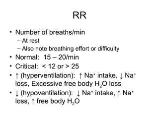
RR
• Number ofbreaths/min
– At rest
– Also note breathing effort or difficulty
• Normal: 15 – 20/min
• Critical: < 12 or > 25
• ↑ (hyperventilation): ↑ Na+
intake, ↓ Na+
loss, Excessive free body H2O loss
• ↓ (hypoventilation): ↓ Na+
intake, ↑ Na+
loss, ↑ free body H2O
7.
Bp
• Measures theforce of blood against the arterial vessel
walls
– Measured while seated, after resting for 5 mins, arm resting @
heart level (if possible)
– Reported as a fraction (systolic/diastolic) & consists of 2
separate measurements:
• Systolic – pressure within artery during cardiac contraction
• Diastolic – pressure within artery during cardiac relaxation and filling
• Normal: < 120 mm Hg systolic and < 80 mm Hg diastolic
• Critical: > 220 mm Hg systolic or > 125 mm Hg diastolic
• ↑ (hypertension [htn]): ↑ Na+
intake, ↓ Na+
loss,
Excessive free body H2O loss
• ↓ (hypotention): ↓ Na+
intake, ↑ Na+
loss, ↑ free body
H2O
8.
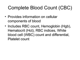
Complete Blood Count(CBC)
• Provides information on cellular
components of blood
• Includes RBC count, Hemoglobin (Hgb),
Hematocrit (Hct), RBC indices, White
blood cell (WBC) count and differential,
Platelet count
9.
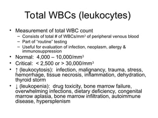
Total WBCs (leukocytes)
•Measurement of total WBC count
– Consists of total # of WBCs/mm3
of peripheral venous blood
– Part of “routine” testing
– Useful for evaluation of infection, neoplasm, allergy &
immunosuppression
• Normal: 4,000 – 10,000/mm3
• Critical: < 2,500 or > 30,000/mm3
• ↑ (leukocytosis): infection, malignancy, trauma, stress,
hemorrhage, tissue necrosis, inflammation, dehydration,
thyroid storm
• ↓ (leukopenia): drug toxicity, bone marrow failure,
overwhelming infections, dietary deficiency, congenital
marrow aplasia, bone marrow infiltration, autoimmune
disease, hypersplenism
10.
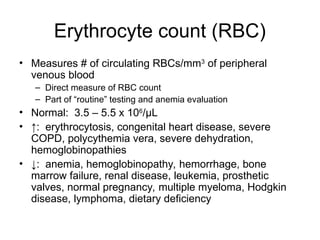
Erythrocyte count (RBC)
•Measures # of circulating RBCs/mm3
of peripheral
venous blood
– Direct measure of RBC count
– Part of “routine” testing and anemia evaluation
• Normal: 3.5 – 5.5 x 10⁶
/μL
• ↑: erythrocytosis, congenital heart disease, severe
COPD, polycythemia vera, severe dehydration,
hemoglobinopathies
• ↓: anemia, hemoglobinopathy, hemorrhage, bone
marrow failure, renal disease, leukemia, prosthetic
valves, normal pregnancy, multiple myeloma, Hodgkin
disease, lymphoma, dietary deficiency
11.
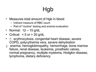
Hgb
• Measures totalamount of Hgb in blood
– Indirect measure of RBC count
– Part of “routine” testing and anemia evaluation
• Normal: 12 – 15 g/dL
• Critical: < 5 or > 20 g/dL
• ↑: erythrocytosis, congenital heart disease, severe
COPD, polycythemia vera, severe dehydration
↓: anemia, hemoglobinopathy, hemorrhage, bone marrow
failure, renal disease, leukemia, prosthetic valves,
normal pregnancy, multiple myeloma, Hodgkin disease,
lymphoma, dietary deficiency
12.
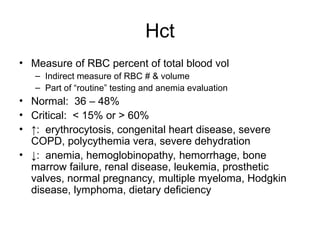
Hct
• Measure ofRBC percent of total blood vol
– Indirect measure of RBC # & volume
– Part of “routine” testing and anemia evaluation
• Normal: 36 – 48%
• Critical: < 15% or > 60%
• ↑: erythrocytosis, congenital heart disease, severe
COPD, polycythemia vera, severe dehydration
• ↓: anemia, hemoglobinopathy, hemorrhage, bone
marrow failure, renal disease, leukemia, prosthetic
valves, normal pregnancy, multiple myeloma, Hodgkin
disease, lymphoma, dietary deficiency
13.
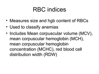
RBC indices
• Measuressize and hgb content of RBCs
• Used to classify anemias
• Includes Mean corpuscular volume (MCV),
mean corpuscular hemoglobin (MCH),
mean corpuscular hemoglobin
concentration (MCHC), red blood cell
distribution width (RDW)
14.
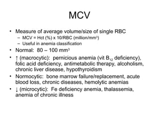
MCV
• Measure ofaverage volume/size of single RBC
– MCV = Hct (%) x 10/RBC (million/mm3
)
– Useful in anemia classification
• Normal: 80 – 100 mm3
• ↑ (macrocytic): pernicious anemia (vit B12 deficiency),
folic acid deficiency, antimetabolic therapy, alcoholism,
chronic liver disease, hypothyroidism
• Normocytic: bone marrow failure/replacement, acute
blood loss, chronic diseases, hemolytic anemias
• ↓ (microcytic): Fe deficiency anemia, thalassemia,
anemia of chronic illness
15.
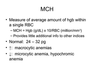
MCH
• Measure ofaverage amount of hgb within
a single RBC
– MCH = Hgb (g/dL) x 10/RBC (million/mm3
)
– Provides little additional info to other indices
• Normal: 24 – 32 pg
• ↑: macrocytic anemias
• ↓: microcytic anemia, hypochromic
anemia
16.
MCHC
• Measure ofaverage [hgb] within a single RBC
– MCHC = Hgb (g/dL) x 100/Hct (%)
– 37 g/dL = maximum Hgb able to fit into an RBC
(cannot be hyperchromic)
• Normal (normochromic): 32 – 36 g/dL
• ↑: spherocytosis, intravascular hemolysis, cold
agglutinins
• ↓ (hypochromic): Fe deficiency anemia,
thalassemia
17.
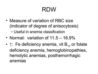
RDW
• Measure ofvariation of RBC size
(indicator of degree of anisocytosis)
– Useful in anemia classification
• Normal: variation of 11.5 – 16.9%
• ↑: Fe deficiency anemia, vit B12 or folate
deficiency anemia, hemoglobinopathies,
hemolytic anemias, posthemorrhagic
anemias
18.
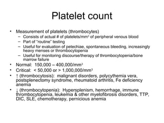
Platelet count
• Measurementof platelets (thrombocytes)
– Consists of actual # of platelets/mm3
of peripheral venous blood
– Part of “routine” testing
– Useful for evaluation of petechiae, spontaneous bleeding, increasingly
heavy menses or thrombocytopenia
– Useful for monitoring discourse/therapy of thrombocytopenia/bone
marrow failure
• Normal: 150,000 – 400,000/mm3
• Critical: < 50,000 or > 1,000,000/mm3
• ↑ (thrombocytosis): malignant disorders, polycythemia vera,
postsplenectomy syndrome, rheumatoid arthritis, Fe deficiency
anemia
• ↓ (thrombocytopenia): Hypersplenism, hemorrhage, immune
thrombocytopenia, leukemia & other myelofibrosis disorders, TTP,
DIC, SLE, chemotherapy, pernicious anemia
19.
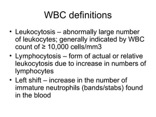
WBC definitions
• Leukocytosis– abnormally large number
of leukocytes; generally indicated by WBC
count of ≥ 10,000 cells/mm3
• Lymphocytosis – form of actual or relative
leukocytosis due to increase in numbers of
lymphocytes
• Left shift – increase in the number of
immature neutrophils (bands/stabs) found
in the blood
20.
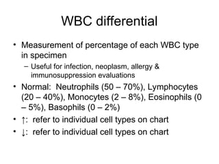
WBC differential
• Measurementof percentage of each WBC type
in specimen
– Useful for infection, neoplasm, allergy &
immunosuppression evaluations
• Normal: Neutrophils (50 – 70%), Lymphocytes
(20 – 40%), Monocytes (2 – 8%), Eosinophils (0
– 5%), Basophils (0 – 2%)
• ↑: refer to individual cell types on chart
• ↓: refer to individual cell types on chart
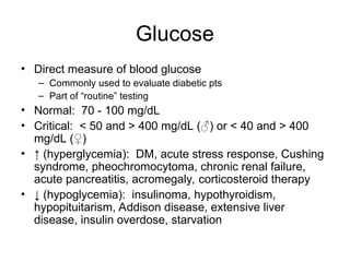
Glucose
• Direct measureof blood glucose
– Commonly used to evaluate diabetic pts
– Part of “routine” testing
• Normal: 70 - 100 mg/dL
• Critical: < 50 and > 400 mg/dL (♂) or < 40 and > 400
mg/dL (♀)
• ↑ (hyperglycemia): DM, acute stress response, Cushing
syndrome, pheochromocytoma, chronic renal failure,
acute pancreatitis, acromegaly, corticosteroid therapy
• ↓ (hypoglycemia): insulinoma, hypothyroidism,
hypopituitarism, Addison disease, extensive liver
disease, insulin overdose, starvation
23.
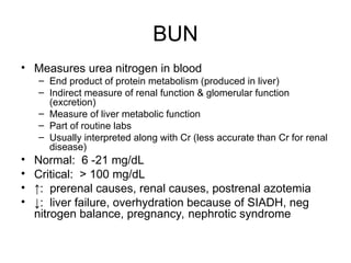
BUN
• Measures ureanitrogen in blood
– End product of protein metabolism (produced in liver)
– Indirect measure of renal function & glomerular function
(excretion)
– Measure of liver metabolic function
– Part of routine labs
– Usually interpreted along with Cr (less accurate than Cr for renal
disease)
• Normal: 6 -21 mg/dL
• Critical: > 100 mg/dL
• ↑: prerenal causes, renal causes, postrenal azotemia
• ↓: liver failure, overhydration because of SIADH, neg
nitrogen balance, pregnancy, nephrotic syndrome
24.
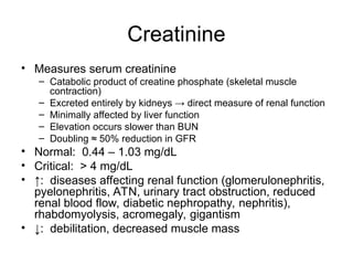
Creatinine
• Measures serumcreatinine
– Catabolic product of creatine phosphate (skeletal muscle
contraction)
– Excreted entirely by kidneys → direct measure of renal function
– Minimally affected by liver function
– Elevation occurs slower than BUN
– Doubling ≈ 50% reduction in GFR
• Normal: 0.44 – 1.03 mg/dL
• Critical: > 4 mg/dL
• ↑: diseases affecting renal function (glomerulonephritis,
pyelonephritis, ATN, urinary tract obstruction, reduced
renal blood flow, diabetic nephropathy, nephritis),
rhabdomyolysis, acromegaly, gigantism
• ↓: debilitation, decreased muscle mass
25.
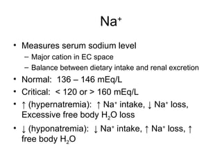
Na+
• Measures serumsodium level
– Major cation in EC space
– Balance between dietary intake and renal excretion
• Normal: 136 – 146 mEq/L
• Critical: < 120 or > 160 mEq/L
• ↑ (hypernatremia): ↑ Na+
intake, ↓ Na+
loss,
Excessive free body H2O loss
• ↓ (hyponatremia): ↓ Na+
intake, ↑ Na+
loss, ↑
free body H2O
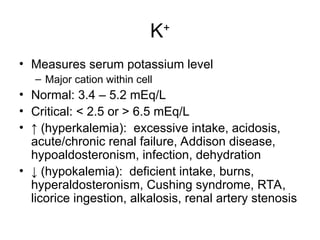
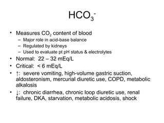
HCO3
-
• Measures CO2content of blood
– Major role in acid-base balance
– Regulated by kidneys
– Used to evaluate pt pH status & electrolytes
• Normal: 22 – 32 mEq/L
• Critical: < 6 mEq/L
• ↑: severe vomiting, high-volume gastric suction,
aldosteronism, mercurial diuretic use, COPD, metabolic
alkalosis
• ↓: chronic diarrhea, chronic loop diuretic use, renal
failure, DKA, starvation, metabolic acidosis, shock
29.
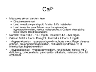
Ca2+
• Measures serumcalcium level
– Direct measurement
– Used to evaluate parathyroid function & Ca metabolism
– Used to monitor renal failure, renal transplantation,
hyperparathyroidism, various malignancies, & Ca level when giving
large-volume blood transfusions
• Normal: Total = 8.3 – 10.3 mg/dL, Ionized = 4.5 – 5.6 mg/dL
• Critical: Total < 6 or > 13 mg/dL, Ionized < 2.2 or > 7 mg/dL
• ↑ (hypercalcemia): hyperparathyroidism, bone mets, Paget disease
of bone, prolonged immobilization, milk-alkali syndrome, vit D
intoxication, hyperthyroidism
• ↓ (hypocalcemia): hypoparathyroidism, renal failure, rickets, vit D
deficiency, osteomalacia, pancreatitis, alkalosis, malabsorption, fat
embolism
30.
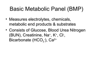
Comprehensive Metabolic Panel
(CMP)
•Includes all components of BMP plus
Albumin, Total protein, Alkaline
phosphatase (ALP), Alanine
aminotransferase (ALT), Aspartate
aminotransferase (AST) and Bilirubin
31.
Albumin
• Measures amountof albumin in blood
– Formed within liver & comprises 60% of total protein in blood
– Maintains colloidal osmotic pressure & transports blood
constituents
– Measure of both hepatic function and nutritional state
• Normal: 3.5 – 5 g/dL
• ↑: dehydration
• ↓: malnutrition, pregnancy, liver disease, protein-losing
enteropathies, protein-losing nephropathies, 3rd
space
losses, overhydration, ↑ capillary permeability,
inflammatory disease, familial idiopathic dysproteinemia
32.
Total Protein
• Measurestotal protein in blood
– Combination of prealbumin, albumin &
globulins
• Normal: 6.4 – 8.3 g/dL
33.
ALP
• Measures serumALP concentration
– Detect & monitor liver and bone disease
• Normal: 30 -120 units/L
• ↑: 1° cirrhosis, intrahepatic/extrahepatic biliary
obstruction, 1°/metastic liver tumor,
hyperparathyroidism, Paget disease, normal
growing bones in children, bone mets, RA, MI,
sarcoidosis, healing fracture, normal pregnancy,
intestinal ischemia or infarction
• ↓: hypophosphatemia, malnutrition, milk-alkali
syndrome, pernicious anemia, scurvy
34.
ALT
• Found predominantlyin liver
– Injury/disease to parenchyma → release into blood
– ID & monitor hepatocellular diseases of liver
– If jaundiced, implicates liver rather than RBC hemolysis
• Normal: 4 – 36 international units/L @ 37°C
• Sig ↑: hepatitis, hepatic necrosis, hepatic ischemia
• Mod ↑: cirrhosis, cholestasis, hepatic tumor, hepatotoxic
drugs, obstructive jaundice, severe burns, trauma to
striated muscle
• Mild ↑: myositis, pancreatitis, MI, infectious mono, shock
35.
AST
• Found inhighly metabolic tissue (cardiac &
skeletal muscle, liver cells)
– Disease/injury → lysing of cells & release into blood
– Elevation proportional to # of cells injured
– Used for evaluation of suspected coronary artery
disease or hepatocellular disease
• Normal: 0 – 35 units/L
• ↑: heart diseases, liver diseases, skeletal
muscle diseases
• ↓: acute renal disease, beriberi, DKA,
pregnancy, chronic renal dialysis
36.
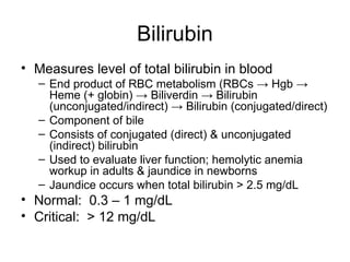
Bilirubin
• Measures levelof total bilirubin in blood
– End product of RBC metabolism (RBCs → Hgb →
Heme (+ globin) → Biliverdin → Bilirubin
(unconjugated/indirect) → Bilirubin (conjugated/direct)
– Component of bile
– Consists of conjugated (direct) & unconjugated
(indirect) bilirubin
– Used to evaluate liver function; hemolytic anemia
workup in adults & jaundice in newborns
– Jaundice occurs when total bilirubin > 2.5 mg/dL
• Normal: 0.3 – 1 mg/dL
• Critical: > 12 mg/dL
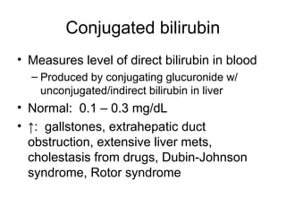
Conjugated bilirubin
• Measureslevel of direct bilirubin in blood
– Produced by conjugating glucuronide w/
unconjugated/indirect bilirubin in liver
• Normal: 0.1 – 0.3 mg/dL
• ↑: gallstones, extrahepatic duct
obstruction, extensive liver mets,
cholestasis from drugs, Dubin-Johnson
syndrome, Rotor syndrome
39.
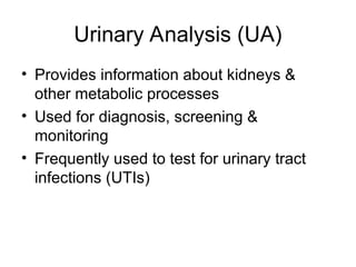
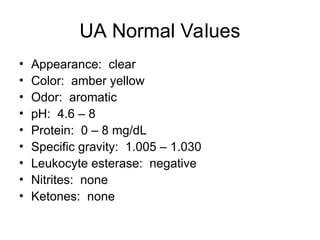
Urinary Analysis (UA)
•Provides information about kidneys &
other metabolic processes
• Used for diagnosis, screening &
monitoring
• Frequently used to test for urinary tract
infections (UTIs)
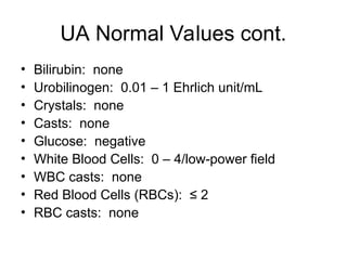
UA Normal Valuescont.
• Bilirubin: none
• Urobilinogen: 0.01 – 1 Ehrlich unit/mL
• Crystals: none
• Casts: none
• Glucose: negative
• White Blood Cells: 0 – 4/low-power field
• WBC casts: none
• Red Blood Cells (RBCs): ≤ 2
• RBC casts: none
42.
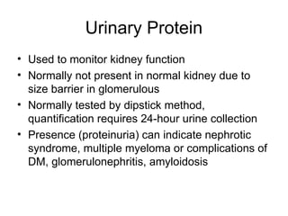
Urinary Protein
• Usedto monitor kidney function
• Normally not present in normal kidney due to
size barrier in glomerulous
• Normally tested by dipstick method,
quantification requires 24-hour urine collection
• Presence (proteinuria) can indicate nephrotic
syndrome, multiple myeloma or complications of
DM, glomerulonephritis, amyloidosis
43.
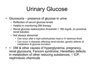
Urinary Glucose
• Glucosuria– presence of glucose in urine
– Reflection of serum glucose levels
– Helpful in monitoring DM therapy
– Renal glucose reabsorption threshold = 180 mg/dL (in proximal
renal tubules)
– Not always abnormal
• Can occur after a high-carbohydrate meal or IV dextrose fluids
• Can occur in diseases affecting renal tubules; genetic defects of
metabolism & glucose excretion
• ↑: DM & other causes of hyperglycemia, pregnancy,
renal glycosuria, Fanconi syndrome, Hereditary defects
in metabolism of other reducing substances, ↑ ICP,
nephrotoxic chemicals
44.
Urinary Leukocyte esterase
•Screen to detect leukocytes in urine
(dipstick method)
• Presence indicates UTI
• 90% accurate
45.
Urinary Ketones
• Endproducts of fatty acid catabolism
• Examples: β-hydroxybutyric acid,
acetoacetic acid, acetone
• Associated with poorly controlled diabetes
• Used to evaluate ketoacidosis associated
w/ alcoholism, fasting, starvation, high-
protein diets, isopropanol ingestion
46.
Urinary Nitrites
• Screenfor UTI (dipstick method)
• Test based on chemical rxn by bacterial
reductase (reduces nitrate to nitrite)
• 50% accurate
• Enhances leukocyte esterase sensitivity
47.
Urinary Casts
• Hyaline– conglomerations of protein; indicative
of proteinuria; few = normal especially after
exercise
• Cellular – conglomerations of degenerated cells
– Granular – glomerular disease
– Fatty – nephrotic syndrome
– Waxy – chronic renal disease
– Epithelial cells & casts (renal tubular casts)
– WBCs & casts – acute pyelonephritis
– RBCs & casts – glomerular diseases
48.
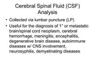
Cerebral Spinal Fluid(CSF)
Analysis
• Collected via lumbar puncture (LP)
• Useful for the diagnosis of 1° or metastatic
brain/spinal cord neoplasm, cerebral
hemorrhage, meningitis, encephalitis,
degenerative brain disease, autoimmune
diseases w/ CNS involvement,
neurosyphilis, demyelinating diseases
References
• Pagana, K.D.& Pagna, T.J. (2006). Mosby’s Manual of
Diagnostic and Laboratory Tests. St. Louis: Mosby
Elsevier.
• 27th
edition (2000). Stedman’s Medical Dictionary.
Baltimore: Lippincott Williams & Wilkins.
• UpToDate. Retrieved July 26, 2009, from
http://www.uptodateonline.com
• Urinalysis. Retrieved July 17, 2009, from
http://library.med.utah.edu/WebPath/TUTORIAL/URINE/
URINE.html
• Vital Signs. Retrieved July 17, 2009, from
http://www.healthsystem.virginia.edu/uvahealth/adult_no
ntrauma/vital.cfm
57.
Additional Resources
• Corbett,J.V. (2008). Laboratory Tests and Diagnostic Procedures
with Nursing Diagnoses 7th
Edition. Upper Saddle River: Prentice
Hall.
• Fischbach, F.T. & Dunning, M.B. (2008). A Manual of Laboratory &
Diagnostic Tests 8th
Edition. Philadelphia: Lippincott Williams &
Wilkins.
• Jacobs, D.S., De Mott, W.R. & Oxley, D.K. (2001). Jacobs & DeMott
Laboratory Test Handbook with Key Word Index 5th
Edition. Hudson:
Lexi Comp, Inc.
• Wu, A. (2006). Tietz Clinical Guide to Laboratory Tests 4th
Edition.
St. Louis: Saunders Elsevier.
• Young, R.H. & Hicks, J. (2002). Directory of Rare Analyses 2000-
2002. St. Louis: AACC Press.
• http://www.labtestsonline.org/
58.
Special Thanks
• Dr.Amira F. Gohara, M.D.
• Dr. Carol Bennett-Clarke, Ph.D.
• Dr. Constance Shriner, Ph.D.
• Cynthia R. O’Connell, BSMT (ASCP)

![Temp
• Measure of body’s core temp (temp of
internal organs)
– in ° F (or ° C)
– Locations: oral, rectum, axilla, ear
– Rectal = 0.5 – 0.7° F higher than oral temp
– Axilla = 0.3 – 0.4° F lower than oral temp
• Normal: 97.8 – 99° F (36.5 – 37.2° C)
• Critical: > 98.6° F orally or 99.8° F rectally
(pyrexia [fever]); < 95° F (hypothermia)](https://image.slidesharecdn.com/introductiontolaboratorytestshandoutversion-250218211748-f5f7f3ca/85/Introduction-to-Laboratory-Tests-Handout-Version-ppt-4-320.jpg)
![Bp
• Measures the force of blood against the arterial vessel
walls
– Measured while seated, after resting for 5 mins, arm resting @
heart level (if possible)
– Reported as a fraction (systolic/diastolic) & consists of 2
separate measurements:
• Systolic – pressure within artery during cardiac contraction
• Diastolic – pressure within artery during cardiac relaxation and filling
• Normal: < 120 mm Hg systolic and < 80 mm Hg diastolic
• Critical: > 220 mm Hg systolic or > 125 mm Hg diastolic
• ↑ (hypertension [htn]): ↑ Na+
intake, ↓ Na+
loss,
Excessive free body H2O loss
• ↓ (hypotention): ↓ Na+
intake, ↑ Na+
loss, ↑ free body
H2O](https://image.slidesharecdn.com/introductiontolaboratorytestshandoutversion-250218211748-f5f7f3ca/85/Introduction-to-Laboratory-Tests-Handout-Version-ppt-7-320.jpg)
![MCHC
• Measure of average [hgb] within a single RBC
– MCHC = Hgb (g/dL) x 100/Hct (%)
– 37 g/dL = maximum Hgb able to fit into an RBC
(cannot be hyperchromic)
• Normal (normochromic): 32 – 36 g/dL
• ↑: spherocytosis, intravascular hemolysis, cold
agglutinins
• ↓ (hypochromic): Fe deficiency anemia,
thalassemia](https://image.slidesharecdn.com/introductiontolaboratorytestshandoutversion-250218211748-f5f7f3ca/85/Introduction-to-Laboratory-Tests-Handout-Version-ppt-16-320.jpg)


![bipin_ppt_most_important[1] - Read-Only.pptx](https://cdn.slidesharecdn.com/ss_thumbnails/bipinpptmostimportant1-read-only-251220122435-b3a2f11c-thumbnail.jpg?width=640&height=640&fit=bounds)


