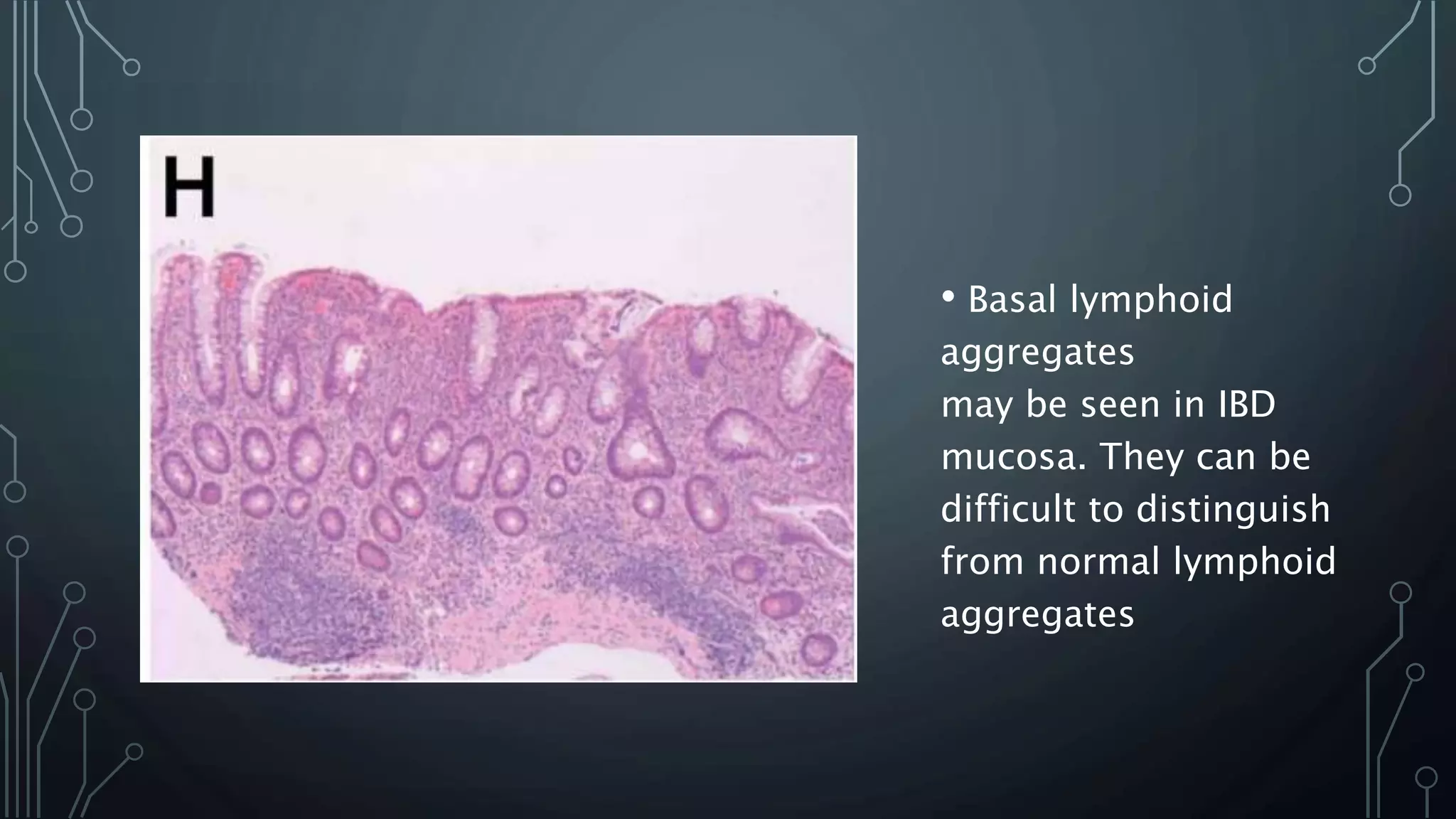
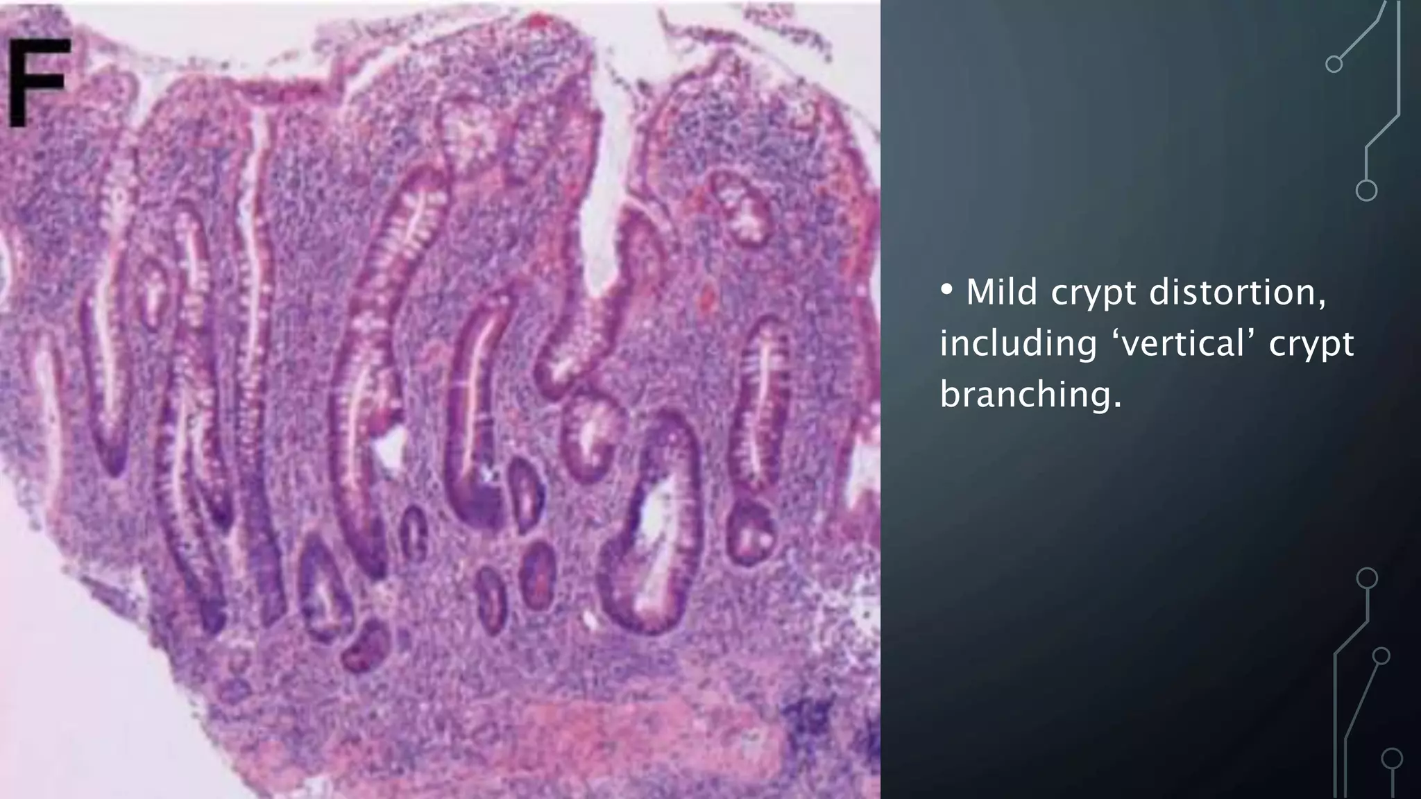
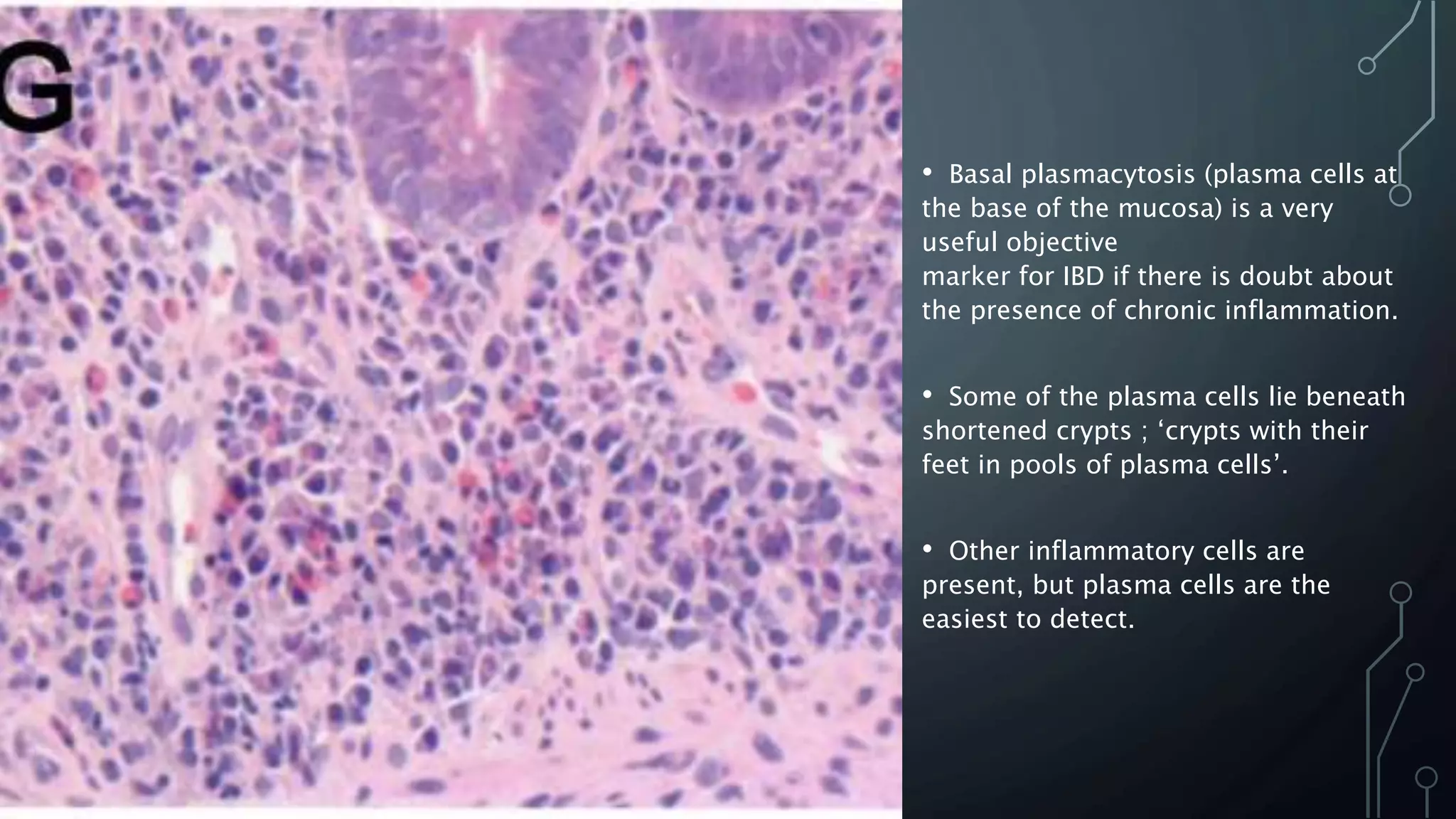
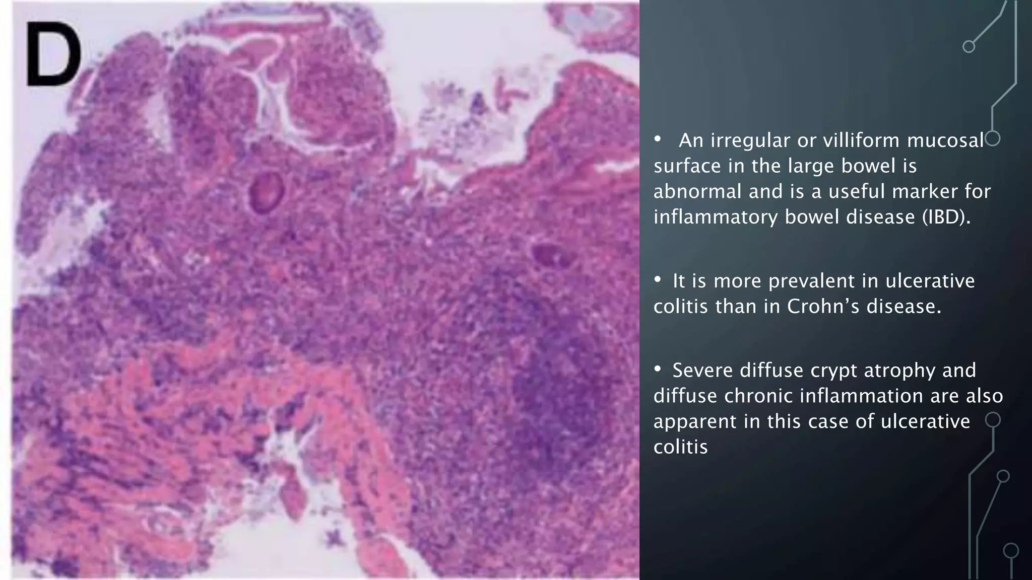
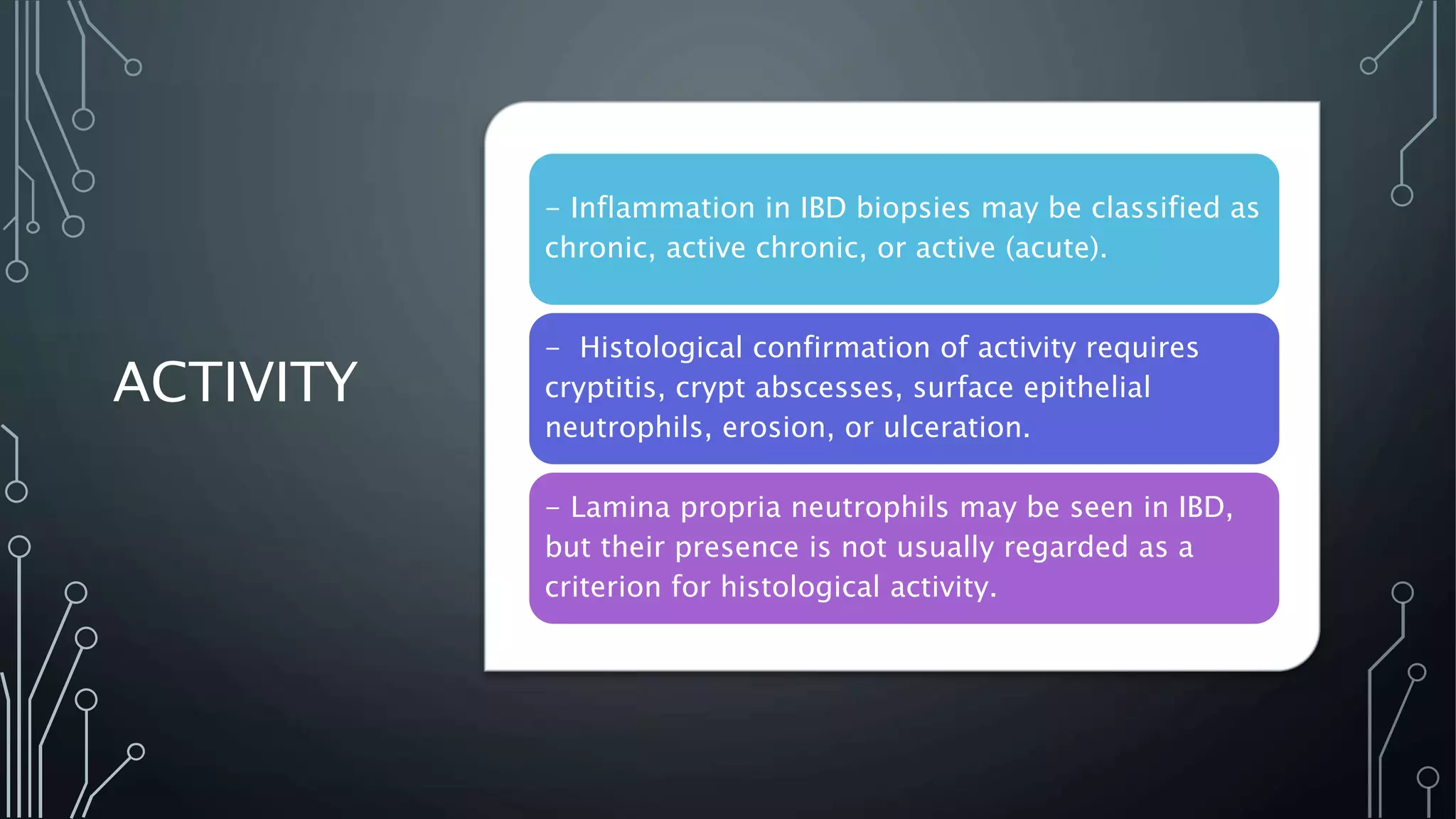
INFLAMMATO
RY BOWEL
DISEASE
PATHOLOGY
BY:
DR. MONA M. RASHED
CONSULTANT HISTOPATHOLOGIST
CLINICAL
CONSIDERATIO
NS
Reasons for taking a biopsy include:
1. Confirmation of the diagnosis of IBD
2. Distinction between UC and CD
3. Exclusion of dysplasia
4. Exclusion of coexistent conditions or complications.
5. The following may also be assessed: disease activity and
disease extent.
CLINICAL
DETAILS:
Diagnosis and classification of IBD depend on a
combination of clinical, endoscopic and
pathological features.
Several entities can mimic IBD clinically and
histologically.
Correlation of histological features with clinical and
endoscopic findings is essential.
TISSUE
SAMPLING
Biopsies from endoscopically normal
and abnormal mucosa should be
available.
For optimal diagnosis and
classification of IBD there should be
samples of the ileum, at least four
colonic sites, and the rectum.
DIAGNOSTIC CATEGORIES
The main questions asked by the histopathologist when
assessing an initial biopsy for IBD are :
• Is the mucosa inflamed?
• If the mucosa is inflamed: is it IBD or not?
• If it is IBD: is it UC, CD, or IBD unclassified?
CLINICAL
CONSIDERATIO
NS
Accurate histopathological assessment is important for
optimum diagnosis, subclassification and management of
IBD.
Biopsies for the diagnosis of IBD should be accompanied
by a full clinical history and a record of endoscopic
findings. The history should include the duration of
disease and details of medical and surgical treatment.
A regular clinicopathological meeting for discussion of
IBD cases should be available.
FEATURES OF NORMAL AND
ABNORMAL MUCOSA
NORMAL
MUCOSA
1. Chronic inflammatory cell density:
•Normally, chronic inflammatory cells are most dense in the upper
third of the mucosa. Their density decreases towards the base,
resulting in the ‘plasma cell gradient’.
•Cellularity is highest in the caecum/ascending colon and may be
low in the rectum
•Dense lymphoid tissue may be found in the normal large bowel,
particularly in the rectum
2. Histiocytes/macrophages:
•Recognition of histiocytes is difficult unless they contain pigment
or mucin more common in the rectum seen in other conditions,
such as atypical mycobacterial infection and Whipple’s disease.
Normal large bowel mucosa with a
decrease in plasma cell density from the
upper third of the lamina propria to the
lower third, the ‘plasma cell gradient’.
Basal plasma cells are infrequent or
absent.
Note also that crypt architecture is
preserved; the crypts are parallel to each
other and extend from the lumenal surface
to the muscularis mucosae.
 The caecum and ascending
colon, overall lamina propria
cell density is higher than
elsewhere.
 Basal plasma cells may be
seen at these sites (arrow)
and should not necessarily be
interpreted as chronic
inflammation.
• Basal lymphoid
aggregates
may be seen in IBD
mucosa. They can be
difficult to distinguish
from normal lymphoid
aggregates
NORMAL
MUCOSA
•The normal lamina propria contains
eosinophils.
3. Eosinophils
•A few intraepithelial neutrophils may reflect
bowel preparation presence of any
intraepithelial neutrophils is probably not
normal and should trigger a search for other
evidence of inflammation.
4. Acute inflammatory cells:
FEATURES
OF
ABNORMALI
TY IN
COLORECTA
L BIOPSIES:
1- Architectural changes:
Crypt distortion; Crypt branching subclassified as vertical and
horizontal
Crypt atrophy; crypt shortening and wider crypt spacing.
2- Inflammatory changes:
Chronic inflammation and lymphoid aggregates Basal plasmacytosis
and ‘loss of the plasma cell gradient’ are objective markers of IBD.
Granulomas and giant cells; It has been suggested that a granuloma
should include at least five epithelioid macrophages
Branched crypts.
• Crypt architectural distortion.
Crypt distortion includes crypt
branching and loss of
parallelism.
• The centrally placed crypt
shows ‘horizontal’ branching
(arrow).
• Mild crypt distortion,
including ‘vertical’ crypt
branching.
• Basal plasmacytosis (plasma cells at
the base of the mucosa) is a very
useful objective
marker for IBD if there is doubt about
the presence of chronic inflammation.
• Some of the plasma cells lie beneath
shortened crypts ; ‘crypts with their
feet in pools of plasma cells’.
• Other inflammatory cells are
present, but plasma cells are the
easiest to detect.
• An irregular or villiform mucosal
surface in the large bowel is
abnormal and is a useful marker for
inflammatory bowel disease (IBD).
• It is more prevalent in ulcerative
colitis than in Crohn’s disease.
• Severe diffuse crypt atrophy and
diffuse chronic inflammation are also
apparent in this case of ulcerative
colitis
FEATURES
OF
ABNORMALI
TY IN
COLORECTA
L BIOPSIES:
3-Epithelial changes:
 Mucin depletion, Depletion can be
graded as mild, moderate, or severe,
 Paneth cell metaplasia, only significant
if it is distal to the splenic flexure, it is
probably a marker of longstanding
disease
Paneth cell metaplasia
(arrows) with supranuclear
eosinophilic granules. This is
a case of longstanding
ulcerative colitis
CHANGES THAT MAY BE RELATED TO
BOWEL PREPARATION OR BIOPSY:
Include pseudo-lipomatosis, sparse acute inflammatory
cells, mucin depletion, damage to the surface epithelium, mild
haemorrhage, basal crypt epithelial cell apoptosis and oedema.
IBD VERSUS NON-IBD:
HISTOLOGY
IBD
VERSUS
NORMAL
Features that are more frequent
in IBD than in normal mucosa, for
example, abnormal crypt
architecture, crypt distortion, crypt
atrophy, basal plasmacytosis, lamina
propria hypercellularity, mucin
depletion, granulomas, crypt
abscesses, cryptitis and ulceration.
IBD VERSUS ACUTE INFECTIVE
COLITIS/ACUTE SELF-LIMITING
COLITIS/NON-IBD COLITIS
Acute infective-type colitis associated
symptoms, lasting for a short time (e.g., less
than 1 month) secondary to common infections
such as Campylobacter, Salmonella, and Shigella.
FEATURES FAVOURING INFECTIVE COLITIS
OVER IBD
Acute inflammatory changes, for example, cryptitis,
crypt abscesses, lamina proprial neutrophils and
oedema, mainly in the upper two-thirds of the mucosa,
with a slight increase in lamina proprial and crypt
epithelial neutrophils.
IBD VERSUS
INFECTION/NON
-IBD
The microscopic features that favor IBD over
infective colitis include basal plasmacytosis
and architectural changes.
Combinations of changes are more useful
than single features.
Correlation with the clinical picture is
essential.
• Probable infective
colitis.
• There is upper lamina
propria hypercellularity
but no basal
plasmacytosis or loss of
the plasma cell gradient.
• Cryptitis is unusually
extensive for an infective
colitis.
UC VERSUS
CD:
HISTOLOGY
The microscopic features that help distinguish UC and CD
from one another include granulomas, architectural changes
and distribution of disease.
Non-crypto lytic granulomas are more useful than any other
feature for discriminating between CD and UC.
Crypto lytic granulomas occur in many colitides and probably
have little or no discriminant value.
If granuloma formation is the sole abnormality, cautious
interpretation is advised.
Distribution of changes
within a biopsy:
 Patchy chronic
inflammation in
Crohn’s colitis.
 Note the variation in
lamina propria
cellularity and very
focal crypt distortion
• An irregular or villiform mucosal
surface in the large bowel is
abnormal and is a useful marker for
inflammatory bowel disease (IBD).
• It is more prevalent in ulcerative
colitis than in Crohn’s disease.
• Severe diffuse crypt atrophy and
diffuse chronic inflammation are also
apparent in this case of ulcerative
colitis
GRANULOMA
S
Granulomas unrelated to crypt injury (non-crypto
lytic granulomas) are very reliable for
distinguishing CD from UC However, they also
occur in TB and, rarely, in other infective colitides.
Cryptolytic granulomas occur as a response to
crypt injury crypt rupture should be excluded as a
cause of granulomas,
• A cryptolytic
granuloma resulting
from rupture of a crypt
abscess.
• Cryptolytic granulomas
are not discriminatory
ILEAL BIOPSIES IN IBD
• ▸ In the setting of IBD, ileal
inflammation strongly favours CD
over UC
• ▸ Granulomas (non-crypto lytic) in
inflamed ileal biopsies help
discriminate CD from UC
UPPER GASTROINTESTINAL
BIOPSIES IN IBD
• Upper gastrointestinal involvement by
CD is considerably more frequent than
involvement by UC.
• Other more common causes of upper
gastrointestinal inflammation, especially
gastro-oesophageal reflux and Hpylori-
associated gastritis, should be excluded
before involvement by IBD is suggested.
• In the setting of IBD, upper
gastrointestinal granulomas strongly
favour CD over UC.
• Upper gastrointestinal granulomas may
raise the possibility of new CD, but
caution is advised.
MIMICS OF
IBD
There are numerous histological mimics of IBD.
Diverticular colitis and Diversion proctocolitis may
closely resemble IBD.
A full clinical and endoscopic history is necessary
before the histological mimics of IBD can be
excluded confidently.
Diversion proctocolitis showing
diffuse chronic inflammation and
crypt distortion.
This inflammatory bowel disease
(IBD)-like pattern can be seen
whether or not there is a past
history of IBD, but tends to be
most severe in the setting of
ulcerative colitis.
Collagenous colitis.
Basal plasma cells and loss of the
usual plasma cell gradient might
raise the possibility of IBD, but the
correct diagnosis is indicated by
the presence of a thickened
subepithelial collagen band,
epithelial degenerative changes
and preserved crypt architecture.
Diverticular colitis
Showing features
reminiscent of ulcerative
colitis, including diffuse
chronic inflammation and
extensive crypt distortion.
This is a relatively
common mimic of IBD in
biopsy diagnosis
EFFECTS
OF TIME
AND
TREATMEN
T
Early untreated IBD may show few or no histological changes. There is some
correlation between the onset of each abnormality and duration of symptoms.
Basal plasmacytosis is the earliest feature. Crypt changes typically appear later.
Time and treatment may modify the histology, distribution, and extent of IBD.
Longstanding UC not infrequently shows patchiness, discontinuity and/or rectal
sparing.
Interpretation of biopsies from early or longstanding IBD should be cautious.
Modification of an established diagnosis of UC or CD should only be made if
there is convincing evidence for a change, ideally after review of previous
histology and of clinical data in a multidisciplinary setting.
REPORTING
SCHEME
FOR IBD
BIOPSIES
(PAID)
PAID’ is a suggested aide memoire for the layout of the
conclusion to an IBD biopsy report.
P = Pattern: of chronic changes: presence, distribution,
extent
A = Activity: maximum grade, location
I = Interpretation: probability of IBD and, if relevant,
probability of UC or CD
D = Dysplasia: location, severity
ACTIVITY
- Inflammation in IBD biopsies may be classified as
chronic, active chronic, or active (acute).
- Histological confirmation of activity requires
cryptitis, crypt abscesses, surface epithelial
neutrophils, erosion, or ulceration.
- Lamina propria neutrophils may be seen in IBD,
but their presence is not usually regarded as a
criterion for histological activity.
DYSPLASIA
Diagnosis and grading of dysplasia relies on
architecture and cytology.
Dysplasia is graded as low grade or high grade.
The term ‘indefinite for dysplasia’ can be used
when distinction between dysplastic and non-
dysplastic atypia is not possible.
Double reporting of cases with dysplasia or
suspected dysplasia is recommended
REFERENCE: ROGER M FEAKINS;
INFLAMMATORY BOWEL DISEASE
BIOPSIES: UPDATED
BRITISH SOCIETY OF
GASTROENTEROLOGY
REPORTING
GUIDELINES. REVIEW; JCP
ONLINE FIRST, PUBLISHED ON
SEPTEMBER 25, 2013 AS
10.1136/JCLINPATH-2013-
201885
THANK YOU
VERY MUCH 🌷🌷
🌷

INFLAMMATORY BOWEL DISEASE PATHOLOGY.pptx

  • 1.
    INFLAMMATO RY BOWEL DISEASE PATHOLOGY BY: DR. MONAM. RASHED CONSULTANT HISTOPATHOLOGIST
  • 2.
    CLINICAL CONSIDERATIO NS Reasons for takinga biopsy include: 1. Confirmation of the diagnosis of IBD 2. Distinction between UC and CD 3. Exclusion of dysplasia 4. Exclusion of coexistent conditions or complications. 5. The following may also be assessed: disease activity and disease extent.
  • 3.
    CLINICAL DETAILS: Diagnosis and classificationof IBD depend on a combination of clinical, endoscopic and pathological features. Several entities can mimic IBD clinically and histologically. Correlation of histological features with clinical and endoscopic findings is essential.
  • 4.
    TISSUE SAMPLING Biopsies from endoscopicallynormal and abnormal mucosa should be available. For optimal diagnosis and classification of IBD there should be samples of the ileum, at least four colonic sites, and the rectum.
  • 5.
    DIAGNOSTIC CATEGORIES The mainquestions asked by the histopathologist when assessing an initial biopsy for IBD are : • Is the mucosa inflamed? • If the mucosa is inflamed: is it IBD or not? • If it is IBD: is it UC, CD, or IBD unclassified?
  • 7.
    CLINICAL CONSIDERATIO NS Accurate histopathological assessmentis important for optimum diagnosis, subclassification and management of IBD. Biopsies for the diagnosis of IBD should be accompanied by a full clinical history and a record of endoscopic findings. The history should include the duration of disease and details of medical and surgical treatment. A regular clinicopathological meeting for discussion of IBD cases should be available.
  • 8.
    FEATURES OF NORMALAND ABNORMAL MUCOSA
  • 9.
    NORMAL MUCOSA 1. Chronic inflammatorycell density: •Normally, chronic inflammatory cells are most dense in the upper third of the mucosa. Their density decreases towards the base, resulting in the ‘plasma cell gradient’. •Cellularity is highest in the caecum/ascending colon and may be low in the rectum •Dense lymphoid tissue may be found in the normal large bowel, particularly in the rectum 2. Histiocytes/macrophages: •Recognition of histiocytes is difficult unless they contain pigment or mucin more common in the rectum seen in other conditions, such as atypical mycobacterial infection and Whipple’s disease.
  • 10.
    Normal large bowelmucosa with a decrease in plasma cell density from the upper third of the lamina propria to the lower third, the ‘plasma cell gradient’. Basal plasma cells are infrequent or absent. Note also that crypt architecture is preserved; the crypts are parallel to each other and extend from the lumenal surface to the muscularis mucosae.
  • 11.
     The caecumand ascending colon, overall lamina propria cell density is higher than elsewhere.  Basal plasma cells may be seen at these sites (arrow) and should not necessarily be interpreted as chronic inflammation.
  • 12.
    • Basal lymphoid aggregates maybe seen in IBD mucosa. They can be difficult to distinguish from normal lymphoid aggregates
  • 13.
    NORMAL MUCOSA •The normal laminapropria contains eosinophils. 3. Eosinophils •A few intraepithelial neutrophils may reflect bowel preparation presence of any intraepithelial neutrophils is probably not normal and should trigger a search for other evidence of inflammation. 4. Acute inflammatory cells:
  • 14.
    FEATURES OF ABNORMALI TY IN COLORECTA L BIOPSIES: 1-Architectural changes: Crypt distortion; Crypt branching subclassified as vertical and horizontal Crypt atrophy; crypt shortening and wider crypt spacing. 2- Inflammatory changes: Chronic inflammation and lymphoid aggregates Basal plasmacytosis and ‘loss of the plasma cell gradient’ are objective markers of IBD. Granulomas and giant cells; It has been suggested that a granuloma should include at least five epithelioid macrophages
  • 15.
  • 16.
    • Crypt architecturaldistortion. Crypt distortion includes crypt branching and loss of parallelism. • The centrally placed crypt shows ‘horizontal’ branching (arrow).
  • 17.
    • Mild cryptdistortion, including ‘vertical’ crypt branching.
  • 18.
    • Basal plasmacytosis(plasma cells at the base of the mucosa) is a very useful objective marker for IBD if there is doubt about the presence of chronic inflammation. • Some of the plasma cells lie beneath shortened crypts ; ‘crypts with their feet in pools of plasma cells’. • Other inflammatory cells are present, but plasma cells are the easiest to detect.
  • 20.
    • An irregularor villiform mucosal surface in the large bowel is abnormal and is a useful marker for inflammatory bowel disease (IBD). • It is more prevalent in ulcerative colitis than in Crohn’s disease. • Severe diffuse crypt atrophy and diffuse chronic inflammation are also apparent in this case of ulcerative colitis
  • 21.
    FEATURES OF ABNORMALI TY IN COLORECTA L BIOPSIES: 3-Epithelialchanges:  Mucin depletion, Depletion can be graded as mild, moderate, or severe,  Paneth cell metaplasia, only significant if it is distal to the splenic flexure, it is probably a marker of longstanding disease
  • 22.
    Paneth cell metaplasia (arrows)with supranuclear eosinophilic granules. This is a case of longstanding ulcerative colitis
  • 23.
    CHANGES THAT MAYBE RELATED TO BOWEL PREPARATION OR BIOPSY: Include pseudo-lipomatosis, sparse acute inflammatory cells, mucin depletion, damage to the surface epithelium, mild haemorrhage, basal crypt epithelial cell apoptosis and oedema.
  • 24.
  • 25.
    IBD VERSUS NORMAL Features that aremore frequent in IBD than in normal mucosa, for example, abnormal crypt architecture, crypt distortion, crypt atrophy, basal plasmacytosis, lamina propria hypercellularity, mucin depletion, granulomas, crypt abscesses, cryptitis and ulceration.
  • 26.
    IBD VERSUS ACUTEINFECTIVE COLITIS/ACUTE SELF-LIMITING COLITIS/NON-IBD COLITIS Acute infective-type colitis associated symptoms, lasting for a short time (e.g., less than 1 month) secondary to common infections such as Campylobacter, Salmonella, and Shigella.
  • 27.
    FEATURES FAVOURING INFECTIVECOLITIS OVER IBD Acute inflammatory changes, for example, cryptitis, crypt abscesses, lamina proprial neutrophils and oedema, mainly in the upper two-thirds of the mucosa, with a slight increase in lamina proprial and crypt epithelial neutrophils.
  • 28.
    IBD VERSUS INFECTION/NON -IBD The microscopicfeatures that favor IBD over infective colitis include basal plasmacytosis and architectural changes. Combinations of changes are more useful than single features. Correlation with the clinical picture is essential.
  • 29.
    • Probable infective colitis. •There is upper lamina propria hypercellularity but no basal plasmacytosis or loss of the plasma cell gradient. • Cryptitis is unusually extensive for an infective colitis.
  • 30.
    UC VERSUS CD: HISTOLOGY The microscopicfeatures that help distinguish UC and CD from one another include granulomas, architectural changes and distribution of disease. Non-crypto lytic granulomas are more useful than any other feature for discriminating between CD and UC. Crypto lytic granulomas occur in many colitides and probably have little or no discriminant value. If granuloma formation is the sole abnormality, cautious interpretation is advised.
  • 32.
    Distribution of changes withina biopsy:  Patchy chronic inflammation in Crohn’s colitis.  Note the variation in lamina propria cellularity and very focal crypt distortion
  • 33.
    • An irregularor villiform mucosal surface in the large bowel is abnormal and is a useful marker for inflammatory bowel disease (IBD). • It is more prevalent in ulcerative colitis than in Crohn’s disease. • Severe diffuse crypt atrophy and diffuse chronic inflammation are also apparent in this case of ulcerative colitis
  • 34.
    GRANULOMA S Granulomas unrelated tocrypt injury (non-crypto lytic granulomas) are very reliable for distinguishing CD from UC However, they also occur in TB and, rarely, in other infective colitides. Cryptolytic granulomas occur as a response to crypt injury crypt rupture should be excluded as a cause of granulomas,
  • 35.
    • A cryptolytic granulomaresulting from rupture of a crypt abscess. • Cryptolytic granulomas are not discriminatory
  • 37.
    ILEAL BIOPSIES INIBD • ▸ In the setting of IBD, ileal inflammation strongly favours CD over UC • ▸ Granulomas (non-crypto lytic) in inflamed ileal biopsies help discriminate CD from UC UPPER GASTROINTESTINAL BIOPSIES IN IBD • Upper gastrointestinal involvement by CD is considerably more frequent than involvement by UC. • Other more common causes of upper gastrointestinal inflammation, especially gastro-oesophageal reflux and Hpylori- associated gastritis, should be excluded before involvement by IBD is suggested. • In the setting of IBD, upper gastrointestinal granulomas strongly favour CD over UC. • Upper gastrointestinal granulomas may raise the possibility of new CD, but caution is advised.
  • 38.
    MIMICS OF IBD There arenumerous histological mimics of IBD. Diverticular colitis and Diversion proctocolitis may closely resemble IBD. A full clinical and endoscopic history is necessary before the histological mimics of IBD can be excluded confidently.
  • 39.
    Diversion proctocolitis showing diffusechronic inflammation and crypt distortion. This inflammatory bowel disease (IBD)-like pattern can be seen whether or not there is a past history of IBD, but tends to be most severe in the setting of ulcerative colitis.
  • 40.
    Collagenous colitis. Basal plasmacells and loss of the usual plasma cell gradient might raise the possibility of IBD, but the correct diagnosis is indicated by the presence of a thickened subepithelial collagen band, epithelial degenerative changes and preserved crypt architecture.
  • 41.
    Diverticular colitis Showing features reminiscentof ulcerative colitis, including diffuse chronic inflammation and extensive crypt distortion. This is a relatively common mimic of IBD in biopsy diagnosis
  • 42.
    EFFECTS OF TIME AND TREATMEN T Early untreatedIBD may show few or no histological changes. There is some correlation between the onset of each abnormality and duration of symptoms. Basal plasmacytosis is the earliest feature. Crypt changes typically appear later. Time and treatment may modify the histology, distribution, and extent of IBD. Longstanding UC not infrequently shows patchiness, discontinuity and/or rectal sparing. Interpretation of biopsies from early or longstanding IBD should be cautious. Modification of an established diagnosis of UC or CD should only be made if there is convincing evidence for a change, ideally after review of previous histology and of clinical data in a multidisciplinary setting.
  • 43.
    REPORTING SCHEME FOR IBD BIOPSIES (PAID) PAID’ isa suggested aide memoire for the layout of the conclusion to an IBD biopsy report. P = Pattern: of chronic changes: presence, distribution, extent A = Activity: maximum grade, location I = Interpretation: probability of IBD and, if relevant, probability of UC or CD D = Dysplasia: location, severity
  • 44.
    ACTIVITY - Inflammation inIBD biopsies may be classified as chronic, active chronic, or active (acute). - Histological confirmation of activity requires cryptitis, crypt abscesses, surface epithelial neutrophils, erosion, or ulceration. - Lamina propria neutrophils may be seen in IBD, but their presence is not usually regarded as a criterion for histological activity.
  • 45.
    DYSPLASIA Diagnosis and gradingof dysplasia relies on architecture and cytology. Dysplasia is graded as low grade or high grade. The term ‘indefinite for dysplasia’ can be used when distinction between dysplastic and non- dysplastic atypia is not possible. Double reporting of cases with dysplasia or suspected dysplasia is recommended
  • 46.
    REFERENCE: ROGER MFEAKINS; INFLAMMATORY BOWEL DISEASE BIOPSIES: UPDATED BRITISH SOCIETY OF GASTROENTEROLOGY REPORTING GUIDELINES. REVIEW; JCP ONLINE FIRST, PUBLISHED ON SEPTEMBER 25, 2013 AS 10.1136/JCLINPATH-2013- 201885
  • 47.
    THANK YOU VERY MUCH🌷🌷 🌷