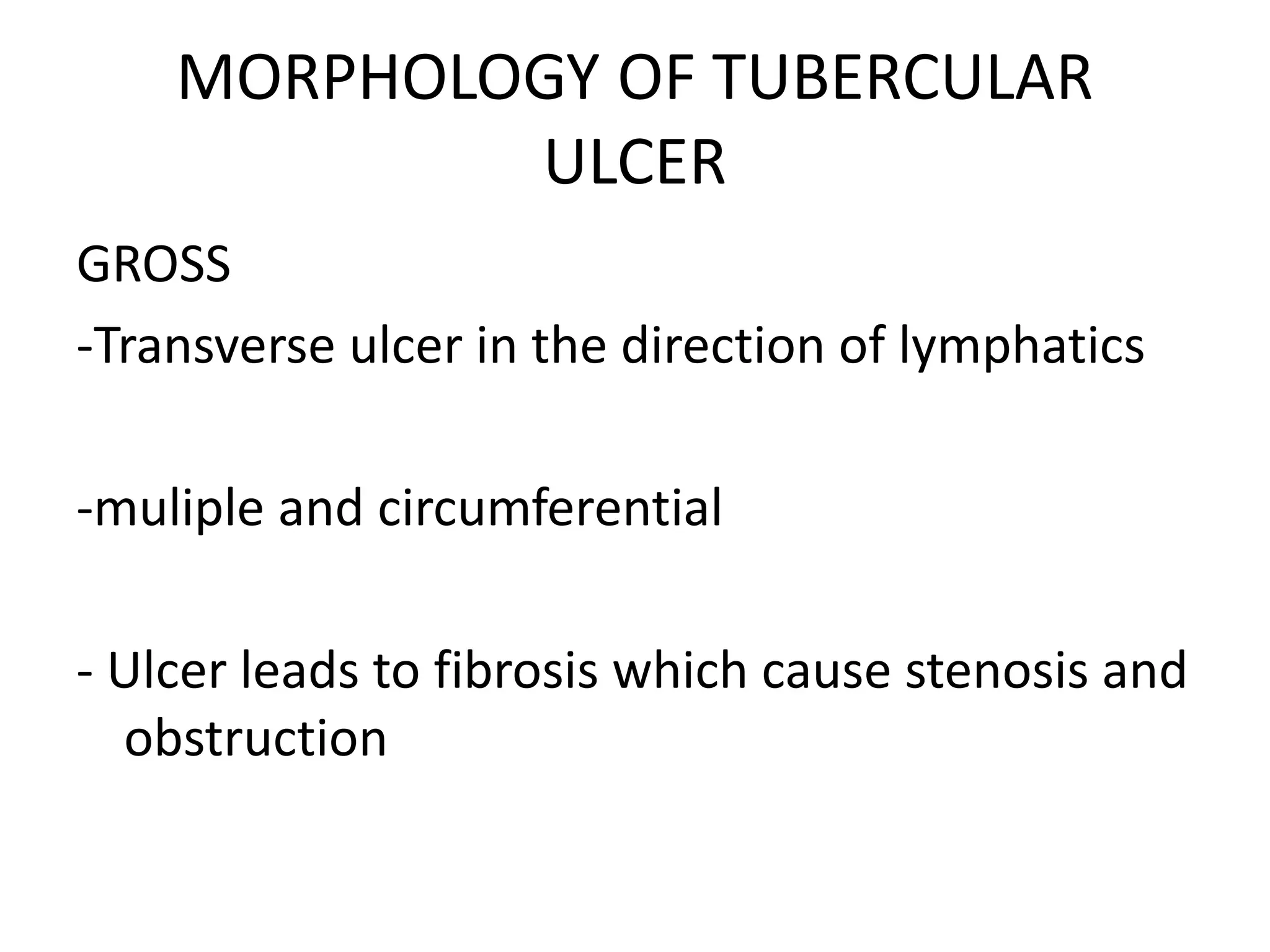
ULCERATIVE LESIONS OF
INTESTINE
Dr. Roopa Urs
Assistant professor
Histology of duodenum
Histology of jejunum
Histology of ileum
Payer’s patches
Electron microscopy- villi
Colonic mucosa
What are the ulcerative lesions?
• Duodenal Ulcer
• Amoebic ulcer
• Typhoid ulcer
• Tuberculous ulcer
• Bacillary dysentry
• Viral
-CMV
-AIDS
• IBD
-Ulcerative colitis
-Crohns disease
• Carcinoma
• Others
GVHD- Graft Verses Host Disease
Hyperplastic tuberculosis
Behcet’s syndrome
Duodenal Peptic Ulcers
• Young men
• Causes- H.pylori
Stress, spicy food
NSAID- aspirin
Cigarette smoking
Heavy drinking
Chemotherapy/Radiation therapy
• Symptoms- Burning sensation, bloating, hunger,
• Pathogenesis
Alteration in the balance b/w acid production
and mucosal defence barrier resulting in
damage to the lining epithelium.
GROSS
Chronic duodenal ulcer
Proximal duodenum
 few centimeters of
pyloric valve n involve
anterior duodenal valve
Solitary
<0.3cm
Shallow
0.6cm- deeper ulcers
Round, oval, sharply
punched out defect
Mucosal margin may
overhang the base slightly
Base is smooth and
clean as a result of peptic
digestion of exudate
Blood vessels+
MICROSCOPY
• Active ulcers the base may have thin layer of
fibrinoid debris underlaid by neutrophilic
inflammatory infiltrate.
• Beneath this active granulation tissue infiltrated
with mononuclear leucocyte and a fibrous or
collagenous scar forms the ulcer base
• Vessel wall thickened, occasionally thrombosed
• Scarring – entire thickness, pucker surrounding
mucosa
MICROSCOPY
Low-power cross-section view of a duodenal
ulcer crater with an acute inflammatory
exudate in the base
Complications
Bleeding
Perforation
Cancer
AMOEBIC ULCER
• Amoebiasis is caused by Entamoeba histolytica
(ameba)
• Dysentery-causing protozoan parasite spread by fecal-
oral transmission.
• Pathogenesis.
-E. histolytica cysts, which have a chitin wall and
four nuclei, are the infectious form because they are
resistant to gastric acid.
-Cysts release trophozoites, the ameboid forms,
which reproduce under anaerobic conditions without
harming the host.
Morphology of amoebic ulcer
• Cecum and ascending colon, followed by the
sigmoid colon, rectum, and appendix.
Discrete areas of ulceration
covered by exudate, with
normal intervening mucosa
• MICROSCOPY- Ulcer shows inflammatory infiltrates and
granulation tissues.
• Trophozoites of amoebae can mimic the appearance of
macrophages because of their comparable size and large
number of vacuoles.
• Amoebic ulcer-fan out laterally to create a flask-shaped ulcer
with a narrow neck and broad base.
• Erythrophagocytosis.
• Special stains:
- PAS- positive.
- Iron Hematoxylin -positive.
Entamoeba histolytica
COMPLICATIONS:
• Parasites may penetrate splanchnic vessels and
embolize to the liver to produce amoebic liver
abcesses.
• Amebic liver abscesses, which can exceed 10 cm
in diameter, have a scant inflammatory reaction
at their margins and a shaggy fibrin lining.
• Amebae may also spread via the bloodstream
into the kidneys and brain.
SALMONELLOSIS- Typhoid ulcer
Causative agent- S. typhimureum.
S. paratyphi.
• PATHOGENESIS:
Salmonella possess virulence genes that
encode a type III secretion system capable of
transferring bacterial proteins into enterocytes.
The transferred proteins activate host cell
Rho GTPases, bacterial uptake that allow bacterial
growth within phagosomes.
In addition, flagellin, the core protein of
bacterial flagellae, activates TLR5 on host cells and
increases the local inflammatory response
GROSS
• Usually affects ileum and colon.
• It present as longitudinal ulcer over the peyers
patches.
Typhoid ulcer
MICROSCOPY
- Neutrophils accumulate within the superficial
LP & macrophages containing bacteria, RBCs,
nuclear debris mix with lymphocytes n plasma
cells in LP.
- Mucosal shedding create oval longitudinal
ulcers
- Blunting of villi
- Mucosal congestion and edema
CLINICAL FEATURES
Anorexia, abdominal pain, bloating, nausea,
vomiting, and bloody diarrhea.
Blood cultures are positive in >90% of affected
individuals during the febrile phase.
In chronic phase- Rose spots, small
erythematous maculopapular lesions, are seen
on the chest and abdomen.
• Extra-intestinal complications including
encephalopathy, meningitis, seizures,
endocarditis, myocarditis, pneumonia,
cholecystitis and Salmonella osteomyelitis.
TUBERCULAR ULCER
• Causative agent- Mycobacterium tuberculosis
• Mostly affects the ileum or ileocaecal junction
• May or may not be associated with TB elsewhere
• Intestinal tuberculosis contracted by the drinking
of contaminated milk.
• In countries where milk is pasteurized, intestinal
TB is caused by the swallowing of coughed-up
infective material in patients with advanced
pulmonary disease
MORPHOLOGY OF TUBERCULAR
ULCER
GROSS
-Transverse ulcer in the direction of lymphatics
-muliple and circumferential
- Ulcer leads to fibrosis which cause stenosis and
obstruction
MICROSCOPY
• CASEATING GRANULOMA
- Shows central caseous necrosis
surrounded by granuloma
- Granuloma composed of epitheloid cells,
langhans giant cells and lymphocytes.
- AFB/ ZN Stain positivity.
TB- microscopy
BACILLARY DYSENTRY-
Clostridium toxin
• It affects colon
• CAP LESION- Small yellow white plaque like
lesions
• PSUEDOMEMBRANE FORMATION- fibrino
purulent necrotic debris
GROSS
MICROSCOPY
Epithelium- denuded
Superficial LP- dense
neutrophilic infiltrate,
occasional fibrin thrombi
within capillaries
Damaged crypts are
distended by mucopurulent
exudate forming
Mushroom/ Volcano
fibrinous exudates.
BACILLARY DYSENTRY-
Campylobacter jejuni
• It affects jejunum to ileum.
• Muiltiple superficial ulcer over the mucosa
• Microscopy -Cryptitis
-Crypt abscess
-Congestion
-Ulceration
BACILLARY DYSENTRY-
Campylobacter jejuni
Crypt abscess
ULCERATIVE COLITIS
• It is a inflammatory bowel disease.
• It usually affects the rectosigmoid junction.
• Gross:
- Irregular multiple ulcer
-Multiple pseudopolyp
-Continuos lesion with retrograde
progression
UC- Inflammatory polyp
• Microscopy :
-Mucosal edema and hyperemia.
-Crypt distortion
-Crypt abscess
-Mucosal depletion
UC- crypt abscess
CROHN’S DISEASE
• It is another inflammatory bowel disease.
• Affects ileum and proximal colon.
• Gross:
-Long serpentine linear ulcer.
-Cobblestone appearance.
-Skip lesions.
-Fissure and fistula.
• Microscopy shows non-caseating granuloma.
CROHNS DISEASE- Non caseating
granuloma
CARCINOMATOUS ULCER
• Commonly affects the rectosigmoid area and
affects any part of colon.
• Gross:
- Single, firm, everted, large cauliflower
shaped ulcer.
• Microscopy:
-Shows malignant glands infiltrating to
deep muscle area.
Carcinoma- gross
Carcinoma- microscopy
GRAFT VERSUS HOST DISEASE
• Usually occurs after bone marrow
transplantation and rarely occurs following
blood transfusion
• Symptoms include fever, skin rash, hepatitis,
bone marrow suppression and the outcome is
fatal.
• Pre-transfusion irradiation of all blood
components containing lymphocytes will
prevent GVHD
GVHD-Graft Verses Host Disease
• Any where in gastrointestinal tract.
• Features :
-Crypt abscesses.
-Inflammatory polyp.
-Fibrosis.
-Chronic inflammation.
BEHCET’S SYNDROME
• Affects large bowel.
• Shows multiple ulcers of various sizes, shape and
depth.
• Microscopy shows lymphocytic vasculitis of
submucosal veins.
Ulcerative lesion 4 6-2016

Ulcerative lesion 4 6-2016

  • 1.
    ULCERATIVE LESIONS OF INTESTINE Dr.Roopa Urs Assistant professor
  • 4.
  • 5.
  • 6.
  • 7.
  • 8.
  • 10.
  • 11.
    What are theulcerative lesions? • Duodenal Ulcer • Amoebic ulcer • Typhoid ulcer • Tuberculous ulcer • Bacillary dysentry • Viral -CMV -AIDS
  • 12.
    • IBD -Ulcerative colitis -Crohnsdisease • Carcinoma • Others GVHD- Graft Verses Host Disease Hyperplastic tuberculosis Behcet’s syndrome
  • 13.
    Duodenal Peptic Ulcers •Young men • Causes- H.pylori Stress, spicy food NSAID- aspirin Cigarette smoking Heavy drinking Chemotherapy/Radiation therapy • Symptoms- Burning sensation, bloating, hunger,
  • 14.
    • Pathogenesis Alteration inthe balance b/w acid production and mucosal defence barrier resulting in damage to the lining epithelium.
  • 15.
    GROSS Chronic duodenal ulcer Proximalduodenum  few centimeters of pyloric valve n involve anterior duodenal valve Solitary <0.3cm Shallow 0.6cm- deeper ulcers Round, oval, sharply punched out defect Mucosal margin may overhang the base slightly Base is smooth and clean as a result of peptic digestion of exudate Blood vessels+
  • 16.
    MICROSCOPY • Active ulcersthe base may have thin layer of fibrinoid debris underlaid by neutrophilic inflammatory infiltrate. • Beneath this active granulation tissue infiltrated with mononuclear leucocyte and a fibrous or collagenous scar forms the ulcer base • Vessel wall thickened, occasionally thrombosed • Scarring – entire thickness, pucker surrounding mucosa
  • 17.
    MICROSCOPY Low-power cross-section viewof a duodenal ulcer crater with an acute inflammatory exudate in the base
  • 18.
  • 19.
    AMOEBIC ULCER • Amoebiasisis caused by Entamoeba histolytica (ameba) • Dysentery-causing protozoan parasite spread by fecal- oral transmission. • Pathogenesis. -E. histolytica cysts, which have a chitin wall and four nuclei, are the infectious form because they are resistant to gastric acid. -Cysts release trophozoites, the ameboid forms, which reproduce under anaerobic conditions without harming the host.
  • 21.
    Morphology of amoebiculcer • Cecum and ascending colon, followed by the sigmoid colon, rectum, and appendix.
  • 22.
    Discrete areas ofulceration covered by exudate, with normal intervening mucosa
  • 23.
    • MICROSCOPY- Ulcershows inflammatory infiltrates and granulation tissues. • Trophozoites of amoebae can mimic the appearance of macrophages because of their comparable size and large number of vacuoles. • Amoebic ulcer-fan out laterally to create a flask-shaped ulcer with a narrow neck and broad base. • Erythrophagocytosis. • Special stains: - PAS- positive. - Iron Hematoxylin -positive.
  • 24.
  • 25.
    COMPLICATIONS: • Parasites maypenetrate splanchnic vessels and embolize to the liver to produce amoebic liver abcesses. • Amebic liver abscesses, which can exceed 10 cm in diameter, have a scant inflammatory reaction at their margins and a shaggy fibrin lining. • Amebae may also spread via the bloodstream into the kidneys and brain.
  • 26.
    SALMONELLOSIS- Typhoid ulcer Causativeagent- S. typhimureum. S. paratyphi.
  • 27.
    • PATHOGENESIS: Salmonella possessvirulence genes that encode a type III secretion system capable of transferring bacterial proteins into enterocytes. The transferred proteins activate host cell Rho GTPases, bacterial uptake that allow bacterial growth within phagosomes. In addition, flagellin, the core protein of bacterial flagellae, activates TLR5 on host cells and increases the local inflammatory response
  • 29.
    GROSS • Usually affectsileum and colon. • It present as longitudinal ulcer over the peyers patches. Typhoid ulcer
  • 30.
    MICROSCOPY - Neutrophils accumulatewithin the superficial LP & macrophages containing bacteria, RBCs, nuclear debris mix with lymphocytes n plasma cells in LP. - Mucosal shedding create oval longitudinal ulcers - Blunting of villi - Mucosal congestion and edema
  • 31.
    CLINICAL FEATURES Anorexia, abdominalpain, bloating, nausea, vomiting, and bloody diarrhea. Blood cultures are positive in >90% of affected individuals during the febrile phase. In chronic phase- Rose spots, small erythematous maculopapular lesions, are seen on the chest and abdomen.
  • 32.
    • Extra-intestinal complicationsincluding encephalopathy, meningitis, seizures, endocarditis, myocarditis, pneumonia, cholecystitis and Salmonella osteomyelitis.
  • 33.
    TUBERCULAR ULCER • Causativeagent- Mycobacterium tuberculosis • Mostly affects the ileum or ileocaecal junction • May or may not be associated with TB elsewhere • Intestinal tuberculosis contracted by the drinking of contaminated milk. • In countries where milk is pasteurized, intestinal TB is caused by the swallowing of coughed-up infective material in patients with advanced pulmonary disease
  • 34.
    MORPHOLOGY OF TUBERCULAR ULCER GROSS -Transverseulcer in the direction of lymphatics -muliple and circumferential - Ulcer leads to fibrosis which cause stenosis and obstruction
  • 36.
    MICROSCOPY • CASEATING GRANULOMA -Shows central caseous necrosis surrounded by granuloma - Granuloma composed of epitheloid cells, langhans giant cells and lymphocytes. - AFB/ ZN Stain positivity.
  • 37.
  • 38.
    BACILLARY DYSENTRY- Clostridium toxin •It affects colon • CAP LESION- Small yellow white plaque like lesions • PSUEDOMEMBRANE FORMATION- fibrino purulent necrotic debris
  • 39.
  • 40.
    MICROSCOPY Epithelium- denuded Superficial LP-dense neutrophilic infiltrate, occasional fibrin thrombi within capillaries Damaged crypts are distended by mucopurulent exudate forming Mushroom/ Volcano fibrinous exudates.
  • 41.
    BACILLARY DYSENTRY- Campylobacter jejuni •It affects jejunum to ileum. • Muiltiple superficial ulcer over the mucosa • Microscopy -Cryptitis -Crypt abscess -Congestion -Ulceration
  • 42.
  • 43.
    ULCERATIVE COLITIS • Itis a inflammatory bowel disease. • It usually affects the rectosigmoid junction. • Gross: - Irregular multiple ulcer -Multiple pseudopolyp -Continuos lesion with retrograde progression
  • 44.
  • 45.
    • Microscopy : -Mucosaledema and hyperemia. -Crypt distortion -Crypt abscess -Mucosal depletion
  • 46.
  • 47.
    CROHN’S DISEASE • Itis another inflammatory bowel disease. • Affects ileum and proximal colon. • Gross: -Long serpentine linear ulcer. -Cobblestone appearance. -Skip lesions. -Fissure and fistula. • Microscopy shows non-caseating granuloma.
  • 48.
    CROHNS DISEASE- Noncaseating granuloma
  • 51.
    CARCINOMATOUS ULCER • Commonlyaffects the rectosigmoid area and affects any part of colon. • Gross: - Single, firm, everted, large cauliflower shaped ulcer. • Microscopy: -Shows malignant glands infiltrating to deep muscle area.
  • 52.
  • 53.
  • 54.
    GRAFT VERSUS HOSTDISEASE • Usually occurs after bone marrow transplantation and rarely occurs following blood transfusion • Symptoms include fever, skin rash, hepatitis, bone marrow suppression and the outcome is fatal. • Pre-transfusion irradiation of all blood components containing lymphocytes will prevent GVHD
  • 55.
    GVHD-Graft Verses HostDisease • Any where in gastrointestinal tract. • Features : -Crypt abscesses. -Inflammatory polyp. -Fibrosis. -Chronic inflammation.
  • 56.
    BEHCET’S SYNDROME • Affectslarge bowel. • Shows multiple ulcers of various sizes, shape and depth. • Microscopy shows lymphocytic vasculitis of submucosal veins.

Editor's Notes

  • #16 NSAIDS- causes direct chemical irritation while suppressing prostaglandin synthesis which is necessary for mucosal protection Cigrette smoking impairs mucosal blood flow n healing High dose corticosteroids suppresses prostaglandin synthesis n impair healing
  • #31 SPLLEN- enlarged n soft with uniform pale red pulp, obliterated follicular markings n prominent phagocytic hyperplasia LIVER- small, randomly scattered foci of parenchymal necrosis in which hepatocytes are replaced by macrophage aggregates called typhoid nodules Typhoid nodules also seen in BM n LN
  • #41 Due to antibiotic assoiated colitis or antibiotic associated diarrhoea
  • #42 a.Composed of dead epithelial cells n inflammatory debris b. Pseudomembrane formation