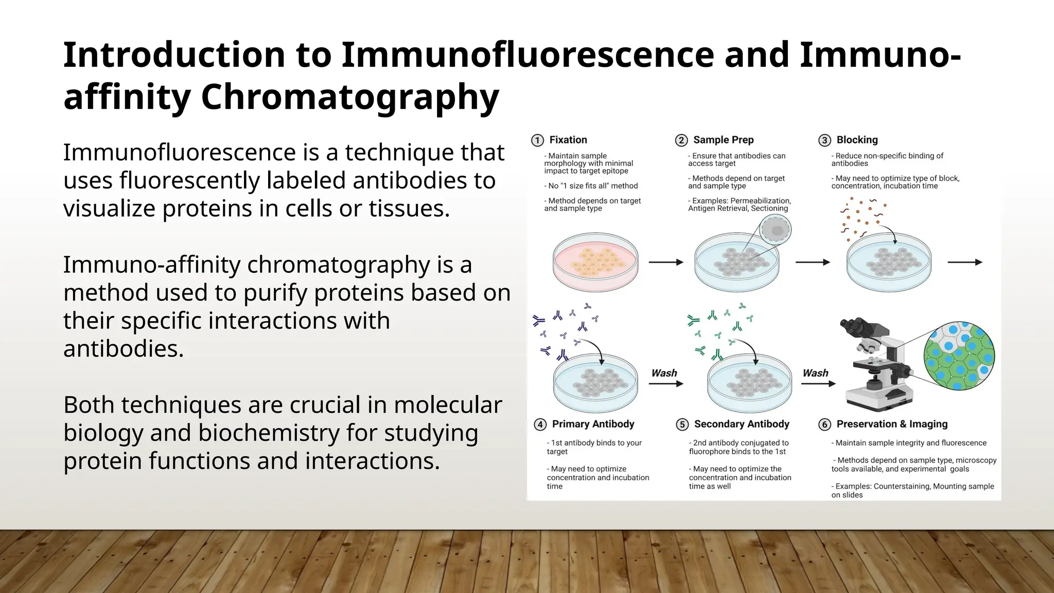
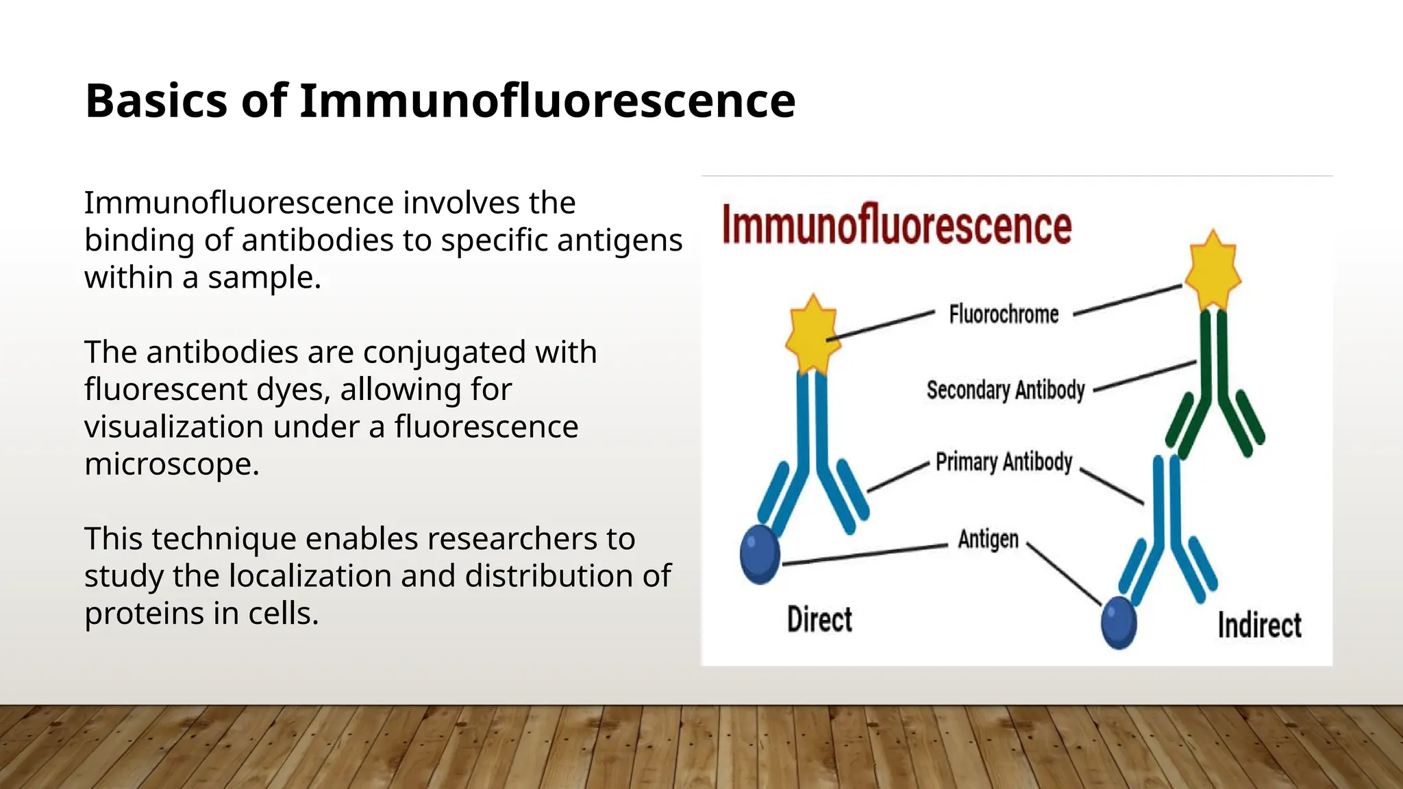
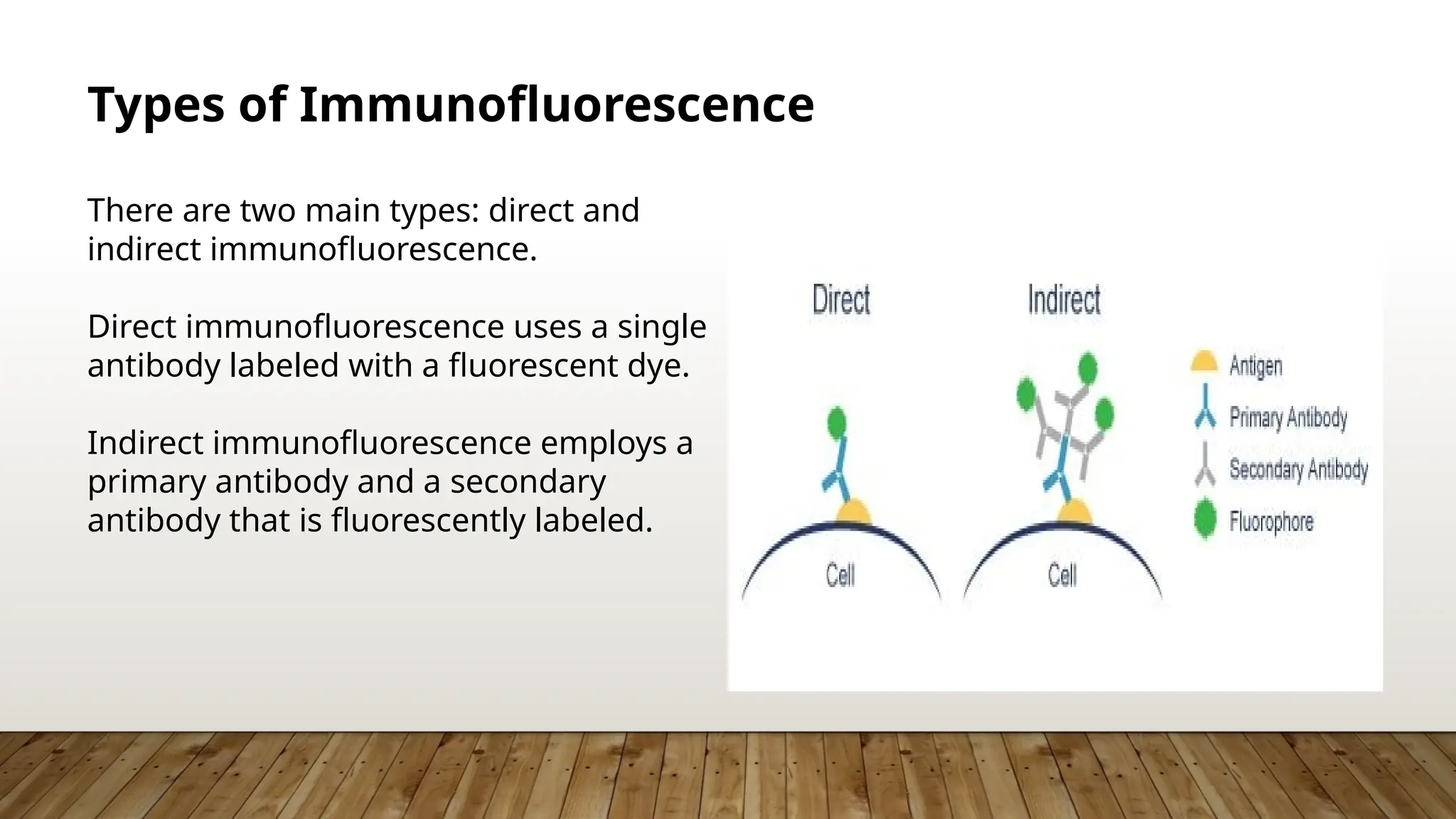
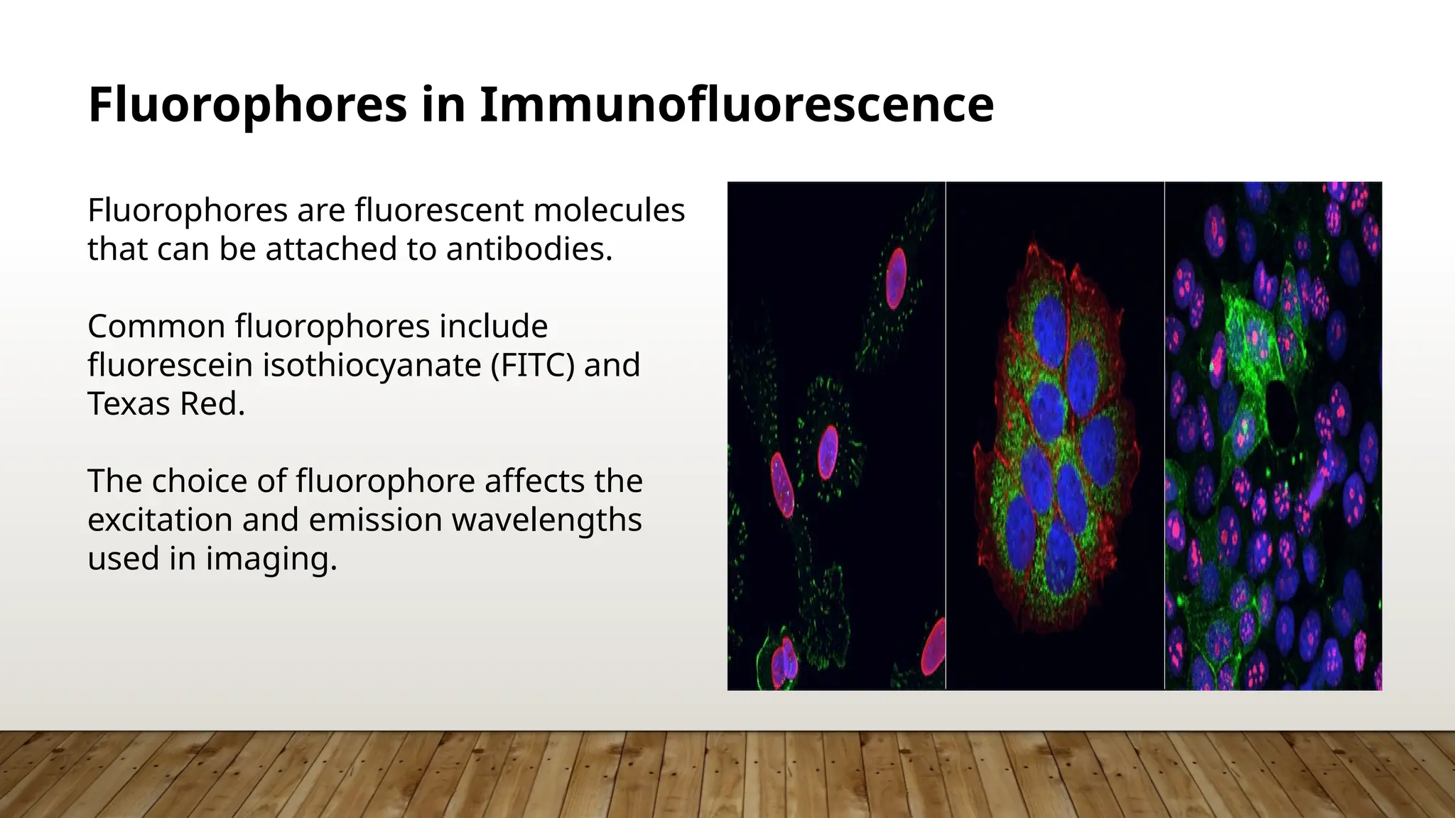
Immunofluorescence Reactions And
Immuno-affinity Chromatography
BY-:ADITYA KOKANE (3403)
Introduction to Immunofluorescence and Immuno-
affinity Chromatography
Immunofluorescence is a technique that
uses fluorescently labeled antibodies to
visualize proteins in cells or tissues.
Immuno-affinity chromatography is a
method used to purify proteins based on
their specific interactions with
antibodies.
Both techniques are crucial in molecular
biology and biochemistry for studying
protein functions and interactions.
Basics of Immunofluorescence
Immunofluorescence involves the
binding of antibodies to specific antigens
within a sample.
The antibodies are conjugated with
fluorescent dyes, allowing for
visualization under a fluorescence
microscope.
This technique enables researchers to
study the localization and distribution of
proteins in cells.
Types of Immunofluorescence
There are two main types: direct and
indirect immunofluorescence.
Direct immunofluorescence uses a single
antibody labeled with a fluorescent dye.
Indirect immunofluorescence employs a
primary antibody and a secondary
antibody that is fluorescently labeled.
Direct Immunofluorescence
Direct immunofluorescence is quicker
and simpler, as it requires only one
antibody.
It is commonly used for detecting
specific antigens in clinical diagnostics.
However, it may have lower sensitivity
compared to indirect methods.
Indirect Immunofluorescence
Indirect immunofluorescence enhances
signal amplification through the use of a
secondary antibody.
This method allows for greater flexibility
in choosing primary antibodies.
It is widely used in research to detect
low-abundance proteins.
Fluorophores in Immunofluorescence
Fluorophores are fluorescent molecules
that can be attached to antibodies.
Common fluorophores include
fluorescein isothiocyanate (FITC) and
Texas Red.
The choice of fluorophore affects the
excitation and emission wavelengths
used in imaging.
Sample Preparation for Immunofluorescence
Sample preparation is crucial for
successful immunofluorescence.
Cells or tissues must be fixed to preserve
their structure and immobilize proteins.
Permeabilization may be necessary to
allow antibodies to penetrate cell
membranes.
Imaging Techniques in Immunofluorescence
Fluorescence microscopy is the primary
imaging technique used in
immunofluorescence.
Confocal microscopy provides enhanced
resolution and the ability to image in
three dimensions.
Advanced imaging techniques, such as
super-resolution microscopy, allow for
even finer details.
Applications of Immunofluorescence
Immunofluorescence is widely used in
cell biology to study protein localization.
It plays a vital role in diagnosing diseases
by detecting specific antigens in patient
samples.
Researchers use it to investigate cellular
responses to treatments and
environmental changes.
Introduction to Immuno-affinity Chromatography
Immuno-affinity chromatography is a
powerful technique for purifying
proteins.
It relies on the specific binding between
an antigen and its corresponding
antibody.
This method is often used to isolate
proteins from complex mixtures, such as
cell lysates.
Principle of Immuno-affinity Chromatography
The principle is based on the interaction
between an immobilized antibody and
target proteins.
Once the sample is passed through the
column, the target protein binds while
unbound proteins are washed away.
Elution of the bound protein can be
achieved using a specific buffer or by
altering conditions.
Types of Immuno-affinity Chromatography
There are several types, including protein
A/G affinity and antigen-specific affinity
chromatography.
Protein A/G affinity is commonly used for
immunoglobulins, while antigen-specific
methods target particular proteins.
Each type has its advantages and
tailored applications based on the
protein of interest.
Materials Used in Immuno-affinity Chromatography
Common materials include agarose or
sepharose beads for immobilizing
antibodies.
The choice of beads can influence the
efficiency and specificity of the
purification process.
Proper selection of buffers is essential
for maintaining protein stability during
purification.
Steps in Immuno-affinity Chromatography
The process begins with the preparation
of the antibody-coupled resin.
The sample is then loaded onto the
column, allowing the target protein to
bind.
Finally, elution buffers are used to
retrieve the purified protein from the
column.
Applications of Immuno-affinity Chromatography
This technique is widely used for
purifying antibodies, enzymes, and other
proteins.
It is essential in biopharmaceutical
production for the development of
therapeutic proteins.
Researchers also use it to study protein-
protein interactions and functional
assays.
Comparison of Immunofluorescence and Immuno-
affinity Chromatography
Both techniques utilize the specificity of
antibodies for their targets.
Immunofluorescence focuses on
localization, while immuno-affinity
chromatography is centered on
purification.
Each method complements the other in
studying proteins from different
perspectives.
Challenges in Immunofluorescence
Non-specific binding of antibodies can
lead to background signals and false
positives.
Optimization of antibody concentrations
and sample preparation is critical for
reliable results.
Proper controls are necessary to validate
the findings of immunofluorescence
experiments.
Challenges in Immuno-affinity Chromatography
One challenge is the potential for the
denaturation of proteins during the
purification process.
Antibody stability and binding efficiency
can vary, affecting purification yield.
It may require extensive optimization to
achieve desired specificity and purity
levels.
Future Directions in Immunofluorescence and
Immuno-affinity Chromatography
Advances in fluorescent probes and
imaging technologies are enhancing
immunofluorescence capabilities.
Novel materials for chromatography are
being developed to improve efficiency
and reduce costs.
Both fields are moving towards higher
sensitivity and specificity in protein
analysis.
Conclusion
Immunofluorescence and immuno-
affinity chromatography are invaluable
techniques in protein analysis.
Their applications span research,
diagnostics, and therapeutic
development.
Continued advancements in these
methodologies promise to further
enhance our understanding of biological
systems.
THANKYOU

Immunofluorescence Reactions And Immuno-affinity Chromatography (6).pptx

  • 1.
    Immunofluorescence Reactions And Immuno-affinityChromatography BY-:ADITYA KOKANE (3403)
  • 2.
    Introduction to Immunofluorescenceand Immuno- affinity Chromatography Immunofluorescence is a technique that uses fluorescently labeled antibodies to visualize proteins in cells or tissues. Immuno-affinity chromatography is a method used to purify proteins based on their specific interactions with antibodies. Both techniques are crucial in molecular biology and biochemistry for studying protein functions and interactions.
  • 3.
    Basics of Immunofluorescence Immunofluorescenceinvolves the binding of antibodies to specific antigens within a sample. The antibodies are conjugated with fluorescent dyes, allowing for visualization under a fluorescence microscope. This technique enables researchers to study the localization and distribution of proteins in cells.
  • 4.
    Types of Immunofluorescence Thereare two main types: direct and indirect immunofluorescence. Direct immunofluorescence uses a single antibody labeled with a fluorescent dye. Indirect immunofluorescence employs a primary antibody and a secondary antibody that is fluorescently labeled.
  • 5.
    Direct Immunofluorescence Direct immunofluorescenceis quicker and simpler, as it requires only one antibody. It is commonly used for detecting specific antigens in clinical diagnostics. However, it may have lower sensitivity compared to indirect methods.
  • 6.
    Indirect Immunofluorescence Indirect immunofluorescenceenhances signal amplification through the use of a secondary antibody. This method allows for greater flexibility in choosing primary antibodies. It is widely used in research to detect low-abundance proteins.
  • 7.
    Fluorophores in Immunofluorescence Fluorophoresare fluorescent molecules that can be attached to antibodies. Common fluorophores include fluorescein isothiocyanate (FITC) and Texas Red. The choice of fluorophore affects the excitation and emission wavelengths used in imaging.
  • 8.
    Sample Preparation forImmunofluorescence Sample preparation is crucial for successful immunofluorescence. Cells or tissues must be fixed to preserve their structure and immobilize proteins. Permeabilization may be necessary to allow antibodies to penetrate cell membranes.
  • 9.
    Imaging Techniques inImmunofluorescence Fluorescence microscopy is the primary imaging technique used in immunofluorescence. Confocal microscopy provides enhanced resolution and the ability to image in three dimensions. Advanced imaging techniques, such as super-resolution microscopy, allow for even finer details.
  • 10.
    Applications of Immunofluorescence Immunofluorescenceis widely used in cell biology to study protein localization. It plays a vital role in diagnosing diseases by detecting specific antigens in patient samples. Researchers use it to investigate cellular responses to treatments and environmental changes.
  • 11.
    Introduction to Immuno-affinityChromatography Immuno-affinity chromatography is a powerful technique for purifying proteins. It relies on the specific binding between an antigen and its corresponding antibody. This method is often used to isolate proteins from complex mixtures, such as cell lysates.
  • 12.
    Principle of Immuno-affinityChromatography The principle is based on the interaction between an immobilized antibody and target proteins. Once the sample is passed through the column, the target protein binds while unbound proteins are washed away. Elution of the bound protein can be achieved using a specific buffer or by altering conditions.
  • 13.
    Types of Immuno-affinityChromatography There are several types, including protein A/G affinity and antigen-specific affinity chromatography. Protein A/G affinity is commonly used for immunoglobulins, while antigen-specific methods target particular proteins. Each type has its advantages and tailored applications based on the protein of interest.
  • 14.
    Materials Used inImmuno-affinity Chromatography Common materials include agarose or sepharose beads for immobilizing antibodies. The choice of beads can influence the efficiency and specificity of the purification process. Proper selection of buffers is essential for maintaining protein stability during purification.
  • 15.
    Steps in Immuno-affinityChromatography The process begins with the preparation of the antibody-coupled resin. The sample is then loaded onto the column, allowing the target protein to bind. Finally, elution buffers are used to retrieve the purified protein from the column.
  • 16.
    Applications of Immuno-affinityChromatography This technique is widely used for purifying antibodies, enzymes, and other proteins. It is essential in biopharmaceutical production for the development of therapeutic proteins. Researchers also use it to study protein- protein interactions and functional assays.
  • 17.
    Comparison of Immunofluorescenceand Immuno- affinity Chromatography Both techniques utilize the specificity of antibodies for their targets. Immunofluorescence focuses on localization, while immuno-affinity chromatography is centered on purification. Each method complements the other in studying proteins from different perspectives.
  • 18.
    Challenges in Immunofluorescence Non-specificbinding of antibodies can lead to background signals and false positives. Optimization of antibody concentrations and sample preparation is critical for reliable results. Proper controls are necessary to validate the findings of immunofluorescence experiments.
  • 19.
    Challenges in Immuno-affinityChromatography One challenge is the potential for the denaturation of proteins during the purification process. Antibody stability and binding efficiency can vary, affecting purification yield. It may require extensive optimization to achieve desired specificity and purity levels.
  • 20.
    Future Directions inImmunofluorescence and Immuno-affinity Chromatography Advances in fluorescent probes and imaging technologies are enhancing immunofluorescence capabilities. Novel materials for chromatography are being developed to improve efficiency and reduce costs. Both fields are moving towards higher sensitivity and specificity in protein analysis.
  • 21.
    Conclusion Immunofluorescence and immuno- affinitychromatography are invaluable techniques in protein analysis. Their applications span research, diagnostics, and therapeutic development. Continued advancements in these methodologies promise to further enhance our understanding of biological systems.
  • 22.

Editor's Notes

  • #2 Image source: https://blog.addgene.org/introduction-to-immunofluorescence
  • #3 Image source: https://microbenotes.com/immunofluorescence/
  • #4 Image source: https://www.abcam.com/secondary-antibodies/direct-vs-indirect-immunofluorescence
  • #5 Image source: https://www.researchgate.net/figure/Principle-for-direct-and-indirect-immunofluorescence-assay-Figure-created-with_fig8_346413375
  • #6 Image source: https://www.alamy.com/stock-image-mechanism-of-indirect-immunofluorescence-test-computer-illustration-164842776.html
  • #7 Image source: https://blog.cellsignal.com/fluorescent-staining-using-multiple-antibodies
  • #8 Image source: https://www.rndsystems.com/resources/protocols/graphic-protocol-preparation-and-fluorescent-ihc-staining-paraffin-embedded
  • #9 Image source: https://bitesizebio.com/33529/fluorescence-microscopy-the-magic-of-fluorophores-and-filters/
  • #10 Image source: https://www.aquaportail.com/dictionnaire/definition/10566/immunofluorescence
  • #11 Image source: https://microbenotes.com/affinity-chromatography/
  • #12 Image source: https://hxeqauecv.blob.core.windows.net/affinity-chromatography-vs-immunoprecipitation.html
  • #13 Image source: https://ar.inspiredpencil.com/pictures-2023/affinity-chromatography-apparatus
  • #14 Image source: https://www.caframolabsolutions.com/application/homogenizing/protein-affinity-chromatography/
  • #15 Image source: https://www.neuromics.com/protein-affinity-chromatography
  • #16 Image source: https://www.iba-lifesciences.com/applications/protein-purification/affinity-chromatography/
  • #17 Image source: https://hxeqauecv.blob.core.windows.net/affinity-chromatography-vs-immunoprecipitation.html
  • #18 Image source: http://www.slideshare.net/InnovaBiosciences/3-antibody-labeling-webinar
  • #19 Image source: https://www.iba-lifesciences.com/applications/protein-purification/affinity-chromatography/
  • #20 Image source: https://www.akoyabio.com/webinar/spatial-biomarkers-in-immuno-oncology-recent-developments-and-future-directions/
  • #21 Image source: https://hxeqauecv.blob.core.windows.net/affinity-chromatography-vs-immunoprecipitation.html