Dr. Nervana Mostafa
MBBS, MD, PhD (UK)
Assistant Professor of Physiology
Consultant Molecular Biology
Director of Academic Quality Unit
College of Medicine, KKUH, KSU
2.
Introduction
working definition ofphysiology:
Physiology is the study of the function of
organisms as integrated systems of molecules,
cells, tissues, and organs, in health and disease.
HUMAN PHYSIOLOGY
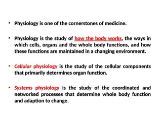
• Physiology isone of the cornerstones of medicine.
• Physiology is the study of how the body works, the ways in
which cells, organs and the whole body functions, and how
these functions are maintained in a changing environment.
• Cellular physiology is the study of the cellular components
that primarily determines organ function.
• Systems physiology is the study of the coordinated and
networked processes that determine whole body function
and adaption to change.
5.
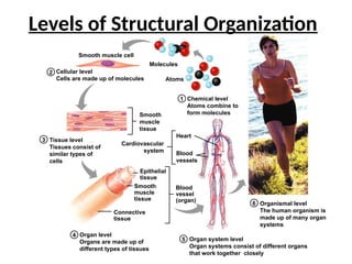
Chemical level
Atoms combineto
form molecules
1
2
3
4
Cellular level
Cells are made up of molecules
Tissue level
Tissues consist of
similar types of
cells
5 Organ system level
Organ systems consist of different organs
that work together closely
Organ level
Organs are made up of
different types of tissues
6 Organismal level
The human organism is
made up of many organ
systems
Atoms
Molecules
Smooth muscle cell
Smooth
muscle
tissue
Connective
tissue
Smooth
muscle
tissue
Epithelial
tissue
Blood
vessel
(organ)
Heart
Blood
vessels
Cardiovascular
system
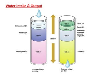
Levels of Structural Organization
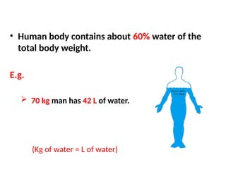
• Human bodycontains about 60% water of the
total body weight.
E.g.
70 kg man has 42 L of water.
(Kg of water = L of water)
9.
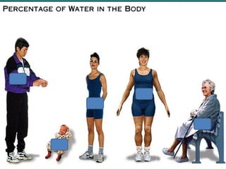
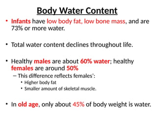
Body Water Content
•Infants have low body fat, low bone mass, and are
73% or more water.
• Total water content declines throughout life.
• Healthy males are about 60% water; healthy
females are around 50%
– This difference reflects females’:
• Higher body fat
• Smaller amount of skeletal muscle.
• In old age, only about 45% of body weight is water.
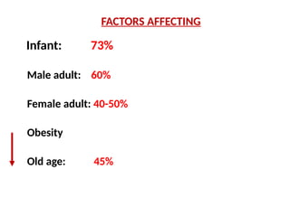
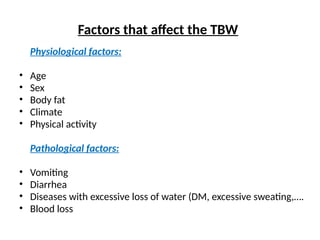
Factors that affectthe TBW
Physiological factors:
• Age
• Sex
• Body fat
• Climate
• Physical activity
Pathological factors:
• Vomiting
• Diarrhea
• Diseases with excessive loss of water (DM, excessive sweating,….
• Blood loss
13.
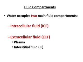
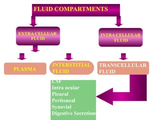
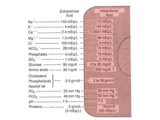
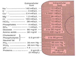
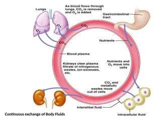
Fluid Compartments
• Wateroccupies two main fluid compartments:
–Intracellular fluid (ICF)
–Extracellular fluid (ECF)
• Plasma
• Interstitial fluid (IF)
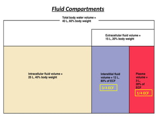
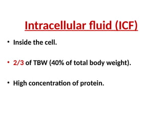
Intracellular fluid (ICF)
•Inside the cell.
• 2/3 of TBW (40% of total body weight).
• High concentration of protein.
17.
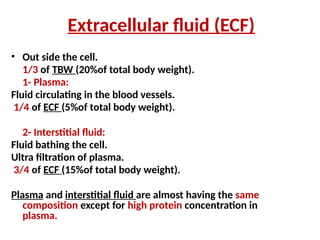
Extracellular fluid (ECF)
•Out side the cell.
1/3 of TBW (20%of total body weight).
1- Plasma:
Fluid circulating in the blood vessels.
1/4 of ECF (5%of total body weight).
2- Interstitial fluid:
Fluid bathing the cell.
Ultra filtration of plasma.
3/4 of ECF (15%of total body weight).
Plasma and interstitial fluid are almost having the same
composition except for high protein concentration in
plasma.
18.
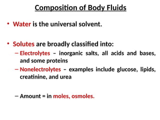
Composition of BodyFluids
• Water is the universal solvent.
• Solutes are broadly classified into:
– Electrolytes – inorganic salts, all acids and bases,
and some proteins
– Nonelectrolytes – examples include glucose, lipids,
creatinine, and urea
– Amount = in moles, osmoles.
21.
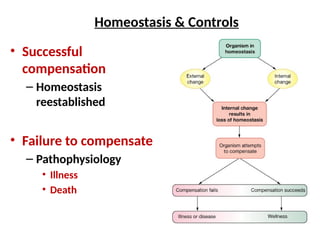
Homeostasis
• Homeostasis isthe ability to maintain a relatively
stable internal environment in an ever-changing
outside world.
• The internal environment of the body (ECF) is in a
dynamic state of equilibrium.
• All different body systems operate in harmony to
provide homeostasis.
22.
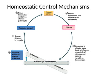
Homeostatic Control Mechanisms
•The variable produces a change in the body
• The three interdependent components of
control mechanisms are:
– Receptor – monitors the environments and responds
to changes (stimuli)
– Control center – determines the set point at which
the variable is maintained
– Effector – provides the means to respond to the
stimulus
23.
Regulation of bodyfunctions
1. Nervous system
- sensory input.
- central nervous system.
- motor out put.
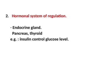
24.
2. Hormonal systemof regulation.
- Endocrine gland.
Pancreas, thyroid
e.g. : insulin control glucose level.
25.
Stimulus:
Produces
change
in variable
1
2
3
Change
detected
by receptor
Input:
Information
sentalong
afferent
pathway to
5 Response of
effector feeds
back to influence
magnitude of
stimulus and
returns
variable to
homeostasis
Variable (in homeostasis)
Imbalance
Imbalance
Receptor (sensor)
Control
center 4 Output:
Information sent
along efferent
pathway to
Effector
Homeostatic Control Mechanisms
objectives
At the endof this session, the students should be able to:
• Describe the fluid mosaic model of membrane structure and
function.
• Define permeability and list factors influencing permeability.
• Identify and describe carried-mediated transport processes:
Primary active transport, secondary active transport,
facilitates diffusion.
31.
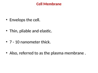
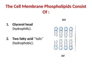
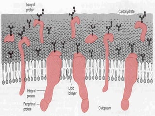
Cell Membrane
• Envelopsthe cell.
• Thin, pliable and elastic.
• 7 - 10 nanometer thick.
• Also, referred to as the plasma membrane .
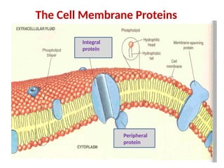
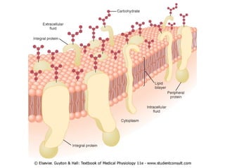
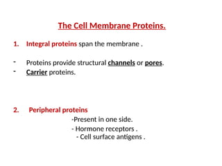
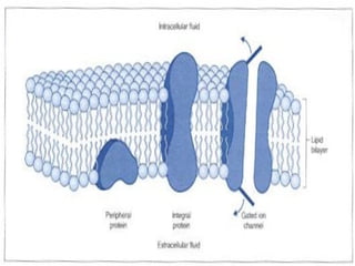
The Cell MembraneProteins.
1. Integral proteins span the membrane .
- Proteins provide structural channels or pores.
- Carrier proteins.
2. Peripheral proteins
-Present in one side.
- Hormone receptors .
- Cell surface antigens .
39.
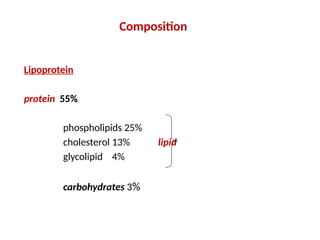
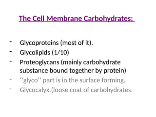
The Cell MembraneCarbohydrates:
- Glycoproteins (most of it).
- Glycolipids (1/10)
- Proteoglycans (mainly carbohydrate
substance bound together by protein)
- ‘’glyco’’ part is in the surface forming.
- Glycocalyx.(loose coat of carbohydrates.
41.
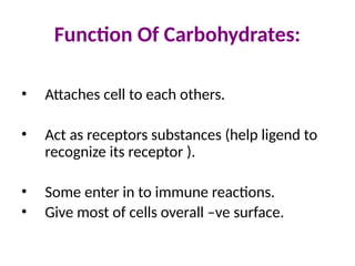
Function Of Carbohydrates:
•Attaches cell to each others.
• Act as receptors substances (help ligend to
recognize its receptor ).
• Some enter in to immune reactions.
• Give most of cells overall –ve surface.
42.
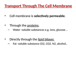
Transport Through TheCell Membrane
• Cell membrane is selectively permeable.
• Through the proteins.
– Water -soluble substances e.g. ions, glucose ..
• Directly through the lipid bilayer.
– Fat -soluble substance (O2, CO2, N2, alcohol..
44.
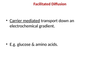
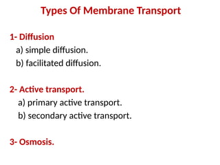
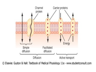
Types Of MembraneTransport
1- Diffusion
a) simple diffusion.
b) facilitated diffusion.
2- Active transport.
a) primary active transport.
b) secondary active transport.
3- Osmosis.
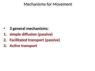
Mechanisms for Movement
•3 general mechanisms:
1. simple diffusion (passive)
2. Facilitated transport (passive)
3. Active transport
47.
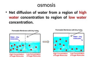
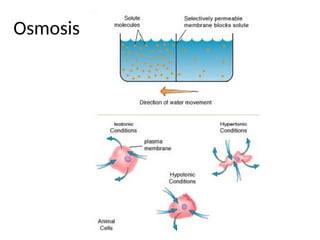
osmosis
• Net diffusionof water from a region of high
water concentration to region of low water
concentration.
48.
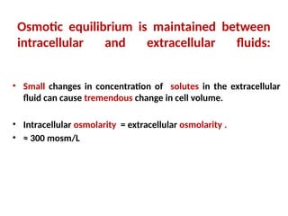
Osmotic equilibrium ismaintained between
intracellular and extracellular fluids:
• Small changes in concentration of solutes in the extracellular
fluid can cause tremendous change in cell volume.
• Intracellular osmolarity = extracellular osmolarity .
• ≈ 300 mosm/L
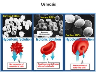
Normal RBCs
Isotonic Solution
Osmosis
Equalmovement of water
into and out of cells
Net movement of
water out of cells Net movement of
water into cells
Shriveled RBCs
Swollen RBCs
Hypertonic Solution Hypotonic Solution
51.
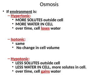
Osmosis
• If environmentis:
– Hypertonic:
• MORE SOLUTES outside cell
• MORE WATER IN CELL
• over time, cell loses water
– Isotonic:
• same
• No change in cell volume
– Hypotonic:
• LESS SOLUTES outside cell
• LESS WATER IN CELL, more solutes in cell.
• over time, cell gains water
52.
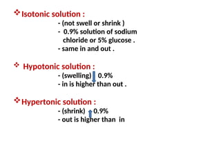
Isotonic solution :
-(not swell or shrink )
- 0.9% solution of sodium
chloride or 5% glucose .
- same in and out .
Hypotonic solution :
- (swelling) 0.9%
- in is higher than out .
Hypertonic solution :
- (shrink) 0.9%
- out is higher than in
53.
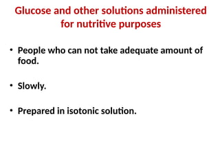
Glucose and othersolutions administered
for nutritive purposes
• People who can not take adequate amount of
food.
• Slowly.
• Prepared in isotonic solution.
55.
Diffusion
Random movement ofsubstance either through the
membrane directly or in combination with carrier
protein down an electrochemical gradient.
1- Simple diffusion.
2- facilitated diffusion.
Simple diffusion & facilitated transport don’t
require input of energy = powered by
concentration gradient or electrical gradient
[Active transport = directly uses ATP]
56.
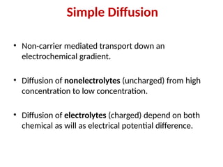
Simple Diffusion
• Non-carriermediated transport down an
electrochemical gradient.
• Diffusion of nonelectrolytes (uncharged) from high
concentration to low concentration.
• Diffusion of electrolytes (charged) depend on both
chemical as will as electrical potential difference.
57.
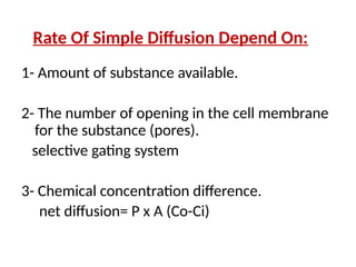
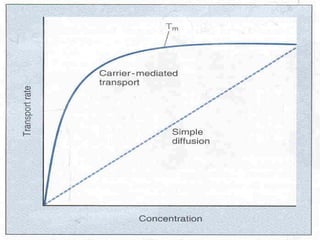
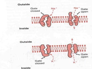
Rate Of SimpleDiffusion Depend On:
1- Amount of substance available.
2- The number of opening in the cell membrane
for the substance (pores).
selective gating system
3- Chemical concentration difference.
net diffusion= P x A (Co-Ci)
58.
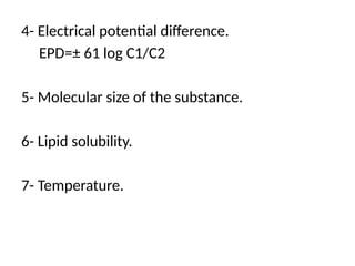
4- Electrical potentialdifference.
EPD=± 61 log C1/C2
5- Molecular size of the substance.
6- Lipid solubility.
7- Temperature.
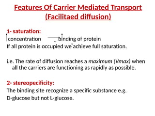
Features Of CarrierMediated Transport
(Facilitaed diffusion)
1- saturation:
concentration binding of protein
If all protein is occupied we achieve full saturation.
i.e. The rate of diffusion reaches a maximum (Vmax) when
all the carriers are functioning as rapidly as possible.
2- stereopecificity:
The binding site recognize a specific substance e.g.
D-glucose but not L-glucose.
62.
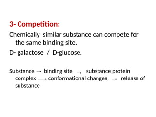
3- Competition:
Chemically similarsubstance can compete for
the same binding site.
D- galactose / D-glucose.
Substance binding site substance protein
complex conformational changes release of
substance
63.
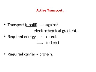
Active Transport:
• Transport(uphill) against
electrochemical gradient.
• Required energy direct.
indirect.
• Required carrier – protein.
64.
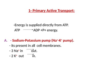
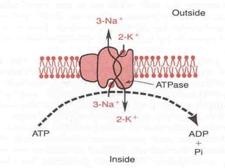
1- Primary ActiveTransport:
-Energy is supplied directly from ATP.
ATP ADP +P+ energy.
A. - Sodium-Potassium pump (Na+
-K+
pump).
- its present in all cell membranes.
- 3 Na+
in out.
- 2 K+
out in.
66.
Characteristic Of ThePump:
1. Carrier protein.
2. Binding site for Na inside the cell.
3. Binding site for K outside the cell.
4. It has ATPase activity.
5. 3 Na out.
6. 2 K in.
67.
Function:
1. Maintaining Na+
andK+
concentration
difference .
2. Maintaining –ve potential inside the cell.
3. Maintains a normal cell volume.
4. It’s the basis of nerve signal transmition .
68.
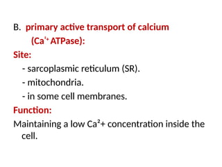
B. primary activetransport of calcium
(Ca²+
ATPase):
Site:
- sarcoplasmic reticulum (SR).
- mitochondria.
- in some cell membranes.
Function:
Maintaining a low Ca²+ concentration inside the
cell.
69.
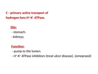
C - primaryactive transport of
hydrogen lons H+
-K+
ATPase.
Site:
- stomach.
- kidneys.
Function:
- pump to the lumen.
- H+
-K+
ATPase inhibitors (treat ulcer disease). (omeprazol)
70.
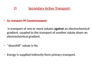
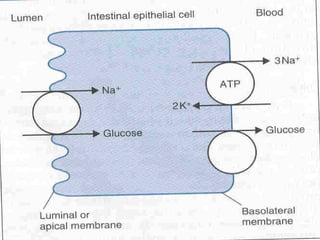
2) Secondary ActiveTransport:
• Co- transport OR Countertransport:
is transport of one or more solutes against an electrochemical
gradient, coupled to the transport of another solute down an
electrochemical gradient.
- ‘’downhill’’ solute is Na.
- Energy is supplied indirectly form primary transport.
71.
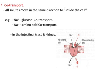
• Co-transport:
- Allsolutes move in the same direction to ‘’inside the cell’’.
- e.g. - Na+
- glucose Co-transport.
- Na+
- amino acid Co-transport.
- in the intestinal tract & kidney.
73.
Countertransport:
• Na+
is movingto the interior causing other
substance to move out.
• Ca²+
- Na+
exchange.
(present in many cell membranes)
• Na+
- H+
exchange in the kidney.
Editor's Notes
#50 Note that molecules of water are constantly moving in BOTH directions across the cell membrane.
Though water is moving in both directions, there is a net gain of water on the side that starts out with less water (more stuff dissolved in the water).
#51 These three terms refer to the concentration of dissolved materials in a cell’s environment compared to the concentration within the cell.
Hypotonic environment:
Cell gains water from surrounding environment.
“Hypo-” means “below” (below the concentration in the cell).
Hypertonic environment:
Cell looses water to surrounding environment.
“Hyper-” means “above” (above the concentration in the cell).
Isotonic environment:
No change in cell volume.
Iso-” means “same as.”


![Diffusion
Random movement of substance either through the
membrane directly or in combination with carrier
protein down an electrochemical gradient.
1- Simple diffusion.
2- facilitated diffusion.
Simple diffusion & facilitated transport don’t
require input of energy = powered by
concentration gradient or electrical gradient
[Active transport = directly uses ATP]](https://image.slidesharecdn.com/youtube-250418173501-a9b2f19d/85/Homeostasis-physiology-complete-lec-pptx-55-320.jpg)