Download to read offline
![Sai Chand et al Int. Journal of Engineering Research and Application
ISSN : 2248-9622, Vol. 3, Issue 5, Sep-Oct 2013, pp.1182-1184
RESEARCH ARTICLE
www.ijera.com
OPEN ACCESS
Microblaze Architecture Development for Medical Image Fusion
Using Wavelet Transform
P.Saichand1, A.Swetha2
M.TECH (VLSI-SD) Gurunanak Institute of Technology,
Assistant Professor, Dept of E.C.E,
Abstract
Now-a-days, almost all areas of medical diagnosis are impacted by the digital image processing. . For medical
diagnosis, Computed Tomography (CT) provides the best information on denser tissue with less distortion.
Magnetic Resonance Image (MRI) provides better information on soft tissue . With more available
multimodality medical images in clinical applications, the idea of fusing images from different modalities
become very important and medical image fusion has emerged as a new promising research field .In this paper
two input images i.e. CT and MRI medical images are converted to header files containing pixel values using
mat lab . header file of image are taken as input to D WT algorithm using lifting scheme . we will take average
value of both input image pixel values for final fusion. We will apply IDWT to get the original fused output of
input images.finally we will get the fused image of input CT and MRI for medical diagnosis .In this paper
hardware implementation of a real-time image fusion is performed. The system is based on an Xilinx Spartan
3 EDK FPGA and implements a configurable linear pixel level D WT algorithm which is able to result in color
fused images using System C language.
Keywords- Fusion, Wavelets Transform
classified into spatial domain fusion and transform
domain fusion. Spatial domain fusion is directly
I.
INTRODUCTION
applied on the source images which in turn reduce the
Image fusion is a technique used to
integrate a high resolution panchromatic image with
signal to-noise ratio of the resultant image with simple
low-resolution multispectral image to produce a highaveraging technique but the spatial distortion still
resolution multispectral image, which contains both
persists in the fused image. To improve on that in
the high-resolution spatial infossssrmation of the
transform domain fusion, firstly the input images are
panchromatic image and the color information of the
decomposed based on transform coefficients. Then the
multispectral image [4].
Although an increasing
fusion technique is applied and the fusion decision map
numbers of high-resolution images are available along
is obtained. Inverse transformation on this decision
with sensor technology development, image fusion is
map yields the fused image. The fused image carries all
still a popular and important method to interpret the
the details of the source images and reduces the spatial
image data for obtaining a more suitable image for
distortion. So, majority of the earlier fusion techniques
a variety of applications,
such
as
visual
were based on wavelet transformation.
interpretation and digital classification [3].
The main objective of medical imaging is to
obtain a high resolution image with as much details
II.
WAVELET BASED IMAGE FUSION
as possible for the sake of diagnosis [7]. MR and CT
TECHNIQUES
imaging are o f main concern for diagnostic purposes
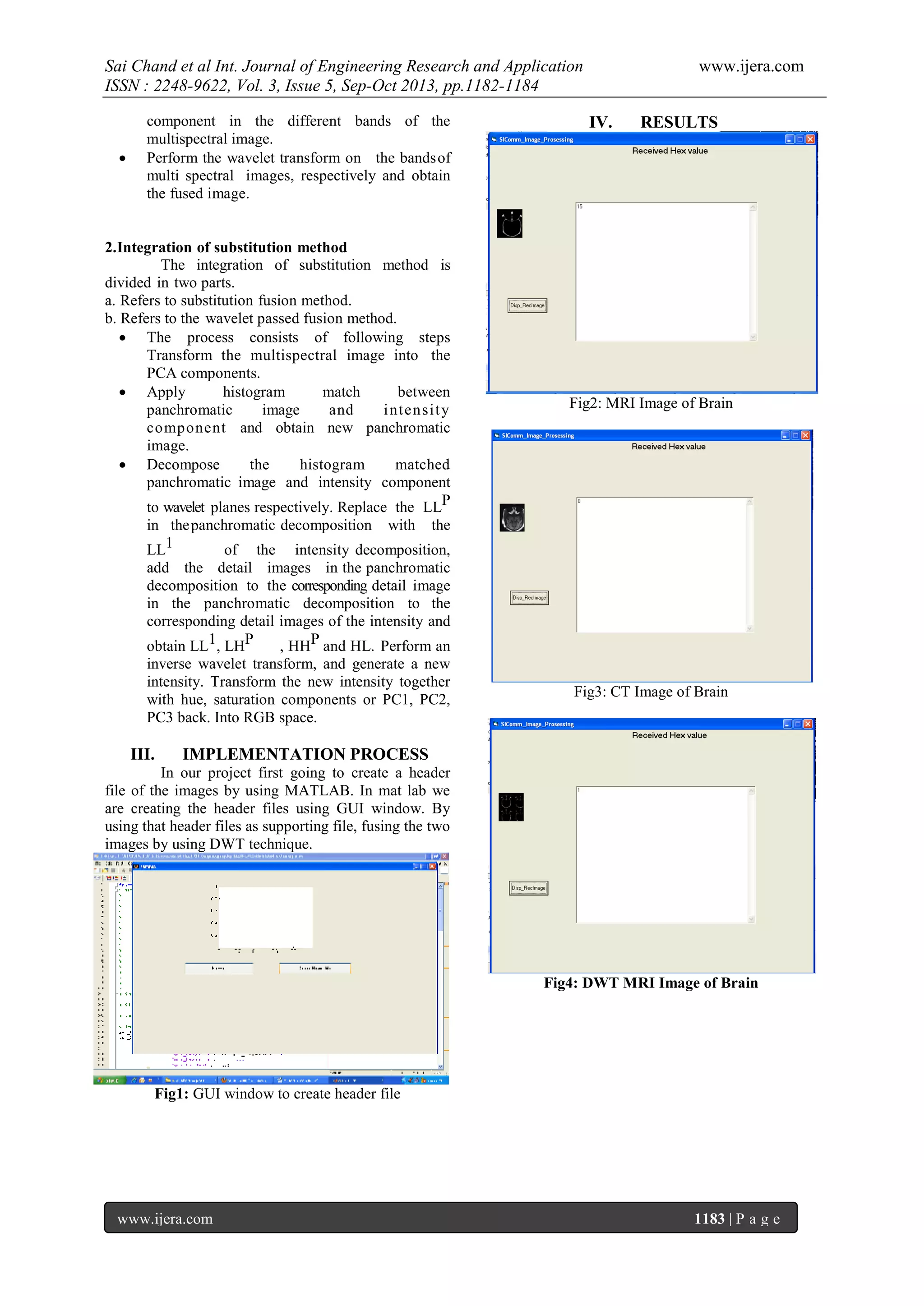
1.
Wavelet based image fusion method
[6].
Both techniques give special sophisticated
The process can be divided into four steps.
characteristics of the organ to be imaged. So, it is
a) Wavelet decomposition
expected that fusion of MR and CT images of the
b) Details information combination
same organ would result in an integrated image of
c) Inverse wavelet transforms
much more details [10].Wavelet transform fusion is
Use the wavelet transform to decompose new
defined as considering the wavelet transforms of
panchromatic images and d i f fe r e n t bands of
the two registered input images together with the
multispectral image twice, respectively.
fusion rule .Then, the inverse wavelet transform is
Add the detail images of the decomposed
computed, and the fused image is reconstructed.
panchromatic images at different levels to the
The actual fusion process can be carried out at
corresponding details of different bands in the
various levels. Under this, in the pixel-level image
multispectral image and obtain the new details
fusion the fused images provided all relevant
component in the different bands of the
information present in original images with no artifacts
multispectral image and obtain the new details
or inconsistencies. The pixel-level image fusions were
www.ijera.com
1182 | P a g e](https://image.slidesharecdn.com/gr3511821184-131015040916-phpapp01/75/Gr3511821184-1-2048.jpg)
![Sai Chand et al Int. Journal of Engineering Research and Application
ISSN : 2248-9622, Vol. 3, Issue 5, Sep-Oct 2013, pp.1182-1184
[6]
[7]
[8]
Fig5: DWT CT Image of Brain
[9]
[10]
www.ijera.com
BrainWeb: Simulated Brain Database.
[Online],available:http://www.bic.mnimcgill.
ca/brainweb/,March, 2004
A. Wang, Haijing Sun, and Yueyang
Guan,
“The
Application
of Wavelet
Transform
to
MultimodalityMedical
Image
Fusion,“ Proceedings of the 2006
IEEE
International
Conference
on
Networking, Sensing and Control, (ICNSC),
pp. 270- 274, 2006.
Chipman, L.J. and Orr, T.M., “Wavelets and
image fusion,” in IEEE International
Conference on Image Processing, 3, pp. 248–
251, 1995.
Wang Z, Bovik A C, Sheikh H R, Simoncelli
E P. Image Quality Assesment: From Error
Visibility to Structual Similarity. IEEE
Transactions on Image Processing, 2004,
13(4):600-612.
Socolinsky D A, Wolff L B. Multispectral
image visualization through first-order fusion.
IEEE Transactions on Image Processing,
2002, 11(8): 923-931.
Fig6: Fused image of brain
V.
Conclusion
A hardware implementation of a real-time
fusion system is done based on an Xilinx Spartan 3
EDK FPGA and implements a configurable linear pixel
level algorithm which is able to result in color fused
images using System C language.
REFERENCS
[1]
[2]
[3]
[4]
[5]
Rafael
c.
Gonzalez,richard
E.woods
Digital image processing, - Addisonwesley.an imprint of pearson education, 1st
edition
Introduction to wavelets and wavelet
transforms BURRUS C.,S.Gopinath, R.A
and GUO[Englewoodcliffs, NT:prentice –
hall]
H. Li, B.S. Manjunath, and S.K. Mitra.
Multisensor image fusion using the wavelet
Graphical Models and Image Processing,
57:235–245, 1995.
S. Udomhunsakul, and P. Wongsita,
“Feature
extraction
in medical MRI
images,“ Proceeding of 2004 IEEE
Conference on Cybernetics and Intelligent
Systems, vol.1, pp. 340- 344, Dec. 2004.
L.J. Chipman, T.M. Orr, and L.N. Lewis.
Wavelets
and
image
fusion. IEEE
Transactions on Image Processing, 3:248–
251.
www.ijera.com
1184 | P a g e](https://image.slidesharecdn.com/gr3511821184-131015040916-phpapp01/75/Gr3511821184-3-2048.jpg)

This research article discusses the development of a hardware implementation for medical image fusion using wavelet transform, focusing on the integration of CT and MRI images to enhance diagnostic quality. The proposed system employs a configurable linear pixel level algorithm on an FPGA, enabling real-time fusion to produce high-resolution medical images. The methodology includes creating header files, applying discrete wavelet transform (DWT), and reconstructing the fused image while minimizing spatial distortion.