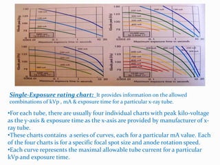
The document discusses the components and functioning of an X-ray tube. It describes how X-ray tubes generate X-rays by accelerating electrons using high voltage and directing them towards a metal target. It explains how factors like voltage, current, target material, filtration and waveform affect the quality and quantity of the emitted X-ray beam. Specifically, it discusses how increasing voltage improves beam quality by producing higher energy photons, while increasing current increases beam quantity by producing more photons. The target material and filtration can also affect the average energy and composition of the X-ray spectrum.
X- Ray physics- X-Ray Tube, Transformer, Generator and Rectifiers by kajalsra...DrKajalLimbad
Â
X-Ray physics including x-ray tube, transformer, generator, and rectifiers. physics made an easy
Note: this ppt has many animations that may not be appreciated over here. Request original ppt at kajalsradiology@gmail.com
Comparing Evolved Extractive Text Summary Scores of Bidirectional Encoder Rep...University of Maribor
Â
Slides from:
11th International Conference on Electrical, Electronics and Computer Engineering (IcETRAN), NiĹĄ, 3-6 June 2024
Track: Artificial Intelligence
https://www.etran.rs/2024/en/home-english/
Cancer cell metabolism: special Reference to Lactate PathwayAADYARAJPANDEY1
Â
Normal Cell Metabolism:
Cellular respiration describes the series of steps that cells use to break down sugar and other  chemicals to get the energy we need to function.
Energy is stored in the bonds of glucose and when glucose is broken down, much of that energy is released. Â
Cell utilize energy in the form of ATP.
The first step of respiration is called glycolysis. In a series of steps, glycolysis breaks glucose into two smaller molecules -Â a chemical called pyruvate. A small amount of ATP is formed during this process.Â
Most healthy cells continue the breakdown in a second process, called the Kreb's cycle. The Kreb's cycle allows cells to âburnâ the pyruvates made in glycolysis to get more ATP.
The last step in the breakdown of glucose is called oxidative phosphorylation (Ox-Phos).
It takes place in specialized cell structures called mitochondria. This process produces a large amount of ATP.  Importantly, cells need oxygen to complete oxidative phosphorylation.
If a cell completes only glycolysis, only 2 molecules of ATP are made per glucose. However, if the cell completes the entire respiration process (glycolysis - Kreb's - oxidative phosphorylation), about 36 molecules of ATP are created, giving it much more energy to use.
IN CANCER CELL:
Unlike healthy cells that "burn" the entire molecule of sugar to capture a large amount of energy as ATP, cancer cells are wasteful.
Cancer cells only partially break down sugar molecules. They overuse the first step of respiration, glycolysis. They frequently do not complete the second step, oxidative phosphorylation.
This results in only 2 molecules of ATP per each glucose molecule instead of the 36 or so ATPs healthy cells gain. As a result, cancer cells need to use a lot more sugar molecules to get enough energy to survive.Â
introduction to WARBERG PHENOMENA:
WARBURG EFFECT Usually, cancer cells are highly glycolytic (glucose addiction) and take up more glucose than do normal cells from outside.
Otto Heinrich Warburg (; 8 October 1883 â 1 August 1970) In 1931 was awarded the Nobel Prize in Physiology for his "discovery of the nature and mode of action of the respiratory enzyme.
WARNBURG EFFECT : Â cancer cells under aerobic (well-oxygenated) conditions to metabolize glucose to lactate (aerobic glycolysis) is known as the Warburg effect. Warburg made the observation that tumor slices consume glucose and secrete lactate at a higher rate than normal tissues.
Deep Behavioral Phenotyping in Systems Neuroscience for Functional Atlasing a...Ana LuĂsa Pinho
Â
Functional Magnetic Resonance Imaging (fMRI) provides means to characterize brain activations in response to behavior. However, cognitive neuroscience has been limited to group-level effects referring to the performance of specific tasks. To obtain the functional profile of elementary cognitive mechanisms, the combination of brain responses to many tasks is required. Yet, to date, both structural atlases and parcellation-based activations do not fully account for cognitive function and still present several limitations. Further, they do not adapt overall to individual characteristics. In this talk, I will give an account of deep-behavioral phenotyping strategies, namely data-driven methods in large task-fMRI datasets, to optimize functional brain-data collection and improve inference of effects-of-interest related to mental processes. Key to this approach is the employment of fast multi-functional paradigms rich on features that can be well parametrized and, consequently, facilitate the creation of psycho-physiological constructs to be modelled with imaging data. Particular emphasis will be given to music stimuli when studying high-order cognitive mechanisms, due to their ecological nature and quality to enable complex behavior compounded by discrete entities. I will also discuss how deep-behavioral phenotyping and individualized models applied to neuroimaging data can better account for the subject-specific organization of domain-general cognitive systems in the human brain. Finally, the accumulation of functional brain signatures brings the possibility to clarify relationships among tasks and create a univocal link between brain systems and mental functions through: (1) the development of ontologies proposing an organization of cognitive processes; and (2) brain-network taxonomies describing functional specialization. To this end, tools to improve commensurability in cognitive science are necessary, such as public repositories, ontology-based platforms and automated meta-analysis tools. I will thus discuss some brain-atlasing resources currently under development, and their applicability in cognitive as well as clinical neuroscience.
What is greenhouse gasses and how many gasses are there to affect the Earth.moosaasad1975
Â
What are greenhouse gasses how they affect the earth and its environment what is the future of the environment and earth how the weather and the climate effects.
This pdf is about the Schizophrenia.
For more details visit on YouTube; @SELF-EXPLANATORY;
https://www.youtube.com/channel/UCAiarMZDNhe1A3Rnpr_WkzA/videos
Thanks...!
Observation of Ioâs Resurfacing via Plume Deposition Using Ground-based Adapt...SĂŠrgio Sacani
Â
Since volcanic activity was first discovered on Io from Voyager images in 1979, changes
on Ioâs surface have been monitored from both spacecraft and ground-based telescopes.
Here, we present the highest spatial resolution images of Io ever obtained from a groundbased telescope. These images, acquired by the SHARK-VIS instrument on the Large
Binocular Telescope, show evidence of a major resurfacing event on Ioâs trailing hemisphere. When compared to the most recent spacecraft images, the SHARK-VIS images
show that a plume deposit from a powerful eruption at Pillan Patera has covered part
of the long-lived Pele plume deposit. Although this type of resurfacing event may be common on Io, few have been detected due to the rarity of spacecraft visits and the previously low spatial resolution available from Earth-based telescopes. The SHARK-VIS instrument ushers in a new era of high resolution imaging of Ioâs surface using adaptive
optics at visible wavelengths.
Slide 1: Title Slide
Extrachromosomal Inheritance
Slide 2: Introduction to Extrachromosomal Inheritance
Definition: Extrachromosomal inheritance refers to the transmission of genetic material that is not found within the nucleus.
Key Components: Involves genes located in mitochondria, chloroplasts, and plasmids.
Slide 3: Mitochondrial Inheritance
Mitochondria: Organelles responsible for energy production.
Mitochondrial DNA (mtDNA): Circular DNA molecule found in mitochondria.
Inheritance Pattern: Maternally inherited, meaning it is passed from mothers to all their offspring.
Diseases: Examples include Leberâs hereditary optic neuropathy (LHON) and mitochondrial myopathy.
Slide 4: Chloroplast Inheritance
Chloroplasts: Organelles responsible for photosynthesis in plants.
Chloroplast DNA (cpDNA): Circular DNA molecule found in chloroplasts.
Inheritance Pattern: Often maternally inherited in most plants, but can vary in some species.
Examples: Variegation in plants, where leaf color patterns are determined by chloroplast DNA.
Slide 5: Plasmid Inheritance
Plasmids: Small, circular DNA molecules found in bacteria and some eukaryotes.
Features: Can carry antibiotic resistance genes and can be transferred between cells through processes like conjugation.
Significance: Important in biotechnology for gene cloning and genetic engineering.
Slide 6: Mechanisms of Extrachromosomal Inheritance
Non-Mendelian Patterns: Do not follow Mendelâs laws of inheritance.
Cytoplasmic Segregation: During cell division, organelles like mitochondria and chloroplasts are randomly distributed to daughter cells.
Heteroplasmy: Presence of more than one type of organellar genome within a cell, leading to variation in expression.
Slide 7: Examples of Extrachromosomal Inheritance
Four Oâclock Plant (Mirabilis jalapa): Shows variegated leaves due to different cpDNA in leaf cells.
Petite Mutants in Yeast: Result from mutations in mitochondrial DNA affecting respiration.
Slide 8: Importance of Extrachromosomal Inheritance
Evolution: Provides insight into the evolution of eukaryotic cells.
Medicine: Understanding mitochondrial inheritance helps in diagnosing and treating mitochondrial diseases.
Agriculture: Chloroplast inheritance can be used in plant breeding and genetic modification.
Slide 9: Recent Research and Advances
Gene Editing: Techniques like CRISPR-Cas9 are being used to edit mitochondrial and chloroplast DNA.
Therapies: Development of mitochondrial replacement therapy (MRT) for preventing mitochondrial diseases.
Slide 10: Conclusion
Summary: Extrachromosomal inheritance involves the transmission of genetic material outside the nucleus and plays a crucial role in genetics, medicine, and biotechnology.
Future Directions: Continued research and technological advancements hold promise for new treatments and applications.
Slide 11: Questions and Discussion
Invite Audience: Open the floor for any questions or further discussion on the topic.
2. X-ray tube
ď X-ray tube is an device for generating X-rays by
accelerating electrons to high energies and causing
them to strike a metal target from which X-rays are
emitted.
3. ď High voltage is applied b/w cathode and anode.
ď The electrons are emitted from the filament by thermionic emission and
accelerated towards the anode and strike the target.
Basic principle of x-ray tube:
- +
4. ď An X-ray tube consists of an anode and cathode mounted inside an evacuated
glass tube.
ď The cathode consists, of a small coil called filament , mounted in a focusing
cup.
ď The anode target is mainly made of Tungsten , separated from filament by a
small gap.
ď Electrons are boiled off the filament by applying an electric current so that it
becomes white hot- the process is called âThermionic emissionâ .
ď A high voltage (HV) of 100 Kv or more applied and the electrons are attracted
across the gap to collide with the anode with the high energies, to produce X-
rays.
ď Bremsstrahlung & Characteristic radiation process are involved here.
X-ray tube
5. Quality & Quantity:
ď Quality refers to the overall energy of the beam
ď As the X-ray beam is polyenergetic, any factors that
increase or decrease the average energy of photons in
the beam affect x-ray beam quality.
ď Quality is directly affected by,
i. Changes in kVP
ii. Changes in the material(atomic number Z) of the
target material.
iii. Changes in the filtration.
iv. The type of waveform used (i.e., 1Ď, 3Ď, or high
frequency).
6. ď Quantity refers to the number of X-ray photons in the
beam .
ď As the number of photons increases, the beam
intensity increases & any factors that affect the
number of x-ray photons in the beam influence x-ray-
beam quantity.
ď Quantity is affected by,
i. Changes in mA (tube current).
ii. Changes in the filtration.
iii. Changes in the material (Z number) of target.
iv. Changes in kVP
v. Changes in type of waveform used.
vi. Changes in distance from the tube (FFD).
7. Effects of changes in kVP:
Peak kilo voltage (kVp ) is the maximum voltage applied
across an X-ray tube, it determines the kinetic energy of
the electrons accelerated in the X-ray tube and the peak
energy of the X-ray spectrum.
â˘The changes in the kVp varies the energy of
the incoming electrons prior to maximum
energy of X-ray photons in Bremsstrahlung
spectrum.
â˘Hence as kVp increases more higher energy
photons are included in the beam , raising
the average energy of the beam.
â˘In addition, the increase in kVp increases
the speed with which incoming electrons
strike the target.
â˘Each time an incoming electron undergoes
a deceleration about a target atom nucleus,
Bremsstrahlung photons are produced,
resulting in overall Bremsstrahlung photons
8. ď Exposure is approximately proportional to the square of kVp in the
diagnostic energy range
Exposure Îą (kVp) 2
Eg: the relative exposure of a beam generated with 80 kVp compared with that
of 60 kVp for the same tube current and exposure time is calculated as follows:
(80)2 â 1.78
(60)2
Therefore, the output exposure increases by approximately 78%.
An increase in kVp increases the efficiency of x-ray production and the quantity
and quality of the x-ray beam.
9. Effect of changes in mA:
Tube current (mA) is the rate of electron flow from filament
to target (electron/sec) and measured in miliamperes.
â˘As the tube current (mA) increases, the
number of incoming electrons striking
target increases.
â˘In addition the number of X-ray photons
produced in both Bremsstrahlung and
characteristic x-ray portions of x-ray
spectrum is altered.
â˘The relationship between the mA and the
number of photons produced directly
affects the amplitude of the x-ray
emission spectrum.
â˘Eg: if we double the tube current (mA)
the number of the x-ray photons will be
doubled.
Number
of
X-rays
emitted
(per
Kev)
â˘The changes in the mA does not produces energy shifts in either portion of
spectrum .
10. Effect of changes in the target materials:
The target (anode) material affects the efficiency of Bremsstrahlung
radiation production, with output exposure roughly proportional to
atomic number.
â˘As the Z number of target increases the
amount of Bremsstrahlung radiation
produced also increases.
â˘Although the effect of using a higher Z
number target is more apparent on the
high-energy side of the Bremsstrahlung
peak, the spectra are same in many ways.
â˘The minimum & maximum energies are
the same.
â˘The peak of Bremsstrahlung spectrum
(greatest no. of X-rays production) occurs
at the same position on energy axis.
11. Effect of changes in the target materials:
â˘Hence in the Bremsstrahlung portion of x-ray spectrum there
is a change in the quantity not in quality of x-ray photons.
â˘As the Z number becomes larger the binding energy of shell
electrons also become larger. This increase in electronic
binding energies results in higher-energy characteristics x-rays.
â˘Low Z number target materials, such as molybdenum(Z=42) ,
tend to produce not only low energy characteristic x-rays but
also Bremsstrahlung x-rays.
â˘Low energy characteristic x-rays of molybdenum are useful in
soft tissue imaging such as mammography.
12. Effects of changes in mAs:
ď Coulombs refers to charge carried by
individual electrons as they move towards
target plate.
ď The exact number of electrons flowing
between the tube electrodes can be
determined by total charge in coulombs by
the charge on individual electron.
â˘Hence as mAs is increased the number of electrons striking the target
increases, the more electrons hit the target, the more x-ray are produced in
both complete & discrete portions of x-ray spectrum.
â˘Changes in mAs do not affect quality.
The exposure time is the duration of x-ray production. The quantity of x-rays is
directly proportional to the product of tube current and exposure time (mAs).
(mA in Ampere ) X (exposure in seconds)= coulombs X sec
sec
=coulombs
13. Effects of changes in added filtration:
ď Filtration refers to the process by which photons in a
polyenergitic beam are selectively removed by
materials placed in the path of emerging x-ray
beam.
ď Filtration materials may be part of the equipment such as,
1. Glass envelope of the tube
2. The insulating oil surrounding the tube
3. Tube housing window
Filtration by the parts of the equipment is called
Inherent filtration.
14. Effects of changes in added filtration
â˘Beryllium (Z=4) windows are used in X-
ray tubes when it is not desirable to
completely filter out low-energy photons
(mammography).
â˘Added filtration consists of additional
filters placed in the path of emerging
beam to absorb low-energy photons(as
these low energy photons contribute
radiation dose only to the patient).
Inherent filtration in most tubes is approximately the same
as 1-mm aluminium equivalent ( 1 mm of Al).
15. There are several effects of added filtration:
1. Increases the average energy of the x-ray beam::
ď As low-energy photons are removed by the added filters.
ď High energy photons constitutes a greater percentage of
the beam.
ď This increases the average energy of the beam which is
referred as âBeam hardeningâ.
Beam hardening: It refers the process in which the quality, or
energy, of an x-ray beam is increased by removing lower-
energy photons with appropriate filtration.
16. Effect of changes in voltage waveform :
SINGLE PHASE (1Ď) vs. THREE PHASE (3Ď) :
ďśThe generator waveform affects the quality of the emitted
x-ray spectrum.
ďśFor the same kVp, a single-phase generator provides a lower
average potential difference than does a three-phase or
high-frequency generator.
17. ď As the voltage across the tube electrodes fluctuate, the number of x-rays
produced also rises & falls.
ď As voltage rises from zero , x-ray intensity gradually increases & intensity
reaches a peak as peak voltage (KVp) when voltage becomes even higher.
ď As voltage increases there is an increased probability of multiple
interactions between incoming electrons and target atoms. each interaction
produces x-rays.
ď During second half-cycle , X-ray intensity drops sharply until the voltage
reaches zero.
ďSingle phase x-ray generators
produces a voltage waveform that
begins at zero, reaches maximum
and again drops back to zero
(cycle repeated throughout the
exposure).
Variation of x-ray production with tube voltage
SINGLE PHASE (1Ď):
18. ďIn three phase (3Ď) generator the high voltage is applied
to the x-ray tube.
ďThree phase power line is supplied through three
separate wires and is stepped up by an transformer.
ďThe voltage waveform in each wire is kept slightly out of
phase to each other, so that the voltage across tube is
always at maximum.
ďWith three-phase power & full wave rectification, six
voltage pulses are applied to the x-ray tube during each
power cycle. This is known as three-phase six pulse
system.
ďThe voltage ripple
[(Vmax âVmin)/ Vmax] x 100
is 13% to 25% for 3Ď six pulse system.
ďWith voltage across tube remains significantly higher
throughout the exposure time, more electrons are
produced(affects quantity) & also average energy of beam
produced is also higher( affects quality)
Three phase generator (3Ď):
20. Ratings of X-ray tubes:
ď Rating is used to describe the practical limits which
are inherent in any device.
ď The rating of an X-ray unit is the combination of
exposure settings which the unit can withstand
without incurring unacceptable damage.
Selectable & non-selectable factors affecting ratings of a X-ray unit
Selectable factors kVp, mA ,exposure time ,focal spot.
Non-Selectable factors
(Rotating anode)
Rectification, thermal capacity of
anode & tube shield, diameter of
anode, rate of anode rotation, ratings
of high-tension cables & transformers.
21. X-RAY EXPOSURE RATING CHARTS :
ď Rating charts are the charts which
determine operational limits of the x-ray tube
for single & multiple exposure & permissible
heat load of anode & tube housing.
ď single exposure
ď multiple rapid exposure (Angiographic capability)
22. Single-Exposure rating chart: It provides information on the allowed
combinations of kVp , mA & exposure time for a particular x-ray tube.
â˘For each tube, there are usually four individual charts with peak kilo-voltage
as the y-axis & exposure time as the x-axis are provided by manufacturer of x-
ray tube.
â˘These charts contains a series of curves, each for a particular mA value. Each
of the four charts is for a specific focal spot size and anode rotation speed.
â˘Each curve represents the maximal allowable tube current for a particular
kVp and exposure time.
23. X-RAY EXPOSURE RATING CHART:
In graph,
ď Using a tube current (mA) of 300 mA & 110 kVp any exposure up to 0.036
sec can be used with single phase(1Ď) & 0.2 sec with 3 phase (3Ď) using high
speed rotor.
ďIf a low speed rotator is employed, the maximum voltage permissible for the
above exposures at 300 mA are about are about 55kVp for single phase(1Ď) &
69KvP for 3 phase (3Ď).
ďHence there is a great advantage of 3 phase power & improvement achieved
by the use of high speed rotor.
24. Multiple-exposure rating charts:
ď§ Multiple-exposure rating charts are used in angiography and in
other imaging applications for which sequential exposures are
necessary.
ď§ These charts can be helpful in the planning of an examination,
but they are used infrequently because of the variations in
imaging requirements.
ď§ Most modern angiographic equipment includes a computerized
heat loading system to indicate the anode status and determine
the safety of a given exposure or series of exposures.
ď§ Similarly, in CT, the computer system monitors the x-ray tube
exposure sequence.
ď§ When the tube heat limit has been reached (for either the anode
loading or the housing loading), further image acquisition is
prevented until the anode (or housing) has cooled sufficiently to
ensure safety.
25. Heat unit:
ď Heat unit(HU): In X-ray applications the heat is
measured in Heat units
HU = kVp x mA x s
-kVp is the kilo-voltage peak
-mA is the tube current
t-is the exposure time
ď For Single phase : 1HU = 1kVp x 1mA x 1sec
ď For three phase : HU=1.35 x kVp x mA x sec
Single phase Three phase
70 kVp
200 mA
0.25 second
HU= 70 X 200 X 0.25
HU = 3500 heat units
60 kVp
100 mA
0.1 second
HU= 60 X 100 X 0.1 X 1.35
HU= 810 heat units
26. Continuous Heating - Fluoroscopy
Fluoroscopy is an imaging
technique that uses X-rays to
obtain real time moving images of
interior of object.
â˘Fluoroscopy almost always single
phase
To Find appropriate curve
HU/sec = kVp X mA For right fluoro time, the graph
should be followed from current
heat to right.
27. Continuous Heating & cooling
Eg:
â˘For 100 kVp , 6mA & 600 HU , the exposure is started with 50000 KHU &
Exposure time is 3 minutes.
â˘After exposing for 3 minutes the heat will be around 1,10,000 HU.
â˘COOLING:
â˘If the cooling process is started at 1,10,000 HU .
â˘The time taken for the cooling process will be around 2 minutes.
â˘Then the Heat will reach around 40000HU. (point under the curve).
28. Vinay Desai
M.Sc Radiation Physics
Radiation Physics Department
KIDWAI MEMORIAL INSTITUTE OF ONCOLOGY
Bengaluru
E-mail:- vinaydesaimsc@gmail.com