This section provides an overview of key molecular biology concepts:
- DNA exists in a condensed chromatin structure with histones to fit in the nucleus. Histone modifications regulate gene expression.
- Mitochondria have their own circular DNA separate from nuclear DNA.
- DNA methylation and histone modifications like methylation and acetylation can alter gene expression without changing DNA sequence. Dysregulated epigenetic modifications are implicated in diseases.
- Nucleotides are composed of a nitrogenous base, a pentose sugar, and a phosphate. Purines and pyrimidines differ in their ring structures. DNA contains thymine while RNA contains uracil.


![xx
Blood, Plasma, Serum Reference Range SI Reference Intervals
*Alanine aminotransferase (ALT, GPT at 30°C) 10–40 U/L 10–40 U/L
*Alkaline phosphatase 25–100 U/L 25–100 U/L
Amylase, serum 25–125 U/L 25–125 U/L
*Aspartate aminotransferase (AST, GOT at 30°C) 12–38 U/L 12–38 U/L
Bilirubin, serum (adult)
Total // Direct 0.1–1.0 mg/dL // 0.0–0.3 mg/dL 2–17 µmol/L // 0–5 µmol/L
*Calcium, serum (Total) 8.4–10.2 mg/dL 2.1–2.6 mmol/L
*Cholesterol, serum (Total) Rec: < 200 mg/dL < 5.2 mmol/L
*Creatinine, serum (Total) 0.6–1.2 mg/dL 53–106 µmol/L
*
Electrolytes, serum
Sodium (Na+
)
Chloride (Cl–
)
Potassium (K+
)
Bicarbonate (HCO3
–
)
Magnesium (Mg2+
)
136–146 mEq/L
95–105 mEq/L
3.5–5.0 mEq/L
22–28 mEq/L
1.5–2 mEq/L
136–146 mmol/L
95–105 mmol/L
3.5–5.0 mmol/L
22–28 mmol/L
0.75–1.0 mmol/L
Gases, arterial blood (room air)
PO2
PCO2
pH
75–105 mm Hg
33–45 mm Hg
7.35–7.45
10.0–14.0 kPa
4.4–5.9 kPa
[H+
] 36–44 nmol/L
*Glucose, serum Fasting: 70–100 mg/dL 3.8–6.1 mmol/L
Growth hormone − arginine stimulation Fasting: < 5 ng/mL
Provocative stimuli: > 7 ng/mL
< 5 µg/L
> 7 µg/L
Osmolality, serum 275–295 mOsmol/kg H2
O 275–295 mOsmol/kg H2
O
*Phosphorus (inorganic), serum 3.0–4.5 mg/dL 1.0–1.5 mmol/L
Prolactin, serum (hPRL) Male: < 17 ng/mL
Female: < 25 ng/mL
< 17 µg/L
< 25 µg/L
*Proteins, serum
Total (recumbent)
Albumin
Globulins
6.0–7.8 g/dL
3.5–5.5 g/dL
2.3–3.5 g/dL
60–78 g/L
35–55 g/L
23–35 g/L
Thyroid-stimulating hormone, serum or plasma 0.4–4.0 µU/mL 0.4–4.0 mIU/L
*Urea nitrogen, serum (BUN) 7–18 mg/dL 25–64 nmol/L
*Uric acid, serum 3.0–8.2 mg/dL 0.18–0.48 mmol/L
(continues)
Selected USMLE Laboratory Values
* = Included in the Biochemical Profile (SMA-12)
FAS1_2021_00_Frontmatter.indd 20 11/11/20 3:03 PM](https://image.slidesharecdn.com/fa2022-220817133603-fbebe1cc/75/FA-2022-pdf-4-2048.jpg)


![Biochemistry
BIOCHEMISTRY—Molecular
SECTION II
34
De novo pyrimidine
and purine synthesis
Various immunosuppressive, antineoplastic, and antibiotic drugs function by interfering with
nucleotide synthesis:
Ribose 5-P
PRPP
IMP
AMP GMP
Orotic
acid
Aspartate
UMP synthase
(impaired in
orotic aciduria)
UMP
UDP
dUDP CTP
dUMP
dTMP
DHF
THF
N5
N10
-
Glutamine + CO2
methylene THF
Thymidylate
synthase
R
ib
o
n
u
c
l
e
o
t
id
e
r
e
d
u
c
t
a
s
e
Carbamoyl
phosphate
CPS2 (carbamoyl
phosphate
synthetase II) PRPP (phosphoribosyl
pyrophosphate) synthetase
(requires aspartate)
(de novo requires aspartate,
glycine, glutamine, and THF)
Purine base production or
reuse from salvage pathway
Pyrimidine base production
Dihydrofolate
reductase
MTX, TMP,
pyrimethamine
5-FU,
capecitabine
Hydroxyurea
6-MP,
azathioprine
Leflunomide
Mycophenolate,
ribavirin
2 ADP + Pi
+
Glutamate
2 ATP
Pyrimidine synthesis:
ƒ Leflunomide: inhibits dihydroorotate
dehydrogenase
ƒ 5-fluorouracil (5-FU) and its prodrug
capecitabine: form 5-F-dUMP, which inhibits
thymidylate synthase ( dTMP)
Purine synthesis:
ƒ 6-mercaptopurine (6-MP) and its prodrug
azathioprine: inhibit de novo purine
synthesis; azathioprine is metabolized via
purine degradation pathway and can lead to
immunosuppression when administered with
xanthine oxidase inhibitor
ƒ Mycophenolate and ribavirin: inhibit inosine
monophosphate dehydrogenase
Purine and pyrimidine synthesis:
ƒ Hydroxyurea: inhibits ribonucleotide
reductase
ƒ Methotrexate (MTX), trimethoprim (TMP),
and pyrimethamine: inhibit dihydrofolate
reductase ( deoxythymidine monophosphate
[dTMP]) in humans (methotrexate),
bacteria (trimethoprim), and protozoa
(pyrimethamine)
CPS1 = m1tochondria, urea cycle, found in liver
and kidney cells
CPS2 = cytwosol, pyrimidine synthesis, found in
most cells
FAS1_2022_01-Biochem.indd 34
FAS1_2022_01-Biochem.indd 34 11/4/21 11:59 AM
11/4/21 11:59 AM](https://image.slidesharecdn.com/fa2022-220817133603-fbebe1cc/75/FA-2022-pdf-9-2048.jpg)
![Biochemistry
BIOCHEMISTRY—Molecular SECTION II 35
Purine salvage deficiencies
ADA, adenosine deaminase; APRT, adenine phosphoribosyltransferase; HGPRT, hypoxanthine guanine phosphoribosyltransferase, XO, xanthine
oxidase; SCID, severe combined immune deficiency (autosomal recessive inheritance)
Nucleotides
Nucleic acids
GMP
De novo synthesis
Ribose 5-phosphate
Nucleosides Guanosine
Free bases
Allantoin
Allopurinol
Febuxostat
Excretion
Urate oxidase (rasburicase)
Uric acid
Xanthine
XO
XO
Lesch-Nyhan
syndrome
HGPRT
IMP
Inosine
Cladribine, pentostatin
ADA
SCID
Adenosine
AMP
Nucleic acids
PRPP synthetase
PRPP
PRPP
APRT
Degradation and salvage
Guanine Hypoxanthine Adenine
Adenosine deaminase
deficiency
ADA is required for degradation of adenosine
and deoxyadenosine. ADA Ž dATP
Ž ribonucleotide reductase activity
Ž DNA precursors in cells Ž lymphocytes.
One of the major causes of autosomal recessive
SCID.
Lesch-Nyhan
syndrome
Defective purine salvage due to absent HGPRT,
which converts hypoxanthine to IMP and
guanine to GMP. Compensatory in purine
synthesis ( PRPP amidotransferase activity)
Ž excess uric acid production. X-linked
recessive.
Findings: intellectual disability, self-mutilation,
aggression, hyperuricemia (red/orange “sand”
[sodium urate crystals] in diaper), gout,
dystonia, macrocytosis.
HGPRT:
Hyperuricemia
Gout
Pissed off (aggression, self-mutilation)
Red/orange crystals in urine
Tense muscles (dystonia)
Treatment: allopurinol, febuxostat.
Genetic code features
Unambiguous Each codon specifies only 1 amino acid.
Degenerate/
redundant
Most amino acids are coded by multiple codons.
Wobble—codons that differ in 3rd (“wobble”)
position may code for the same tRNA/amino
acid. Specific base pairing is usually required
only in the first 2 nucleotide positions of
mRNA codon.
Exceptions: methionine (AUG) and tryptophan
(UGG) encoded by only 1 codon.
Commaless,
nonoverlapping
Read from a fixed starting point as a continuous
sequence of bases.
Exceptions: some viruses.
Universal Genetic code is conserved throughout
evolution.
Exception in humans: mitochondria.
FAS1_2022_01-Biochem.indd 35
FAS1_2022_01-Biochem.indd 35 11/4/21 11:59 AM
11/4/21 11:59 AM](https://image.slidesharecdn.com/fa2022-220817133603-fbebe1cc/75/FA-2022-pdf-10-2048.jpg)

![Biochemistry
BIOCHEMISTRY—Molecular SECTION II 37
DNA repair
Double strand
Nonhomologous end
joining
Brings together 2 ends of DNA fragments to
repair double-stranded breaks.
Homology not required. Part of the DNA may be
lost or translocated.
Double strand break
5´
3´
3´
5´
5´
3´
5´
3´
Nonhomologous end joining
Double strand break
Homologous recombination
Homologous
recombination
Requires 2 homologous DNA duplexes. A
strand from damaged dsDNA is repaired
using a complementary strand from intact
homologous dsDNA as a template.
Defective in breast/ovarian cancers with BRCA1
or BRCA2 mutations and in Fanconi anemia.
Restores duplexes accurately without loss of
nucleotides.
Double strand break
5´
3´
3´
5´
5´
3´
5´
3´
3´
5´
3´
5´
Nonhomologous end joining
Double strand break
Homologous recombination
Single strand
Nucleotide excision
repair
Specific endonucleases remove the
oligonucleotides containing damaged bases;
DNA polymerase and ligase fill and reseal the
gap, respectively. Repairs bulky helix-distorting
lesions (eg, pyrimidine dimers).
Occurs in G1 phase of cell cycle.
Defective in xeroderma pigmentosum
(inability to repair DNA pyrimidine dimers
caused by UV exposure). Presents with dry
skin, photosensitivity, skin cancer.
Base excision repair Base-specific Glycosylase removes altered base
and creates AP site (apurinic/apyrimidinic).
One or more nucleotides are removed by
AP-Endonuclease, which cleaves 5′ end. AP-
Lyase cleaves 3′ end. DNA Polymerase-β fills
the gap and DNA ligase seals it.
Occurs throughout cell cycle.
Important in repair of spontaneous/toxic
deamination.
“GEL Please.”
Mismatch repair Mismatched nucleotides in newly synthesized
strand are removed and gap is filled and
resealed.
Occurs predominantly in S phase of cell cycle.
Defective in Lynch syndrome (hereditary
nonpolyposis colorectal cancer [HNPCC]).
UV exposure
Newly replaced segment
Nucleotide excision repair Base excision repair
Glycosylase removes base
(AP site)
Endonuclease and lyase
remove backbone segment
Mismatch repair
Pyrimidine dimer
AP
site
T T
T T
A A
A A
A A
T T Endonucleases remove
damaged segment
Deaminated C
U
G
G
G
G
C
U
G
A
T
A
G
A
Mismatched segment
removed
FAS1_2022_01-Biochem.indd 37
FAS1_2022_01-Biochem.indd 37 11/4/21 11:59 AM
11/4/21 11:59 AM](https://image.slidesharecdn.com/fa2022-220817133603-fbebe1cc/75/FA-2022-pdf-12-2048.jpg)


![Biochemistry
BIOCHEMISTRY—Molecular
SECTION II
40
RNA polymerases
Eukaryotes RNA polymerase I makes rRNA, the most
common (rampant) type; present only in
nucleolus.
RNA polymerase II makes mRNA (massive),
microRNA (miRNA), and small nuclear RNA
(snRNA).
RNA polymerase III makes 5S rRNA, tRNA
(tiny).
No proofreading function, but can initiate
chains. RNA polymerase II opens DNA at
promoter site.
I, II, and III are numbered in the same order
that their products are used in protein
synthesis: rRNA, mRNA, then tRNA.
α-amanitin, found in Amanita phalloides (death
cap mushrooms), inhibits RNA polymerase II.
Causes dysentery and severe hepatotoxicity if
ingested.
Dactinomycin inhibits RNA polymerase in both
prokaryotes and eukaryotes.
Prokaryotes 1 RNA polymerase (multisubunit complex)
makes all 3 kinds of RNA.
Rifamycins (rifampin, rifabutin) inhibit DNA-
dependent RNA polymerase in prokaryotes.
Splicing of pre-mRNA Part of process by which precursor mRNA (pre-mRNA) is transformed into mature mRNA. Introns
typically begin with GU and end with AG. Alterations in snRNP assembly can cause clinical
disease; eg, in spinal muscular atrophy, snRNP assembly is affected due to SMN protein
Ž congenital degeneration of anterior horns of spinal cord Ž symmetric weakness (hypotonia, or
“floppy baby syndrome”).
snRNPs are snRNA bound to proteins (eg, Smith [Sm]) to form a spliceosome that cleaves pre-
mRNA. Anti-U1 snRNP antibodies are associated with SLE, mixed connective tissue disease,
other rheumatic diseases.
P
P
P
O O
O
OH 3′
OH 3′
GU
U1 snRNP
Primary transcript combines with
small nuclear ribonucleoproteins
(snRNPs) and other proteins to
form spliceosome.
Cleavage at 5′ splice site; lariat-
shaped (loop) intermediate is
generated.
Cleavage at 3′ splice site; lariat
is released to precisely remove
intron and join 2 exons.
Exon 1
Exon 1 Exon 2
Exon 2
Intron
Spliceosome
+
Branch point
P
P
AG
U G
A
AG
A
5′splice site
5′ 3′
3′splice site
U2 snRNP
Exon 1
Mature mRNA
Exon 2
P
U G
A
AG
FAS1_2022_01-Biochem.indd 40
FAS1_2022_01-Biochem.indd 40 11/4/21 11:59 AM
11/4/21 11:59 AM](https://image.slidesharecdn.com/fa2022-220817133603-fbebe1cc/75/FA-2022-pdf-15-2048.jpg)


![Biochemistry
BIOCHEMISTRY—Cellular SECTION II 45
Rough endoplasmic
reticulum
Site of synthesis of secretory (exported) proteins
and of N-linked oligosaccharide addition to
lysosomal and other proteins.
Nissl bodies (RER in neurons)—synthesize
peptide neurotransmitters for secretion.
Free ribosomes—unattached to any membrane;
site of synthesis of cytosolic, peroxisomal, and
mitochondrial proteins.
N-linked glycosylation occurs in the
eNdoplasmic reticulum.
Mucus-secreting goblet cells of small intestine
and antibody-secreting plasma cells are rich in
RER.
Proteins within organelles (eg, ER, Golgi bodies,
lysosomes) are formed in RER.
Smooth endoplasmic
reticulum
Site of steroid synthesis and detoxification of
drugs and poisons. Lacks surface ribosomes.
Location of glucose-6-phosphatase (last step in
both glycogenolysis and gluconeogenesis).
Hepatocytes and steroid hormone–producing
cells of the adrenal cortex and gonads are rich
in SER.
Cell trafficking Golgi is distribution center for proteins and lipids from ER to vesicles and plasma membrane.
Posttranslational events in GOlgi include modifying N-oligosaccharides on asparagine, adding
O-oligosaccharides on serine and threonine, and adding mannose-6-phosphate to proteins for
lysosomal and other proteins.
Endosomes are sorting centers for material from outside the cell or from the Golgi, sending it to
lysosomes for destruction or back to the membrane/Golgi for further use.
I-cell disease (inclusion cell disease/mucolipidosis type II)—inherited lysosomal storage disorder
(autosomal recessive); defect in N-acetylglucosaminyl-1-phosphotransferase Ž failure of the
Golgi to phosphorylate mannose residues ( mannose-6-phosphate) on glycoproteins Ž enzymes
secreted extracellularly rather than delivered to lysosomes Ž lysosomes deficient in digestive
enzymes Ž buildup of cellular debris in lysosomes (inclusion bodies). Results in coarse facial
features, gingival hyperplasia, corneal clouding, restricted joint movements, claw hand deformities,
kyphoscoliosis, and plasma levels of lysosomal enzymes. Symptoms similar to but more severe
than Hurler syndrome. Often fatal in childhood.
Secretory
vesicle
Golgi
apparatus
Early
endosome
Late
endosome
Lysosome
cis
trans
Key:
Clathrin
Retrograde
Anterograde
COPI
COPII
Rough
endoplasmic
reticulum
Nuclear envelope
Plasma membrane
Signal recognition particle (SRP)—abundant,
cytosolic ribonucleoprotein that traffics
polypeptide-ribosome complex from the
cytosol to the RER. Absent or dysfunctional
SRP Ž accumulation of protein in cytosol.
Vesicular trafficking proteins
ƒ COPI: Golgi Ž Golgi (retrograde); cis-Golgi
Ž ER.
ƒ COPII: ER Ž cis-Golgi (anterograde). “Two
(COPII) steps forward (anterograde); one
(COPI) step back (retrograde).”
ƒ Clathrin: trans-Golgi Ž lysosomes; plasma
membrane Ž endosomes (receptor-mediated
endocytosis [eg, LDL receptor activity]).
FAS1_2022_01-Biochem.indd 45
FAS1_2022_01-Biochem.indd 45 11/4/21 11:59 AM
11/4/21 11:59 AM](https://image.slidesharecdn.com/fa2022-220817133603-fbebe1cc/75/FA-2022-pdf-20-2048.jpg)

![Biochemistry
BIOCHEMISTRY—Cellular SECTION II 47
Cilia structure Motile cilia consist of 9 doublet + 2 singlet arrangement of microtubules (axoneme) A .
Basal body (base of cilium below cell membrane) consists of 9 microtubule triplets B with no
central microtubules.
Nonmotile (primary) cilia work as chemical signal sensors and have a role in signal transduction
and cell growth control. Dysgenesis may lead to polycystic kidney disease, mitral valve prolapse,
or retinal degeneration.
Axonemal dynein—ATPase that links peripheral 9 doublets and causes bending of cilium by
differential sliding of doublets.
Gap junctions enable coordinated ciliary movement.
Dynein
arm
Microtubule
A
Microtubule
B
Nexin
Doublets
Triplets
A B
Primary ciliary
dyskinesia
R L
A
Also called Kartagener syndrome. Autosomal recessive. Dynein arm defect Ž immotile cilia Ž
dysfunctional ciliated epithelia.
Developmental abnormalities due to impaired migration and orientation (eg, situs inversus A , hearing
loss due to dysfunctional eustachian tube cilia); recurrent infections (eg, sinusitis, ear infections,
bronchiectasis due to impaired ciliary clearance of debris/pathogens); infertility ( risk of ectopic
pregnancy due to dysfunctional fallopian tube cilia, immotile spermatozoa).
Lab findings: nasal nitric oxide (used as screening test).
Sodium-potassium
pump
Na+
/K+
-ATPase is located in the plasma
membrane with ATP site on cytosolic side. For
each ATP consumed, 2 K+
go in to the cell
(pump dephosphorylated) and 3 Na+
go out of
the cell (pump phosphorylated).
2 strikes? K, you’re still in. 3 strikes? Nah, you’re
out!
Cardiac glycosides (digoxin and digitoxin)
directly inhibit Na+
/K+
-ATPase Ž indirect
inhibition of Na+
/Ca2+
exchange Ž [Ca2+
]i Ž
cardiac contractility.
3Na+ ATP ADP
2K+
2K+
3Na+
P
P
Extracellular
space
Cytosol
Plasma
membrane
FAS1_2022_01-Biochem.indd 47
FAS1_2022_01-Biochem.indd 47 11/4/21 11:59 AM
11/4/21 11:59 AM](https://image.slidesharecdn.com/fa2022-220817133603-fbebe1cc/75/FA-2022-pdf-22-2048.jpg)

![Biochemistry
BIOCHEMISTRY—Cellular SECTION II 49
Osteogenesis
imperfecta
A
Upper
extremity
Genetic bone disorder (brittle bone
disease) caused by a variety of gene defects
(most commonly COL1A1 and COL1A2).
Most common form is autosomal dominant
with production of otherwise normal type I
collagen (altered triple helix formation).
Manifestations include:
ƒ Multiple fractures and bone deformities
(arrows in A ) after minimal trauma (eg,
during birth)
ƒ Blue sclerae B due to the translucent
connective tissue over choroidal veins
ƒ Some forms have tooth abnormalities,
including opalescent teeth that wear easily
due to lack of dentin (dentinogenesis
imperfecta)
ƒ Conductive hearing loss (abnormal ossicles)
May be confused with child abuse.
Treat with bisphosphonates to fracture risk.
Patients can’t BITE:
Bones = multiple fractures
I (eye) = blue sclerae
Teeth = dental imperfections
Ear = hearing loss
B
Ehlers-Danlos
syndrome
Faulty collagen synthesis causing
hyperextensible skin A , hypermobile joints B ,
and tendency to bleed (easy bruising).
Multiple types. Inheritance and severity vary.
Can be autosomal dominant or recessive. May
be associated with joint dislocation, berry and
aortic aneurysms, organ rupture.
Hypermobility type (joint instability): most
common type.
Classical type (joint and skin symptoms):
caused by a mutation in type V collagen (eg,
COL5A1, COL5A2).
Vascular type (fragile tissues including vessels
[eg, aorta], muscles, and organs that are prone
to rupture [eg, gravid uterus]): mutations in
type III procollagen (eg, COL3A1).
A B
Menkes disease X-linked recessive connective tissue disease caused by impaired copper absorption and transport
due to defective Menkes protein ATP7A (Absent copper), vs ATP7B in Wilson disease (copper
Buildup). Leads to activity of lysyl oxidase (copper is a necessary cofactor) Ž defective collagen
cross-linking. Results in brittle, “kinky” hair, growth and developmental delay, hypotonia, risk of
cerebral aneurysms.
FAS1_2022_01-Biochem.indd 49
FAS1_2022_01-Biochem.indd 49 11/4/21 11:59 AM
11/4/21 11:59 AM](https://image.slidesharecdn.com/fa2022-220817133603-fbebe1cc/75/FA-2022-pdf-24-2048.jpg)
![Biochemistry
Biochemistry—Laboratory Techniques
SECTION II
50
Elastin
Single
elastin
molecule
Stretch
Relax Cross-link
A
Stretchy protein within skin, lungs, large arteries, elastic ligaments, vocal cords, epiglottis,
ligamenta flava (connect vertebrae Ž relaxed and stretched conformations).
Rich in nonhydroxylated proline, glycine, and lysine residues, vs the hydroxylated residues of
collagen.
Tropoelastin with fibrillin scaffolding.
Cross-linking occurs extracellularly via lysyl oxidase and gives elastin its elastic properties.
Broken down by elastase, which is normally inhibited by α1-antitrypsin.
α1-Antitrypsin deficiency results in unopposed elastase activity, which can cause COPD.
Marfan syndrome—autosomal dominant (with variable expression) connective tissue disorder
affecting skeleton, heart, and eyes. FBN1 gene mutation on chromosome 15 (fifteen) results in
defective fibrillin-1, a glycoprotein that forms a sheath around elastin and sequesters TGF-β.
Findings: tall with long extremities; chest wall deformity (pectus carinatum [pigeon chest] or
pectus excavatum A ); hypermobile joints; long, tapering fingers and toes (arachnodactyly); cystic
medial necrosis of aorta; aortic root aneurysm rupture or dissection (most common cause of
death); mitral valve prolapse; risk of spontaneous pneumothorax.
Homocystinuria—most commonly due to cystathionine synthase deficiency leading to
homocysteine buildup. Presentation similar to Marfan syndrome with pectus deformity, tall
stature, arm:height ratio, upper:lower body segment ratio, arachnodactyly, joint hyperlaxity,
skin hyperelasticity, scoliosis.
Marfan syndrome Homocystinuria
INHERITANCE Autosomal dominant Autosomal recessive
INTELLECT Normal Decreased
VASCULAR COMPLICATIONS Aortic root dilatation Thrombosis
LENS DISLOCATION Upward/temporal (Marfan fans out) Downward/nasal
` BIOCHEMISTRY—LABORATORY TECHNIQUES
Polymerase chain
reaction
Molecular biology lab procedure used to amplify a desired fragment of DNA. Useful as a diagnostic
tool (eg, neonatal HIV, herpes encephalitis).
Double-stranded DNA
Repeat
DNA primer
5' 3'
5' 3'
3' 5'
3' 5'
5' 3' 5' 3'
3' 5'
3' 5' 3' 5'
5' 3'
dNTP
Denaturation—DNA template, DNA primers, a heat-stable DNA polymerase, and
deoxynucleotide triphosphates (dNTPs) are heated to ~ 95ºC to separate the DNA strands.
Annealing—sample is cooled to ~ 55ºC. DNA primers anneal to the specific sequence to be
amplified on the DNA template.
Elongation—temperature is increased to ~ 72ºC. DNA polymerase adds dNTPs to the strand to
replicate the sequence after each primer.
Heating and cooling cycles continue until the amount of DNA is sufficient.
FAS1_2022_01-Biochem.indd 50
FAS1_2022_01-Biochem.indd 50 11/4/21 11:59 AM
11/4/21 11:59 AM](https://image.slidesharecdn.com/fa2022-220817133603-fbebe1cc/75/FA-2022-pdf-25-2048.jpg)
![Biochemistry
Biochemistry—Laboratory Techniques SECTION II 51
CRISPR/Cas9 A genome editing tool derived from bacteria. Consists of a guide RNA (gRNA) , which is
complementary to a target DNA sequence, and an endonuclease (Cas9), which makes a single- or
double-strand break at the target site . Imperfectly cut segments are repaired by nonhomologous
end joining (NHEJ) Ž accidental frameshift mutations (“knock-out”) , or a donor DNA
sequence can be added to fill in the gap using homology-directed repair (HDR) .
Potential applications include removing virulence factors from pathogens, replacing disease-causing
alleles of genes with healthy variants (in clinical trials for sickle cell disease), and specifically targeting
tumor cells.
B
3
A
3
gRNA
Cas9
Frameshift/inactivation
(”knock-out”)
Edited sequence
(”knock-in”)
NHEJ
Donor DNA
+
HDR
Blotting procedures
Southern blot 1. DNA sample is enzymatically cleaved into
smaller pieces, which are separated on a gel
by electrophoresis, and then transferred to a
membrane.
2. Membrane is exposed to labeled DNA probe
that anneals to its complementary strand.
3. Resulting double-stranded, labeled piece
of DNA is visualized when membrane is
exposed to film or digital imager.
I: Parents
I: Parents
I: Parents
II: Children
II: Children
II: Children
Genotype
Genotype
Genotype
Mutant
Mutant
Mutant
Normal
Normal
Normal
PEDIGREE
PEDIGREE
PEDIGREE
SOUTHERN
BLOT
SOUTHERN
BLOT
SOUTHERN
BLOT
Aa Aa Aa AA
aa
SNoW DRoP:
Southern = DNA
Northern = RNA
Western = Protein
Northern blots detect splicing errors.
Northern blot Similar to Southern blot, except that an RNA
sample is electrophoresed. Useful for studying
mRNA levels, which are reflective of gene
expression.
Western blot Sample protein is separated via gel electrophoresis
and transferred to a membrane. Labeled
antibody is used to bind to relevant protein.
Southwestern blot Identifies DNA-binding proteins (eg, c-Jun,
c-Fos [leucine zipper motif]) using labeled
double-stranded DNA probes.
Southern (DNA) + Western (protein) =
Southwestern (DNA-binding protein).
FAS1_2022_01-Biochem.indd 51
FAS1_2022_01-Biochem.indd 51 11/4/21 11:59 AM
11/4/21 11:59 AM](https://image.slidesharecdn.com/fa2022-220817133603-fbebe1cc/75/FA-2022-pdf-26-2048.jpg)


![Biochemistry
BIOCHEMISTRY—Genetics
SECTION II
58
Autosomal dominant
diseases
Achondroplasia, autosomal dominant polycystic kidney disease, familial adenomatous polyposis,
familial hypercholesterolemia, hereditary hemorrhagic telangiectasia (Osler-Weber-Rendu
syndrome), hereditary spherocytosis, Huntington disease, Li-Fraumeni syndrome, Marfan syndrome,
multiple endocrine neoplasias, myotonic muscular dystrophy, neurofibromatosis type 1 (von
Recklinghausen disease), neurofibromatosis type 2, tuberous sclerosis, von Hippel-Lindau disease.
Autosomal recessive
diseases
Mostly consist of enzyme defects. Oculocutaneous albinism, phenylketonuria, cystic fibrosis, sickle
cell disease, Wilson disease, sphingolipidoses (except Fabry disease), hemochromatosis, glycogen
storage diseases, thalassemia, mucopolysaccharidoses (except Hunter syndrome), Friedreich
ataxia, Kartagener syndrome, ARPKD. Oh, please! Can students who score high grades tell me
features of the kidney disorder Autosomal Recessive Polycystic Kidney Disease?
Cystic fibrosis
GENETICS Autosomal recessive; defect in CFTR gene on chromosome 7; commonly a deletion of gene on
chromosome 7 (ΔF508). Most common lethal genetic disease in patients with European ancestry.
PATHOPHYSIOLOGY CFTR encodes an ATP-gated Cl−
channel that secretes Cl−
in lungs and GI tract, and reabsorbs
Cl−
in sweat glands. Phe508 deletion Ž misfolded protein Ž improper protein trafficking and
protein retention in RER Ž protein absent from cell membrane Ž Cl−
(and H2O) secretion;
intracellular Cl−
results in compensatory Na+
reabsorption via epithelial Na+
channels
(ENaC) Ž H2O reabsorption Ž abnormally thick mucus secreted into lungs and GI tract. Na+
reabsorption also causes more negative transepithelial potential difference.
DIAGNOSIS Cl−
concentration in pilocarpine-induced sweat test is diagnostic. Can present with contraction
alkalosis and hypokalemia (ECF effects analogous to a patient taking a loop diuretic) because
of ECF H2O/Na+
losses via sweating and concomitant renal K+
/H+
wasting. immunoreactive
trypsinogen (newborn screening) due to clogging of pancreatic duct.
COMPLICATIONS Recurrent pulmonary infections (eg, S aureus [infancy and early childhood], P aeruginosa
[adulthood], allergic bronchopulmonary aspergillosis [ABPA]), chronic bronchitis and bronchiectasis
Ž reticulonodular pattern on CXR, opacification of sinuses. Nasal polyps, nail clubbing.
Pancreatic insufficiency, malabsorption with steatorrhea, and fat-soluble vitamin deficiencies (A, D,
E, K) progressing to endocrine dysfunction (CF-related diabetes), biliary cirrhosis, liver disease.
Meconium ileus in newborns.
Infertility in males (absence of vas deferens, spermatogenesis may be unaffected) and subfertility in
females (amenorrhea, abnormally thick cervical mucus).
TREATMENT Multifactorial: chest physiotherapy, albuterol, aerosolized dornase alfa (DNase), and inhaled
hypertonic saline facilitate mucus clearance. Azithromycin used as anti-inflammatory agent.
Ibuprofen slows disease progression. Pancreatic enzyme replacement therapy (pancrelipase) for
pancreatic insufficiency.
Combination of lumacaftor or tezacaftor (each corrects misfolded proteins and improves their
transport to cell surface) with ivacaftor. (opens Cl–
channels Ž improved chloride transport).
CI– CI–
CFTR ENaC
Na+
Na+
CF Normal CF
Normal mucus Dehydrated mucus
Lumen
Interstitium
H₂O
CI– CI–
Na+ H₂O Na+
Normal Sweat duct Airway
FAS1_2022_01-Biochem.indd 58
FAS1_2022_01-Biochem.indd 58 11/4/21 11:59 AM
11/4/21 11:59 AM](https://image.slidesharecdn.com/fa2022-220817133603-fbebe1cc/75/FA-2022-pdf-33-2048.jpg)

![Biochemistry
BIOCHEMISTRY—Genetics
SECTION II
60
Mitochondrial diseases Rare disorders arising 2° to failure in oxidative phosphorylation. Tissues with energy requirements
are preferentially affected (eg, CNS, skeletal muscle).
Mitochondrial myopathies—include MELAS (mitochondrial encephalomyopathy with lactic
acidosis and strokelike episodes) and MERRF (myoclonic epilepsy with ragged red fibers). Light
microscopy with stain: ragged red fibers due to compensatory proliferation of mitochondria.
Electron microscopy: mitochondrial crystalline inclusions.
Leber hereditary optic neuropathy—mutations in complex I of ETC Ž neuronal death in retina
and optic nerve Ž subacute bilateral vision loss in teens/young adults (males females). Usually
permanent. May be accompanied by neurologic dysfunction (eg, tremors, multiple sclerosis–like
illness).
Rett syndrome Sporadic disorder seen almost exclusively in females (affected males die in utero or shortly after
birth). Most cases are caused by de novo mutation of MECP2 on X chromosome. Symptoms of
Rett syndrome usually appear between ages 1–4 and are characterized by regression (“retturn”) in
motor, verbal, and cognitive abilities; ataxia; seizures; growth deceleration; and stereotyped hand-
wringing.
Fragile X syndrome X-linked dominant inheritance. Trinucleotide
repeats in FMR1 Ž hypermethylation of
cytosine residues Ž expression.
Most common inherited cause of intellectual
disability (Down syndrome is most common
genetic cause, but most cases occur
sporadically).
Trinucleotide repeat expansion [(CGG)n] occurs
during oogenesis.
Premutation (50-200 repeats) Ž tremor, ataxia,
1° ovarian insufficiency.
Full mutation (200 repeats) Ž postpubertal
macroorchidism (enlarged testes), long face
with large jaw, large everted ears, autism,
mitral valve prolapse, hypermobile joints.
Self-mutilation is common and can be confused
with Lesch-Nyhan syndrome.
Trinucleotide repeat
expansion diseases
May show genetic anticipation (disease severity and age of onset in successive generations).
DISEASE TRINUCLEOTIDE REPEAT MODE OF INHERITANCE MNEMONIC
Huntington disease (CAG)n AD Caudate has ACh and GABA
Myotonic dystrophy (CTG)n AD Cataracts, Toupee (early balding in males),
Gonadal atrophy in males, reduced fertility in
females
Fragile X syndrome (CGG)n XD Chin (protruding), Giant Gonads
Friedreich ataxia (GAA)n AR Ataxic GAAit
FAS1_2022_01-Biochem.indd 60
FAS1_2022_01-Biochem.indd 60 11/16/21 7:10 AM
11/16/21 7:10 AM](https://image.slidesharecdn.com/fa2022-220817133603-fbebe1cc/75/FA-2022-pdf-35-2048.jpg)


![Biochemistry
BIOCHEMISTRY—Nutrition SECTION II 65
Vitamin B2 Also called riboflavin.
FUNCTION Component of flavins FAD and FMN, used as
cofactors in redox reactions, eg, the succinate
dehydrogenase reaction in the TCA cycle.
FAD and FMN are derived from riboFlavin
(B2 ≈ 2 ATP).
DEFICIENCY Cheilosis (inflammation of lips, scaling
and fissures at the corners of the mouth),
“magenta” tongue, corneal vascularization.
The 2 C’s of B2.
Vitamin B3 Also called niacin, nicotinic acid.
FUNCTION Constituent of NAD+
, NADP+
(used in redox
reactions and as cofactor by dehydrogenases).
Derived from tryptophan. Synthesis requires
vitamins B2 and B6. Used to treat dyslipidemia
( VLDL, HDL).
NAD derived from Niacin (B3 ≈ 3 ATP).
DEFICIENCY
A
Glossitis. Severe deficiency of B3 leads to
pellagra, which can also be caused by Hartnup
disease, malignant carcinoid syndrome
( tryptophan metabolism Ž serotonin
synthesis), and isoniazid ( vitamin B6).
Symptoms of B3 deficiency (pellagra) (the 3
D’s): diarrhea, dementia (also hallucinations),
dermatitis (C3/C4 dermatome circumferential
“broad collar” rash [Casal necklace],
hyperpigmentation of sun-exposed limbs A ).
Hartnup disease—autosomal recessive.
Deficiency of neutral amino acid (eg,
tryptophan) transporters in proximal renal
tubular cells and on enterocytes Ž neutral
aminoaciduria and absorption from the
gut Ž tryptophan for conversion to niacin
Ž pellagra-like symptoms. Treat with high-
protein diet and nicotinic acid.
Pellagra = vitamin B3 levels fell.
EXCESS Facial flushing (induced by prostaglandin, not
histamine; can avoid by taking aspirin with
niacin), hyperglycemia, hyperuricemia.
Podagra = vitamin B3 OD (overdose).
Vitamin B5 Also called pantothenic acid. B5 is “pento”thenic acid.
FUNCTION Component of coenzyme A (CoA, a cofactor for acyl transfers) and fatty acid synthase.
DEFICIENCY Dermatitis, enteritis, alopecia, adrenal insufficiency may lead to burning sensation of feet
(“burning feet syndrome”; distal paresthesias, dysesthesia).
Vitamin B6 Also called pyridoxine.
FUNCTION Converted to pyridoxal phosphate (PLP), a cofactor used in transamination (eg, ALT and AST),
decarboxylation reactions, glycogen phosphorylase. Synthesis of glutathione, cystathionine, heme,
niacin, histamine, and neurotransmitters including serotonin, epinephrine, norepinephrine (NE),
dopamine, and GABA.
DEFICIENCY Convulsions, hyperirritability, peripheral neuropathy (deficiency inducible by isoniazid and oral
contraceptives), sideroblastic anemia (due to impaired hemoglobin synthesis and iron excess).
FAS1_2022_01-Biochem.indd 65
FAS1_2022_01-Biochem.indd 65 11/4/21 11:59 AM
11/4/21 11:59 AM](https://image.slidesharecdn.com/fa2022-220817133603-fbebe1cc/75/FA-2022-pdf-40-2048.jpg)


![Immunology
Immunology—Immune Responses
SECTION II
108
Cell surface proteins
T cells TCR (binds antigen-MHC complex), CD3
(associated with TCR for signal transduction),
CD28 (binds B7 on APC)
Helper T cells CD4, CD40L, CXCR4/CCR5 (coreceptors for
HIV)
Cytotoxic T cells CD8
Regulatory T cells CD4, CD25
B cells Ig (binds antigen), CD19, CD20, CD21
(receptor for Epstein-Barr virus), CD40,
MHC II, B7 (CD80/86)
Must be 21 to drink at a Barr
NK cells CD16 (binds Fc of IgG), CD56 (suggestive
marker for NK cells)
Macrophages CD14 (receptor for PAMPs [eg, LPS]), CD40,
CCR5, MHC II, B7, Fc and C3b receptors
(enhanced phagocytosis)
Hematopoietic
stem cells
CD34
Anergy State during which a cell cannot become activated by exposure to its antigen. T and B cells
become anergic when exposed to their antigen without costimulatory signal (signal 2). Another
mechanism of self-tolerance.
Passive vs active immunity
Passive Active
MEANS OF ACQUISITION Receiving preformed antibodies Exposure to exogenous antigens
ONSET Rapid Slow
DURATION Short span of antibodies (half-life = 3 weeks) Long-lasting protection (memory)
EXAMPLES IgA in breast milk, maternal IgG crossing
placenta, antitoxin, humanized monoclonal
antibody
Natural infection, vaccines, toxoid
NOTES After exposure to tetanus toxin, HBV, varicella,
rabies virus, botulinum toxin, or diphtheria
toxin, unvaccinated patients are given
preformed antibodies (passive)—“to Heal very
rapidly before dying”
Combined passive and active immunizations
can be given for hepatitis B or rabies exposure
FAS1_2022_02-Immunology.indd 108
FAS1_2022_02-Immunology.indd 108 11/4/21 12:02 PM
11/4/21 12:02 PM](https://image.slidesharecdn.com/fa2022-220817133603-fbebe1cc/75/FA-2022-pdf-83-2048.jpg)


![Immunology
Immunology—Immune Responses
SECTION II
112
Immunologic blood transfusion reactions
TYPE PATHOGENESIS TIMING CLINICAL PRESENTATION DONOR BLOOD HOST BLOOD
Allergic/
anaphylactic
reaction
Type I hypersensitivity
reaction against plasma
proteins in transfused
blood
IgA-deficient individuals
should receive blood
products without IgA
Within minutes
to 2–3 hr (due to
release of preformed
inflammatory
mediators in
degranulating mast
cells)
Allergies: urticaria,
pruritus
Anaphylaxis:
wheezing,
hypotension,
respiratory arrest,
shock
Donor plasma proteins,
including IgA
Host mast cell
IgE
(anti-IgA)
Acute
hemolytic
transfusion
reaction
Type II hypersensitivity
reaction
Typically causes
intravascular hemolysis
(ABO blood group
incompatibility)
During transfusion
or within 24 hr
(due to preformed
antibodies)
Fever, hypotension,
tachypnea,
tachycardia,
flank pain,
hemoglobinuria
(intravascular),
jaundice
(extravascular)
Donor RBC with A and/
or B group antigens
Host anti-A, anti-B IgG,
IgM
Febrile
nonhemolytic
transfusion
reaction
Cytokines created by
donor WBCs accumulate
during storage of blood
products
Reactions prevented by
leukoreduction of blood
products
Within 1–6 hr (due
to preformed
cytokines)
Fever, headaches,
chills, flushing
More common in
children Donor WBC releases
preformed cytokines
Host anti-HLA, anti-
leukocyte IgG
Transfusion-
related acute
lung injury
Two-hit mechanism:
ƒ Neutrophils are
sequestered and
primed in pulmonary
vasculature due to
recipient risk factors
ƒ Neutrophils are
activated by a product
(eg, antileukocyte
antibodies) in the
transfused blood and
release inflammatory
mediators Ž capillary
permeability
Ž pulmonary edema
Within minutes to
6 hr
Respiratory distress,
noncardiogenic
pulmonary edema
Donor antileukocyte
antibody
Host
neutrophils
Delayed
hemolytic
transfusion
reaction
Anamnestic response to
a foreign antigen on
donor RBCs (Rh [D] or
other minor blood group
antigens) previously
encountered by recipient
Typically causes
extravascular hemolysis
Onset over 24 hr
Usually presents
within 1–2 wk
(due to slow
destruction by
reticuloendothelial
system)
Generally self limited
and clinically silent
Mild fever,
hyperbilirubinemia
FAS1_2022_02-Immunology.indd 112
FAS1_2022_02-Immunology.indd 112 11/4/21 12:02 PM
11/4/21 12:02 PM](https://image.slidesharecdn.com/fa2022-220817133603-fbebe1cc/75/FA-2022-pdf-87-2048.jpg)

![Immunology
Immunology—Immune Responses
SECTION II
114
Immunodeficiencies
DISEASE DEFECT PRESENTATION FINDINGS
B-cell disorders
X-linked (Bruton)
agammaglobulinemia
Defect in BTK, a tyrosine
kinase gene Ž no B-cell
maturation; X-linked recessive
( in Boys)
Recurrent bacterial and
enteroviral infections after 6
months ( maternal IgG)
Absent B cells in peripheral
blood, Ig of all classes.
Absent/scanty lymph nodes
and tonsils (1° follicles
and germinal centers
absent) Ž live vaccines
contraindicated
Selective IgA
deficiency
Cause unknown
Most common 1°
immunodeficiency
Majority Asymptomatic
Can see Airway and GI
infections, Autoimmune
disease, Atopy, Anaphylaxis to
IgA in blood products
IgA with normal IgG, IgM
levels
susceptibility to giardiasis
Can cause false-negative celiac
disease test
Common variable
immunodeficiency
Defect in B-cell differentiation.
Cause unknown in most cases
May present in childhood
but usually diagnosed after
puberty
risk of autoimmune disease,
bronchiectasis, lymphoma,
sinopulmonary infections
plasma cells,
immunoglobulins
T-cell disorders
Thymic aplasia 22q11 microdeletion; failure
to develop 3rd and 4th
pharyngeal pouches Ž absent
thymus and parathyroids
DiGeorge syndrome—thymic,
parathyroid, cardiac defects
Velocardiofacial syndrome—
palate, facial, cardiac defects
CATCH-22: Cardiac defects
(conotruncal abnormalities
[eg, tetralogy of Fallot, truncus
arteriosus]), Abnormal facies,
Thymic hypoplasia Ž T-cell
deficiency (recurrent viral/
fungal infections), Cleft
palate, Hypocalcemia 2° to
parathyroid aplasia Ž tetany
T cells, PTH, Ca2+
Thymic shadow absent on
CXR
IL-12 receptor
deficiency
Th1 response; autosomal
recessive
Disseminated mycobacterial
and fungal infections; may
present after administration of
BCG vaccine
IFN-γ
Most common cause of
Mendelian susceptibility
to mycobacterial diseases
(MSMD)
Autosomal dominant
hyper-IgE syndrome
(Job syndrome)
Deficiency of Th17 cells due to
STAT3 mutation Ž impaired
recruitment of neutrophils to
sites of infection
Cold (noninflamed)
staphylococcal Abscesses,
retained Baby teeth, Coarse
facies, Dermatologic problems
(eczema), IgE, bone
Fractures from minor trauma
IgE
eosinophils
Learn the ABCDEF’s to get a
Job!
Chronic
mucocutaneous
candidiasis
T-cell dysfunction
Impaired cell-mediated
immunity against Candida sp
Classic form caused by defects
in AIRE
Persistent noninvasive Candida
albicans infections of skin and
mucous membranes
Absent in vitro T-cell
proliferation in response to
Candida antigens
Absent cutaneous reaction to
Candida antigens
FAS1_2022_02-Immunology.indd 114
FAS1_2022_02-Immunology.indd 114 11/4/21 12:02 PM
11/4/21 12:02 PM](https://image.slidesharecdn.com/fa2022-220817133603-fbebe1cc/75/FA-2022-pdf-89-2048.jpg)


![Microbiology
microbiology—Basic Bacteriology
SECTION II
122
` MICROBIOLOGY—BASIC BACTERIOLOGY
Bacterial structures
STRUCTURE CHEMICAL COMPOSITION FUNCTION
Appendages
Flagellum Proteins Motility
Pilus/fimbria Glycoprotein Mediate adherence of bacteria to cell surface;
sex pilus forms during conjugation
Specialized structures
Spore Keratinlike coat; dipicolinic acid; peptidoglycan,
DNA
Gram ⊕ only
Survival: resist dehydration, heat, chemicals
Cell envelope
Capsule Discrete layer usually made of polysaccharides
(and rarely proteins)
Protects against phagocytosis
Slime (S) layer Loose network of polysaccharides Mediates adherence to surfaces, plays a role in
biofilm formation (eg, indwelling catheters)
Outer membrane Outer leaflet: contains endotoxin (LPS/LOS)
Embedded proteins: porins and other outer
membrane proteins (OMPs)
Inner leaflet: phospholipids
Gram ⊝ only
Endotoxin: lipid A induces TNF and IL-1;
antigenic O polysaccharide component
Most OMPs are antigenic
Porins: transport across outer membrane
Periplasm Space between cytoplasmic membrane
and outer membrane in gram ⊝ bacteria
(peptidoglycan in middle)
Accumulates components exiting gram
⊝ cells, including hydrolytic enzymes
(eg, β-lactamases)
Cell wall Peptidoglycan is a sugar backbone with peptide
side chains cross-linked by transpeptidase
Netlike structure gives rigid support, protects
against osmotic pressure damage
Cytoplasmic
membrane
Phospholipid bilayer sac with embedded
proteins (eg, penicillin-binding proteins
[PBPs]) and other enzymes
Lipoteichoic acids (gram positive) only extend
from membrane to exterior
Site of oxidative and transport enzymes; PBPs
involved in cell wall synthesis
Lipoteichoic acids induce TNF-α and IL-1
Cell envelope
Gram ⊕ Gram ⊝
Flagellum
Unique to
gram ⊕
Lipoteichoic acid
Common to both
Peptidoglycan
Pilus
Cytoplasmic
membrane
Cell wall
Capsule
Porin
Endotoxin/LPS
Wide periplasmic space
containing β-lactamase
Unique to
gram ⊝
Outer
membrane
FAS1_2022_03-Microbiology.indd 122
FAS1_2022_03-Microbiology.indd 122 11/4/21 12:05 PM
11/4/21 12:05 PM](https://image.slidesharecdn.com/fa2022-220817133603-fbebe1cc/75/FA-2022-pdf-97-2048.jpg)


![Microbiology
microbiology—Basic Bacteriology
SECTION II
130
Bacteria with exotoxins
BACTERIA TOXIN MECHANISM MANIFESTATION
Inhibit protein synthesis
Corynebacterium
diphtheriae
Diphtheria toxina
Inactivate elongation factor
(EF-2)
Pharyngitis with pseudomembranes in throat
and severe lymphadenopathy (bull neck),
myocarditis
Pseudomonas
aeruginosa
Exotoxin Aa
Host cell death
Shigella spp Shiga toxina
Inactivate 60S ribosome by
removing adenine from
rRNA
Damages GI mucosa Ž dysentery
Enhances cytokine release Ž hemolytic-uremic
syndrome (HUS; prototypically in EHEC
serotype O157:H7)
Unlike Shigella, EHEC does not invade host
cells
Enterohemorrhagic
E coli
Increase fluid secretion
Enterotoxigenic
E coli
Heat-labile
toxin (LT)a
Heat-stable
toxin (ST)
Overactivates adenylate
cyclase ( cAMP) Ž Cl−
secretion in gut and H2O
efflux
Overactivates guanylate
cyclase ( cGMP)
Ž resorption of NaCl
and H2O in gut
Watery diarrhea: “labile in the Air (Adenylate
cyclase), stable on the Ground (Guanylate
cyclase)”
Bacteria that cAMP include Cholera,
Anthracis, Pertussis, E coli; “Increase cAMP
with CAPE
Bacillus anthracis Anthrax toxina
Mimics adenylate cyclase
( cAMP)
Likely responsible for characteristic edematous
borders of black eschar in cutaneous anthrax
Vibrio cholerae Cholera toxina
Overactivates adenylate
cyclase ( cAMP) by
permanently activating Gs
Voluminous “rice-water” diarrhea
Inhibit phagocytic ability
Bordetella pertussis Pertussis toxina
Activates adenylate cyclase
( cAMP) by inactivating
inhibitory subunit (Gi).
Whooping cough—child coughs on expiration
and “whoops” on inspiration; can cause
“100-day cough” in adults; associated with
posttussive emesis
Inhibit release of neurotransmitter
Clostridium tetani Tetanospasmina
Both are proteases that
cleave SNARE (soluble
NSF attachment
protein receptor), a set
of proteins required for
neurotransmitter release
via vesicular fusion
Toxin prevents release of inhibitory (GABA
and glycine) neurotransmitters from Renshaw
cells in spinal cord Ž spastic paralysis, risus
sardonicus, trismus (lockjaw), opisthotonos
Clostridium
botulinum
Botulinum toxina
Infant botulism—caused by ingestion of spores
(eg, from soil, raw honey). Toxin produced in
vivo
Foodborne botulism—caused by ingestion of
preformed toxin (eg, from canned foods)
a
An AB toxin (also called two-component toxin [or three for anthrax]) with B enabling Binding and triggering uptake
(endocytosis) of the Active A component. The A components are usually ADP ribosyltransferases; others have enzymatic
activities as listed in chart.
FAS1_2022_03-Microbiology.indd 130
FAS1_2022_03-Microbiology.indd 130 11/4/21 12:05 PM
11/4/21 12:05 PM](https://image.slidesharecdn.com/fa2022-220817133603-fbebe1cc/75/FA-2022-pdf-105-2048.jpg)


![Microbiology
microbiology—Clinical Bacteriology
SECTION II
142
Salmonella vs Shigella Both Salmonella and Shigella are gram ⊝ rods, non-lactose fermenters, oxidase ⊝, and can invade
the GI tract via M cells of Peyer patches.
Salmonella typhi (ty-Vi) Salmonella spp.
except S typhi
Shigella
RESERVOIRS Humans only Humans and animals Humans only
SPREAD Hematogenous spread Hematogenous spread Cell to cell; no hematogenous spread
H2S PRODUCTION Yes Yes No
FLAGELLA Yes (salmon swim) Yes (salmon swim) No
VIRULENCE FACTORS Endotoxin; Vi capsule
(pronounce “tyVi”)
Endotoxin Endotoxin; Shiga toxin (enterotoxin)
INFECTIOUS DOSE (ID50) High—large inoculum
required; acid-labile
(inactivated by gastric acids)
High Low—very small inoculum required;
acid stable (resistant to gastric acids)
EFFECT OF ANTIBIOTICS ON FECAL
EXCRETION
Prolongs duration Prolongs duration Shortens duration (shortens Shigella)
IMMUNE RESPONSE Primarily monocytes PMNs in disseminated
disease
Primarily PMN infiltration
GI MANIFESTATIONS Constipation, followed by
diarrhea
Diarrhea (possibly bloody) Crampy abdominal pain Ž tenesmus,
bloody mucoid stools (bacillary
dysentery)
VACCINE Oral vaccine contains live
attenuated S typhi
IM vaccine contains Vi
capsular polysaccharide
No vaccine No vaccine
UNIQUE PROPERTIES
ƒ Causes typhoid fever
(rose spots on abdomen,
constipation, abdominal
pain, fever [pulse-
temperature dissociation];
later GI ulceration
and hemorrhage); treat
with ceftriaxone or
fluoroquinolone
ƒ Carrier state with
gallbladder colonization
ƒ Poultry, eggs, pets, and
turtles are common
sources
ƒ Antibiotics not
indicated
ƒ Gastroenteritis is
usually caused by non-
typhoidal Salmonella
ƒ 4 F’s: fingers, flies, food, feces
ƒ In order of decreasing severity (less
toxin produced): S dysenteriae,
S flexneri, S boydii, S sonnei
ƒ Invasion of M cells is key to
pathogenicity: organisms that
produce little toxin can cause disease
Yersinia enterocolitica Gram ⊝ pleomorphic rod/coccobacillus with bipolar staining. Usually transmitted from pet feces
(eg, cats, dogs), contaminated milk, or pork. Can cause acute bloody diarrhea, pseudoappendicitis
(right lower abdominal pain due to mesenteric adenitis and/or terminal ileitis), reactive arthritis in
adults.
Lactose-fermenting
enteric bacteria
Fermentation of lactose Ž pink colonies
on MacConkey agar. Examples include
Citrobacter, E coli, Enterobacter, Klebsiella,
Serratia.
McCowkey CEEKS milk.
EMB agar—lactose fermenters grow as purple/
black colonies. E coli grows colonies with a
green sheen.
FAS1_2022_03-Microbiology.indd 142
FAS1_2022_03-Microbiology.indd 142 11/4/21 12:05 PM
11/4/21 12:05 PM](https://image.slidesharecdn.com/fa2022-220817133603-fbebe1cc/75/FA-2022-pdf-117-2048.jpg)


![Microbiology
microbiology—Antimicrobials SECTION II 189
Tetracyclines Tetracycline, doxycycline, minocycline.
MECHANISM Bacteriostatic; bind to 30S and prevent attachment of aminoacyl-tRNA. Limited CNS penetration.
Doxycycline is fecally eliminated and can be used in patients with renal failure. Do not take
tetracyclines with milk (Ca2+
), antacids (eg, Ca2+
or Mg2+
), or iron-containing preparations
because divalent cations inhibit drugs’ absorption in the gut.
CLINICAL USE Borrelia burgdorferi, M pneumoniae. Drugs’ ability to accumulate intracellularly makes them very
effective against Rickettsia and Chlamydia. Also used to treat acne. Doxycycline effective against
community-acquired MRSA.
ADVERSE EFFECTS GI distress, discoloration of teeth and inhibition of bone growth in children, photosensitivity.
“Teratocylines” are teratogenic; generally avoided in pregnancy and in children (except
doxycycline).
MECHANISM OF RESISTANCE uptake or efflux out of bacterial cells by plasmid-encoded transport pumps.
Tigecycline
MECHANISM Tetracycline derivative. Binds to 30S, inhibiting protein synthesis. Generally bacteriostatic.
CLINICAL USE Broad-spectrum anaerobic, gram ⊝, and gram ⊕ coverage. Multidrug-resistant organisms (eg,
MRSA, VRE).
ADVERSE EFFECTS Nausea, vomiting.
Chloramphenicol
MECHANISM Blocks peptidyltransferase at 50S ribosomal subunit. Bacteriostatic.
CLINICAL USE Meningitis (Haemophilus influenzae, Neisseria meningitidis, Streptococcus pneumoniae) and
rickettsial diseases (eg, Rocky Mountain spotted fever [Rickettsia rickettsii]).
Limited use due to toxicity but often still used in developing countries because of low cost.
ADVERSE EFFECTS Anemia (dose dependent), aplastic anemia (dose independent), gray baby syndrome (in premature
infants because they lack liver UDP-glucuronosyltransferase).
MECHANISM OF RESISTANCE Plasmid-encoded acetyltransferase inactivates the drug.
Clindamycin
MECHANISM Blocks peptide transfer (translocation) at 50S ribosomal subunit. Bacteriostatic.
CLINICAL USE Anaerobic infections (eg, Bacteroides spp., Clostridium perfringens) in aspiration pneumonia, lung
abscesses, and oral infections. Also effective against invasive group A streptococcal infection.
Treats anaerobic infections above the diaphragm vs metronidazole (anaerobic infections below
diaphragm).
ADVERSE EFFECTS Pseudomembranous colitis (C difficile overgrowth), fever, diarrhea.
FAS1_2022_03-Microbiology.indd 189
FAS1_2022_03-Microbiology.indd 189 11/4/21 12:06 PM
11/4/21 12:06 PM](https://image.slidesharecdn.com/fa2022-220817133603-fbebe1cc/75/FA-2022-pdf-164-2048.jpg)
![Microbiology
microbiology—Antimicrobials SECTION II 191
Sulfonamides Sulfamethoxazole (SMX), sulfisoxazole,
sulfadiazine.
MECHANISM Inhibit dihydropteroate synthase, thus inhibiting
folate synthesis. Bacteriostatic (bactericidal
when combined with trimethoprim).
CLINICAL USE Gram ⊕, gram ⊝, Nocardia. TMP-SMX for
simple UTI.
ADVERSE EFFECTS Hypersensitivity reactions, hemolysis if G6PD
deficient, nephrotoxicity (tubulointerstitial
nephritis), photosensitivity, Stevens-Johnson
syndrome, kernicterus in infants, displace
other drugs from albumin (eg, warfarin).
MECHANISM OF RESISTANCE Altered enzyme (bacterial dihydropteroate
synthase), uptake, or PABA synthesis.
Dapsone
MECHANISM Similar to sulfonamides, but structurally distinct
agent.
CLINICAL USE Leprosy (lepromatous and tuberculoid),
Pneumocystis jirovecii prophylaxis, or treatment
when used in combination with TMP.
ADVERSE EFFECTS Hemolysis if G6PD deficient,
methemoglobinemia, agranulocytosis.
Trimethoprim
MECHANISM Inhibits bacterial dihydrofolate reductase.
Bacteriostatic.
CLINICAL USE Used in combination with sulfonamides
(trimethoprim-sulfamethoxazole [TMP-
SMX]), causing sequential block of folate
synthesis. Combination used for UTIs,
Shigella, Salmonella, Pneumocystis jirovecii
pneumonia treatment and prophylaxis,
toxoplasmosis prophylaxis.
ADVERSE EFFECTS Hyperkalemia (high doses), megaloblastic
anemia, leukopenia, granulocytopenia, which
may be avoided with coadministration of
leucovorin (folinic acid). TMP Treats Marrow
Poorly.
Dihydropteroate
synthase
Dihydrofolate
reductase
PABA + Pteridine
Dihydropteroic acid
Dihydrofolic acid
Tetrahydrofolic acid
Purines
Protein
Thymidine Methionine
DNA
DNA, RNA
Sulfonamides,
dapsone
Trimethoprim,
pyrimethamine
FAS1_2022_03-Microbiology.indd 191
FAS1_2022_03-Microbiology.indd 191 11/4/21 12:06 PM
11/4/21 12:06 PM](https://image.slidesharecdn.com/fa2022-220817133603-fbebe1cc/75/FA-2022-pdf-166-2048.jpg)


![Microbiology
microbiology—Antimicrobials
SECTION II
200
HIV therapy Antiretroviral therapy (ART): often initiated at the time of HIV diagnosis.
Strongest indication for use with patients presenting with AIDS-defining illness, low CD4+ cell
counts ( 500 cells/mm3
), or high viral load. Regimen consists of 3 drugs to prevent resistance:
2 NRTIs and preferably an integrase inhibitor.
Most ARTs are active against both HIV-1 and HIV-2 (exceptions: NNRTIs and enfuvirtide not
effective against HIV-2).
DRUG MECHANISM TOXICITY
NRTIs
Abacavir (ABC)
Emtricitabine (FTC)
Lamivudine (3TC)
Tenofovir (TDF)
Zidovudine (ZDV,
formerly AZT)
Competitively inhibit nucleotide binding to
reverse transcriptase and terminate the DNA
chain (lack a 3′ OH group). Tenofovir is a
nucleoTide; the others are nucleosides. All
need to be phosphorylated to be active.
ZDV can be used for general prophylaxis and
during pregnancy to risk of fetal transmission.
Have you dined (vudine) with my nuclear
(nucleosides) family?
Myelosuppression (can be reversed with
granulocyte colony-stimulating factor
[G-CSF] and erythropoietin), nephrotoxicity.
Abacavir contraindicated if patient has
HLA-B*5701 mutation due to risk of
hypersensitivity.
NNRTIs
Efavirenz
Nevirapine
Bind to reverse transcriptase at site different
from NRTIs. Do not require phosphorylation
to be active or compete with nucleotides.
Rash and hepatotoxicity are common to all
NNRTIs. Vivid dreams and CNS symptoms
are common with efavirenz.
Integrase inhibitors
Bictegravir
Dolutegravir
Elvitegravir
Raltegravir
Inhibits HIV genome integration into host cell
chromosome by reversibly inhibiting HIV
integrase.
creatine kinase, weight gain.
Protease inhibitors
Atazanavir
Darunavir
Lopinavir
Ritonavir
Assembly of virions depends on HIV-1 protease
(pol gene), which cleaves the polypeptide
products of HIV mRNA into their functional
parts. Thus, protease inhibitors prevent
maturation of new viruses.
Navir (never) tease a protease.
Hyperglycemia, GI intolerance (nausea,
diarrhea).
Rifampin (potent CYP/UGT inducer) protease
inhibitor concentrations; use rifabutin instead.
Ritonavir (cytochrome P-450 inhibitor), other
drug concentrations.
Entry inhibitors
Enfuvirtide Binds gp41, inhibiting viral entry. Skin reaction at injection sites.
Enfuvirtide inhibits fusion.
Maraviroc Binds CCR-5 on surface of T cells/monocytes,
inhibiting interaction with gp120.
Maraviroc inhibits docking.
Pre-exposure
prophylaxis
Administered to patients with a negative HIV test, normal renal function, and any of the following
indications:
ƒ Men who have sex with men without protection
ƒ Sexual activity with an HIV ⊕ partner or multiple partners of unknown HIV status
ƒ Injection drug use with high-risk needle behavior
Treatment: tenofovir + emtricitabine.
Counsel on adherence and risk reduction with follow-up HIV testing every 3 months.
FAS1_2022_03-Microbiology.indd 200
FAS1_2022_03-Microbiology.indd 200 11/4/21 12:06 PM
11/4/21 12:06 PM](https://image.slidesharecdn.com/fa2022-220817133603-fbebe1cc/75/FA-2022-pdf-175-2048.jpg)


![Pathology
PATHOLOGY—Cellular Injury
SECTION II
206
Apoptosis ATP-dependent programmed cell death.
Intrinsic and extrinsic pathways; both pathways activate caspases (cytosolic proteases) Ž cellular
breakdown including cell shrinkage, chromatin condensation, membrane blebbing, and
formation of apoptotic bodies, which are then phagocytosed.
Characterized by deeply eosinophilic cytoplasm and basophilic nucleus, pyknosis, and karyorrhexis.
Cell membrane typically remains intact without significant inflammation (unlike necrosis).
DNA laddering (fragments in multiples of 180 bp) is a sensitive indicator of apoptosis.
Intrinsic
(mitochondrial)
pathway
Involved in tissue remodeling in embryogenesis. Occurs when a regulating factor is withdrawn
from a proliferating cell population (eg, IL-2 after a completed immunologic reaction
Ž apoptosis of proliferating effector cells). Also occurs after exposure to injurious stimuli (eg,
radiation, toxins, hypoxia).
Regulated by Bcl-2 family of proteins. BAX and BAK are proapoptotic (BAd for survival), while
Bcl-2 and Bcl-xL are antiapoptotic (Be clever, live).
BAX and BAK form pores in the mitochondrial membrane Ž release of cytochrome C from inner
mitochondrial membrane into the cytoplasm Ž activation of caspases.
Bcl-2 keeps the mitochondrial membrane impermeable, thereby preventing cytochrome C release.
Bcl-2 overexpression (eg, follicular lymphoma t[14;18]) Ž caspase activation Ž tumorigenesis.
Extrinsic (death
receptor) pathway
2 pathways:
ƒ Ligand receptor interactions (FasL binding to Fas [CD95] or TNF-α binding to its receptor)
ƒ Immune cell (cytotoxic T-cell release of perforin and granzyme B)
Fas-FasL interaction is necessary in thymic medullary negative selection.
Autoimmune lymphoproliferative syndrome—caused by defective Fas-FasL interaction Ž failure
of clonal deletion Ž numbers of self-reacting lymphocytes. Presents with lymphadenopathy,
hepatosplenomegaly, autoimmune cytopenias.
Executioner
caspases
Cytoplasmic
bleb Apoptotic
body
Ligands for
macrophage cell
receptors
Macrophage
Cytotoxic T cell
Perforin
Granzyme B
FasL
TNF-α
TNFR
Fas
Misfolded proteins
Hypoxia
p53 activation
BAX/BAK
Cytochrome C
Bcl-2
Initiator
caspases
Initiator
caspases
Nuclear
fragmentation
Intrinsic
(mitochondrial) pathway
Extrinsic
(death receptor) pathway
DNA damage
Radiation, ROS, toxins
Cytoskeletal dispersion
FAS1_2022_04-Pathol.indd 206
FAS1_2022_04-Pathol.indd 206 11/9/21 12:47 PM
11/9/21 12:47 PM](https://image.slidesharecdn.com/fa2022-220817133603-fbebe1cc/75/FA-2022-pdf-180-2048.jpg)

![Pathology
PATHOLOGY—Cellular Injury
SECTION II
208
Ischemia
A
Inadequate blood supply to meet demand. Mechanisms include arterial perfusion (eg,
atherosclerosis), venous drainage (eg, testicular torsion, Budd-Chiari syndrome), shock.
Regions most vulnerable to hypoxia/ischemia and subsequent infarction:
ORGAN REGION
Brain ACA/MCA/PCA boundary areasa,b
Heart Subendocardium of LV (yellow lines in A
outline a subendocardial infarction)
Kidney Straight segment of proximal tubule (medulla)
Thick ascending limb (medulla)
Liver Area around central vein (zone III)
Colon Splenic flexure (Griffith point),a
rectosigmoid
junction (Sudeck point)a
a
Watershed areas (border zones) receive blood supply from most distal branches of 2 arteries with
limited collateral vascularity. These areas are susceptible to ischemia from hypoperfusion.
b
Neurons most vulnerable to hypoxic-ischemic insults include Purkinje cells of the cerebellum and
pyramidal cells of the hippocampus and neocortex (zones 3, 5, 6).
Types of infarcts
Red infarct Occurs in venous occlusion and tissues with
multiple blood supplies (eg, liver, lung A ,
intestine, testes), and with reperfusion (eg,
after angioplasty). Reperfusion injury is due to
damage by free radicals.
A B
Pale infarct Occurs in solid organs with a single (end-
arterial) blood supply (eg, heart, kidney B ).
Free radical injury Free radicals damage cells via membrane lipid peroxidation, protein modification, DNA breakage.
Initiated via radiation exposure (eg, cancer therapy), metabolism of drugs (phase I), redox
reactions, nitric oxide (eg, inflammation), transition metals (eg, iron, copper; form free radicals
via Fenton reaction), WBC (eg, neutrophils, macrophages) oxidative burst.
Free radicals can be eliminated by scavenging enzymes (eg, catalase, superoxide dismutase,
glutathione peroxidase), spontaneous decay, antioxidants (eg, vitamins A, C, E), and certain metal
carrier proteins (eg, transferrin, ceruloplasmin).
Examples:
ƒ Oxygen toxicity: retinopathy of prematurity (abnormal vascularization), bronchopulmonary
dysplasia, reperfusion injury after thrombolytic therapy
ƒ Drug/chemical toxicity: acetaminophen overdose (hepatotoxicity), carbon tetrachloride
(converted by cytochrome P-450 into CCl3 free radical Ž fatty liver [cell injury
Ž apolipoprotein synthesis Ž fatty change], centrilobular necrosis)
ƒ Metal storage diseases: hemochromatosis (iron) and Wilson disease (copper)
FAS1_2022_04-Pathol.indd 208
FAS1_2022_04-Pathol.indd 208 11/4/21 12:08 PM
11/4/21 12:08 PM](https://image.slidesharecdn.com/fa2022-220817133603-fbebe1cc/75/FA-2022-pdf-182-2048.jpg)

![Pathology
PATHOLOGY—Cellular Injury
SECTION II
210
Amyloidosis Abnormal aggregation of proteins (or their
fragments) into β-pleated linear sheets
Ž insoluble fibrils Ž cellular damage and
apoptosis. Amyloid deposits visualized by
Congo red stain (red/orange on nonpolarized
light [arrows in A ], apple-green birefringence
on polarized light [arrows in B ]), and HE
stain.
A B
COMMON TYPES FIBRIL PROTEIN DESCRIPTION NOTES
Systemic
Primary amyloidosis AL (from Ig Light chains) Seen in plasma cell disorders
(eg, multiple myeloma)
Manifestations include:
ƒ Cardiac (eg, restrictive
cardiomyopathy)
ƒ GI (eg, macroglossia,
hepatomegaly)
ƒ Renal (eg, nephrotic
syndrome)
ƒ Hematologic (eg, easy
bruising, splenomegaly)
ƒ Neurologic (eg, neuropathy)
ƒ Musculoskeletal (eg, carpal
tunnel syndrome)
Secondary
amyloidosis
Serum Amyloid A
(AA)
Seen in chronic inflammatory
conditions, (eg, rheumatoid
arthritis, IBD, familial
Mediterranean fever, protracted
infection)
Dialysis-related
amyloidosis
β2-microglobulin Seen in patients with ESRD
and/or on long-term dialysis
Localized
Alzheimer disease β-amyloid protein Cleaved from amyloid precursor
protein (APP)
Type 2 diabetes
mellitus
Islet amyloid polypeptide
(IAPP)
Caused by deposition of amylin
in pancreatic islets
Medullary thyroid
cancer
Calcitonin
Isolated atrial
amyloidosis
ANP Common in normal aging
risk of atrial fibrillation
Systemic senile (age-
related) amyloidosis
Normal (wild-type)
transthyretin (TTR)
Seen predominantly in cardiac
ventricles
Cardiac dysfunction more
insidious than in AL
amyloidosis
Hereditary
Familial amyloid
cardiomyopathy
Mutated transthyretin
(ATTR)
Ventricular endomyocardium
deposition Ž restrictive
cardiomyopathy, arrhythmias
Familial amyloid
polyneuropathies
Mutated transthyretin
(ATTR)
Due to transthyretin gene
mutation
FAS1_2022_04-Pathol.indd 210
FAS1_2022_04-Pathol.indd 210 11/16/21 7:58 AM
11/16/21 7:58 AM](https://image.slidesharecdn.com/fa2022-220817133603-fbebe1cc/75/FA-2022-pdf-184-2048.jpg)

![Pathology
PATHOLOGY—Inflammation
SECTION II
212
Erythrocyte
sedimentation rate
RBCs normally remain separated via ⊝ charges. Products of inflammation (eg, fibrinogen) coat
RBCs Ž ⊝ charge Ž RBC aggregation. Denser RBC aggregates fall at a faster rate within a
pipette tube Ž ESR. Often co-tested with CRP (more specific marker of inflammation).
ESR ESRa
Most anemias
Infections
Inflammation (eg, giant cell [temporal] arteritis,
polymyalgia rheumatica)
Cancer (eg, metastases, multiple myeloma)
Renal disease (end-stage or nephrotic syndrome)
Pregnancy
Sickle cell anemia (altered shape)
Polycythemia ( RBCs “dilute” aggregation
factors)
HF
Microcytosis
Hypofibrinogenemia
a
Lower than expected.
Acute inflammation
A
Transient and early response to injury or infection. Characterized by neutrophils in tissue A ,
often with associated edema. Rapid onset (seconds to minutes) and short duration (minutes to
days). Represents a reaction of the innate immune system (ie, less specific response than chronic
inflammation).
STIMULI Infections, trauma, necrosis, foreign bodies.
MEDIATORS Toll-like receptors, arachidonic acid metabolites,
neutrophils, eosinophils, antibodies (pre-
existing), mast cells, basophils, complement,
Hageman factor (factor XII).
Inflammasome—Cytoplasmic protein complex
that recognizes products of dead cells,
microbial products, and crystals (eg, uric acid
crystals) Ž activation of IL-1 and inflammatory
response.
COMPONENTS
ƒ Vascular: vasodilation (Ž blood flow
and stasis) and endothelial permeability
(contraction of endothelial cells opens
interendothelial junctions)
ƒ Cellular: extravasation of leukocytes (mainly
neutrophils) from postcapillary venules
Ž accumulation of leukocytes in focus of
injury Ž leukocyte activation
To bring cells and proteins to site of injury or
infection.
Leukocyte extravasation has 4 steps: margination
and rolling, adhesion, transmigration, and
migration (chemoattraction).
OUTCOMES
ƒ Resolution and healing (IL-10, TGF-β)
ƒ Persistent acute inflammation (IL-8)
ƒ Abscess (acute inflammation walled off by
fibrosis)
ƒ Chronic inflammation (antigen presentation
by macrophages and other APCs
Ž activation of CD4+ Th cells)
ƒ Scarring
Macrophages predominate in the late stages of
acute inflammation (peak 2–3 days after onset)
and influence outcome by secreting cytokines.
FAS1_2022_04-Pathol.indd 212
FAS1_2022_04-Pathol.indd 212 11/4/21 12:08 PM
11/4/21 12:08 PM](https://image.slidesharecdn.com/fa2022-220817133603-fbebe1cc/75/FA-2022-pdf-186-2048.jpg)


![Pathology
PATHOLOGY—Neoplasia
SECTION II
218
Tumor nomenclature Carcinoma implies epithelial origin, whereas sarcoma denotes mesenchymal origin. Both terms
generally imply malignancy.
Benign tumors are usually well-differentiated and well-demarcated, with low mitotic activity, no
metastases, and no necrosis.
Malignant tumors (cancers) may show poor differentiation, erratic growth, local invasion,
metastasis, and apoptosis.
Terms for non-neoplastic malformations include hamartoma (disorganized overgrowth of tissues in
their native location, eg, Peutz-Jeghers polyps) and choristoma (normal tissue in a foreign location,
eg, gastric tissue located in distal ileum in Meckel diverticulum).
CELL TYPE BENIGN MALIGNANT
Epithelium Adenoma, papilloma Adenocarcinoma, papillary carcinoma
Mesenchyme
Blood cells Leukemia, lymphoma
Blood vessels Hemangioma Angiosarcoma
Smooth muscle Leiomyoma Leiomyosarcoma
Striated muscle Rhabdomyoma Rhabdomyosarcoma
Connective tissue Fibroma Fibrosarcoma
Bone Osteoma Osteosarcoma
Fat Lipoma Liposarcoma
Melanocyte Nevus/mole Melanoma
Tumor grade vs stage
Grade Degree of cell differentiation (tissue of origin
resemblance) and mitotic activity on histology.
Ranges from low-grade (well differentiated)
to high-grade (poorly differentiated or
undifferentiated [anaplastic]).
Higher grade often correlates with higher
aggressiveness.
Low grade High grade
N
T
M
Lymph
node
Spread to other
organs and tissues
Blood or
lymphatic vessel
Stage Degree of invasion and spread from initial site.
Based on clinical (c) or pathologic (p) findings.
TNM staging system (importance: M N T):
ƒ Primary tumor size/invasion.
ƒ Regional lymph node metastasis.
ƒ Distant metastasis.
Stage generally has more prognostic value than
grade (eg, a high-stage yet low-grade tumor is
usually worse than a low-stage yet high-grade
tumor). Stage (spread) determines survival.
Low grade High grade
N
T
M
Lymph
node
Spread to other
organs and tissues
Blood or
lymphatic vessel
FAS1_2022_04-Pathol.indd 218
FAS1_2022_04-Pathol.indd 218 11/4/21 12:08 PM
11/4/21 12:08 PM](https://image.slidesharecdn.com/fa2022-220817133603-fbebe1cc/75/FA-2022-pdf-192-2048.jpg)


![Pharmacology
PHARMACOLOGY—PHARMACOKINETICS and Pharmacodynamics
SECTION II
230
` PHARMACOLOGY—PHARMACOKINETICS AND PHARMACODYNAMICS
Enzyme kinetics
Michaelis-Menten
kinetics
Km is inversely related to the affinity of the
enzyme for its substrate.
Vmax is directly proportional to the enzyme
concentration.
Most enzymatic reactions follow a hyperbolic
curve (ie, Michaelis-Menten kinetics);
however, enzymatic reactions that exhibit a
sigmoid curve usually indicate cooperative
kinetics (eg, hemoglobin).
[S] = concentration of substrate; V = velocity.
Noncompetitive inhibitor
Saturation
Uninhibited
Competitive inhibitor
slope =
Km
Km
Km
1
1
[S]
[S]
1
[S]
1
V
1
V
Velocity
(V)
= [S] at 1⁄2 Vmax
Vmax
1⁄2 Vmax
Vmax
Vmax
1
−Km
Uninhibited
Effects of enzyme inhibition
Saturation
Km
[S]
1
[S]
1
V
Velocity
(V)
1
−Km
Vmax
1⁄2 Vmax
1
Vmax
Noncompetitive inhibitor
Noncompetitive inhibitor
Competitive inhibitor (reversible)
Uninhibited
Competitive inhibitor (reversible)
Lineweaver-Burk plot The closer to 0 on the Y-axis, the higher the
Vmax.
The closer to 0 on the X-axis, the higher the Km.
The higher the Km, the lower the affinity.
Competitive inhibitors cross each other,
whereas noncompetitive inhibitors do not.
Kompetitive inhibitors increase Km.
Noncompetitive inhibitor
Saturation
Uninhibited
Competitive inhibitor
slope =
Km
Km
Km
1
1
[S]
[S]
1
[S]
1
V
1
V
Velocity
(V)
= [S] at 1⁄2 Vmax
Vmax
1⁄2 Vmax
Vmax
Vmax
1
−Km
Uninhibited
Effects of enzyme inhibition
Saturation
Km
[S]
1
[S]
1
V
Velocity
(V)
1
−Km
Vmax
1⁄2 Vmax
1
Vmax
Noncompetitive inhibitor
Noncompetitive inhibitor
Competitive inhibitor (reversible)
Uninhibited
Competitive inhibitor (reversible)
Competitive
inhibitors,
reversible
Competitive
inhibitors,
irreversible
Noncompetitive
inhibitors
Resemble substrate Yes Yes No
Overcome by [S] Yes No No
Bind active site Yes Yes No
Effect on Vmax Unchanged
Effect on Km Unchanged Unchanged
Pharmacodynamics potency efficacy efficacy
FAS1_2022_05-Pharmacology.indd 230
FAS1_2022_05-Pharmacology.indd 230 11/4/21 12:10 PM
11/4/21 12:10 PM](https://image.slidesharecdn.com/fa2022-220817133603-fbebe1cc/75/FA-2022-pdf-203-2048.jpg)
![Pharmacology
PHARMACOLOGY—PHARMACOKINETICS and Pharmacodynamics SECTION II 231
Pharmacokinetics
Bioavailability (F) Fraction of administered drug
reaching systemic circulation
unchanged. For an IV dose,
F = 100%.
Orally: F typically 100% due to
incomplete absorption and first-pass
metabolism. Can be calculated
from the area under the curve in
a plot of plasma concentration
over time.
Time
Plasma
concentration
AUCIV
AUCOral
x100
F =
[AUCoral
x DoseIV
]
[Doseoral
x AUCIV
]
Volume of distribution
(Vd)
Theoretical volume occupied by the total amount of drug in the body relative to its plasma
concentration. Apparent Vd of plasma protein–bound drugs can be altered by liver and kidney
disease ( protein binding, Vd). Drugs may distribute in more than one compartment.
Hemodialysis is most effective for drugs with a low Vd.
Vd =
amount of drug in the body
plasma drug concentration
Vd COMPARTMENT DRUG TYPES
Low Intravascular Large/charged molecules; plasma protein bound
Medium ECF Small hydrophilic molecules
High All tissues including
fat
Small lipophilic molecules, especially if bound
to tissue protein
Clearance (CL) The volume of plasma cleared of drug per unit time. Clearance may be impaired with defects in
cardiac, hepatic, or renal function.
CL =
rate of elimination of drug
= Vd × Ke (elimination constant)
plasma drug concentration
Half-life (t1/2) The time required to change the amount of drug in the body by 1
⁄2 during elimination.
Steady state is a dynamic equilibrium in which drug concentration stays constant (ie, rate of drug
elimination = rate of drug administration).
In first-order kinetics, a drug infused at a constant rate takes 4–5 half-lives to reach steady state. It
takes 3.3 half-lives to reach 90% of the steady-state level.
t1/2 =
0.7 × Vd
in first-order elimination
CL
Dosage calculations
Loading dose =
Cp × Vd
F
Maintenance dose =
Cp × CL × τ
F
Cp = target plasma concentration
τ = dosage interval (time between doses), if not
administered continuously
In renal or liver disease, maintenance dose and
loading dose is usually unchanged.
Time to steady state depends primarily on
t1/2 and is independent of dose and dosing
frequency.
# of half-lives 1 2 3 4
% remaining 50% 25% 12.5% 6.25%
FAS1_2022_05-Pharmacology.indd 231
FAS1_2022_05-Pharmacology.indd 231 11/4/21 12:10 PM
11/4/21 12:10 PM](https://image.slidesharecdn.com/fa2022-220817133603-fbebe1cc/75/FA-2022-pdf-204-2048.jpg)


![Pharmacology
PHARMACOLOGY—Autonomic Drugs SECTION II 239
G-protein–linked second messengers
RECEPTOR G-PROTEIN CLASS MAJOR FUNCTIONS
Adrenergic
α
α1 q
vascular smooth muscle contraction, pupillary dilator muscle contraction
(mydriasis), intestinal and bladder sphincter muscle contraction
α
α2 i
sympathetic (adrenergic) outflow, insulin release, lipolysis, platelet
aggregation, aqueous humor production
β
β1 s
heart rate, contractility (one heart), renin release, lipolysis
β
β2 s Vasodilation, bronchodilation (two lungs), lipolysis, insulin release,
glycogenolysis, uterine tone (tocolysis), aqueous humor production,
cellular K+
uptake
β
β3 s
lipolysis, thermogenesis in skeletal muscle, bladder relaxation
Cholinergic
M1 q Mediates higher cognitive functions, stimulates enteric nervous system
M2 i heart rate and contractility of atria
M3 q
exocrine gland secretions (eg, lacrimal, sweat, salivary, gastric acid),
gut peristalsis, bladder contraction, bronchoconstriction, pupillary
sphincter muscle contraction (miosis), ciliary muscle contraction
(accommodation), insulin release, endothelium-mediated vasodilation
Dopamine
D1 s Relaxes renal vascular smooth muscle, activates direct pathway of striatum
D2 i Modulates transmitter release, especially in brain, inhibits indirect
pathway of striatum
Histamine
H1 q
nasal and bronchial mucus production, vascular permeability and
vasodilation, bronchoconstriction, pruritus, pain
H2 s
gastric acid secretion
Vasopressin
V1 q
vascular smooth muscle contraction
V2 s H2O permeability and reabsorption via upregulating aquaporin-2 in
collecting twobules (tubules) of kidney, release of vWF
Receptor
Lipids PIP2
Gq
H1, α1, V1,
M1, M3
IP3
DAG Protein
kinase C
Phospholipase C
Gs
β1, β2, β3, D1,
H2, V2
Protein kinase A
ATP
Receptor
Adenylate cyclase
M2, α2, D2
Gi
cAMP
Receptor
People who are too (2) MAD inhibit themselves.
[Ca2+
]in (heart)
Myosin light-chain
kinase (smooth
muscle contraction)
[Ca2+]in
Smooth muscle contraction
+
–
–
Cutie (q-tie), HAVe 1 MM!
FAS1_2022_05-Pharmacology.indd 239
FAS1_2022_05-Pharmacology.indd 239 11/4/21 12:10 PM
11/4/21 12:10 PM](https://image.slidesharecdn.com/fa2022-220817133603-fbebe1cc/75/FA-2022-pdf-212-2048.jpg)


![Pharmacology
PHARMACOLOGY—Toxicities and ADVERSE Effects SECTION II 249
Specific toxicity
treatments
TOXIN TREATMENT
Acetaminophen N-acetylcysteine (replenishes glutathione)
AChE inhibitors, organophosphates Atropine pralidoxime
Antimuscarinic, anticholinergic agents Physostigmine (crosses BBB), control
hyperthermia
Arsenic Dimercaprol, succimer
Benzodiazepines Flumazenil
β-blockers Atropine, glucagon, saline
Carbon monoxide 100% O2, hyperbaric O2
Copper “Penny”cillamine (penicillamine), trientine
(3 copper pennies)
Cyanide Hydroxocobalamin, nitrites + sodium
thiosulfate
Dabigatran Idarucizumab
Digoxin Digoxin-specific antibody fragments
Direct factor Xa inhibitors (eg, apixaban) Andexanet alfa
Heparin Protamine sulfate
Iron (Fe) Deferoxamine, deferasirox, deferiprone
Lead Calcium disodium EDTA, dimercaprol,
succimer, penicillamine
Mercury Dimercaprol, succimer
Methanol, ethylene glycol (antifreeze) Fomepizole ethanol, dialysis
Methemoglobin Methylene blue, vitamin C (reducing agent)
Methotrexate Leucovorin
Opioids Naloxone
Salicylates NaHCO3 (alkalinize urine), dialysis
TCAs NaHCO3 (stabilizes cardiac cell membrane)
Warfarin Vitamin K (delayed effect), PCC (prothrombin
complex concentrate)/FFP (immediate effect)
Drug reactions—cardiovascular
DRUG REACTION CAUSAL AGENTS
Coronary vasospasm Cocaine, Amphetamines, Sumatriptan, Ergot alkaloids (CASE)
Cutaneous flushing Vancomycin, Adenosine, Niacin, Ca2+
channel blockers, Echinocandins, Nitrates (flushed from
VANCEN [dancing])
Vancomycin infusion reaction (formerly called red man syndrome)—rate-dependent infusion
reaction to vancomycin causing widespread pruritic erythema due to histamine release. Manage
with diphenhydramine, slower infusion rate.
Dilatedcardiomyopathy Alcohol, anthracycline (eg, doxorubicin, daunorubicin; prevent with dexrazoxane), trastuzumab
Torsades de pointes Agents that prolong QT interval: antiArrhythmics (class IA, III), antiBiotics (eg, macrolides,
fluoroquinolones), anti“C”ychotics (eg, ziprasidone), antiDepressants (eg, TCAs), antiEmetics (eg,
ondansetron), antiFungals (eg, fluconazole) (ABCDEF)
FAS1_2022_05-Pharmacology.indd 249
FAS1_2022_05-Pharmacology.indd 249 11/4/21 12:10 PM
11/4/21 12:10 PM](https://image.slidesharecdn.com/fa2022-220817133603-fbebe1cc/75/FA-2022-pdf-222-2048.jpg)


![Public Health Sciences
PUBLIC HEALTH SCIENCES—QUALITY AND SAFETY
SECTION II
278
` PUBLIC HEALTH SCIENCES—QUALITY AND SAFETY
Safety culture Organizational environment in which everyone can freely bring up safety concerns without fear of
penalty.
Human factors design Forcing functions (those that prevent
undesirable actions [eg, connecting feeding
syringe to IV tubing]) are the most effective.
Standardization improves process reliability (eg,
clinical pathways, guidelines, checklists).
Simplification reduces wasteful activities (eg,
consolidating electronic medical records).
Deficient designs hinder workflow and lead to
staff workarounds that bypass safety features
(eg, patient ID barcodes affixed to computers
due to unreadable wristbands).
PDSA cycle Process improvement model to test changes in
real clinical setting. Impact on patients:
ƒ Plan—define problem and solution
ƒ Do—test new process
ƒ Study—measure and analyze data
ƒ Act—integrate new process into workflow
S
t
u
d
y
P
l
a
n
Do
Act
Quality measurements
MEASURE EXAMPLE
Structural Physical equipment, resources, facilities Number of diabetes educators
Process Performance of system as planned Percentage of patients with diabetes whose
HbA1c was measured in the past 6 months
Outcome Impact on patients Average HbA1c of patients with diabetes
Balancing Impact on other systems/outcomes Incidence of hypoglycemia among patients who
tried an intervention to lower HbA1c
Swiss cheese model Focuses on systems and conditions rather than
an individual’s error. The risk of a threat
becoming a reality is mitigated by differing
layers and types of defenses. Patient harm can
occur despite multiple safeguards when “the
holes in the cheese line up.”
Potential failures
in defense strategy
Defense
strategies
Hazard
Harm
FAS1_2022_06-PubHealth.indd 278
FAS1_2022_06-PubHealth.indd 278 11/4/21 12:19 PM
11/4/21 12:19 PM](https://image.slidesharecdn.com/fa2022-220817133603-fbebe1cc/75/FA-2022-pdf-251-2048.jpg)


![Cardiovascular
cardiovascular—Physiology SECTION III 303
Capillary fluid
exchange
Starling forces determine fluid movement through capillary membranes:
ƒ Pc = capillary hydrostatic pressure—pushes fluid out of capillary
ƒ Pi = interstitial hydrostatic pressure—pushes fluid into capillary
ƒ πc = plasma oncotic pressure—pulls fluid into capillary
ƒ πi = interstitial fluid oncotic pressure—pulls fluid out of capillary
Jv = net fluid flow = Kf [(Pc − Pi) − σ(πc − πi)]
Kf = capillary permeability to fluid
σ = reflection coefficient (measure of capillary impermeability to protein)
Edema—excess fluid outflow into interstitium commonly caused by:
ƒ capillary pressure ( Pc; eg, HF)
ƒ capillary permeability ( Kf ; eg, toxins, infections, burns)
ƒ interstitial fluid oncotic pressure ( πi; eg, lymphatic blockage)
ƒ plasma proteins ( πc; eg, nephrotic syndrome, liver failure, protein malnutrition)
Osmotic pressure
Hydrostatic pressure
Arteriole end
of capillary
Venous end
of capillary
Interstitium
Interstitium
Filtration
Net flow out of capillary
Reabsorption
Net flow into capillary
Net fluid flow = Kf [ (Pc i
- P i
) - π
σ ( -
c π ) ]
FAS1_2022_07-Cardio.indd 303
FAS1_2022_07-Cardio.indd 303 11/5/21 9:55 AM
11/5/21 9:55 AM](https://image.slidesharecdn.com/fa2022-220817133603-fbebe1cc/75/FA-2022-pdf-271-2048.jpg)


![Cardiovascular
CARDIOVASCULAR—Pathology SECTION III 307
Hyperlipidemia signs
Xanthomas Plaques or nodules composed of lipid-laden histiocytes in skin A , especially the eyelids
(xanthelasma B ).
Tendinous xanthoma Lipid deposit in tendon C , especially Achilles tendon and finger extensors.
Corneal arcus Lipid deposit in cornea. Common in older adults (arcus senilis D ), but appears earlier in life with
hypercholesterolemia.
A B C D
Arteriosclerosis Hardening of arteries, with arterial wall thickening and loss of elasticity.
Arteriolosclerosis Common. Affects small arteries and arterioles. Two types:
ƒ Hyaline—thickening of vessel walls 2° to plasma protein leak into endothelium in essential
hypertension or diabetes mellitus A .
ƒ Hyperplastic—“onion skinning” B in severe hypertension with proliferation of smooth muscle
cells.
Mönckeberg sclerosis Also called medial calcific sclerosis. Uncommon. Affects medium-sized arteries. Calcification
of internal elastic lamina and media of arteries Ž vascular stiffening without obstruction.
“Pipestem” appearance on x-ray C . Does not obstruct blood flow; intima not involved.
B C
A
Cholesterol emboli
syndrome
Microembolization of cholesterol displaced from atherosclerotic plaques in large arteries (usually
the aorta). Results in end-organ damage due to small artery emboli and an inflammatory response
(eg, livedo reticularis, digital ischemia [blue toe syndrome], acute renal failure, cerebrovascular
accident, gut ischemia). Pulses remain palpable because larger arteries are unaffected. May follow
invasive vascular procedures (angiography, angioplasty, endovascular grafting).
FAS1_2022_07-Cardio.indd 307
FAS1_2022_07-Cardio.indd 307 11/4/21 12:22 PM
11/4/21 12:22 PM](https://image.slidesharecdn.com/fa2022-220817133603-fbebe1cc/75/FA-2022-pdf-275-2048.jpg)


![Cardiovascular
CARDIOVASCULAR—Pathology
SECTION III
310
Ischemic heart disease manifestations
Angina Chest pain due to ischemic myocardium 2° to coronary artery narrowing or spasm; no necrosis.
ƒ Stable—usually 2° to atherosclerosis (≥ 70% occlusion); exertional chest pain in classic
distribution (possibly with ST depression on ECG), resolving with rest or nitroglycerin.
ƒ Vasospastic (formerly Prinzmetal or variant)—occurs at rest 2° to coronary artery spasm;
transient ST elevation on ECG. Tobacco smoking is a risk factor; hypertension and
hypercholesterolemia are not. Triggers include cocaine, alcohol, and triptans. Treat with Ca2+
channel blockers, nitrates, and smoking cessation (if applicable).
ƒ Unstable—thrombosis with incomplete coronary artery occlusion; +/− ST depression and/or
T-wave inversion on ECG but no cardiac biomarker elevation (unlike non–ST-segment
elevation MI [STEMI]); in frequency or intensity of chest pain or any chest pain at rest.
Stable Angina Unstable Angina NSTEMI STEMI
PAIN On exertion Mild exertion or at rest At rest At rest
TROPONIN LEVEL No elevation No elevation Elevated Elevated
INFARCTION None None
Subendocardial
RV LV
Transmural
RV LV
ECG CHANGES None Possible ST
depressions or T-wave
inversions
ST depression ST elevation
Coronary steal
syndrome
Distal to coronary stenosis, vessels are maximally dilated at baseline. Administration of vasodilators
(eg, dipyridamole, regadenoson) dilates normal vessels Ž blood is shunted toward well-perfused
areas Ž ischemia in myocardium perfused by stenosed vessels. Principle behind pharmacologic
stress tests with coronary vasodilators.
Sudden cardiac death Death occurs within 1 hour of symptoms, most commonly due to lethal arrhythmia (eg, ventricular
fibrillation). Associated with CAD (up to 70% of cases), cardiomyopathy (hypertrophic, dilated),
and hereditary ion channelopathies (eg, long QT syndrome, Brugada syndrome). Prevent with ICD.
Chronic ischemic
heart disease
Progressive onset of HF over many years due to chronic ischemic myocardial damage.
Myocardial hibernation—potentially reversible LV systolic dysfunction in the setting of chronic
ischemia. Contrast with myocardial stunning, a transient LV systolic dysfunction after a brief
episode of acute ischemia.
Myocardial infarction Most often due to rupture of coronary artery atherosclerotic plaque Ž acute thrombosis. cardiac
biomarkers (CK-MB, troponins) are diagnostic.
NSTEMI STEMI
INFARCT LOCATION Subendocardial Transmural
LAYERS INVOLVED Subendocardium (inner 1/3) especially
vulnerable to ischemia
Full thickness of myocardial wall
ECG CHANGES ST-segment depression ST-segment elevation, pathologic Q waves
FAS1_2022_07-Cardio.indd 310
FAS1_2022_07-Cardio.indd 310 11/4/21 12:22 PM
11/4/21 12:22 PM](https://image.slidesharecdn.com/fa2022-220817133603-fbebe1cc/75/FA-2022-pdf-278-2048.jpg)


![Cardiovascular
CARDIOVASCULAR—Pathology
SECTION III
320
Infective endocarditis Infection of the endocardial surface of the
heart, typically involving ≥1 heart valves.
Caused by bacteria fungi. Forms:
ƒ Acute—classically S aureus (high virulence).
Large destructive vegetations A on
previously normal valves. Rapid onset.
ƒ Subacute—classically viridans streptococci
(low virulence). Smaller vegetations on
congenitally abnormal or diseased valves.
Sequela of dental procedures. Gradual
onset.
Presents with fever (most common), new
murmur, vascular and immunologic
phenomena.
Vascular phenomena—septic embolism,
petechiae, splinter hemorrhages (linear
hemorrhagic lesions on nail bed B ), Janeway
lesions (painless, flat, erythematous lesions on
palms or soles C ).
Immunologic phenomena—immune complex
deposition, glomerulonephritis, Osler nodes
(painful [“Ouchy”], raised, violaceous lesions
on finger or toe pads D ), Roth spots (Retinal
hemorrhagic lesions with pale centers E ).
Mitral valve (most common) aortic valve.
Tricuspid valve involvement is associated with
injection drug use (don’t “tri” drugs).
Common associations:
ƒ Prosthetic valves—S epidermidis
ƒ GI/GU procedures—Enterococcus
ƒ Colon cancer—S gallolyticus (formerly S
bovis)
ƒ Gram ⊝—HACEK organisms
(Haemophilus, Aggregatibacter [formerly
Actinobacillus], Cardiobacterium, Eikenella,
Kingella)
ƒ Culture ⊝—Coxiella, Bartonella
ƒ Injection drug use—S aureus, Pseudomonas,
Candida
Endothelial injury Ž formation of vegetations
consisting of platelets, fibrin, and microbes
on heart valves Ž valve regurgitation, septic
embolism (systemic circulation in left-sided
endocarditis, pulmonary in right-sided).
Diagnosis requires multiple blood cultures and
echocardiography.
E
B C D
A
Nonbacterial
thrombotic
endocarditis
Also called marantic endocarditis. Rare,
noninfective. Vegetations typically arise on
mitral or aortic valve and consist of sterile,
platelet-rich thrombi that dislodge easily.
Usually asymptomatic until embolism occurs.
Associated with the hypercoagulable state seen
in advanced malignancy (especially pancreatic
adenocarcinoma) or SLE (called Libman-
Sacks endocarditis in this setting).
FAS1_2022_07-Cardio.indd 320
FAS1_2022_07-Cardio.indd 320 11/4/21 12:22 PM
11/4/21 12:22 PM](https://image.slidesharecdn.com/fa2022-220817133603-fbebe1cc/75/FA-2022-pdf-288-2048.jpg)
![Cardiovascular
CARDIOVASCULAR—Pathology SECTION III 321
Rheumatic fever
A
A consequence of pharyngeal infection with
group A β-hemolytic streptococci. Late
sequelae include rheumatic heart disease,
which affects heart valves—mitral aortic
tricuspid (high-pressure valves affected most).
Early valvular regurgitation, late valvular
stenosis.
Associated with Aschoff bodies (granuloma with
giant cells [blue arrows in A ]), Anitschkow
cells (enlarged macrophages with ovoid,
wavy, rodlike nucleus [red arrow in A ]),
anti-streptolysin O (ASO) and anti-DNase B
titers.
Immune mediated (type II hypersensitivity);
not a direct effect of bacteria. Antibodies to
M protein cross-react with self antigens, often
myosin (molecular mimicry).
Treatment/prophylaxis: penicillin.
J♥NES (major criteria):
Joint (migratory polyarthritis)
♥ (carditis)
Nodules in skin (subcutaneous)
Erythema marginatum (evanescent rash with
ring margin)
Sydenham chorea (involuntary irregular
movements of limbs and face)
Syphilitic heart
disease
3° syphilis disrupts the vasa vasorum of the
aorta with consequent atrophy of vessel wall
and dilation of aorta and valve ring.
May see calcification of aortic root, ascending
aortic arch, and thoracic aorta. Leads to “tree
bark” appearance of aorta.
Can result in aneurysm of ascending aorta or
aortic arch, aortic insufficiency.
FAS1_2022_07-Cardio.indd 321
FAS1_2022_07-Cardio.indd 321 11/4/21 12:22 PM
11/4/21 12:22 PM](https://image.slidesharecdn.com/fa2022-220817133603-fbebe1cc/75/FA-2022-pdf-289-2048.jpg)


![Cardiovascular
Cardiovascular—pharmacology
SECTION III
328
Cardiac glycosides Digoxin.
MECHANISM Direct inhibition of Na+
/K+
-ATPase.
Ž indirect inhibition of Na+
/Ca2+
exchanger.
[Ca2+
]i Ž positive inotropy. Stimulates vagus
nerve Ž HR.
Ca2+
↑↑Ca2+
Na+
/Ca2+
exchanger
Na -
+
/K+
ATPase
↑TnC Ca2+
binding
Na+
Digoxin
K+
SR
↑ cardiac
contraction
CLINICAL USE HF ( contractility); atrial fibrillation ( conduction at AV node and depression of SA node).
ADVERSE EFFECTS Cholinergic effects (nausea, vomiting, diarrhea), blurry yellow vision (“van Glow”), arrhythmias,
AV block.
Can lead to hyperkalemia, which indicates poor prognosis.
Factors predisposing to toxicity: renal failure ( excretion), hypokalemia (permissive for digoxin
binding at K+
-binding site on Na+
/K+
-ATPase), drugs that displace digoxin from tissue-binding
sites, and clearance (eg, verapamil, amiodarone, quinidine).
ANTIDOTE Slowly normalize K+
, cardiac pacer, anti-digoxin Fab fragments, Mg2+
.
Antiarrhythmics—
sodium channel
blockers (class I)
Slow or block conduction (especially in depolarized cells). slope of phase 0 depolarization.
action at faster HR. State dependent HR shorter diastole, Na+
channels spend less time in
resting state (drugs dissociate during this state) less time for drug to dissociate from receptor.
Effect most pronounced in ICIAIB due to relative binding strength. Fast taxi CAB.
Class IA Quinidine, procainamide, disopyramide.
“The queen proclaims Diso’s pyramid.”
0 mV
0 mV
0 mV
Slope of
phase 0
INa
Slope of
phase 0
INa
Slope of
phase 0
INa
MECHANISM Moderate Na+
channel blockade.
AP duration, effective refractory period
(ERP) in ventricular action potential, QT
interval, some K+
channel blocking effects.
CLINICAL USE Both atrial and ventricular arrhythmias,
especially reentrant and ectopic SVT and VT.
ADVERSE EFFECTS Cinchonism (headache, tinnitus with
quinidine), reversible SLE-like syndrome
(procainamide), HF (disopyramide),
thrombocytopenia, torsades de pointes due to
QT interval.
Class IB Lidocaine, phenytoin, mexiletine.
“I’d Buy Liddy’s phine Mexican tacos.”
0 mV
0 mV
0 mV
Slope of
phase 0
INa
Slope of
phase 0
INa
Slope of
phase 0
INa
MECHANISM Weak Na+
channel blockade.
AP duration. Preferentially affect ischemic or
depolarized Purkinje and ventricular tissue.
CLINICAL USE Acute ventricular arrhythmias (especially post-
MI), digitalis-induced arrhythmias.
IB is Best post-MI.
ADVERSE EFFECTS CNS stimulation/depression, cardiovascular
depression.
FAS1_2022_07-Cardio.indd 328
FAS1_2022_07-Cardio.indd 328 11/4/21 12:22 PM
11/4/21 12:22 PM](https://image.slidesharecdn.com/fa2022-220817133603-fbebe1cc/75/FA-2022-pdf-296-2048.jpg)


![Endocrine
endocrine—physiology
SECTION III
340
Insulin
SYNTHESIS
S S
Insulin C-peptide
Proinsulin
Preproinsulin (synthesized in RER of pancreatic β cells) Ž cleavage of “presignal” Ž proinsulin
(stored in secretory granules) Ž cleavage of proinsulin Ž exocytosis of insulin and C-peptide
equally. Both insulin and C-peptide are in endogenous insulin secretion (eg, type 2 DM, insulin
secretagogues, insulinoma), whereas exogenous insulin lacks C-peptide.
FUNCTION Binds insulin receptors (tyrosine kinase
activity ), inducing glucose uptake (carrier-
mediated transport) into insulin-dependent
tissue and gene transcription.
Anabolic effects of insulin:
ƒ glucose transport in skeletal muscle and
adipose tissue
ƒ glycogen synthesis and storage
ƒ triglyceride synthesis
ƒ Na+
retention (kidneys)
ƒ protein synthesis (muscles)
ƒ cellular uptake of K+
and amino acids
ƒ glucagon release
ƒ lipolysis in adipose tissue
Unlike glucose, insulin does not cross placenta.
Insulin-dependent glucose transporters:
ƒ GLUT4: adipose tissue, striated muscle
(exercise can also GLUT4 expression)
Insulin-independent transporters:
ƒ GLUT1: RBCs, brain, cornea, placenta
ƒ GLUT2 (bidirectional): β islet cells, liver,
kidney, GI tract (think 2-way street)
ƒ GLUT3: brain, placenta
ƒ GLUT5 (fructose): spermatocytes, GI tract
ƒ SGLT1/SGLT2 (Na+
-glucose cotransporters):
kidney, small intestine
Brain prefers glucose, but may use ketone bodies
during starvation. RBCs utilize only glucose, as
they lack mitochondria for aerobic metabolism.
BRICK LIPS (insulin-independent glucose
uptake): Brain, RBCs, Intestine, Cornea, Kidney,
Liver, Islet (β) cells, Placenta, Spermatocytes.
REGULATION Glucose is the major regulator of insulin release. insulin response with oral vs IV glucose due
to incretins (eg, glucagonlike peptide 1 [GLP-1], glucose-dependent insulinotropic polypeptide
[GIP]), which are released after meals and β cell sensitivity to glucose. Release by α2, by β2
stimulation (2 = regulates insulin).
Glucose enters β cells Ž ATP generated from glucose metabolism closes K+
channels (target
of sulfonylureas) and depolarizes β cell membrane . Voltage-gated Ca2+
channels open
Ž Ca2+
influx and stimulation of insulin exocytosis .
ATP-sensitive
K+
channels close K+
Blood
vessel
Insulin
Glucose
GLUT2
ATP/ADP ratio Intracellular
Ca2+
Glycolysis
Glucose
Exocytosis
of insulin
granules
Depolarization
ATP
Insulin secretion by pancreatic β cells
Voltage-gated
Ca2+
channels
open
GLUT4
Glucose
Insulin
Insulin
Tyrosine
phosphorylation
Phosphoinositide-3
kinase pathway
RAS/MAP
kinase
pathway
Vesicles
containing
GLUT4
Glycogen,
lipid, protein
synthesis
Cell growth,
DNA
synthesis
Insulin-dependent glucose uptake
FAS1_2022_08-Endocrine.indd 340
FAS1_2022_08-Endocrine.indd 340 11/5/21 10:54 AM
11/5/21 10:54 AM](https://image.slidesharecdn.com/fa2022-220817133603-fbebe1cc/75/FA-2022-pdf-308-2048.jpg)
![Endocrine
endocrine—physiology SECTION III 341
Adrenal steroids and congenital adrenal hyperplasias
Cholesterol desmolase
Aldosterone synthase
21-hydroxylation
11β-hydroxylation
ZONA GLOMERULOSA
Mineralocorticoids
ZONA FASCICULATA
Glucocorticoids
Adrenal cortex Peripheral tissue
ZONA RETICULARIS
Androgens Estrogens, DHT
Cholesterol (via StARa)
Pregnenolone
Progesterone
11-deoxycorticosterone
Corticosterone
Aldosterone
17-hydroxyprogesterone
17-hydroxypregnenolone
11-deoxycortisol
Cortisone
Glycyrrhetinic acid
Cortisol
Dehydroepiandrosterone (DHEA)
Anastrozole, letrozole, exemestane
Finasteride
Androstenedione
Testosterone
Dihydrotestosterone
(DHT)
Angiotensin II
3β-hydroxysteroid
dehydrogenase
Estrone
Estradiol
Aromatase
Aromatase
17α-hydroxylase 17,20-lyase
17,20-lyase
17α-hydroxylase
ACTH Ketoconazole (blocks several steps in steroidogenesis)
5α-reductase
C
A
B
C Metyrapone
aRate-limiting step.
ENZYME DEFICIENCY MINERALOCORTICOIDS [K+
] BP CORTISOL
SEX
HORMONES LABS PRESENTATION
17α-hydroxylasea androstenedione XY: atypical genitalia,
undescended testes
XX: lacks 2° sexual
development
21-hydroxylasea renin activity
17-hydroxy
progesterone
Most common
Presents in infancy (salt
wasting) or childhood
(precocious puberty)
XX: virilization
11β-hydroxylasea aldosterone
11-deoxycorti
costerone
(results in
BP)
renin activity Presents in infancy
(severe hypertension)
or childhood
(precocious puberty)
XX: virilization
a
All congenital adrenal enzyme deficiencies are autosomal recessive disorders and most are characterized by skin
hyperpigmentation (due to MSH production, which is coproduced and secreted with ACTH) and bilateral adrenal gland
enlargement (due to ACTH stimulation).
If deficient enzyme starts with 1, it causes hypertension; if deficient enzyme ends with 1, it causes virilization in females.
FAS1_2022_08-Endocrine.indd 341
FAS1_2022_08-Endocrine.indd 341 11/4/21 12:27 PM
11/4/21 12:27 PM](https://image.slidesharecdn.com/fa2022-220817133603-fbebe1cc/75/FA-2022-pdf-309-2048.jpg)


![Endocrine
endocrine—Pathology SECTION III 349
Thyroid cancer Typically diagnosed with fine needle aspiration; treated with thyroidectomy. Complications of
surgery include hypocalcemia (due to removal of parathyroid glands), transection of recurrent
laryngeal nerve during ligation of inferior thyroid artery (leads to dysphagia and dysphonia
[hoarseness]), and injury to the external branch of the superior laryngeal nerve during ligation of
superior thyroid vascular pedicle (may lead to loss of tenor usually noticeable in professional voice
users).
Papillary carcinoma
A
Most common. Empty-appearing nuclei with central clearing (“Orphan Annie” eyes) A ,
psamMoma bodies, nuclear grooves (Papi and Moma adopted Orphan Annie). risk with RET/
PTC rearrangements and BRAF mutations, childhood irradiation.
Papillary carcinoma: most prevalent, palpable lymph nodes. Good prognosis.
Follicular carcinoma Good prognosis. Invades thyroid capsule and vasculature (unlike follicular adenoma), uniform
follicles; hematogenous spread is common. Associated with RAS mutation and PAX8-PPAR-γ
translocations. Fine needle aspiration cytology may not be able to distinguish between follicular
adenoma and carcinoma.
Medullary carcinoma
B
From parafollicular “C cells”; produces calcitonin, sheets of polygonal cells in an amyloid stroma
B (stains with Congo red). Associated with MEN 2A and 2B (RET mutations).
Undifferentiated/
anaplastic carcinoma
Older patients; presents with rapidly enlarging neck mass Ž compressive symptoms (eg, dyspnea,
dysphagia, hoarseness); very poor prognosis. Associated with TP53 mutation.
FAS1_2022_08-Endocrine.indd 349
FAS1_2022_08-Endocrine.indd 349 11/4/21 12:27 PM
11/4/21 12:27 PM](https://image.slidesharecdn.com/fa2022-220817133603-fbebe1cc/75/FA-2022-pdf-317-2048.jpg)


![Endocrine
endocrine—Pathology
SECTION III
356
Hyperaldosteronism Increased secretion of aldosterone from adrenal gland. Clinical features include hypertension,
or normal K+
, metabolic alkalosis. 1° hyperaldosteronism does not directly cause edema due
to aldosterone escape mechanism. However, certain 2° causes of hyperaldosteronism (eg, heart
failure) impair the aldosterone escape mechanism, leading to worsening of edema.
Primary
hyperaldosteronism
Seen in patients with bilateral adrenal hyperplasia or adrenal adenoma (Conn syndrome).
aldosterone, renin. Leads to treatment-resistant hypertension.
Secondary
hyperaldosteronism
Seen in patients with renovascular hypertension, juxtaglomerular cell tumors (renin-producing),
and edema (eg, cirrhosis, heart failure, nephrotic syndrome).
Neuroendocrine
tumors
Heterogeneous group of neoplasms originating from neuroendocrine cells (which have traits similar
to nerve cells and hormone-producing cells).
Most neoplasms occur in the GI system (eg, carcinoid, gastrinoma), pancreas (eg, insulinoma,
glucagonoma), and lungs (eg, small cell carcinoma). Also in thyroid (eg, medullary carcinoma)
and adrenals (eg, pheochromocytoma).
Neuroendocrine cells (eg, pancreatic β cells, enterochromaffin cells) share a common biologic
function through amine precursor uptake decarboxylase (APUD) despite differences in
embryologic origin, anatomic site, and secretory products (eg, chromogranin A, neuron-specific
enolase [NSE], synaptophysin, serotonin, histamine, calcitonin). Treatment: surgical resection,
somatostatin analogs.
Neuroblastoma
A
Most common tumor of the adrenal medulla in children, usually 4 years old. Originates from
neural crest cells. Occurs anywhere along the sympathetic chain.
Most common presentation is abdominal distension and a firm, irregular mass that can cross the
midline (vs Wilms tumor, which is smooth and unilateral). Less likely to develop hypertension
than with pheochromocytoma (neuroblastoma is normotensive). Can also present with
opsoclonus-myoclonus syndrome (“dancing eyes-dancing feet”).
HVA and VMA (catecholamine metabolites) in urine. Homer-Wright rosettes (neuroblasts
surrounding a central area of neuropil A ) characteristic of neuroblastoma and medulloblastoma.
Bombesin and NSE ⊕. Associated with amplification of N-myc oncogene.
FAS1_2022_08-Endocrine.indd 356
FAS1_2022_08-Endocrine.indd 356 11/4/21 12:27 PM
11/4/21 12:27 PM](https://image.slidesharecdn.com/fa2022-220817133603-fbebe1cc/75/FA-2022-pdf-324-2048.jpg)
![Endocrine
endocrine—Pathology SECTION III 357
Pheochromocytoma
ETIOLOGY
A
Most common tumor of the adrenal medulla in
adults A . Derived from chromaffin cells (arise
from neural crest).
May be associated with germline mutations (eg,
NF-1, VHL, RET [MEN 2A, 2B]).
Rule of 10’s:
10% malignant
10% bilateral
10% extra-adrenal (eg, bladder wall, organ of
Zuckerkandl)
10% calcify
10% kids
SYMPTOMS Most tumors secrete epinephrine,
norepinephrine, and dopamine, which can
cause episodic hypertension. May also secrete
EPO Ž polycythemia.
Symptoms occur in “spells”—relapse and remit.
Episodic hyperadrenergic symptoms (5 P’s):
Pressure ( BP)
Pain (headache)
Perspiration
Palpitations (tachycardia)
Pallor
FINDINGS catecholamines and metanephrines (eg,
homovanillic acid, vanillylmandelic acid) in
urine and plasma.
Chromogranin, synaptophysin and NSE ⊕.
TREATMENT Irreversible α-antagonists (eg,
phenoxybenzamine) followed by β-blockers
prior to tumor resection. α-blockade must be
achieved before giving β-blockers to avoid a
hypertensive crisis. A before B.
Phenoxybenzamine for pheochromocytoma.
FAS1_2022_08-Endocrine.indd 357
FAS1_2022_08-Endocrine.indd 357 11/4/21 12:27 PM
11/4/21 12:27 PM](https://image.slidesharecdn.com/fa2022-220817133603-fbebe1cc/75/FA-2022-pdf-325-2048.jpg)


![Gastrointestinal
gastrointestinal—Embryology SECTION III 367
Ventral wall defects Developmental defects due to failure of rostral fold closure (eg, sternal defects [ectopia cordis]),
lateral fold closure (eg, omphalocele, gastroschisis), or caudal fold closure (eg, bladder exstrophy).
Gastroschisis Omphalocele
PRESENTATION Extrusion of abdominal contents through
abdominal wall defect
Herniation of abdominal contents through
umbilicus
COVERAGE Not covered by peritoneum or amnion A ;
“the guts come out of the gap (schism) in the
letter G”
Covered by peritoneum and amnion B (light
gray shiny sac); “abdominal contents are sealed
in the letter O”
ASSOCIATIONS Not associated with chromosome abnormalities;
good prognosis
Associated with congenital anomalies (eg,
trisomies 13 and 18, Beckwith-Wiedemann
syndrome) and other structural abnormalities
(eg, cardiac, GU, neural tube)
B
A
Congenital umbilical
hernia
C
Failure of umbilical ring to close after physiologic herniation of midgut. Covered by skin C .
Protrudes with intra-abdominal pressure (eg, crying). May be associated with congenital
disorders (eg, Down syndrome, congenital hypothyroidism). Small defects usually close
spontaneously.
FAS1_2022_09-Gastrointestinal.indd 367
FAS1_2022_09-Gastrointestinal.indd 367 11/4/21 12:29 PM
11/4/21 12:29 PM](https://image.slidesharecdn.com/fa2022-220817133603-fbebe1cc/75/FA-2022-pdf-334-2048.jpg)

![Gastrointestinal
gastrointestinal—Anatomy SECTION III 369
Pancreas and spleen
embryology
A
stomach
Pancreas—derived from foregut. Ventral pancreatic bud contributes to uncinate process. Both
ventral and dorsal buds contribute to pancreatic head and main pancreatic duct.
Annular pancreas—abnormal rotation of ventral pancreatic bud forms a ring of pancreatic tissue
Ž encircles 2nd part of duodenum; may cause duodenal narrowing (arrows in A ) and vomiting.
Associated with Down syndrome.
Pancreas divisum—ventral and dorsal parts fail to fuse at 7 weeks of development. Common
anomaly; mostly asymptomatic, but may cause chronic abdominal pain and/or pancreatitis.
Spleen—arises in mesentery of stomach (hence is mesodermal) but has foregut supply (celiac trunk
Ž splenic artery).
Gallbladder
Ventral
pancreatic bud
Main pancreatic duct
Uncinate process
Dorsal
pancreatic bud
Accessory
pancreatic duct
Minor papilla
Major papilla
` GASTROINTESTINAL—ANATOMY
Retroperitoneal
structures
Retroperitoneal structures A are posterior to
(and outside of) the peritoneal cavity. Injuries
to retroperitoneal structures can cause blood
or gas accumulation in retroperitoneal space.
SAD PUCKER:
Suprarenal (adrenal) glands [not shown]
Aorta and IVC
Duodenum (2nd through 4th parts)
Pancreas (except tail)
Ureters [not shown]
Colon (descending and ascending)
Kidneys
Esophagus (thoracic portion) [not shown]
Rectum (partially) [not shown]
A
Asc
Colon
Desc
Colon
Ao
IVC
L. Kid
R. Kid
Right Left
Liver Kidney
Aorta
IVC
Pancreas
Ascending
colon
Descending
colon
Duodenum Duodenum/jejunum
Peritoneum
FAS1_2022_09-Gastrointestinal.indd 369
FAS1_2022_09-Gastrointestinal.indd 369 11/4/21 12:29 PM
11/4/21 12:29 PM](https://image.slidesharecdn.com/fa2022-220817133603-fbebe1cc/75/FA-2022-pdf-336-2048.jpg)

![Gastrointestinal
gastrointestinal—Anatomy SECTION III 371
Digestive tract
anatomy
Layers of gut wall A (inside to outside—MSMS):
ƒ Mucosa—epithelium, lamina propria, muscularis mucosa
ƒ Submucosa—includes submucosal nerve plexus (Meissner), secretes fluid
ƒ Muscularis externa—includes myenteric nerve plexus (Auerbach), motility
ƒ Serosa (when intraperitoneal), adventitia (when retroperitoneal)
Ulcers can extend into submucosa, inner or outer muscular layer. Erosions are in mucosa only.
Frequency of basal electric rhythm (slow waves), which originate in the interstitial cells of Cajal:
duodenum ileum stomach.
Mucosa
Epithelium
Inner circular layer
Outer longitudinal layer
Myenteric nerve plexus
(Auerbach)
Submucosa
Serosa
Muscularis
Submucosal gland
Lumen
Vein
Submucosal nerve
plexus (Meissner)
Lamina propria
Muscularis mucosa
Artery
Lymph vessel
Mesentery
Tunica mucosa
Tunica serosa
(peritoneum)
Tunica muscularis
Tunica submucosa
A
Digestive tract histology
Esophagus Nonkeratinized stratified squamous epithelium. Upper 1/3, striated muscle; middle and lower 2/3
smooth muscle, with some overlap at the transition.
Stomach Gastric glands A . Parietal cells are eosinophilic (pink, red arrow in B ), chief cells are basophilic
(black arrow in B ).
Duodenum Villi C and microvilli absorptive surface. Brunner glands (bicarbonate-secreting cells of
submucosa), crypts of Lieberkühn (contain stem cells that replace enterocytes/goblet cells and
Paneth cells that secrete defensins, lysozyme, and TNF), and plicae circulares (distal duodenum).
Jejunum Villi, crypts of Lieberkühn, and plicae circulares (taller, more prominent, numerous [vs ileum]) Ž
feathered appearance with oral contrast and surface area.
Ileum Villi, Peyer patches (arrow in D ; lymphoid aggregates in lamina propria, submucosa), plicae
circulares (proximal ileum), crypts of Lieberkühn. Largest number of goblet cells in small
intestine.
Colon Crypts of Lieberkühn with abundant goblet cells, but no villi E .
A B C D E
FAS1_2022_09-Gastrointestinal.indd 371
FAS1_2022_09-Gastrointestinal.indd 371 11/4/21 12:30 PM
11/4/21 12:30 PM](https://image.slidesharecdn.com/fa2022-220817133603-fbebe1cc/75/FA-2022-pdf-338-2048.jpg)


![Gastrointestinal
gastrointestinal—Anatomy SECTION III 375
Pectinate line Also called dentate line. Formed where endoderm (hindgut) meets ectoderm.
Nerves
Visceral innervation
(inferior hypogastric
plexus [T12–L3])
Arteries
Superior rectal
artery (branch
of IMA)
Veins
Superior rectal vein
→ IMV → splenic
vein → portal vein
Lymphatics
Drain to internal
iliac LN
Somatic innervation
(pudendal nerve
[S2–S4])
Inferior rectal artery
(branch of internal
pudendal artery)
Drain to superficial
inguinal LN
Inferior rectal vein
→ internal pudendal
vein → internal iliac
vein → common iliac
vein → IVC
Pectinate line
Above pectinate line: internal hemorrhoids,
adenocarcinoma.
Internal hemorrhoids—abnormal distention of
anal venous plexus. Risk factors include older
age and chronic constipation. Receive visceral
innervation and are therefore not painful.
Below pectinate line: external hemorrhoids,
anal fissures, squamous cell carcinoma.
External hemorrhoids—receive somatic
innervation (inferior rectal branch of
pudendal nerve) and are therefore painful if
thrombosed.
Anal fissure—tear in anoderm below
pectinate line. Pain while pooping; blood
on toilet paper. Located in the posterior
midline because this area is poorly
perfused. Associated with low-fiber diets and
constipation.
FAS1_2022_09-Gastrointestinal.indd 375
FAS1_2022_09-Gastrointestinal.indd 375 11/4/21 12:30 PM
11/4/21 12:30 PM](https://image.slidesharecdn.com/fa2022-220817133603-fbebe1cc/75/FA-2022-pdf-342-2048.jpg)


![Gastrointestinal
gastrointestinal—Pathology SECTION III 387
Barrett esophagus
A
B
Specialized intestinal metaplasia (arrow in A )—replacement of nonkeratinized stratified
squamous epithelium with intestinal epithelium (nonciliated columnar with goblet cells [arrows
in B ]) in distal esophagus. Due to chronic gastroesophageal reflux disease (GERD). Associated
with risk of esophageal adenocarcinoma.
Stomach
Squamocolumnar
(epithelial) junction
(SCJ or Z line)
Esophagus
Lower
esophageal
sphincter
Esophageal cancer Typically presents with progressive dysphagia (first solids, then liquids) and weight loss. Aggressive
course due to lack of serosa in esophageal wall, allowing rapid extension. Poor prognosis due to
advanced disease at presentation.
CANCER PART OF ESOPHAGUS AFFECTED RISK FACTORS PREVALENCE
Squamous cell
carcinoma
Upper 2/3 Alcohol, hot liquids, caustic
strictures, smoking, achalasia
More common worldwide
Adenocarcinoma Lower 1/3 Chronic GERD, Barrett
esophagus, obesity, tobacco
smoking
More common in America
FAS1_2022_09-Gastrointestinal.indd 387
FAS1_2022_09-Gastrointestinal.indd 387 11/16/21 8:14 AM
11/16/21 8:14 AM](https://image.slidesharecdn.com/fa2022-220817133603-fbebe1cc/75/FA-2022-pdf-354-2048.jpg)


![Gastrointestinal
gastrointestinal—Pathology
SECTION III
406
Cholangiocarcinoma Malignant tumor of bile duct epithelium. Most common location is convergence of right and
left hepatic ducts. Risk factors include 1° sclerosing cholangitis, liver fluke infections. Usually
presents late with fatigue, weight loss, abdominal pain, jaundice. Imaging may show biliary tract
obstruction. Histology: infiltrating neoplastic glands associated with desmoplastic stroma.
Acute pancreatitis
A
B
Autodigestion of pancreas by pancreatic enzymes ( A shows pancreas [yellow arrows] surrounded by
edema [red arrows]).
Causes: Idiopathic, Gallstones, Ethanol, Trauma, Steroids, Mumps, Autoimmune disease,
Scorpion sting, Hypercalcemia/Hypertriglyceridemia ( 1000 mg/dL), ERCP, Drugs (eg, sulfa
drugs, NRTIs, protease inhibitors). I GET SMASHED.
Diagnosis by 2 of 3 criteria: acute epigastric pain often radiating to the back, serum amylase or
lipase (more specific) to 3× upper limit of normal, or characteristic imaging findings.
Complications: pseudocyst B (lined by granulation tissue, not epithelium), abscess, necrosis,
hemorrhage, infection, organ failure (ALI/ARDS, shock, renal failure), hypocalcemia
(precipitation of Ca2+
soaps).
Chronic pancreatitis
A
Chronic inflammation, atrophy, calcification of the pancreas A . Major risk factors include alcohol
use disorder and genetic predisposition (eg, cystic fibrosis); can be idiopathic. Complications
include pancreatic insufficiency and pseudocysts.
Pancreatic insufficiency (typically when 10% pancreatic function) may manifest with steatorrhea,
fat-soluble vitamin deficiency, diabetes mellitus.
Amylase and lipase may or may not be elevated (almost always elevated in acute pancreatitis).
FAS1_2022_09-Gastrointestinal.indd 406
FAS1_2022_09-Gastrointestinal.indd 406 11/4/21 12:30 PM
11/4/21 12:30 PM](https://image.slidesharecdn.com/fa2022-220817133603-fbebe1cc/75/FA-2022-pdf-373-2048.jpg)


![Hematology and Oncology
hematology and oncology—Pathology SECTION III 427
Microcytic, hypochromic anemias (continued)
Lead poisoning Lead inhibits ferrochelatase and ALA dehydratase Ž heme synthesis and RBC protoporphyrin.
Also inhibits rRNA degradation Ž RBCs retain aggregates of rRNA (basophilic stippling).
Symptoms of LEAD poisoning:
ƒ Lead Lines on gingivae (Burton lines) and on metaphyses of long bones D on x-ray.
ƒ Encephalopathy and Erythrocyte basophilic stippling.
ƒ Abdominal colic and sideroblastic Anemia.
ƒ Drops—wrist and foot drop.
Treatment: chelation with succimer, EDTA, dimercaprol.
Exposure risk in old houses (built before 1978) with chipped paint (children) and workplace
(adults).
Sideroblastic anemia Causes: genetic (eg, X-linked defect in ALA synthase gene), acquired (myelodysplastic syndromes),
and reversible (alcohol is most common; also lead poisoning, vitamin B6
deficiency, copper
deficiency, drugs [eg, isoniazid, linezolid]).
Lab findings: iron, normal/ TIBC, ferritin. Ringed sideroblasts (with iron-laden, Prussian
blue–stained mitochondria) seen in bone marrow E . Peripheral blood smear: basophilic stippling
of RBCs. Some acquired variants may be normocytic or macrocytic.
Treatment: pyridoxine (B6
, cofactor for ALA synthase).
A B C D E
Interpretation of iron studies
Iron
deficiency
Chronic
disease Hemochromatosis
Pregnancy/
OCP use
Serum iron —
Transferrin or TIBC a
Ferritin —
% transferrin saturation
(serum iron/TIBC)
—/
= 1° disturbance.
Transferrin—transports iron in blood.
TIBC—indirectly measures transferrin.
Ferritin—1° iron storage protein of body.
a
Evolutionary reasoning—pathogens use circulating iron to thrive. The body has adapted a system in which iron is stored
within the cells of the body and prevents pathogens from acquiring circulating iron.
FAS1_2022_10-HemaOncol.indd 427
FAS1_2022_10-HemaOncol.indd 427 11/4/21 12:34 PM
11/4/21 12:34 PM](https://image.slidesharecdn.com/fa2022-220817133603-fbebe1cc/75/FA-2022-pdf-394-2048.jpg)


![Hematology and Oncology
hematology and oncology—Pathology
SECTION III
432
Heme synthesis,
porphyrias, and lead
poisoning
The porphyrias are hereditary or acquired conditions of defective heme synthesis that lead to the
accumulation of heme precursors. Lead inhibits specific enzymes needed in heme synthesis,
leading to a similar condition.
CONDITION AFFECTED ENZYME ACCUMULATED SUBSTRATE PRESENTING SYMPTOMS
Lead poisoning
A
Ferrochelatase and
ALA dehydratase
Protoporphyrin, ALA
(blood)
Microcytic anemia (basophilic stippling in
peripheral smear A , ringed sideroblasts in
bone marrow), GI and kidney disease.
Children—exposure to lead paint Ž mental
deterioration.
Adults—environmental exposure (eg, batteries,
ammunition) Ž headache, memory loss,
demyelination (peripheral neuropathy).
Acute intermittent
porphyria
Porphobilinogen
deaminase (autosomal
dominant mutation)
Porphobilinogen, ALA Symptoms (5 P’s):
ƒ Painful abdomen
ƒ Port wine–colored Pee
ƒ Polyneuropathy
ƒ Psychological disturbances
ƒ Precipitated by factors that ALA synthase
(eg, drugs [CYP450 inducers], alcohol,
starvation)
Treatment: hemin and glucose.
Porphyria cutanea
tarda
B
Uroporphyrinogen
decarboxylase
Uroporphyrin (tea-
colored urine)
Blistering cutaneous photosensitivity and
hyperpigmentation B .
Most common porphyria. Exacerbated with
alcohol consumption.
Causes: familial, hepatitis C.
Treatment: phlebotomy, sun avoidance,
antimalarials (eg, hydroxychloroquine).
Coproporphyrinogen III
Succinyl CoA + glycine
Glucose, hemin
Sideroblastic anemia (X-linked)
ALA synthase
(rate-limiting step)
Aminolevulinic
acid
Lead poisoning
MITOCHONDRIA
Porphobilinogen
Porphobilinogen
deaminase
Acute
intermittent
porphyria
Hydroxymethylbilane
Uroporphyrinogen III
Mitochondrial
membrane
Uroporphyrinogen
decarboxylase
Porphyria cutanea tarda
Protoporphyrin
Ferrochelatase
Lead
poisoning
Heme
Fe2+
CYTOPLASM
ALA dehydratase
B₆
FAS1_2022_10-HemaOncol.indd 432
FAS1_2022_10-HemaOncol.indd 432 11/4/21 12:34 PM
11/4/21 12:34 PM](https://image.slidesharecdn.com/fa2022-220817133603-fbebe1cc/75/FA-2022-pdf-399-2048.jpg)
![Hematology and Oncology
hematology and oncology—Pathology SECTION III 433
Iron poisoning
Acute Chronic
FINDINGS High mortality rate associated with accidental
ingestion by children (adult iron tablets may
look like candy).
Seen in patients with 1° (hereditary) or 2° (eg,
chronic blood transfusions for thalassemia or
sickle cell disease) hemochromatosis.
MECHANISM Cell death due to formation of free radicals and
peroxidation of membrane lipids.
SYMPTOMS/SIGNS Abdominal pain, vomiting, GI bleeding.
Radiopaque pill seen on x-ray. May progress to
anion gap metabolic acidosis and multiorgan
failure. Leads to scarring with GI obstruction.
Arthropathy, cirrhosis, cardiomyopathy, diabetes
mellitus and skin pigmentation (“bronze
diabetes”), hypogonadism.
TREATMENT Chelation (eg, deferoxamine, deferasirox),
gastric lavage.
Phlebotomy (patients without anemia) or
chelation.
Coagulation disorders PT—tests function of common and extrinsic pathway (factors I, II, V, VII, and X). Defect Ž PT
(Play Tennis outside [extrinsic pathway]).
INR (international normalized ratio) = patient PT/control PT. 1 = normal, 1 = prolonged. Most
common test used to follow patients on warfarin, which prolongs INR.
PTT—tests function of common and intrinsic pathway (all factors except VII and XIII). Defect
Ž PTT (Play Table Tennis inside).
Coagulation disorders can be due to clotting factor deficiencies or acquired factor inhibitors (most
commonly against factor VIII). Diagnosed with a mixing study, in which normal plasma is added
to patient’s plasma. Clotting factor deficiencies should correct (the PT or PTT returns to within
the appropriate normal range), whereas factor inhibitors will not correct.
DISORDER PT PTT MECHANISM AND COMMENTS
Hemophilia A, B, or C
A
— Intrinsic pathway coagulation defect ( PTT).
ƒ A: deficiency of factor VIII; X-linked recessive. Pronounce “hemophilia
eight.”
ƒ B: deficiency of factor IX; X-linked recessive.
ƒ C: deficiency of factor XI; autosomal recessive.
Hemorrhage in hemophilia—hemarthroses (bleeding into joints, eg, knee A ),
easy bruising, bleeding after trauma or surgery (eg, dental procedures).
Treatment: desmopressin, factor VIII concentrate, emicizumab (A); factor IX
concentrate (B); factor XI concentrate (C).
Vitamin K deficiency General coagulation defect. Bleeding time normal.
activity of factors II, VII, IX, X, protein C, protein S.
FAS1_2022_10-HemaOncol.indd 433
FAS1_2022_10-HemaOncol.indd 433 11/4/21 12:34 PM
11/4/21 12:34 PM](https://image.slidesharecdn.com/fa2022-220817133603-fbebe1cc/75/FA-2022-pdf-400-2048.jpg)


![Hematology and Oncology
hematology and oncology—Pathology SECTION III 437
Non-Hodgkin lymphoma
TYPE OCCURS IN GENETICS COMMENTS
Neoplasms of mature B cells
Burkitt lymphoma Adolescents or young
adults
t(8;14)—translocation
of c-myc (8) and
heavy-chain Ig (14)
“Starry sky” appearance (StarBurst), sheets of
lymphocytes with interspersed “tingible body”
macrophages (arrows in A ). Associated with
EBV.
Jaw lesion B in endemic form in Africa; pelvis
or abdomen in sporadic form.
Diffuse large B-cell
lymphoma
Usually older adults,
but 20% in children
Mutations in BCL-2,
BCL-6
Most common type of non-Hodgkin lymphoma
in adults.
Follicular lymphoma Adults t(14;18)—translocation
of heavy-chain Ig (14)
and BCL-2 (18)
Indolent course with painless “waxing and
waning” lymphadenopathy. Bcl-2 normally
inhibits apoptosis.
Mantle cell lymphoma Adult males adult
females
t(11;14)—translocation
of cyclin D1 (11) and
heavy-chain Ig (14),
CD5+
Very aggressive, patients typically present with
late-stage disease.
Marginal zone
lymphoma
Adults t(11;18) Associated with chronic inflammation (eg,
Sjögren syndrome, chronic gastritis [MALT
lymphoma; may regress with H pylori
eradication]).
Primary central
nervous system
lymphoma
Adults EBV related;
associated with HIV/
AIDS
Considered an AIDS-defining illness. Variable
presentation: confusion, memory loss, seizures.
CNS mass (often single, ring-enhancing lesion
on MRI) in immunocompromised patients C ,
needs to be distinguished from toxoplasmosis
via CSF analysis or other lab tests.
Neoplasms of mature T cells
Adult T-cell lymphoma Adults Caused by HTLV
(associated with IV
drug use)
Adults present with cutaneous lesions; common
in Japan (T-cell in Tokyo), West Africa, and the
Caribbean.
Lytic bone lesions, hypercalcemia.
Mycosis fungoides/
Sézary syndrome
Adults Mycosis fungoides: skin patches and plaques D
(cutaneous T-cell lymphoma), characterized by
atypical CD4+ cells with “cerebriform” nuclei
and intraepidermal neoplastic cell aggregates
(Pautrier microabscess). May progress to Sézary
syndrome (T-cell leukemia).
A B C D
FAS1_2022_10-HemaOncol.indd 437
FAS1_2022_10-HemaOncol.indd 437 11/4/21 12:34 PM
11/4/21 12:34 PM](https://image.slidesharecdn.com/fa2022-220817133603-fbebe1cc/75/FA-2022-pdf-404-2048.jpg)
![Hematology and Oncology
hematology and oncology—Pathology
SECTION III
438
Plasma cell disorders
Albumin α1 α2 β
M spike
γ
Characterized by monoclonal immunoglobulin (paraprotein) overproduction due to plasma cell
disorder.
Labs: serum protein electrophoresis (SPEP) or free light chain (FLC) assay for initial tests (M
spike on SPEP represents overproduction of a monoclonal Ig fragment). For urinalysis, use 24-hr
urine protein electrophoresis (UPEP) to detect light chain, as routine urine dipstick detects only
albumin.
Confirm with bone marrow biopsy.
Multiple myeloma Overproduction of IgG (55% of cases) IgA.
Clinical features: CRAB
ƒ HyperCalcemia ( secretion of cytokines [eg, IL-1, TNF-a, RANK-L] by malignant plasma cells
Ž osteoclast activity)
ƒ Renal involvement
ƒ Anemia
ƒ Bone lytic lesions (“punched out” on X-ray A ) Ž back pain.
Peripheral blood smear shows rouleaux formation B (RBCs stacked like poker chips).
Urinalysis shows Ig light chains (Bence Jones proteinuria) with ⊖ urine dipstick.
Bone marrow analysis shows 10% monoclonal plasma cells with clock-face chromatin C and
intracytoplasmic inclusions containing IgG.
Complications: infection risk, 1° amyloidosis (AL).
Waldenstrom
macroglobulinemia
Overproduction of IgM (macroglobulinemia because IgM is the largest Ig).
Clinical features:
ƒ Peripheral neuropathy
ƒ No CRAB findings
ƒ Hyperviscosity syndrome:
ƒ Headache
ƒ Blurry vision
ƒ Raynaud phenomenon
ƒ Retinal hemorrhages
Bone marrow analysis shows 10% small lymphocytes with intranuclear pseudoinclusions
containing IgM (lymphoplasmacytic lymphoma).
Complication: thrombosis.
Monoclonal
gammopathy of
undetermined
significance
Overproduction of any Ig type.
Usually asymptomatic. No CRAB findings.
Bone marrow analysis shows 10% monoclonal plasma cells.
Complication: 1–2% risk per year of transitioning to multiple myeloma.
A B C
FAS1_2022_10-HemaOncol.indd 438
FAS1_2022_10-HemaOncol.indd 438 11/4/21 12:34 PM
11/4/21 12:34 PM](https://image.slidesharecdn.com/fa2022-220817133603-fbebe1cc/75/FA-2022-pdf-405-2048.jpg)

![Hematology and Oncology
hematology and oncology—Pathology
SECTION III
440
Leukemias Unregulated growth and differentiation of WBCs in bone marrow Ž marrow failure Ž anemia
( RBCs), infections ( mature WBCs), and hemorrhage ( platelets). Usually presents with
circulating WBCs (malignant leukocytes in blood), although some cases present with
normal/ WBCs.
Leukemic cell infiltration of liver, spleen, lymph nodes, and skin (leukemia cutis) possible.
TYPE NOTES
Lymphoid neoplasms
Acute lymphoblastic
leukemia/lymphoma
Most frequently occurs in children; less common in adults (worse prognosis). T-cell ALL can
present as mediastinal mass (presenting as SVC-like syndrome). Associated with Down syndrome.
Peripheral blood and bone marrow have lymphoblasts A .
TdT+ (marker of pre-T and pre-B cells), CD10+ (marker of pre-B cells).
Most responsive to therapy.
May spread to CNS and testes.
t(12;21) Ž better prognosis; t(9;22) (Philadelphia chromosome) Ž worse prognosis.
Chronic lymphocytic
leukemia/small
lymphocytic
lymphoma
Age 60 years. Most common adult leukemia. CD20+, CD23+, CD5+ B-cell neoplasm. Often
asymptomatic, progresses slowly; smudge cells B in peripheral blood smear; autoimmune
hemolytic anemia. CLL = Crushed Little Lymphocytes (smudge cells).
Richter transformation—CLL/SLL transformation into an aggressive lymphoma, most commonly
diffuse large B-cell lymphoma (DLBCL).
Hairy cell leukemia Adult males. Mature B-cell tumor. Cells have filamentous, hairlike projections (fuzzy appearing on
LM C ). Peripheral lymphadenopathy is uncommon.
Causes marrow fibrosis Ž dry tap on aspiration. Patients usually present with massive splenomegaly
and pancytopenia.
Stains TRAP (Tartrate-Resistant Acid Phosphatase) ⊕ (TRAPped in a hairy situation). TRAP stain
largely replaced with flow cytometry. Associated with BRAF mutations.
Treatment: purine analogs (cladribine, pentostatin).
Myeloid neoplasms
Acute myelogenous
leukemia
Median onset 65 years. Auer rods D ; myeloperoxidase ⊕ cytoplasmic inclusions seen mostly in
APL (formerly M3 AML); circulating myeloblasts on peripheral smear. May present with
leukostasis (capillary occlusion by malignant, nondistensible cells Ž organ damage).
Risk factors: prior exposure to alkylating chemotherapy, radiation, benzene, myeloproliferative
disorders, Down syndrome (typically acute megakaryoblastic leukemia [formerly M7 AML]).
APL: t(15;17), responds to all-trans retinoic acid (vitamin A) and arsenic trioxide, which induce
differentiation of promyelocytes; DIC is a common presentation.
Chronic myelogenous
leukemia
Peak incidence: 45–85 years; median age: 64 years. Defined by the Philadelphia chromosome
(t[9;22], BCR-ABL) and myeloid stem cell proliferation. Presents with dysregulated production of
mature and maturing granulocytes (eg, neutrophils, metamyelocytes, myelocytes, basophils E )
and splenomegaly. May accelerate and transform to AML or ALL (“blast crisis”).
Responds to BCR-ABL tyrosine kinase inhibitors (eg, imatinib).
A B C D E
FAS1_2022_10-HemaOncol.indd 440
FAS1_2022_10-HemaOncol.indd 440 11/4/21 12:34 PM
11/4/21 12:34 PM](https://image.slidesharecdn.com/fa2022-220817133603-fbebe1cc/75/FA-2022-pdf-407-2048.jpg)


![Hematology and Oncology
hematology and oncology—pharmacology
SECTION III
444
Warfarin
MECHANISM Inhibits vitamin K epoxide reductase by competing with vitamin K Ž inhibition of vitamin K–
dependent γ-carboxylation of clotting factors II, VII, IX, and X and proteins C and S. Metabolism
affected by polymorphisms in the gene for vitamin K epoxide reductase complex (VKORC1). In
laboratory assay, has effect on extrinsic pathway and PT. Long half-life.
“The ex-PresidenT went to war(farin).”
CLINICAL USE Chronic anticoagulation (eg, venous thromboembolism prophylaxis and prevention of stroke
in atrial fibrillation). Not used in pregnant patients (because warfarin, unlike heparin, crosses
placenta). Monitor PT/INR.
ADVERSE EFFECTS
A
Bleeding, teratogenic effects, skin/tissue necrosis A , drug-drug interactions (metabolized by
cytochrome P-450 [CYP2C9]).
Initial risk of hypercoagulation: protein C has shorter half-life than factors II and X. Existing
protein C depletes before existing factors II and X deplete, and before warfarin can reduce factors
II and X production Ž hypercoagulation. Skin/tissue necrosis within first few days of large doses
believed to be due to small vessel microthrombosis.
Heparin “bridging”: heparin frequently used when starting warfarin. Heparin’s activation of
antithrombin enables anticoagulation during initial, transient hypercoagulable state caused by
warfarin. Initial heparin therapy reduces risk of recurrent venous thromboembolism and skin/
tissue necrosis.
For reversal of warfarin, give vitamin K. For rapid reversal, give FFP or PCC.
Heparin vs warfarin
Heparin Warfarin
ROUTE OF ADMINISTRATION Parenteral (IV, SC) Oral
SITE OF ACTION Blood Liver
ONSET OF ACTION Rapid (seconds) Slow, limited by half-lives of normal clotting
factors
DURATION OF ACTION Hours Days
MONITORING PTT (intrinsic pathway) PT/INR (extrinsic pathway)
CROSSES PLACENTA No Yes (teratogenic)
Direct coagulation factor inhibitors
DRUG MECHANISM CLINICAL USE ADVERSE EFFECTS
Bivalirudin,
argatroban,
dabigatran
Directly inhibit thrombin
(factor IIa)
Venous thromboembolism,
atrial fibrillation. Can be used
in HIT, when heparin is BAD
for the patient
Bleeding (reverse dabigatran
with idarucizumab)
Dabigatran is the only oral
agent in class
Do not require lab monitoring
Apixaban, edoxaban,
rivaroxaban
Directly inhibit factor Xa Treatment and prophylaxis
for DVT and PE; stroke
prophylaxis in patients with
atrial fibrillation
Bleeding (reverse with
andexanet alfa)
Oral agents that do not usually
require lab monitoring
FAS1_2022_10-HemaOncol.indd 444
FAS1_2022_10-HemaOncol.indd 444 11/4/21 12:34 PM
11/4/21 12:34 PM](https://image.slidesharecdn.com/fa2022-220817133603-fbebe1cc/75/FA-2022-pdf-411-2048.jpg)


![Hematology and Oncology
hematology and oncology—pharmacology SECTION III 451
Anticancer small molecule inhibitors
AGENT TARGET CLINICAL USE ADVERSE EFFECTS
Alectinib, crizotinib ALK Non–small cell lung cancer Edema, rash, diarrhea
Erlotinib, gefitinib,
afatinib
EGFR Non–small cell lung cancer Rash, diarrhea
Imatinib, dasatinib,
nilotinib
BCR-ABL (also other tyrosine
kinases [eg, c-KIT])
CML, ALL, GISTs Myelosuppression, LFTs,
edema, myalgias
Ruxolitinib JAK1/2 Polycythemia vera Bruises, LFTs
Bortezomib, ixazomib,
carfilzomib
Proteasome (induce
arrest at G2-M phase via
accumulation of abnormal
proteins Ž apoptosis)
Multiple myeloma, mantle cell
lymphoma
Peripheral neuropathy, herpes
zoster reactivation ( T-cell
activation Ž cell-mediated
immunity)
Vemurafenib,
encorafenib,
dabrafenib
BRAF Melanoma
Often co-administered
with MEK inhibitors (eg,
trametinib)
Rash, fatigue, nausea, diarrhea
Palbociclib Cyclin-dependent kinase 4/6
(induces arrest at G1-S phase
Ž apoptosis)
Breast cancer Myelosuppression, pneumonitis
Olaparib Poly(ADP-ribose) polymerase
( DNA repair)
Breast, ovarian, pancreatic, and
prostate cancers
Myelosuppression, edema,
diarrhea
Chemotoxicity amelioration
DRUG MECHANISM CLINICAL USE
Amifostine Free radical scavenger Nephrotoxicity from platinum compounds
Dexrazoxane Iron chelator Cardiotoxicity from anthracyclines
Leucovorin (folinic
acid)
Tetrahydrofolate precursor Myelosuppression from methotrexate (leucovorin
“rescue”); also enhances the effects of 5-FU
Mesna Sulfhydryl compound that binds acrolein (toxic
metabolite of cyclophosphamide/ifosfamide)
Hemorrhagic cystitis from cyclophosphamide/
ifosfamide
Rasburicase Recombinant uricase that catalyzes metabolism
of uric acid to allantoin
Tumor lysis syndrome
Ondansetron,
granisetron
5-HT3
receptor antagonists
Acute nausea and vomiting (usually within
1-2 hr after chemotherapy)
Prochlorperazine,
metoclopramide
D2
receptor antagonists
Aprepitant,
fosaprepitant
NK1
receptor antagonists Delayed nausea and vomiting (24 hr after
chemotherapy)
Filgrastim,
sargramostim
Recombinant G(M)-CSF Neutropenia
Epoetin alfa Recombinant erythropoietin Anemia
FAS1_2022_10-HemaOncol.indd 451
FAS1_2022_10-HemaOncol.indd 451 11/4/21 12:34 PM
11/4/21 12:34 PM](https://image.slidesharecdn.com/fa2022-220817133603-fbebe1cc/75/FA-2022-pdf-418-2048.jpg)


![Musculoskeletal,
S
kin, and
C
onnective
T
issue
anatomy and physiology
SECTION
III
454
` MUSCULOSKELETAL, SKIN, AND CONNECTIVE TISSUE—ANATOMY AND PHYSIOLOGY
Rotator cuff muscles
Glenoid
Humerus
A
Shoulder muscles that form the rotator cuff:
ƒ Supraspinatus (suprascapular nerve)—
abducts arm initially (before the action
of the deltoid); most common rotator
cuff injury (trauma or degeneration and
impingement Ž tendinopathy or tear [arrow
in A ]), assessed by “empty/full can” test
ƒ Infraspinatus (suprascapular nerve)—
externally rotates arm; pitching injury
ƒ teres minor (axillary nerve)—adducts and
externally rotates arm
ƒ Subscapularis (upper and lower subscapular
nerves)—internally rotates and adducts arm
Innervated primarily by C5-C6.
SItS (small t is for teres minor).
Supraspinatus
Acromion
Coracoid
Greater tubercle
Humerus
Infraspinatus
Teres minor
Lesser tubercle Subscapularis
Arm abduction
DEGREE MUSCLE NERVE
0°–15° Supraspinatus Suprascapular
15°–90° Deltoid Axillary
90° Trapezius Accessory
90° Serratus Anterior Long Thoracic (SALT)
Upper extremity nerves
NERVE CAUSES O F IN JURY PRESENTATION
Axillary (C5-C6) Fractured surgical neck of humerus
Anterior dislocation of humerus
Flattened deltoid
Loss of arm abduction at shoulder ( 15°)
Loss of sensation over deltoid and lateral arm
Musculocutaneous
(C5-C7)
Upper trunk compression biceps (C5-6) reflex
Loss of forearm flexion and supination
Loss of sensation over radial and dorsal forearm
Radial (C5-T1) Compression of axilla, eg, due to crutches or
sleeping with arm over chair (“Saturday night
palsy”)
Midshaft fracture of humerus
Repetitive pronation/supination of forearm, eg,
due to screwdriver use (“finger drop”)
Injuries above the elbow cause loss of sensation
over posterior arm/forearm and dorsal hand,
wrist drop (loss of elbow, wrist, and finger
extension) with grip strength (wrist extension
necessary for maximal action of flexors)
Injuries below the elbow cause distal
paresthesias without wrist drop
Tricep function and posterior arm sensation
spared in midshaft fracture
FAS1_2022_11-Musculo.indd 454
FAS1_2022_11-Musculo.indd 454 11/4/21 12:37 PM
11/4/21 12:37 PM](https://image.slidesharecdn.com/fa2022-220817133603-fbebe1cc/75/FA-2022-pdf-421-2048.jpg)


![Musculoskeletal, Skin, and Connective Tissue
anatomy and physiology
SECTION III
462
Knee exam Lateral femoral condyle to anterior tibia: ACL.
Medial femoral condyle to posterior tibia: PCL.
LAMP.
Medial
condyle
Medial
meniscus
Tibia
PCL
MCL
Femur
Fibula
Lateral
condyle
Lateral
meniscus
ACL
LCL
TEST PROCEDURE
Anterior drawer sign Bending knee at 90° angle, anterior gliding of
tibia (relative to femur) due to ACL injury
Lachman test also tests ACL, but is more
sensitive ( anterior gliding of tibia [relative to
femur] with knee bent at 30° angle)
ACL tear
Posterior drawer sign Bending knee at 90° angle, posterior gliding of
tibia due to PCL injury
PCL tear
Abnormal passive
abduction
Also called valgus stress test.
Knee either extended or at ~ 30° angle, lateral
(valgus) force Ž medial space widening of
tibia Ž MCL injury
Abduction
force on
ankle
Valgus
force on
knee
MCL tear
Abnormal passive
adduction
Also called varus stress test.
Knee either extended or at ~ 30° angle, medial
(varus) force Ž lateral space widening of tibia
Ž LCL injury
Adduction
(varus)
force
LCL tear
McMurray test During flexion and extension of knee with
rotation of tibia/foot (LIME):
ƒ Pain, “popping” on internal rotation
and varus force Ž Lateral meniscal tear
(Internal rotation stresses lateral meniscus)
ƒ Pain, “popping” on external rotation and
valgus force Ž Medial meniscal tear
(External rotation stresses medial meniscus)
Internal
rotation
and
varus force
Lateral
meniscal
tear
External
rotation
and
valgus force
Medial
meniscal
tear
FAS1_2022_11-Musculo.indd 462
FAS1_2022_11-Musculo.indd 462 11/4/21 12:37 PM
11/4/21 12:37 PM](https://image.slidesharecdn.com/fa2022-220817133603-fbebe1cc/75/FA-2022-pdf-429-2048.jpg)


![Musculoskeletal, Skin, and Connective Tissue
Pathology
SECTION III
468
Clavicle fractures Common in children and as birth trauma.
Usually caused by a fall on outstretched hand
or by direct trauma to shoulder. Weakest point
at the junction of middle and lateral thirds;
fractures at the middle third segment are
most common A . Presents as shoulder drop,
shortened clavicle (lateral fragment is depressed
due to arm weight and medially rotated by arm
adductors [eg, pectoralis major]).
A
Wrist and hand injuries
Guyon canal
syndrome
Compression of ulnar nerve at wrist. Classically
seen in cyclists due to pressure from
handlebars.
May also be seen with fracture/dislocation of
the hook of hamate.
Carpal tunnel
syndrome
A
Entrapment of median nerve in carpal tunnel
(between transverse carpal ligament and carpal
bones) Ž nerve compression Ž paresthesia,
pain, and numbness in distribution of median
nerve. Thenar eminence atrophies A but
sensation spared, because palmar cutaneous
branch enters hand external to carpal tunnel.
Suggested by ⊕ Tinel sign (percussion of wrist
causes tingling) and Phalen maneuver (90°
flexion of wrist causes tingling).
Associated with pregnancy (due to edema),
rheumatoid arthritis, hypothyroidism, diabetes,
acromegaly, dialysis-related amyloidosis; may
be associated with repetitive use.
Metacarpal neck
fracture
B
Also called boxer’s fracture. Common fracture
caused by direct blow with a closed fist (eg,
from punching a wall). Most commonly seen
in the 5th metacarpal B .
Iliopsoas abscess Collection of pus in iliopsoas compartment. May spread from blood (hematogenous) or from
adjacent structures (eg, vertebral osteomyelitis, tuberculous spondylitis [also called Pott disease],
pyelonephritis). Associated with Crohn disease, diabetes, and immunocompromised states.
Staphylococcus aureus most commonly isolated, but may also occur 2° to tuberculosis.
Findings: flank pain, fever, flank bulge, inguinal mass, ⊕ psoas sign (hip extension exacerbates lower
abdominal pain).
Labs: leukocytosis. Imaging (CT/MRI) will show focal hypodense lesion within the muscle plane.
Treatment: antibiotics based on culture, CT-guided percutaneous drainage (PCD), or surgical
drainage.
Don’t delete
FAS1_2022_11-Musculo.indd 468
FAS1_2022_11-Musculo.indd 468 11/9/21 2:13 PM
11/9/21 2:13 PM](https://image.slidesharecdn.com/fa2022-220817133603-fbebe1cc/75/FA-2022-pdf-435-2048.jpg)


![Musculoskeletal, Skin, and Connective Tissue
Pathology SECTION III 475
Primary bone tumors (continued)
TUMOR TYPE EPIDEMIOLOGY LOCATION CHARACTERISTICS
Malignant tumors
Osteosarcoma
(osteogenic sarcoma)
Accounts for 20% of 1°
bone cancers.
Peak incidence of 1° tumor
in males 20 years.
Less common in older
adults; usually 2° to
predisposing factors,
such as Paget disease
of bone, bone infarcts,
radiation, familial
retinoblastoma,
Li-Fraumeni syndrome.
Metaphysis of long bones
(often in knee region).
Pleomorphic osteoid-producing cells
(malignant osteoblasts).
Presents as painful enlarging mass or
pathologic fractures.
Codman triangle D (from elevation of
periosteum) or sunburst pattern on
x-ray E (think of an osteocod [bone
fish] swimming in the sun).
Aggressive. 1° usually responsive to
treatment (surgery, chemotherapy),
poor prognosis for 2°.
Chondrosarcoma Most common in adults
50 years old.
Medulla of pelvis, proximal
femur and humerus.
Tumor of malignant chondrocytes.
Lytic ( 50%) cases with intralesional
calcifications, endosteal erosion,
cortex breach.
Ewing sarcoma Most common in White
patients, generally males
15 years old.
Diaphysis of long bones
(especially femur), pelvic
flat bones.
Anaplastic small blue cells of
neuroectodermal (mesenchymal)
origin (resemble lymphocytes) F .
Differentiate from conditions with
similar morphology (eg, lymphoma,
chronic osteomyelitis) by testing for
t(11;22) (fusion protein EWS-FLI1).
“Onion skin” periosteal reaction.
Aggressive with early metastases, but
responsive to chemotherapy.
11 + 22 = 33 (Patrick Ewing’s jersey
number).
D E
A C
B
Diaphysis
Round cell lesions
Ewing sarcoma
Myeloma Fibrous dysplasia
Simple bone cyst
Osteochondroma
Physis
Giant cell tumor
Osteosarcoma
Metaphysis
Epiphysis
Osteoid osteoma F
FAS1_2022_11-Musculo.indd 475
FAS1_2022_11-Musculo.indd 475 11/4/21 12:38 PM
11/4/21 12:38 PM](https://image.slidesharecdn.com/fa2022-220817133603-fbebe1cc/75/FA-2022-pdf-442-2048.jpg)

![Musculoskeletal, Skin, and Connective Tissue
Pathology SECTION III 477
Gout
FINDINGS Acute inflammatory monoarthritis caused by precipitation of monosodium urate crystals in
joints A . Risk factors: male sex, hypertension, obesity, diabetes, dyslipidemia, alcohol use.
Strongest risk factor is hyperuricemia, which can be caused by:
ƒ Underexcretion of uric acid (90% of patients)—largely idiopathic, potentiated by renal failure;
can be exacerbated by alcohol and certain medications (eg, thiazide diuretics).
ƒ Overproduction of uric acid (10% of patients)—Lesch-Nyhan syndrome, PRPP excess, cell
turnover (eg, tumor lysis syndrome), von Gierke disease.
Crystals are needle shaped and ⊝ birefringent under polarized light (yellow under parallel light,
blue under perpendicular light B ). Serum uric acid levels may be normal during an acute attack.
SYMPTOMS Asymmetric joint distribution. Joint is swollen, red, and painful. Classic manifestation is painful
MTP joint of big toe (podagra). Tophus formation C (often on external ear, olecranon bursa, or
Achilles tendon). Acute attack tends to occur after a large meal with foods rich in purines (eg, red
meat, seafood), trauma, surgery, dehydration, diuresis, or alcohol consumption (alcohol [beer
spirits] metabolites compete for same excretion sites in kidney as uric acid Ž uric acid secretion
and subsequent buildup in blood).
TREATMENT Acute: NSAIDs (eg, indomethacin), glucocorticoids, colchicine.
Chronic (preventive): xanthine oxidase inhibitors (eg, allopurinol, febuxostat).
B C
A
Calcium
pyrophosphate
deposition disease
A
Formerly called pseudogout. Deposition of
calcium pyrophosphate crystals within the
joint space. Occurs in patients 50 years old;
both sexes affected equally. Usually idiopathic,
sometimes associated with hemochromatosis,
hyperparathyroidism, joint trauma.
Pain and swelling with acute inflammation
(pseudogout) and/or chronic degeneration
(pseudo-osteoarthritis). Most commonly
affected joint is the knee.
Chondrocalcinosis (cartilage calcification) on
x-ray.
Crystals are rhomboid and weakly ⊕
birefringent under polarized light (blue when
parallel to light) A .
Acute treatment: NSAIDs, colchicine,
glucocorticoids.
Prophylaxis: colchicine.
The blue P’s of CPPD—blue (when parallel),
positive birefringence, calcium pyrophosphate,
pseudogout.
FAS1_2022_11-Musculo.indd 477
FAS1_2022_11-Musculo.indd 477 11/4/21 12:38 PM
11/4/21 12:38 PM](https://image.slidesharecdn.com/fa2022-220817133603-fbebe1cc/75/FA-2022-pdf-444-2048.jpg)


![Musculoskeletal, Skin, and Connective Tissue
Pathology SECTION III 481
Polymyalgia rheumatica
SYMPTOMS Pain and stiffness in proximal muscles (eg, shoulders, hips), often with fever, malaise, weight loss.
Does not cause muscular weakness. More common in females 50 years old; associated with
giant cell (temporal) arteritis.
FINDINGS ESR, CRP, normal CK.
TREATMENT Rapid response to low-dose glucocorticoids.
Fibromyalgia Most common in females 20–50 years old. Chronic, widespread musculoskeletal pain associated
with “tender points,” stiffness, paresthesias, poor sleep, fatigue, cognitive disturbance (“fibro fog”).
Normal inflammatory markers like ESR. Treatment: regular exercise, antidepressants (TCAs,
SNRIs), neuropathic pain agents (eg, gabapentin).
Polymyositis/
dermatomyositis
Nonspecific: ⊕ ANA, CK. Specific: ⊕ anti-Jo-1 (histidyl-tRNA synthetase), ⊕ anti-SRP (signal
recognition particle), ⊕ anti-Mi-2 (helicase).
Polymyositis Progressive symmetric proximal muscle weakness, characterized by endomysial inflammation with
CD8+ T cells. Most often involves shoulders.
Dermatomyositis Clinically similar to polymyositis, but also involves Gottron papules A , photodistributed facial
erythema (eg, heliotrope [violaceous] edema of the eyelids B ), “shawl and face” rash C ,
mechanic’s hands (thickening, cracking, irregular “dirty”-appearing marks due to hyperkeratosis
of digital skin D. risk of occult malignancy. Perimysial inflammation and atrophy with CD4+ T
cells.
D
C
A B
Myositis ossificans Heterotopic ossification involving skeletal muscle (eg, quadriceps). Associated with blunt muscle
trauma. Presents as painful soft tissue mass. Imaging: eggshell calcification. Histology: metaplastic
bone surrounding area of fibroblastic proliferation. Benign, but may be mistaken for sarcoma.
FAS1_2022_11-Musculo.indd 481
FAS1_2022_11-Musculo.indd 481 11/4/21 12:38 PM
11/4/21 12:38 PM](https://image.slidesharecdn.com/fa2022-220817133603-fbebe1cc/75/FA-2022-pdf-448-2048.jpg)


![503
“We are all now connected by the Internet, like neurons in a giant brain.”
—Stephen Hawking
“Exactly how [the brain] operates remains one of the biggest unsolved
mysteries, and it seems the more we probe its secrets, the more surprises we
find.”
—Neil deGrasse Tyson
“It’s not enough to be nice in life. You’ve got to have nerve.”
—Georgia O’Keeffe
“I not only use all the brains that I have, but all that I can borrow.”
—Woodrow Wilson
“The chief function of the body is to carry the brain around.”
—Thomas Edison
“I opened two gifts this morning. They were my eyes.”
—Hilary Hinton “Zig” Ziglar
Understand the difference between the findings and underlying anatomy
of upper motor neuron and lower motor neuron lesions. Know the
major motor, sensory, cerebellar and visual pathways and their respective
locations in the CNS. Connect key neurological associations with certain
pathologies (eg, cerebellar lesions, stroke manifestations, Brown-Séquard
syndrome). Recognize common findings on MRI/CT (eg, ischemic and
hemorrhagic stroke) and on neuropathology (eg, neurofibrillary tangles
and Lewy bodies). High-yield medications include those used to treat
epilepsy, Parkinson disease, migraine, and pain (eg, opioids).
`Embryology 504
`Anatomy and
Physiology 507
`Pathology 528
`Otology 551
`Ophthalmology 553
`Pharmacology 564
H I G H - Y I E L D S Y S T E M S
Neurology and
Special Senses
FAS1_2022_12-Neurol.indd 503
FAS1_2022_12-Neurol.indd 503 11/4/21 12:42 PM
11/4/21 12:42 PM](https://image.slidesharecdn.com/fa2022-220817133603-fbebe1cc/75/FA-2022-pdf-469-2048.jpg)
![Neurology and Special Senses
neurology—Embryology
SECTION III
504
` NEUROLOGY—EMBRYOLOGY
Neural development
Neural plate
Neural fold
Neural
crest
cells
Neural tube
Notochord
Notochord induces overlying ectoderm to differentiate into neuroectoderm and form neural plate.
Notochord becomes nucleus pulposus of intervertebral disc in adults.
Neural plate gives rise to neural tube and neural crest cells.
Lateral walls of neural tube are divided into alar and basal plates.
Alar plate (dorsal): sensory; induced by bone
morphogenetic proteins (BMPs)
Same orientation as spinal cord
Basal plate (ventral): motor; induced by
sonic hedgehog (SHH)
Regionalization of
neural tube
Telencephalon is the 1st part. Diencephalon is the 2nd part. The rest are arranged alphabetically:
mesencephalon, metencephalon, myelencephalon.
Forebrain
(prosencephalon)
Three primary
vesicles
Wall Cavity
Midbrain
(mesencephalon)
Hindbrain
(rhombencephalon)
Telencephalon
Diencephalon
Mesencephalon
Metencephalon
Myelencephalon
Cerebral
hemispheres
Basal ganglia
Thalamus
Hypothalamus
Retina
Midbrain
Pons
Cerebellum
Medulla
Spinal cord
Lateral
ventricles
Third
ventricle
Cerebral
aqueduct
Upper part of
fourth ventricle
Lower part of
fourth ventricle
Five secondary
vesicles
Adult derivatives of:
Walls Cavities
Neuro–Regional specification of developing brain.pdf 1 10/19/20 12:34 PM
Central and peripheral
nervous systems
origins
Neuroepithelia in neural tube—CNS neurons, CNS glial cells (astrocytes, oligodendrocytes,
ependymal cells).
Neural crest—PNS neurons (dorsal root ganglia, autonomic ganglia [sympathetic, parasympathetic,
enteric]), PNS glial cells (Schwann cells, satellite cells), adrenal medulla.
Mesoderm—microglia (like macrophages).
FAS1_2022_12-Neurol.indd 504
FAS1_2022_12-Neurol.indd 504 11/4/21 12:42 PM
11/4/21 12:42 PM](https://image.slidesharecdn.com/fa2022-220817133603-fbebe1cc/75/FA-2022-pdf-470-2048.jpg)


![Neurology and Special Senses
neurology—Anatomy and Physiology SECTION III 511
Meninges Three membranes that surround and protect the
brain and spinal cord:
ƒ Dura mater—thick outer layer closest to
skull. Derived from mesoderm.
ƒ Arachnoid mater—middle layer, contains
weblike connections. Derived from neural
crest.
ƒ Pia mater—thin, fibrous inner layer that
firmly adheres to brain and spinal cord.
Derived from neural crest.
CSF flows in the subarachnoid space, located
between arachnoid and pia mater.
Epidural space—potential space between dura
mater and skull/vertebral column containing
fat and blood vessels. Site of blood collection
associated with middle meningeal artery
injury.
Blood-brain barrier Prevents circulating blood substances
(eg, bacteria, drugs) from reaching the CSF/
CNS. Formed by 4 structures:
ƒ Tight junctions between nonfenestrated
capillary endothelial cells
ƒ Basement membrane
ƒ Pericytes
ƒ Astrocyte foot processes
Glucose and amino acids cross slowly by carrier-
mediated transport mechanisms.
Nonpolar/lipid-soluble substances cross rapidly
via diffusion.
Circumventricular organs with fenestrated
capillaries and no blood-brain barrier
allow molecules in blood to affect brain
function (eg, area postrema—vomiting after
chemotherapy; OVLT [organum vasculosum
lamina terminalis]—osmoreceptors) or
neurosecretory products to enter circulation
(eg, neurohypophysis—ADH release).
BBB disruption (eg, stroke) Ž vasogenic edema.
Hyperosmolar agents (eg, mannitol) can disrupt
the BBB Ž permeability of medications.
Astrocyte foot
processes
Tight
junction
Capillary
lumen
Basement
membrane
Vomiting center Coordinated by nucleus tractus solitarius (NTS) in the medulla, which receives information from
the chemoreceptor trigger zone (CTZ, located within area postrema (pronounce “puke”-strema)
in 4th ventricle), GI tract (via vagus nerve), vestibular system, and CNS.
CTZ and adjacent vomiting center nuclei receive input through 5 major receptors: histamine (H1
),
muscarinic (M1
), neurokinin (NK-1), dopamine (D2
), and serotonin (5-HT3
).
ƒ 5-HT3
, D2
, and NK-1 antagonists used to treat chemotherapy-induced vomiting.
ƒ H1
and M1
antagonists treat motion sickness; H1
antagonists treat hyperemesis gravidarum.
FAS1_2022_12-Neurol.indd 511
FAS1_2022_12-Neurol.indd 511 11/4/21 12:43 PM
11/4/21 12:43 PM](https://image.slidesharecdn.com/fa2022-220817133603-fbebe1cc/75/FA-2022-pdf-477-2048.jpg)
![Neurology and Special Senses
neurology—Anatomy and Physiology
SECTION III
512
Sleep physiology Sleep cycle is regulated by the circadian rhythm, which is driven by suprachiasmatic nucleus (SCN)
of the hypothalamus. Circadian rhythm controls nocturnal release of ACTH, prolactin, melatonin,
norepinephrine: SCN Ž norepinephrine release Ž pineal gland Ž melatonin. SCN is regulated
by environment (eg, light).
Two stages: rapid-eye movement (REM) and non-REM.
Alcohol, benzodiazepines, and barbiturates are associated with REM sleep and N3 sleep;
norepinephrine also REM sleep.
Benzodiazepines are useful for night terrors and sleepwalking by N3 and REM sleep.
SLEEP STAGE (% OF TOTAL SLEEP
TIME IN YOUNG ADULTS) DESCRIPTION EEG WAVEFORM AND NOTES
Awake (eyes open) Alert, active mental concentration. Beta (highest frequency, lowest amplitude).
Awake (eyes closed) Alpha.
Non-REM sleep
Stage N1 (5%) Light sleep. Theta.
Stage N2 (45%) Deeper sleep; when bruxism (“twoth” [tooth]
grinding) occurs.
Sleep spindles and K complexes.
Stage N3 (25%) Deepest non-REM sleep (slow-wave sleep);
sleepwalking, night terrors, and bedwetting
occur (wee and flee in N3).
Delta (lowest frequency, highest amplitude),
deepest sleep stage.
REM sleep (25%) Loss of motor tone, brain O2
use, variable
pulse/BP, ACh. REM is when dreaming,
nightmares, and penile/clitoral tumescence
occur; may serve memory processing function.
Extraocular movements due to activity of PPRF
(paramedian pontine reticular formation/
conjugate gaze center).
Occurs every 90 minutes, and duration
through the night.
Beta.
Changes in older adults: REM, N3, sleep
latency, early awakenings.
Changes in depression: REM sleep time,
REM latency, N3, repeated nighttime
awakenings, early morning awakening (terminal
insomnia).
Change in narcolepsy: REM latency.
At night, BATS Drink Blood.
FAS1_2022_12-Neurol.indd 512
FAS1_2022_12-Neurol.indd 512 11/4/21 12:43 PM
11/4/21 12:43 PM](https://image.slidesharecdn.com/fa2022-220817133603-fbebe1cc/75/FA-2022-pdf-478-2048.jpg)


![Neurology and Special Senses
neurology—Anatomy and Physiology SECTION III 525
Spinal cord and
associated tracts
Legs (lumbosacral) are lateral in lateral corticospinal, spinothalamic tracts. Thoracic spinal cord
section in A .
Dorsal columns are organized as you are, with hands at sides. “Arms outside, legs inside.”
Central canal
Dorsal column
Lateral corticospinal tract
Anterior spinothalamic tract
Posterior horn
Anterior white commissure
Anterior horn
A
Lateral spinothalamic tract
Intermediolateral column (sympathetic)
(T1 - L2/L3)
Dorsal column
(pressure, vibration, fine touch, [conscious]
proprioception)
DESCENDING
ASCENDING
ASCENDING
Lateral corticospinal tract
(voluntary motor)
Anterior corticospinal tract
(voluntary motor)
Anterior spinothalamic tract (crude touch, pressure)
Lateral spinothalamic tract (pain, temperature)
• Fasciculus graciLis (Lower body, legs)
• Sacral
• Cervical
Gray matter
Posterior horn
C
e
r
v
i
c
a
l
T
h
o
r
a
c
i
c
Lu
m
b
a
r
Sa
cr
al
• Cervical
• Sacral
Intermediate horn (sympathetic)
(T1 - L2/L3)
• Fasciculus cUneatus (Upper body, arms)
White matter
Anterior horn
Central canal
FAS1_2022_12-Neurol.indd 525
FAS1_2022_12-Neurol.indd 525 11/4/21 12:43 PM
11/4/21 12:43 PM](https://image.slidesharecdn.com/fa2022-220817133603-fbebe1cc/75/FA-2022-pdf-491-2048.jpg)


![Neurology and Special Senses
neurology—Pathology
SECTION III
536
Headaches Pain due to irritation of intra- or extracranial structures (eg, meninges, blood vessels). Primary
headaches include cluster, migraine, and tension; migraine and tension headaches are more
common in females. Secondary headaches include subarachnoid hemorrhage, meningitis,
hydrocephalus, neoplasia, giant cell (temporal) arteritis.
CLASSIFICATION LOCALIZATION DURATION DESCRIPTION TREATMENT
Clustera
Unilateral 15 min–3 hr;
repetitive
Excruciating periorbital pain
(“suicide headache”) with
autonomic symptoms (eg,
lacrimation, rhinorrhea,
conjunctival injection).
May present with Horner
syndrome. More common in
males.
Acute: sumatriptan, 100% O2
.
Prophylaxis: verapamil.
Migraine Unilateral 4–72 hr Pulsating pain with nausea,
photophobia, and/or
phonophobia. May have
“aura.” Due to irritation of
CN V, meninges, or blood
vessels (release of vasoactive
neuropeptides [eg, substance
P, calcitonin gene-related
peptide]).
Acute: NSAIDs, triptans,
dihydroergotamine,
antiemetics (eg,
prochlorperazine,
metoclopramide).
Prophylaxis: lifestyle changes
(eg, sleep, exercise, diet),
β-blockers, amitriptyline,
topiramate, valproate,
botulinum toxin, anti-CGRP
monoclonal antibodies.
POUND–Pulsatile, One-day
duration, Unilateral, Nausea,
Disabling.
Tension Bilateral 30 min
(typically 4–6
hr); constant
Steady, “bandlike” pain. No
photophobia or phonophobia.
No aura.
Acute: analgesics, NSAIDs,
acetaminophen.
Prophylaxis: TCAs (eg,
amitriptyline), behavioral
therapy.
a
Compare with trigeminal neuralgia, which produces repetitive, unilateral, shooting/shocklike pain in the distribution of
CN V. Triggered by chewing, talking, touching certain parts of the face. Lasts (typically) for seconds to minutes, but episodes
often increase in intensity and frequency over time. First-line therapy: carbamazepine.
FAS1_2022_12-Neurol.indd 536
FAS1_2022_12-Neurol.indd 536 11/4/21 12:43 PM
11/4/21 12:43 PM](https://image.slidesharecdn.com/fa2022-220817133603-fbebe1cc/75/FA-2022-pdf-502-2048.jpg)


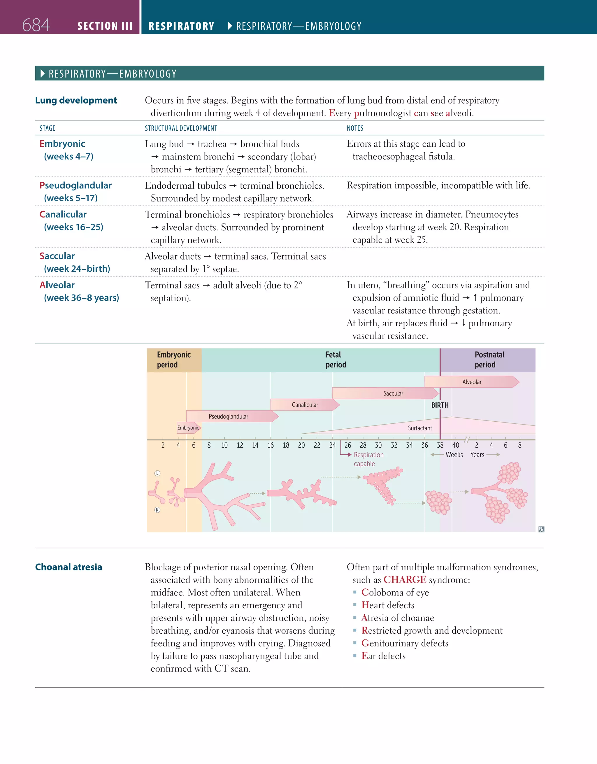


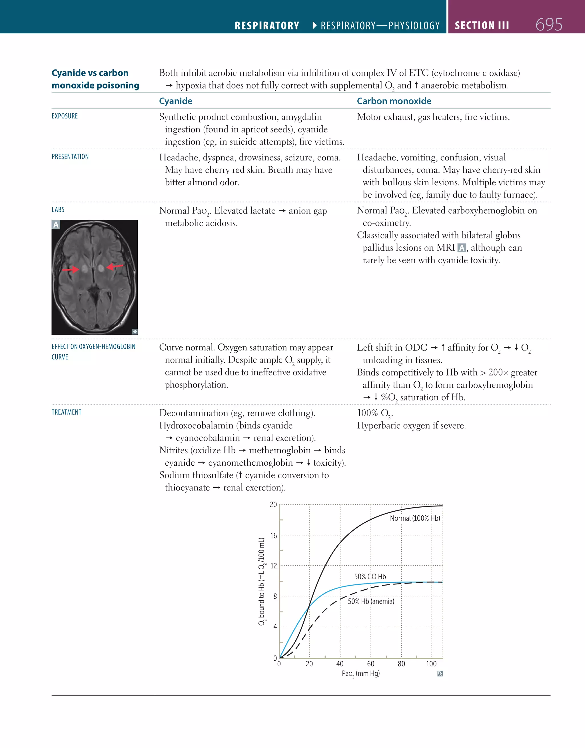


![bipin_ppt_most_important[1] - Read-Only.pptx](https://cdn.slidesharecdn.com/ss_thumbnails/bipinpptmostimportant1-read-only-251220122435-b3a2f11c-thumbnail.jpg?width=640&height=640&fit=bounds)