
Recommended
Recommended
More Related Content
What's hot
What's hot (20)
Similar to Extraction of teeth as part of orthodontics
Similar to Extraction of teeth as part of orthodontics (20)
More from MaherFouda1
More from MaherFouda1 (20)
Recently uploaded
Recently uploaded (20)
Extraction of teeth as part of orthodontics
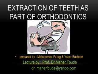
- 1. EXTRACTION OF TEETH AS PART OF ORTHODONTICS • prepared by : Mohammed Farag & Yaser Basheer Lecture by : Prof. Dr Maher Fouda dr_maherfouda@yahoo.com
- 2. Methods of gaining space in the permanent dentition Common methods of gaining space 1) Arch expansion 2) Inter-proximal reduction 3) Molar distalization 4) Selective/ therapeutic extractions
- 3. The upper arch is well suited for expansion than its lower counterpart. Usually done in narrow contracted upper arches with cross bites in the premolar region. Arch expansion
- 4. Arch expansion devices Hyrax expanderQuad helix Haas expanderJackscrew Beidner type.NiTi expander W arch
- 6. •arch expansion by quad helix
- 7. Oclusal view before treatment Beggining of Treatment
- 8. After 4 months After 2 months.
- 9. Before and After Quad-Helix– 5 months total time
- 10. GAINING SPACE BY INTERPROXIMAL REDUCTION • Interproximal reduction is a procedure to create space for crowding and increase stability by flattening curved contact surfaces. • The enamel is removed by using either: • a fine dental bur • a disc in a dental handpiece (drill) or • by hand with an abrasive strip
- 11. Enamel reduction by dental bur Enamel reduction by hand using a abrasivestrip Diamond Strip Abrasive Disc
- 12. maximum of 0.5 mm interproximal reduction per contact is recommended. Therefore, a total of 2.5 mm of tooth-size reduction is possible from cuspid to cuspid
- 13. • Decision to do interproximal reduction is based on the model analysis concerning the overall size discrepancy. • It could also be added up with a Bolton analysis
- 16. GAINING SPACE BY MOLAR DISTALIZATION • An 11-year-old girl was referred to our clinic for orthodontic treatment with a chief complaint of protruding upper anterior teeth and irregular upper and lower • With class1 divission 1 mallocclusion
- 17. Facial and intraoral photographs of the case before treatment (age 11 years)
- 18. Pretreatment lateral cephalometric and panoramic radiographs of the case
- 19. TREATMENT OBJECTIVES • The treatment objectives, based on the clinical examination and the cephalometric analysis, were to • 1. Distalize the maxillary molars to establish a well-intercuspated bilateral Class I molar and canine relationship. • 2. Retract the upper incisors for overjet reduction. • 3. Ideally align the fully erupted lower and upper permanent teeth.
- 20. Occlusal views of the Frog Appliance. A, During activation; B, on the dental cast and immediately after the cementation
- 21. Upper occlusal view of the patient immediately after the distalization (A), and intraoral photographs after cutting of the anchor wires of premolars (B-D) (after 4 months of distalization)
- 22. Lateral cephalometric and panoramic radiographs of the case taken immediately after the distalization
- 23. Facial and intraoral photographs of the case at the end of the fixed orthodontic treatment (age 12 years 4 months).
- 24. EXTRACTION
- 37. As this case illustrates, it may be advantageous to remove second molars instead of premolars in selected patients who cannot be adequately treated without extractions. Second molar extraction can create sufficient space in the posterior segments of patients with crowded arches, providing good long-term facial and dental esthetics. In addition, it is a relatively simple procedure that leaves the patient with the maximum possible number of permanent teeth.
- 45. 11 year old female patient Class II canine and molar relation "". She has a 7mm overjet, a deep bite and5mm tooth- size arch-length discrepancy in the upper arch. The treatment objectives were to align teeth, reduce overjet, achieve aclass I canine relation and accept a class II molar relation.
- 49. Active tiebacks were continued. The patient missed two appointments, shecame back with and edge to edge occlusion . "Arches werere moved to allow the arches to relapse
- 50. Closing the remaining extraction space came from burning anchorage and moving upper molars mesially. Case finished with aclass II molar relation, which was anticipated.
- 53. The dental panoramic tomogram confirmed the presence of all permanent teeth, including developing third molars. The first molars were all restored or carious, and their long-term prognosis was considered to be poor.
- 61. Twelve months into treatment, left maxillary lateral incisor (22) was included for alignment
- 63. Fifteen months into treatment, in the lower arch a 0.016 inch stainless steel archwire was used for further alignment and to start closing spaces
- 64. Eighteen months into treatment, upper and lower 0.019 inch × 0.025 inch stainless steel archwires were. Used for arch coordination and space closure.
- 65. Twenty-seven months into treatment, the case was debonded, the teeth were in well-interdigitated occlusion.
- 68. The side profile X-ray and cephalometric tracing showed: Incisor uprighting (1-NA = 0°); Class II skeletal pattern, ANB angle = 5, (SNA = 80° and SNB = 75) and normal mandibular growth in the vertical orientation (SN-GoGn = 32°, FMA = 23 and Y-axis = 60°).
- 74. The cephalometric analysis showed protrusion in the maxilla and mandible in relation to the cranial base, skeletal Class I malocclusion, dolichofacial pattern, protruding upperand lower incisors with increased axial inclination Normal bone profile, straight facial profile.
- 90. • The patient had already extracted his four first premolars hoping that this would alleviate mild crowding present this had only lead to deep bite and four extraction spaces
- 99. • The four first premolars had already been extracted notice canted maxillary occlusal plane excessive gingival display
- 102. Cephalometric analysis revealed a retrusive mandible (ANB angle 7°) and an increased IMPA angle (94°). The SNA angle was within the normal limits (82º); however, SNB angle was decreased (75º). In other words, patient had a skeletal class II profile accompanied with mandibular dental compensation.
- 111. Mandibular incisor extraction can be an effective treatment option in border line cases with mild crowding in lower arch. In patients with moderate crowding and without excessive mandibular tooth mass, interproximal reduction may be a better alternative. Formation of open gingival embrasures or black triangles is a common side effect of mandibular incisor extraction. Minimal alteration of mandibular arch form is key for success and stable results.
- 120. • a 15-year-old boy who had an Angle Class I malocclusion with a right palatally impacted maxillary canine. The right deciduous canine was also persisted in the mouth. The treatment involved the tunnel traction method, by which the impacted canine was pulled toward the center of the alveolar ridge via the deciduous canine socket.
- 121. PRETREATMENT INTRA ORAL PHOTOGRAPHS
- 124. Intra oral lateral photograph Occlusal photograph
- 125. OCCLUSAL RADIOGRAPH
- 126. PANORAMIC RADIOGRAPH
- 127. DURING DIRECTED TRACTION: CANINE INTO ITS PROPER POSITION
- 130. RADIOGRAPHICALLY, THE RIGHT CANINE DISPLAYED PROPER ROOT INCLINATION AND THE INCISORS REMAINED STABLE AT THE END OF TREATMENT
- 132. .
- 133. Pretreatment extraoral and intraoral photographs
- 134. Transpalatal bar for maintaining arch form, partial canine retraction, and uprighting intrusion force on mandibular posterior teeth
- 135. En masse retraction of 6 maxillary anterior teeth with Ni-Ti coil springs connected from microscrew implants
- 136. Application of intrusion force on maxillary and mandibular posterior teeth.
- 137. Post treatment extraoral and intraoral photographs
- 138. THANX