This document provides guidelines for the prevention and treatment of venous thromboembolism (VTE) in cancer patients from the European Society for Medical Oncology (ESMO). It finds that VTE is a major health problem and leading cause of death in cancer patients. The risk of VTE is higher in cancer patients than those without cancer. Diagnosis of VTE in cancer patients should proceed directly to imaging tests like ultrasound or CT scans without using clinical prediction tools due to their reduced effectiveness in cancer patients. The guidelines provide recommendations for pharmacological and mechanical thromboprophylaxis in surgical cancer patients as well as nonsurgical cancer patients. It finds that low molecular weight heparins are generally recommended for pharmacological prophylaxis but mechanical methods
![CAT complicates the management of anticancer therapies
and is associated with substantial increase of expenditure
for the health care systems.8
Preventing VTE in people with
cancer by pharmacological and non-pharmacological mea-
sures is a challenging and crucial issue. It is important to
identify patients in the highest-risk categories, who can
most benefit from primary thromboprophylaxis.
The efficacy and safety of direct oral anticoagulants
(DOACs) inhibiting activated coagulation factor X (Xa) have
been recently tested for the treatment of CAT and offer an
alternative to low-molecular-weight heparin (LMWH).9,10
The approval status of the agents discussed in this
guideline might differ from country to country. With a focus
on ease of implementation, this updated European Society
for Medical Oncology (ESMO) Clinical Practice Guideline
(CPG) summarises recommendations for prevention and
treatment of VTE in patients with cancer.
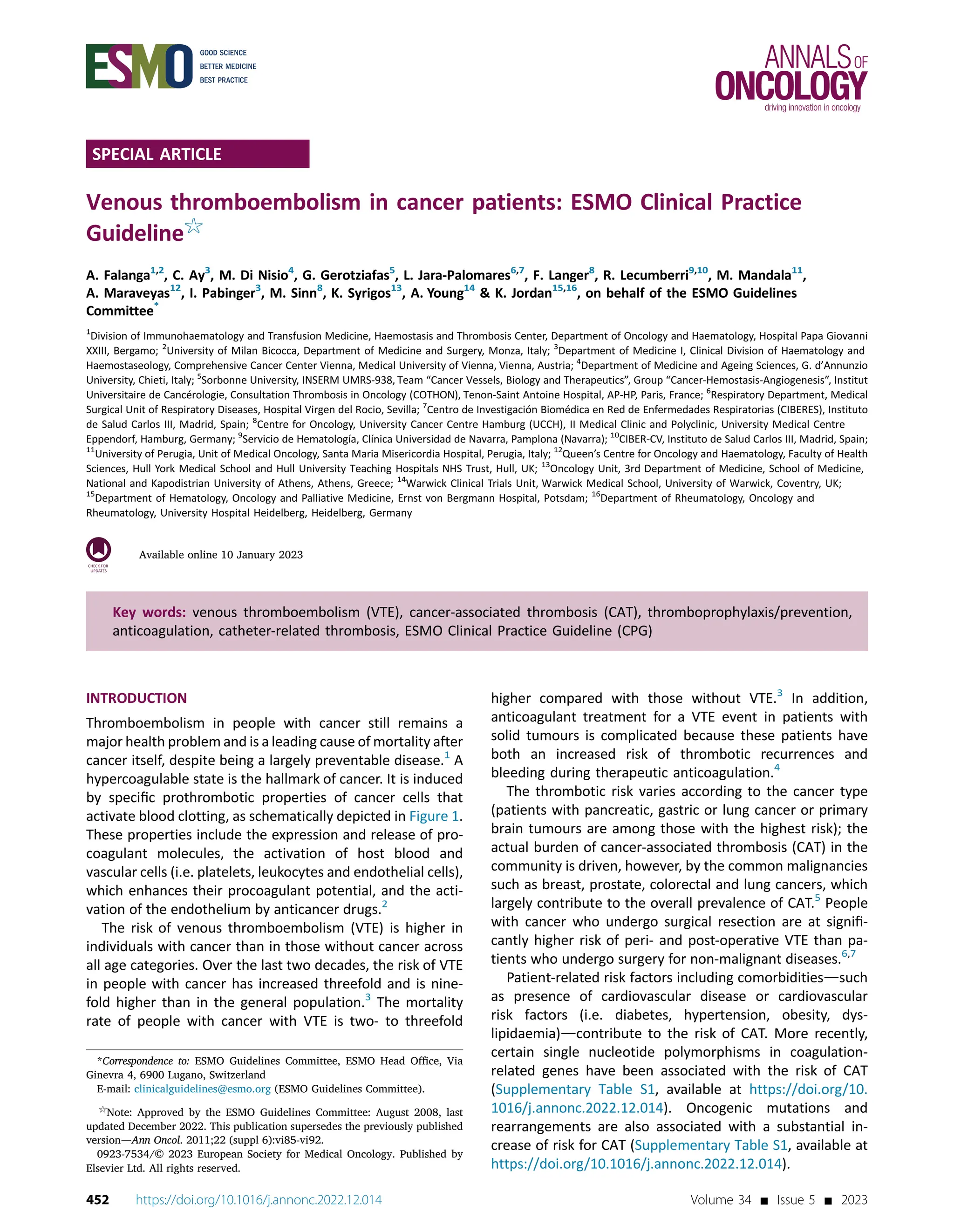
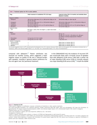
DIAGNOSIS OF VTE
VTE includes deep vein thrombosis (DVT) and pulmonary
embolism (PE). Clinical manifestations of DVT of the legs
include redness, tenderness, swelling, pitting oedema and
appearance of collateral superficial veins, while manifesta-
tions of PE are dyspnoea, chest pain, cough, tachycardia,
cyanosis, dizziness, fainting and excessive sweating.
The diagnosis of VTE, however, cannot rely on the
clinical manifestations alone as the signs and symptoms
are not specific. Imaging is necessary to confirm the
diagnosis. In the general population, diagnostic algorithms
consisting of clinical probability assessment and D-dimer
testing have been established to guide decisions about
who should be referred for compression ultrasonography
(CUS) in case of suspected DVT and computed tomogra-
phy pulmonary angiography (CTPA) in case of suspected
PE.11
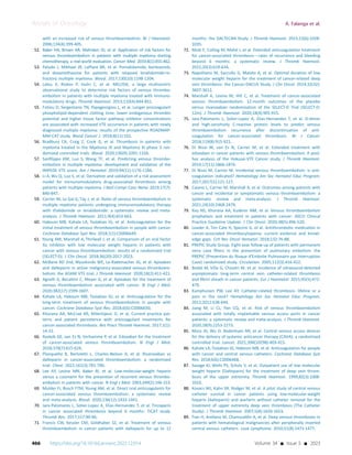
For comparison, the diagnostic algorithm for
non-cancer patients is reported in Supplementary
Figure S1, available at https://doi.org/10.1016/j.annonc.
2022.12.014.
Unfortunately, in cancer patients the performance of
clinical decision rules and D-dimer testing is poor.12
As a
consequence, in these patients, physicians should consider
proceeding to CUS and CTPA directly (Figure 2).
Recommendation
In cancer patients, diagnosis by CUS in case of suspected
DVT and diagnosis by CTPA in case of suspected PE,
without using clinical prediction rules and D-dimer level,
are recommended [I, A].
PRIMARY PREVENTION OF VTE
Table 1 shows available pharmacological and mechanical
VTE prophylaxis options.
Thromboprophylaxis in the surgical setting
General considerations. Assessment of the risk of throm-
bosis and bleeding should be carried out before any surgical
procedure, including cancer surgery. The following factors
are to be considered:
Patient risk factors [i.e. by risk assessment models (RAMs)],
e.g. the Caprini score;13
(see also Supplementary Material,
Tables S2 and S3 and Figure S2, available at https://doi.org/
10.1016/j.annonc.2022.12.014)
Type of intervention (minor surgery, i.e. open or laparo-
scopic of 45-min duration; or major surgery, i.e. open
or laparoscopic of 45-min duration)
Contraindications to pharmacological thromboprophylaxis
(e.g. active bleeding, acute hepatitis or acquired haemo-
philic states, uncontrolled hypertension, acute stroke,
platelet count 25000 ml, lumbar puncture or spinal/
epidural anaesthesia in the next 12 h or in the previous
4 h, ongoing anticoagulant treatment for other indications)
Pharmacological thromboprophylaxis. Pharmacological
thromboprophylaxis with LMWH or unfractionated heparin
(UFH) is standard of care in surgical patients with a high risk
of VTE and a low risk of bleeding. Malignancy is associated
with an increased risk for both thromboembolic and hae-
morrhagic complications. Thus, particular caution is war-
ranted in patients undergoing major cancer surgery, since
the overall riskebenefit ratio of pharmacological throm-
boprophylaxis may be less clear in cancer versus non-cancer
surgical patients.
A recent systematic review and meta-analysis of rando-
mised controlled trials (RCTs) specifically conducted in sur-
gical cancer patients concluded that LMWH, in comparison
with no prophylaxis or mechanical prophylaxis, decreased
the rates of DVT [relative risk (RR) 0.20, 95% confidence
interval (CI) 0.07-0.61] and PE (RR 0.13, 95% CI 0.01-2.25),
but potentially increased the risk of major bleeding (RR
2.47, 95% CI 0.08-74.18).14
No data support the superiority of any LMWH over the
other. Similarly, subcutaneous LMWH once daily (o.d.) and
subcutaneous UFH three times daily (t.d.s.) have comparable
Figure 2. Diagnostic algorithm for suspected DVT and PE in cancer patients.
Purple: general categories or stratification; white: other aspects of management;
turquoise: combination of treatments or other systemic treatments.
CTPA, computed tomography pulmonary angiography; CUS, compression ultra-
sonography; DVT, deep vein thrombosis; PE, pulmonary embolism.
Annals of Oncology A. Falanga et al.
454 https://doi.org/10.1016/j.annonc.2022.12.014 Volume 34 - Issue 5 - 2023](https://image.slidesharecdn.com/esmoguidelines-240120045640-f7f9c8ff/85/esmo-guidelines-pdf-3-320.jpg)
![UFH (1-2 h). To avoid bleeding, the interval between
preoperative subcutaneous injection and surgical procedure
should be generally longer in patients receiving LMWH,
particularly when the highest approved prophylactic dose is
used. Some LMWHs may only be licensed for post-operative
commencement of VTE prophylaxis.
Where different VTE prophylaxis doses are approved for a
given LMWH, the highest dose is recommended to prevent
VTE in cancer patients undergoing major surgery. A pro-
spective, randomised, double-blind, multicentre trial in
patients undergoing surgery demonstrated in the group of
cancer patients that prophylaxis with higher-dose dalteparin
(5000 IU daily) compared with lower-dose dalteparin
(2500 IU daily) reduced DVT rates from 14.9% to 8.5%
(P 0.001), without significant increase in bleeding com-
plications.22
Perioperative prophylaxis with UFH t.d.s. is
equally effective to LMWH o.d. and superior to UFH twice
daily (b.i.d.).16
In a small RCT on 111 cancer patients un-
dergoing oesophagectomy, the incidence of VTE was lower
with nadroparin administered b.i.d. compared with o.d.
(0% versus 9.1%; P ¼ 0.032).23
Nevertheless, more research
is warranted to confirm these findings.
Duration. The duration of post-operative thromboprophy-
laxis should be at least 10 days.24,25
The mean time from
major surgery to VTE occurrence, however, is reported to be
17 days, and in over one-third of patients VTE occurs later
than 21 days after surgery or hospital discharge.7,26
These
data support extended post-operative prophylaxis beyond 10
days in select patients. For example, several meta-analyses
show that extended thromboprophylaxis with LMWH after
major abdominal or pelvic cancer surgery reduces the risk of
VTE compared with conventional duration of 2 weeks or less,
without increasing the risk of major bleeding.14,27
This effect
is not limited to open surgery, but also occurs with laparo-
scopic surgery. In patients undergoing laparoscopic surgery
for colorectal cancer, extended pharmacological prophylaxis
for 4 weeks reduced VTE risk compared with prophylaxis for
1 week with similar bleeding rates.28
Prevention of VTE in non-surgical patients with cancer
General considerations for prevention of VTE in non-surgical
patients with cancer are provided in the Supplementary
Material, available at https://doi.org/10.1016/j.annonc.
2022.12.014.
Ambulatory patients. The risk of VTE is increased in pa-
tients with ‘active cancer’ (as defined in Supplementary
Table S4, available at https://doi.org/10.1016/j.annonc.
2022.12.014).29
This risk is, however, very variable
depending on individual factors (previous history of
thrombosis, immobility, cardiovascular risk factors), the
type and stage of cancer, the time since cancer diagnosis
(within 6 months after first diagnosis and after progression
or recurrence) and the use of systemic anticancer therapy;30
therefore, primary thromboprophylaxis is not justified in all
of these patients. Identifying patients at high risk is of
particular interest in this setting. RAMs and material for the
calculation of the risk of CAT are provided in the
Supplementary Material, Tables S2 and S3 and Figure S2,
available at https://doi.org/10.1016/j.annonc.2022.12.014,
the latter of which exemplifies the inclusion of a biomarker
of hypercoagulability (i.e. D-dimer) to clinical predictors.
Since thrombogenic potential varies depending on the type
of cancer or the presence of certain oncogene mutations or
rearrangements, the authors endorse the development of
cancer-specific RAMs to further refine current risk stratifi-
cation approaches or to develop new models that incor-
porate promising biomarkers. Results of these studies could
alter the approach to risk stratification in the future.
A recent meta-analysis and an individual patient data
meta-analysis including 10 431 ambulatory patients with
cancer who participated in phase III RCTs showed that
prophylactic doses of heparins reduced symptomatic VTE by
w40% without increasing the bleeding risk as compared
with no prophylaxis.31,32
Two studies in pancreatic cancer using higher doses of
LMWHs (150 IU/kg dalteparin or 1 mg/kg enoxaparin) for 3
months in patients receiving systemic anticancer treatment
have shown an 85% and 65% RR reduction of any VTE and
the composite of DVT and PE with number needed to treat
(NNT) of 5 and 11, respectively.33
The recent AVERT and CASSINI studies randomised 574
and 841 patients, respectively, with intermediate-high risk
of VTE [estimated thrombosis risk 9.6% over 6 months
using the Khorana risk score (KRS) 2] to either placebo or
a factor Xa inhibitor for 6 months.34,35
In the AVERT study,
apixaban (2.5 mg b.i.d.) was associated with a lower rate of
VTE [4.2% versus 10.2%; hazard ratio (HR) 0.41, 95% CI 0.26-
0.65, NNT ¼ 17] and a higher rate of major bleeding (3.5%
versus 1.8%; HR 2.00, 95% CI 1.01-3.95; number needed to
harm ¼ 59).34
In the CASSINI study, rivaroxaban (10 mg
o.d.) did not achieve significant VTE risk reduction over
placebo (6.0% versus 8.8%; HR 0.66, 95% CI 0.4-1.09) with a
major bleeding rate of 2% versus 1% (HR 1.96, 95% CI 0.59-
6.49).35
Neither study included patients with severe
thrombocytopaenia (platelet count 50 000/mm3
) or renal
dysfunction [creatinine clearance (CrCl) 30 ml/min].
Shared decision making should take into account utility of
oral route, renal and hepatic function, drugedrug in-
teractions and risk of bleeding, with caution to be taken in
patients with gastrointestinal malignancies, particularly if
the primary lesion is luminal and non-resected.
The duration of pharmacological thromboprophylaxis in
ambulatory cancer patients cannot be firmly determined.
The first 3 months from diagnosis and anticancer treatment
initiation comprise the conventional higher-risk period
during which 50% of VTE episodes occur, and all existing
studies have covered at a minimum this period; the two
DOAC studies34,35
had a predetermined, 6-month throm-
boprophylaxis period and the two pancreatic ductal
adenocarcinoma studies33
explored a maximum of 3
months at a higher dose of LMWH. These patients, however,
also have a KRS of 2 points and very often progressive
disease; hence, prolonging thromboprophylaxis with a
DOAC up to 6 months remains evidence-based. For
Annals of Oncology A. Falanga et al.
456 https://doi.org/10.1016/j.annonc.2022.12.014 Volume 34 - Issue 5 - 2023](https://image.slidesharecdn.com/esmoguidelines-240120045640-f7f9c8ff/85/esmo-guidelines-pdf-5-320.jpg)
![thromboprophylaxis beyond 6 months an individualised
approach should be considered.
Patients with cancer hospitalised for an acute medical
illness. LMWHs represent the agents of choice for VTE
thromboprophylaxis in patients with cancer hospitalised for
an acute medical illness. The recommendation of pharma-
cological thromboprophylaxis to prevent VTE for inpatients
with cancer is based on the results from large clinical trials
of hospitalised medical patients.36
Studies suggest that
thromboprophylaxis in cancer patients hospitalised for
acute medical illness, though frequent, may: (i) not be
appropriately targeted,37
(ii) be based on risk assessment
tools (e.g. Padua score or IMPROVE score) that have limited
accuracy in cancer patients38
(KRS, according to a single
retrospective study,39
might have some value in this setting
and needs to be further investigated) and (iii) not benefit
cancer patients as a subgroup.40
The use of DOACs in this setting, including extended
thromboprophylaxis for 4 weeks after discharge, is currently
not recommended since the reduction of VTE compared
with standard heparin prophylaxis was offset by an increase
in major bleeding.41
Dedicated studies to define optimal pharmacological
prophylaxis in cancer patients hospitalised for acute medical
illness are required.
Patients with multiple myeloma. The incidence of VTE in
patients with multiple myeloma (MM) ranges between 8
and 22 per 1000 person-years, and w8%-10% of patients
will suffer symptomatic VTE during the course of the dis-
ease.42
The risk of VTE in these patients is influenced by:
The characteristics of MM (e.g. time since diagnosis,
levels and type of paraproteinaemia)43-45
The anti-myeloma treatments (e.g. immunomodulatory
drugs with high-dose dexamethasone, multi-agent
chemotherapy or anthracyclines)46-50
The patient-related intrinsic risk factors (e.g. obesity, car-
diovascular disease or cardiovascular risk factors and
age)51
Other triggering risk factors [e.g. central venous cathe-
ters (CVCs), use of erythropoietin or other colony-
stimulating factors, recent hospitalisation for acute
medical illness or surgical interventions].
The International Myeloma Working Group (IMWG), the
National Comprehensive Cancer Network (NCCN) and the
European Myeloma Network (EMN) adopted a simplified
algorithm (in practice since 2014), based on the concept
that treatment with immunomodulatory imide drugs
(IMiDs) is the major determinant of VTE risk in patients with
MM. Nevertheless, this algorithm is based on an empirical
scoring of the VTE risk and its accuracy is limited.52-56
The
clinical decision tool proposed by the IMWG/NCCN/EMN is
shown in Supplementary Table S5, available at https://doi.
org/10.1016/j.annonc.2022.12.014.
The IMPEDE VTE and the SAVED scores have been
recently proposed. These scores were derived from the
retrospective analysis of the Surveillance, Epidemiology and
End Results (SEER)eMedicare database (Supplementary
Table S6, available at https://doi.org/10.1016/j.annonc.
2022.12.014).57,58
Both scores showed borderline accuracy
and need independent validation in order to be proposed in
clinical practice.
There is a need for prospective derivation and validation
of an RAM for VTE in patients with MM. Due to the paucity
of specific clinical trials for thromboprophylaxis in patients
with MM, the recommendations for thromboprophylaxis
stem from those with solid tumours, except for patients
receiving IMiDs.59
Recommendations
Thromboprophylaxis in the surgical setting
Unless contraindicated due to a high risk of bleeding,
pharmacological VTE prophylaxis with LMWH (preferred)
or UFH is recommended in patients undergoing major
cancer surgery [I, A]. Fondaparinux may be used as an
alternative [II, C].
Mechanical methods such as IPC or GCSs are suggested
as an alternative when pharmacological VTE prophylaxis
is contraindicated (e.g. in the presence of active
bleeding) [II, B]. Mechanical methods may be used in
combination with pharmacological VTE prophylaxis in pa-
tients at exceedingly high risk of VTE [II, C].
Depending on the heparin type and dosage, commence-
ment of pharmacological thromboprophylaxis with
LMWH or UFH 2-12 h preoperatively is suggested in can-
cer surgical patients [II, B].
Where several prophylactic dosages are approved for a
given LMWH, the highest prophylactic LMWH dose o.d.
or 5000 IU UFH t.d.s. is recommended [II, A].
Patients undergoing major cancer surgery should receive
pharmacological thromboprophylaxis for at least 10 days
post-operatively [I, A]. In patients with cancer undergo-
ing open abdominal or pelvic surgery or laparoscopic
colorectal cancer surgery, extended post-operative VTE
prophylaxis for 4 weeks with LMWH is recommended
[I, A].
Prevention of VTE in non-surgical patients with cancer
Patient education materials on CAT, including risk fac-
tors, signs and symptoms and information on positive
lifestyle factors, should be one component of the infor-
mation package provided to all ambulatory patients
scheduled to receive systemic anticancer treatment
[III, A].
Cancer patients should be offered a CAT risk assessment
and have an opportunity to discuss their particular risk
[III, B].
VTE risk assessment should be based on validated RAMs
such as the KRS, the Prospective Comparison of Methods
for thromboembolic risk assessment with clinical
Perceptions and AwareneSS in real-life patients-Cancer-
Associated Thrombosis (COMPASS-CAT) or the Vienna
Cancer and Thrombosis Study (Vienna-CATS) nomogram
score [III, C].
A. Falanga et al. Annals of Oncology
Volume 34 - Issue 5 - 2023 https://doi.org/10.1016/j.annonc.2022.12.014 457](https://image.slidesharecdn.com/esmoguidelines-240120045640-f7f9c8ff/85/esmo-guidelines-pdf-6-320.jpg)
![An estimated risk of VTE 8%-10% at 6 months is sug-
gested as threshold for discussing primary thrombopro-
phylaxis [II, C]. This risk is observed in patients with a
KRS 2 and can individually be calculated with the
Vienna-CATS nomogram score and the COMPASS-CAT
score.
For ambulatory pancreatic cancer patients on first-line
systemic anticancer treatment, LMWH given at a higher
dose (150 IU/kg dalteparin or 1 mg/kg enoxaparin) for a
maximum of 3 months may be considered [II, C].
In ambulatory cancer patients starting systemic anti-
cancer treatment who have a high thrombosis risk, apix-
aban, rivaroxaban or LMWH may be considered for
primary thromboprophylaxis for a maximum of 6 months
[I, B].
In hospitalised cancer patients confined to bed with an
acute medical complication, prophylaxis with LMWH,
UFH [I, B] or fondaparinux [II, B] is recommended.
Where concerns of DOAC safety exist and the patient is
perceived as having clinically important risk for VTE,
LMWH at conventional primary thromboprophylaxis
dosing may be administered [II, C].
Patients with MM
All patients with MM should be offered a VTE risk assess-
ment and have the opportunity to discuss their partic-
ular risk [III, B].
Patients with MM scheduled to receive or receiving IMiD
treatment should be assessed for VTE risk with the
IMWG/NCCN/EMN score [III, B].
In ambulatory patients with MM receiving IMiD treat-
ment combined with low-dose dexamethasone and
without additional risk factors, aspirin (100 mg/day) is
recommended [III, B].
In ambulatory patients with MM classified as high risk
for VTE, pharmacological thromboprophylaxis with
LMWH for 3-6 months is recommended [II, B].
Extension of thromboprophylaxis should be considered
on a case-by-case basis [IV, B].
Apixaban 2.5 mg b.i.d. or rivaroxaban 10 mg o.d. are
potential options in patients with CrCl 30 ml/min
who present contraindications or intolerance to LMWH
[IV, C].
TREATMENT OF CAT
For the treatment of VTE in cancer patients, the agents
listed in Table 2 are available options. Treatment of CAT is
usually divided into an acute phase (first 5-10 days after
diagnosis), a long-term phase (first 3-6 months) and an
extended phase (beyond 6 months) (Figure 3).
Acute phase
The evidence on the treatment of CAT during the early
phase is largely indirect and based on RCTs conducted in
non-cancer patients with acute DVT or PE who were
assigned to LMWH versus UFH or LMWH versus fondapar-
inux. All parenteral agents were administered for w5-10
days followed by vitamin K antagonists (VKAs; target in-
ternational normalised ratio range between 2.0 and 3.0). A
meta-analysis of these studies suggests that LMWH may
reduce mortality and recurrent VTE compared with UFH or
fondaparinux with a similar risk of major bleeding.60
The
use of DOACs (rivaroxaban and apixaban) in the acute phase
is supported by three prospective RCTs.61-63
In cancer pa-
tients with severe renal impairment, defined as CrCl 30
ml/min, UFH might be preferable over LMWH or fonda-
parinux, whereas the latter might be considered in patients
with CAT and a prior history of HIT.60
Long-term phase
LMWH has represented the first-line treatment of CAT for
about two decades. A meta-analysis of studies on cancer
patients with DVT or PE found a lower rate of recurrent
thrombosis and similar risk of bleeding with a 6-month
course of LMWH compared with a VKA.64
The main char-
acteristics of these studies are shown in Supplementary
Table S7, available at https://doi.org/10.1016/j.annonc.
2022.12.014.
LMWH requires daily subcutaneous injections, which may
impair the patient’s quality of life due to the persistence of
anticoagulant therapy.65
Five open-label RCTs evaluated the efficacy and safety of
direct oral factor Xa inhibitors (i.e. edoxaban, rivaroxaban
and apixaban) for the treatment of symptomatic or inci-
dental VTE in patients with active cancer.61-63,66,67
These
trials considered study treatment durations of 6-12 months
and all used the same regimen of dalteparin as the
comparator, based on the results of the CLOT study.68
The
main characteristics of these studies are shown in
Supplementary Table S8, available at https://doi.org/10.
1016/j.annonc.2022.12.014.
The Hokusai VTE cancer trial demonstrated that edoxaban
was noninferior to dalteparin for the composite outcome of
recurrent VTE or major bleeding.66
The rate of recurrent VTE
was lower with edoxaban (7.9% versus 11.3%), but the rates
of major bleeding (6.9% versus 4.0%) and clinically relevant
non-major bleeding (14.6% versus 11.1%) were higher
(Supplementary Table S8, available at https://doi.org/10.
1016/j.annonc.2022.12.014). The relative excess of major
bleeding was observed in patients with gastrointestinal
cancer, although the absolute number of severe bleeding
events was low and comparable to dalteparin.
In the pilot SELECT-D trial, the 6-month cumulative rate
of recurrent VTE was lower with rivaroxaban (4% versus
11%), but the rate of major bleeding was numerically higher
(6% versus 4%) and clinically relevant non-major bleeding
was significantly increased (13% versus 4%) with rivarox-
aban.61
The incidence of major bleeding was particularly
increased in patients with oesophageal and gastro-
oesophageal junction cancers (36% versus 11%).
In the ADAM VTE trial, apixaban was associated with
significantly lower incidence of recurrent VTE (0.7% versus
6.3%) with no increase in major bleeding (0% versus 1.4%)
or clinically relevant non-major bleeding (6.2% versus 4.2%)
Annals of Oncology A. Falanga et al.
458 https://doi.org/10.1016/j.annonc.2022.12.014 Volume 34 - Issue 5 - 2023](https://image.slidesharecdn.com/esmoguidelines-240120045640-f7f9c8ff/85/esmo-guidelines-pdf-7-320.jpg)

![Special populations
Data in this setting largely derive from retrospective trials in
the non-cancer population (Figure 5). This topic has been
extensively reviewed elsewhere.9
Renal impairment. Patients with VTE and concomitant renal
impairment are at higher risk of major bleeding and
recurrent VTE during anticoagulant treatment compared
with patients with normal renal function. Post hoc and
subgroup analyses suggest that in patients with CAT and
moderate renal impairment (CrCl 30-60 ml/min), the effi-
cacy and safety of LMWH and DOACs are generally consis-
tent with those of cancer patients without renal
impairment. Patients with CAT and severe renal impairment
(CrCl 30 ml/min) were excluded from the pivotal trials on
the treatment of CAT. In these patients, two options may be
considered: UFH followed by VKAs or LMWH with the dose
adjusted to anti-Xa activity level for the treatment of CAT.
Data on the dosing and safety of DOACs in patients with
CAT and severe renal impairment are lacking.
Obese patients. Based on limited data from observational
and retrospective studies, it is suggested that in patients
with extreme body weight (120 kg or body mass index
40 kg/m2
) the dose of LMWH should be calculated based
on a person’s actual body weight without capping at a
maximum dose. Although subgroup analyses have not
shown any significant reduction in efficacy and safety of
DOACs in obese patients, DOACs should be used cautiously
in patients weighing 120 kg.79
Patients with thrombocytopaenia. In patients with persis-
tent, severe thrombocytopaenia (50 000/ml), two man-
agement strategies have been proposed based on the
underlying risk of thrombosis recurrence or extension.80
In
patients with acute VTE who are at high risk of thrombus
progression (i.e. first 30 days from thromboembolic event,
segmental or more proximal PE, proximal DVT or a history of
recurrent thrombosis), full-dose anticoagulation may be
considered in combination with platelet transfusion support
aiming at platelet count 40-50 000/ml. If the risk of
thrombosis progression is deemed to be low (i.e. 30 days
from thromboembolic event, distal DVT, isolated sub-
segmental PE), intermediate- to prophylactic-dose LMWH
may be considered with temporary discontinuation of
anticoagulation if the platelet count falls below 25 000/ml.
In patients with platelet count 50 000/ml, full therapeutic
dose anticoagulation should be considered. Data on the use
of DOACs for the treatment of CAT in the presence of se-
vere thrombocytopaenia are lacking.
Vena cava filters in CAT
The efficacy and safety of vena cava filters for acute CAT have
not been evaluated in RCTs. In the general population, the
use of vena cava filters in addition to anticoagulant treat-
ment may reduce the incidence of PE, but increases the risk
of DVT and has no survival benefits over anticoagulation
alone.81
Inferior vena cava filters may be indicated in the
acute phase of VTE when there is a contraindication to
anticoagulant treatment. The potential value of vena cava
filters is likely reduced after the acute phase when the risk of
thrombosis recurrence or extension diminishes significantly.
Recommendations
In patients with CAT, LMWH, UFH, fondaparinux, apixa-
ban or rivaroxaban are recommended treatments for
the acute phase [I, A]. LMWH is preferred over UFH or
fondaparinux [V, A]. UFH may be considered in patients
with CAT and severe renal impairment (defined as CrCl
30 ml/min) [IV, C].
Long-term anticoagulation for at least 6 months includes
LMWH, apixaban, edoxaban or rivaroxaban which are
preferred over VKAs [I, A]. VKAs may be used if LMWH
or direct factor Xa inhibitors are not accessible [IV, C].
In patients with luminal gastrointestinal cancer, LMWH is
preferred for treating CAT [II, B]. Similar considerations
potentially apply to patients with urothelial cancer
[II, B]. The use of oral factor Xa inhibitors should consider
patient preferences [IV, C].
In patients at high risk for gastrointestinal bleeding, such
as those with active gastroduodenal ulcers or patients
receiving strong inhibitors or inducers of P-glycoprotein
and CYP3A4, LMWH is preferred [IV, B]. The author panel
acknowledges that only limited evidence is available on
drugedrug interactions between direct factor Xa inhibi-
tors and systemic antineoplastic therapy.
Extended anticoagulation beyond the initial 6 months
with LMWH, apixaban, edoxaban, rivaroxaban or VKAs
should be considered for patients with active cancer in
whom the risk of recurrent thrombosis is higher and
may outweigh that of bleeding [III, B]. The riskebenefit
profile of anticoagulant therapy should be regularly
assessed to ensure a favourable balance [IV, C].
For incidentally detected VTE, the same treatment as for
symptomatic VTE is recommended [II, A].
Anticoagulant therapy is suggested for most of the pa-
tients with subsegmental PE [II, A].
In patients with high risk of bleeding or single incidental
subsegmental PE without concomitant DVT, provided
that there is adequate cardiopulmonary reserve, a
watchful approach or a shorter course of anticoagulation
may be considered [V, C].
The insertion of vena cava filters is suggested in patients
with acute and life-threatening VTEs who have absolute
contraindications to anticoagulant therapy [III, B] or as
an adjunct to anticoagulation in patients with recurrent
VTE or extension of thrombosis despite optimal antico-
agulant therapy [IV, C].
PREVENTION AND MANAGEMENT OF CATHETER-RELATED
VTE IN ADULTS WITH CANCER
CVCs are commonly used for patients with cancer receiving
intravenous chemotherapy or other systemic anticancer
medications or supportive care. Upper-extremity DVT is a
Annals of Oncology A. Falanga et al.
462 https://doi.org/10.1016/j.annonc.2022.12.014 Volume 34 - Issue 5 - 2023](https://image.slidesharecdn.com/esmoguidelines-240120045640-f7f9c8ff/85/esmo-guidelines-pdf-11-320.jpg)
![common complication of having a CVC. Most catheter-
related thromboses (CRTs) are asymptomatic82
and may
go undetected. The overall rate of CRT is estimated to be
14%-18% with w5% becoming symptomatic.83
In a recent
systematic review of 80 studies (39 148 cancer patients with
CVCs), implantable ports had a decreased risk of thrombosis
compared with peripherally inserted central catheters
(PICCs) (OR 0.20, 95% CI 0.09-0.43).84,85
Pharmacological prophylaxis of CRT
A Cochrane review and meta-analysis of people with cancer
and CVCs86
found that LMWH decreased the incidence of
symptomatic CRT up to 3-month follow-up in comparison
with no LMWH (RR 0.43, 95% CI 0.22-0.31), although the
certainty of evidence was moderate due to serious risk of
bias (Supplementary Figure S3, available at https://doi.org/
10.1016/j.annonc.2022.12.014). This review concluded that,
compared with no prophylaxis, LMWH may reduce CRT
without increasing the risk of bleeding (Supplementary
Figure S4, available at https://doi.org/10.1016/j.annonc.
2022.12.014), whereas VKAs may lower the risk of CRT,
but potentially increase the risk of bleeding. Nevertheless,
the absolute effect is low (LMWH: 38 fewer events per
1000, VKA: 31 fewer events per 1000). There is no
improvement in mortality due to anticoagulation
(Supplementary Figure S5, available at https://doi.org/10.
1016/j.annonc.2022.12.014) and the burden of injecting
daily LMWH or intake of VKA is considerable.
Treatment of established catheter-related VTE
Previous recommendations for the treatment of CRT were
mainly based on the extrapolation of the results from
clinical trials evaluating the treatment of lower-limb DVT
and two prospective cohorts and one retrospective cohort
that had evaluated the efficacy and safety of LMWH plus
VKA.87-89
The evidence remains scant.
In one retrospective and two prospective studies rivar-
oxaban was used to treat CRT.90-92
These three studies are
small and may be unreliable. Properly powered randomised
trials are needed to evaluate the safety and efficacy of
DOACs for the treatment of CRT.
Anticoagulation versus catheter removal. In a retrospective
study, 112 cancer patients with CRT underwent a variety of
therapeutic interventions that included anticoagulation, CVC
removal or a combination of both. No patient developed a
PE or compromise of the limb. Only four patients required
delayed catheter removal due to persistence of their symp-
toms.93
In another prospective study, 74 cancer patients with
CRT were treated with anticoagulants for 3 months. No
recurrent DVT events were reported, and no catheter was
removed due to malfunction or thrombosis extension.88
In a
recent retrospective study including 83 patients with PICC-
associated thrombosis, 62 were managed with catheter
removal alone, while 21 underwent PICC removal followed
by therapeutic anticoagulation. No patient in the anti-
coagulation group developed progressive thrombosis
compared with 6.4% of patients treated with catheter
removal alone, although major bleeding was higher in the
anticoagulation group (28.5% versus 4.8%).94
Duration of anticoagulant therapy. A systematic review
including 23 studies (no RCTs) evaluated the efficacy and
safety of different durations of anticoagulant treatment for
CRT.95
Duration of anticoagulation in most studies was be-
tween 3 and 6 months. The heterogeneity of study designs,
populations and outcome definitions does not allow firm
conclusions to be drawn on the optimal duration of anti-
coagulant therapy. Data extrapolated from studies of pa-
tients with provoked lower-limb DVT are frequently used in
the setting of CRT. For this reason, if the CVC is maintained
after completion of 3 months of anticoagulation, the sce-
nario would be comparable to that of a DVT related to a
persistent risk factor. Extended secondary prophylaxis
beyond 3 months may be considered.
Recommendations
Routine pharmacological prophylaxis of CRT is not rec-
ommended [II, D].
For the treatment of symptomatic CRT in cancer pa-
tients, anticoagulant treatment is recommended for a
minimum of 3 months [III, A]. LMWH is suggested,
although, in the absence of direct comparisons between
anticoagulants in this setting, VKAs or DOACs may be
considered alternative options [IV, C].
It is recommended to remove the catheter if it is not
needed or is infected, anticoagulant treatment is contra-
indicated or there is clinical deterioration due to
thrombus extension despite treatment [III, B].
In patients with CRT, who have completed 3 months of
anticoagulant treatment, extended anticoagulation until
catheter removal is suggested, if the patient’s bleeding
risk is low [IV, C].
METHODOLOGY
This CPG was developed in accordance with the ESMO stan-
dard operatingproceduresforCPGdevelopment(http://www.
esmo.org/Guidelines/ESMO-Guidelines-Methodology). The
relevant literature has been selected by the expert authors.
Assessment prioritised recent literature [1 January 2020-
manuscript resubmission (December 2022)] and its evalu-
ation in the context of existing systematic reviews that focus
on earlier knowledge. The lead author (AF) assigned co-
authors to subgroups for evaluating topics, questions and
literature based on their expertise (FL, KS and MS: throm-
boprophylaxis in the surgical setting; AM, CA, GG and
IP: prevention of VTE in non-surgical patients; LJP, MDN
and MM: treatment of CAT; RL, AY and IP: prevention and
management of CRT). Further literature searching and
validation was conducted by the ESMO Guidelines Com-
mittee Subject Editor (KJ). Each co-author subgroup drafted
manuscript text and recommendation statements. The full
manuscript was compiled and revised by AF and KJ.
A. Falanga et al. Annals of Oncology
Volume 34 - Issue 5 - 2023 https://doi.org/10.1016/j.annonc.2022.12.014 463](https://image.slidesharecdn.com/esmoguidelines-240120045640-f7f9c8ff/85/esmo-guidelines-pdf-12-320.jpg)


![PERI-PROSTHETIC FRACTURE NAIL-PLATE CONSTRUCT [NPC].pptx](https://cdn.slidesharecdn.com/ss_thumbnails/drarunkumardrmohamedashrafperiprostheticfrasturenail-plateconstructnpc-260209164459-7e9d15a1-thumbnail.jpg?width=640&height=640&fit=bounds)