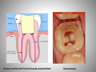
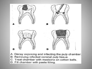
Endodontics for primary teeth
overview
• Diagnosis and case selection for pulp therapy
• Treatment of Deep Caries ( indirect pulp therapy )
• Vital Pulp Exposure ( direct pulp capping, pulpotomy,
partial pulpectomy)
• Pulpless Teeth ( non-vital pulp therapy)
• Failure of pulp therapy
Caries + unhealthy pulp
1. Vital pulp (Irreversible pulpitis / degenerating pulp)
2. Necrotic pulp
Root canal treatment
(pulpectomy)
Caries + healthy pulp
(Reversible pulpitis)
Caries management
Indirect pulp therapy (IPT)
Direct pulp therapy (pulpotomy)
Vital pulp
therapy
Non-vital
pulp
therapy
DIAGNOSTIC AIDS IN THE SELECTION
OF TEETH FOR VITAL PULP
THERAPY
1- HISTORY OF PAIN:
• A severe toothache at night usually signals extensive degeneration
of the pulp and calls for more than a conservative type of pulp
therapy. A spontaneous toothache of more than momentary
duration occurring at any time (unprovoked) usually means that
pulpal disease has progressed too far for treatment with even a
pulpotomy.
• A toothache coincident with or immediately after a meal may not
indicate extensive pulpal inflammation. The pain may be caused by
an accumulation of food within a carious lesion, by pressure, or by a
chemical irritation to vital pulp protected by only a thin layer of
intact dentin.
2- CLINICAL SIGNS AND SYMPTOMS:
• A gingival abscess or a draining fistula associated with a tooth with
a deep carious lesion is an obvious clinical sign of an irreversibly
diseased pulp
• Abnormal tooth mobility is another clinical sign that may indicate a
severely diseased pulp
• Sensitivity to percussion or pressure is a clinical symptom
suggestive of at least some degree of pulpal disease, but the
degenerative stage of the pulp is probably of the acute
inflammatory type
• Tooth mobility or sensitivity to percussion or pressure may be a
clinical signal of other dental problems as well, such as a high
restoration or advanced periodontal disease. However, when this
clinical information is identified in a child and is associated with a
tooth having a deep carious lesion, the problem is most likely to be
caused by pulpal disease and possibly by inflammatory involvement
of the periodontal ligament
• 3- RADIOGRAPHIC INTERPRETATION:
• A recent x-ray film must be available to examine for evidence of
periradicular or periapical changes, such as thickening of the
periodontal ligament or rarefaction of the supporting bone. These
conditions almost always rule out treatment other than an
endodontic procedure or extraction of the tooth.
Remember that:
• The proximity of carious lesions to the pulp cannot always be
determined accurately in the x-ray film.
4- PULP TESTING:
• The value of the electric pulp test in determining the condition of
the pulp of primary teeth is questionable
• Thermal tests have reliability problems in the primary dentition,
too. The lack of reliability is possibly related to the young child’s
inability to understand the tests.
5-PHYSICAL CONDITION OF THE PATIENT :
• In seriously ill children, extraction of the involved tooth after
proper premedication with antibiotics, rather than pulp
therapy, should be the treatment of choice. Children with
conditions that render them susceptible to sub-acute
bacterial endocarditis or those with nephritis, leukemia, solid
tumors, idiopathic cyclic neutropenia, or any condition that
causes cyclic or chronic depression of granulocyte and
polymorphonuclear leukocyte counts should not be subjected
to the possibility of an acute infection resulting from failed
pulp therapy
EVALUATION OF TREATMENT PROGNOSIS
BEFORE PULP THERAPY
• Other factors to consider include the following:
1. The level of patient and parent cooperation and motivation in
receiving the treatment
2. The level of patient and parent desire and motivation in maintaining
oral health and hygiene
3. The caries activity of the patient and the overall prognosis of oral
rehabilitation
4. The stage of dental development of the patient
5. The degree of difficulty anticipated in adequately performing
the pulp therapy (instrumentation) in the particular case
• 6. Space management issues resulting from previous extractions,
preexisting malocclusion, ankylosis, congenitally missing teeth, and
space loss caused by the extensive carious destruction of teeth and
subsequent drifting
• 7. Excessive extrusion of the pulpally involved tooth resulting from
the absence of opposing teeth. These examples, in any
combination, illustrate the almost infinite number of treatment
considerations that could be important in an individual patient with
pulpal pathosis.
TREATMENT OF THE DEEP
CARIOUS LESION
• Indirect pulp treatment: It is a procedure performed in a tooth with
a deep carious lesion approximating the pulp but without signs or
symptoms of pulp degeneration (irreversible pulpitis). The caries
surrounding the pulp is left in place to avoid pulp exposure and is
covered with a biocompatible material. A radiopaque liner such as a
dentin bonding agent, resin modified glass ionomer, calcium
hydroxide, zinc oxide/eugenol, or glass ionomer cement is placed
over the remaining carious dentin to stimulate healing and repair. If
calcium hydroxide is used, a glass ionomer or reinforced zinc
oxide/eugenol material should be placed over it to provide a seal
against microleakage since calcium hydroxide has a high solubility
and poor seal.
• The use of glass ionomer cements or reinforced zinc oxide/eugenol
restorative materials has the additional advantage of inhibitory
activity against cariogenic bacteria. The tooth then is restored with
a material that seals the tooth from microleakage.
• Current literature indicates that there is inconclusive evidence that
it is necessary to reenter the tooth to remove the residual caries. As
long as the tooth remains sealed from bacterial contamination, the
prognosis is good for caries to arrest and reparative dentin to form
to protect the pulp.
• Stepwise caries excavation vs. partial caries removal
VITAL PULP EXPOSURE
• The appropriate procedure should be selected only after a careful
evaluation of the patient’s symptoms, results of diagnostic tests,
and conditions at the exposure site.
• SIZE OF THE EXPOSURE AND PULPAL HEMORRHAGE:
• The size of the exposure, the appearance of the pulp, and the
amount of bleeding are valuable observations in diagnosing the
condition of the primary pulp
• The most favorable condition for vital pulp therapy is the small
pinpoint exposure surrounded by sound dentin. However, a true
carious exposure, even of pinpoint size, will be accompanied by
inflammation of the pulp, the degree of which is usually directly
related to the size of the exposure
• excessive hemorrhage at the point of carious exposure or during
pulp amputation is invariably associated with hyperemia and
generalized inflammation of the pulp. When a generalized
inflammation of the pulp is observed, endodontic therapy or
extraction of the tooth is the treatment of choice
• DIRECT PULP CAPPING:
• It is generally agreed that pulp-capping procedures should be
limited to small exposures that have been produced accidentally by
trauma or during cavity preparation or to true pinpoint carious
exposures that are surrounded by sound dentin.
• Pulp capping should be considered only for teeth in which there is
an absence of pain, with the possible exception of discomfort
caused by the intake of food. In addition, there should be either no
bleeding at the exposure site, as is often the case in a mechanical
exposure, or bleeding in an amount that would be considered
normal in the absence of a hyperemic or inflamed pulp.
• Calcium hydroxide remains the standard material for pulp capping
normal vital pulp tissue.
• In addition, the use of mineral trioxide aggregate (MTA) has shown
promise, but further research would be helpful.
• Many clinicians prefer pulpotomy over direct pulp capping in
primary teeth because of its lower success rate.
• PULPOTOMY:
• It is performed in a primary tooth with extensive caries but without
evidence of radicular pathology when caries removal results in a
carious or mechanical pulp exposure. The coronal pulp is
amputated, and the remaining vital radicular pulp tissue surface is
treated with a long-term clinically-successful medicament such as
Buckley’s Solution of formo-cresol or ferric sulfate. Electrosurgery
also has demonstrated success. Gluteraldehyde and calcium
hydroxide have been used but with less long-term success. MTA is a
more recent material used for pulpotomies with a high rate of
success.
• In the pulpotomy procedure the tooth should first be anesthetized
and isolated with the rubber dam. A surgically clean technique
should be used throughout the procedure. All remaining dental
caries should first be removed, as well as the overhanging enamel,
to provide good access to coronal pulp. Pain during caries removal
and instrumentation may be an indication of faulty anesthetic
technique. More often, however, it indicates pulpal hyperemia and
inflammation, which makes the tooth a poor risk for vital
pulpotomy. If the pulp at the exposure site bleeds excessively after
complete removal of caries, the tooth is also a poor risk for vital
pulpotomy.
• The entire roof of the pulp chamber should be removed. No
overhanging dentin from the roof of the pulp chamber or pulp
horns should remain. No attempt is made to control the
hemorrhage until the coronal pulp has been amputated. A funnel-
shaped access to the entrance of the root canals should be
produced. A sharp discoid spoon excavator, large enough to extend
across the entrance of the individual root canals, may be used to
amputate the coronal pulp at its entrance into the canals. The pulp
stumps should be cleanly excised with no tags of tissue extending
across the floor of the pulp chamber. The pulp chamber should
then be irrigated with a light flow of water from the water syringe
and evacuated. Cotton pellets moistened with water should be
placed in the pulp chamber and allowed to remain over the pulp
stumps until a clot forms
• The pulp chamber is dried with sterile cotton pellets. Next, a pellet
of cotton moistened with a 1:5 concentration of Buckley’s
formocresol and blotted on sterile gauze to remove the excess is
placed in contact with the pulp stumps and is allowed to remain for
5 minutes. Because formocresol is caustic, care must be taken to
avoid contact with the gingival tissues. The pellets are the removed,
and the pulp chamber is dried with new pellets. A thick paste of
hard-setting zinc oxide–eugenol is prepared and placed over the
pulp stumps.
• After the coronal pulp chamber is filled with zinc/oxide eugenol or
other suitable base, the tooth is restored with a restoration that
seals the tooth from microleakage.
Access cavity and Coronal pulp amputation Hemostasis
• PARTIAL PULPECTOMY:
• A partial pulpectomy may be performed on primary teeth when
coronal pulp tissue and the tissue entering the pulp canals are vital
but show clinical evidence of hyperemia. The tooth may or may not
have a history of painful pulpitis, but the contents of the root canals
should not show evidence of necrosis (suppuration). In addition,
there should be no radiographic evidence of a thickened
periodontal ligament or of radicular disease.
• The partial pulpectomy technique, which may be completed in one
appointment, involves the removal of the coronal pulp as described
for the pulpotomy technique. The pulp filaments from the root
canals are removed with a fine barbed broach; considerable
hemorrhage will occur at this point. A Hedström file will be helpful
in the removal of remnants of the pulp tissue. The file removes
tissue only as it is withdrawn and penetrates readily with a
minimum of resistance. Care should be taken to avoid penetrating
the apex of the tooth.
• After the pulp tissue has been removed from the canals, a syringe is
used to irrigate them with 3% hydrogen peroxide followed by
sodium hypochlorite. The canals should then be dried with sterile
paper points. When hemorrhaging is controlled and the canals
remain dry, a thin mix of unreinforced zinc oxide–eugenol paste
may be prepared. Small Kerr files may be used to file the paste into
the walls. A thick mix of the treatment paste should then be
prepared and carried into the canal. Root canal pluggers may be
used to condense the filling material into the canals. An x-ray film
may be necessary to allow evaluation of the success in filling the
canals
• Another popular root canal filling material for primary teeth is
Vitapex a product that has received many favorable anecdotal
reports about its successful use in infected primary teeth. The
primary components of Vitapex are calcium hydroxide and
iodoform.
• COMPLETE PULPECTOMY:
• Is a root canal procedure for pulp tissue that is irreversibly infected
or necrotic due to caries or trauma. The root canals are debrided
and shaped with hand or rotary files and optimally disinfected.
After the canals are dried, a resorbable material such as non-
reinforced zinc/oxide-eugenol, iodoform-based paste (KRI), or a
combination paste of iodoform and calcium hydroxide (Vitapex®,
Endoflax®) is used to fill the canals. The tooth then is restored with
a restoration that seals the tooth from microleakage.
• It is indicated in a primary tooth with irreversible pulpitis or
necrosis or a tooth treatment planned for pulpotomy in which the
radicular pulp exhibits clinical signs of irreversible pulpitis (eg,
excessive hemorrhage that is not controlled with a damp cotton
pellet applied for several minutes) or pulp necrosis (e.g.,
suppuration, purulence). The roots should exhibit minimal or no
resorption
Pre-op
Post-op
SS- Crown
cemented
• Contra indications:
• Non restorability
• Teeth near exfoliation
• Advanced loss of
periodontal or bony support
(excessive mobility)
• Severe external or internal
pathologic root resorption.
• Large periradicular lesion
involving the permanent tooth
follicle.
• Furcal perforation (either carious
or traumatic)
• Medical condition of the child.
Summary
• RESTORATION OF THE PULPALLY INVOLVED
TOOTH
• Application of a layer of hard-setting cement over the capping
material followed by a substantial restoration will adequately
protect the pulp against contaminating oral fluids during the healing
process.
• An amalgam restoration, a composite resin restoration, or a glass
ionomer restoration may serve as the immediate restoration and
often the final restoration for teeth with well-supported crowns. As
soon as it is practical, however, other pulpally treated posterior
teeth should be prepared for stainless steel crowns. Pulp treatment
of a primary molar is often followed by placement of a stainless
steel crown restoration during the same appointment.
FAILURES AFTER VITAL PULP THERAPY
• 1- INTERNAL RESORPTION:
• Radiographic evidence of internal resorption occurring within the
pulp canal several months after the pulpotomy procedure is the
most frequently seen evidence of an abnormal response in primary
teeth. Internal resorption is a destructive process generally believed
to be caused by odontoclastic activity, and it may progress slowly or
rapidly. Occasionally, secondary repair of the resorbed dentinal area
occurs.
• No satisfactory explanation for the post-pulpotomy type of internal
resorption has been given. It has been demonstrated, however, that
with a true carious exposure of the pulp, an inflammatory process
of some degree will be present. The inflammation may be limited to
the exposure site, or it may be diffuse throughout the coronal
portion of the pulp. Amputation of all the pulp that shows the
inflammatory change may be difficult or impossible, and abnormal
pulp tissue may be allowed to remain
Internal resorption
• 2- ALVEOLAR ABSCESS:
• An alveolar abscess occasionally develops some months
after pulp therapy has been completed. The tooth usually
remains asymptomatic, and the child is unaware of the
infection, which may be present in the bone surrounding
the root apices or in the area of the root bifurcation. A
fistulous opening may be present, which indicates the
chronic condition of the infection.
EARLY EXFOLIATION OR OVERRETENTION
OF PRIMARY TEETH WITH PULP
TREATMENTS
• Occasionally a pulpally treated tooth previously believed to be
successfully managed will loosen and exfoliate (or require
extraction) prematurely for no apparent reason. It is believed that
such a condition results from low-grade, chronic, asymptomatic,
localized infection. Usually, abnormal and incomplete root
resorption patterns of the affected teeth are also observed. When
this occurs, space management must be considered. Another
sequel requiring close observation is the tendency for primary teeth
undergoing successful pulpotomies or pulpectomies to be over-
retained
• Close periodic observation of pulpally treated teeth is necessary to
intercept such a developing problem. Extraction of the primary
tooth is usually sufficient

Endodontics for primary teeth II.pdfffff

  • 1.
  • 2.
    overview • Diagnosis andcase selection for pulp therapy • Treatment of Deep Caries ( indirect pulp therapy ) • Vital Pulp Exposure ( direct pulp capping, pulpotomy, partial pulpectomy) • Pulpless Teeth ( non-vital pulp therapy) • Failure of pulp therapy
  • 3.
    Caries + unhealthypulp 1. Vital pulp (Irreversible pulpitis / degenerating pulp) 2. Necrotic pulp Root canal treatment (pulpectomy) Caries + healthy pulp (Reversible pulpitis) Caries management Indirect pulp therapy (IPT) Direct pulp therapy (pulpotomy) Vital pulp therapy Non-vital pulp therapy
  • 4.
    DIAGNOSTIC AIDS INTHE SELECTION OF TEETH FOR VITAL PULP THERAPY 1- HISTORY OF PAIN: • A severe toothache at night usually signals extensive degeneration of the pulp and calls for more than a conservative type of pulp therapy. A spontaneous toothache of more than momentary duration occurring at any time (unprovoked) usually means that pulpal disease has progressed too far for treatment with even a pulpotomy. • A toothache coincident with or immediately after a meal may not indicate extensive pulpal inflammation. The pain may be caused by an accumulation of food within a carious lesion, by pressure, or by a chemical irritation to vital pulp protected by only a thin layer of intact dentin.
  • 5.
    2- CLINICAL SIGNSAND SYMPTOMS: • A gingival abscess or a draining fistula associated with a tooth with a deep carious lesion is an obvious clinical sign of an irreversibly diseased pulp • Abnormal tooth mobility is another clinical sign that may indicate a severely diseased pulp • Sensitivity to percussion or pressure is a clinical symptom suggestive of at least some degree of pulpal disease, but the degenerative stage of the pulp is probably of the acute inflammatory type • Tooth mobility or sensitivity to percussion or pressure may be a clinical signal of other dental problems as well, such as a high restoration or advanced periodontal disease. However, when this clinical information is identified in a child and is associated with a tooth having a deep carious lesion, the problem is most likely to be caused by pulpal disease and possibly by inflammatory involvement of the periodontal ligament
  • 7.
    • 3- RADIOGRAPHICINTERPRETATION: • A recent x-ray film must be available to examine for evidence of periradicular or periapical changes, such as thickening of the periodontal ligament or rarefaction of the supporting bone. These conditions almost always rule out treatment other than an endodontic procedure or extraction of the tooth. Remember that: • The proximity of carious lesions to the pulp cannot always be determined accurately in the x-ray film.
  • 9.
    4- PULP TESTING: •The value of the electric pulp test in determining the condition of the pulp of primary teeth is questionable • Thermal tests have reliability problems in the primary dentition, too. The lack of reliability is possibly related to the young child’s inability to understand the tests.
  • 10.
    5-PHYSICAL CONDITION OFTHE PATIENT : • In seriously ill children, extraction of the involved tooth after proper premedication with antibiotics, rather than pulp therapy, should be the treatment of choice. Children with conditions that render them susceptible to sub-acute bacterial endocarditis or those with nephritis, leukemia, solid tumors, idiopathic cyclic neutropenia, or any condition that causes cyclic or chronic depression of granulocyte and polymorphonuclear leukocyte counts should not be subjected to the possibility of an acute infection resulting from failed pulp therapy
  • 11.
    EVALUATION OF TREATMENTPROGNOSIS BEFORE PULP THERAPY • Other factors to consider include the following: 1. The level of patient and parent cooperation and motivation in receiving the treatment 2. The level of patient and parent desire and motivation in maintaining oral health and hygiene 3. The caries activity of the patient and the overall prognosis of oral rehabilitation 4. The stage of dental development of the patient 5. The degree of difficulty anticipated in adequately performing the pulp therapy (instrumentation) in the particular case
  • 12.
    • 6. Spacemanagement issues resulting from previous extractions, preexisting malocclusion, ankylosis, congenitally missing teeth, and space loss caused by the extensive carious destruction of teeth and subsequent drifting • 7. Excessive extrusion of the pulpally involved tooth resulting from the absence of opposing teeth. These examples, in any combination, illustrate the almost infinite number of treatment considerations that could be important in an individual patient with pulpal pathosis.
  • 13.
    TREATMENT OF THEDEEP CARIOUS LESION • Indirect pulp treatment: It is a procedure performed in a tooth with a deep carious lesion approximating the pulp but without signs or symptoms of pulp degeneration (irreversible pulpitis). The caries surrounding the pulp is left in place to avoid pulp exposure and is covered with a biocompatible material. A radiopaque liner such as a dentin bonding agent, resin modified glass ionomer, calcium hydroxide, zinc oxide/eugenol, or glass ionomer cement is placed over the remaining carious dentin to stimulate healing and repair. If calcium hydroxide is used, a glass ionomer or reinforced zinc oxide/eugenol material should be placed over it to provide a seal against microleakage since calcium hydroxide has a high solubility and poor seal.
  • 14.
    • The useof glass ionomer cements or reinforced zinc oxide/eugenol restorative materials has the additional advantage of inhibitory activity against cariogenic bacteria. The tooth then is restored with a material that seals the tooth from microleakage. • Current literature indicates that there is inconclusive evidence that it is necessary to reenter the tooth to remove the residual caries. As long as the tooth remains sealed from bacterial contamination, the prognosis is good for caries to arrest and reparative dentin to form to protect the pulp. • Stepwise caries excavation vs. partial caries removal
  • 16.
    VITAL PULP EXPOSURE •The appropriate procedure should be selected only after a careful evaluation of the patient’s symptoms, results of diagnostic tests, and conditions at the exposure site. • SIZE OF THE EXPOSURE AND PULPAL HEMORRHAGE: • The size of the exposure, the appearance of the pulp, and the amount of bleeding are valuable observations in diagnosing the condition of the primary pulp • The most favorable condition for vital pulp therapy is the small pinpoint exposure surrounded by sound dentin. However, a true carious exposure, even of pinpoint size, will be accompanied by inflammation of the pulp, the degree of which is usually directly related to the size of the exposure
  • 17.
    • excessive hemorrhageat the point of carious exposure or during pulp amputation is invariably associated with hyperemia and generalized inflammation of the pulp. When a generalized inflammation of the pulp is observed, endodontic therapy or extraction of the tooth is the treatment of choice • DIRECT PULP CAPPING: • It is generally agreed that pulp-capping procedures should be limited to small exposures that have been produced accidentally by trauma or during cavity preparation or to true pinpoint carious exposures that are surrounded by sound dentin. • Pulp capping should be considered only for teeth in which there is an absence of pain, with the possible exception of discomfort caused by the intake of food. In addition, there should be either no bleeding at the exposure site, as is often the case in a mechanical exposure, or bleeding in an amount that would be considered normal in the absence of a hyperemic or inflamed pulp.
  • 18.
    • Calcium hydroxideremains the standard material for pulp capping normal vital pulp tissue. • In addition, the use of mineral trioxide aggregate (MTA) has shown promise, but further research would be helpful. • Many clinicians prefer pulpotomy over direct pulp capping in primary teeth because of its lower success rate.
  • 20.
    • PULPOTOMY: • Itis performed in a primary tooth with extensive caries but without evidence of radicular pathology when caries removal results in a carious or mechanical pulp exposure. The coronal pulp is amputated, and the remaining vital radicular pulp tissue surface is treated with a long-term clinically-successful medicament such as Buckley’s Solution of formo-cresol or ferric sulfate. Electrosurgery also has demonstrated success. Gluteraldehyde and calcium hydroxide have been used but with less long-term success. MTA is a more recent material used for pulpotomies with a high rate of success.
  • 21.
    • In thepulpotomy procedure the tooth should first be anesthetized and isolated with the rubber dam. A surgically clean technique should be used throughout the procedure. All remaining dental caries should first be removed, as well as the overhanging enamel, to provide good access to coronal pulp. Pain during caries removal and instrumentation may be an indication of faulty anesthetic technique. More often, however, it indicates pulpal hyperemia and inflammation, which makes the tooth a poor risk for vital pulpotomy. If the pulp at the exposure site bleeds excessively after complete removal of caries, the tooth is also a poor risk for vital pulpotomy.
  • 22.
    • The entireroof of the pulp chamber should be removed. No overhanging dentin from the roof of the pulp chamber or pulp horns should remain. No attempt is made to control the hemorrhage until the coronal pulp has been amputated. A funnel- shaped access to the entrance of the root canals should be produced. A sharp discoid spoon excavator, large enough to extend across the entrance of the individual root canals, may be used to amputate the coronal pulp at its entrance into the canals. The pulp stumps should be cleanly excised with no tags of tissue extending across the floor of the pulp chamber. The pulp chamber should then be irrigated with a light flow of water from the water syringe and evacuated. Cotton pellets moistened with water should be placed in the pulp chamber and allowed to remain over the pulp stumps until a clot forms
  • 23.
    • The pulpchamber is dried with sterile cotton pellets. Next, a pellet of cotton moistened with a 1:5 concentration of Buckley’s formocresol and blotted on sterile gauze to remove the excess is placed in contact with the pulp stumps and is allowed to remain for 5 minutes. Because formocresol is caustic, care must be taken to avoid contact with the gingival tissues. The pellets are the removed, and the pulp chamber is dried with new pellets. A thick paste of hard-setting zinc oxide–eugenol is prepared and placed over the pulp stumps. • After the coronal pulp chamber is filled with zinc/oxide eugenol or other suitable base, the tooth is restored with a restoration that seals the tooth from microleakage.
  • 24.
    Access cavity andCoronal pulp amputation Hemostasis
  • 26.
    • PARTIAL PULPECTOMY: •A partial pulpectomy may be performed on primary teeth when coronal pulp tissue and the tissue entering the pulp canals are vital but show clinical evidence of hyperemia. The tooth may or may not have a history of painful pulpitis, but the contents of the root canals should not show evidence of necrosis (suppuration). In addition, there should be no radiographic evidence of a thickened periodontal ligament or of radicular disease. • The partial pulpectomy technique, which may be completed in one appointment, involves the removal of the coronal pulp as described for the pulpotomy technique. The pulp filaments from the root canals are removed with a fine barbed broach; considerable hemorrhage will occur at this point. A Hedström file will be helpful in the removal of remnants of the pulp tissue. The file removes tissue only as it is withdrawn and penetrates readily with a minimum of resistance. Care should be taken to avoid penetrating the apex of the tooth.
  • 27.
    • After thepulp tissue has been removed from the canals, a syringe is used to irrigate them with 3% hydrogen peroxide followed by sodium hypochlorite. The canals should then be dried with sterile paper points. When hemorrhaging is controlled and the canals remain dry, a thin mix of unreinforced zinc oxide–eugenol paste may be prepared. Small Kerr files may be used to file the paste into the walls. A thick mix of the treatment paste should then be prepared and carried into the canal. Root canal pluggers may be used to condense the filling material into the canals. An x-ray film may be necessary to allow evaluation of the success in filling the canals • Another popular root canal filling material for primary teeth is Vitapex a product that has received many favorable anecdotal reports about its successful use in infected primary teeth. The primary components of Vitapex are calcium hydroxide and iodoform.
  • 28.
    • COMPLETE PULPECTOMY: •Is a root canal procedure for pulp tissue that is irreversibly infected or necrotic due to caries or trauma. The root canals are debrided and shaped with hand or rotary files and optimally disinfected. After the canals are dried, a resorbable material such as non- reinforced zinc/oxide-eugenol, iodoform-based paste (KRI), or a combination paste of iodoform and calcium hydroxide (Vitapex®, Endoflax®) is used to fill the canals. The tooth then is restored with a restoration that seals the tooth from microleakage. • It is indicated in a primary tooth with irreversible pulpitis or necrosis or a tooth treatment planned for pulpotomy in which the radicular pulp exhibits clinical signs of irreversible pulpitis (eg, excessive hemorrhage that is not controlled with a damp cotton pellet applied for several minutes) or pulp necrosis (e.g., suppuration, purulence). The roots should exhibit minimal or no resorption
  • 29.
  • 30.
    • Contra indications: •Non restorability • Teeth near exfoliation
  • 31.
    • Advanced lossof periodontal or bony support (excessive mobility) • Severe external or internal pathologic root resorption.
  • 32.
    • Large periradicularlesion involving the permanent tooth follicle. • Furcal perforation (either carious or traumatic) • Medical condition of the child.
  • 33.
  • 34.
    • RESTORATION OFTHE PULPALLY INVOLVED TOOTH • Application of a layer of hard-setting cement over the capping material followed by a substantial restoration will adequately protect the pulp against contaminating oral fluids during the healing process. • An amalgam restoration, a composite resin restoration, or a glass ionomer restoration may serve as the immediate restoration and often the final restoration for teeth with well-supported crowns. As soon as it is practical, however, other pulpally treated posterior teeth should be prepared for stainless steel crowns. Pulp treatment of a primary molar is often followed by placement of a stainless steel crown restoration during the same appointment.
  • 36.
    FAILURES AFTER VITALPULP THERAPY • 1- INTERNAL RESORPTION: • Radiographic evidence of internal resorption occurring within the pulp canal several months after the pulpotomy procedure is the most frequently seen evidence of an abnormal response in primary teeth. Internal resorption is a destructive process generally believed to be caused by odontoclastic activity, and it may progress slowly or rapidly. Occasionally, secondary repair of the resorbed dentinal area occurs. • No satisfactory explanation for the post-pulpotomy type of internal resorption has been given. It has been demonstrated, however, that with a true carious exposure of the pulp, an inflammatory process of some degree will be present. The inflammation may be limited to the exposure site, or it may be diffuse throughout the coronal portion of the pulp. Amputation of all the pulp that shows the inflammatory change may be difficult or impossible, and abnormal pulp tissue may be allowed to remain
  • 37.
  • 38.
    • 2- ALVEOLARABSCESS: • An alveolar abscess occasionally develops some months after pulp therapy has been completed. The tooth usually remains asymptomatic, and the child is unaware of the infection, which may be present in the bone surrounding the root apices or in the area of the root bifurcation. A fistulous opening may be present, which indicates the chronic condition of the infection.
  • 39.
    EARLY EXFOLIATION OROVERRETENTION OF PRIMARY TEETH WITH PULP TREATMENTS • Occasionally a pulpally treated tooth previously believed to be successfully managed will loosen and exfoliate (or require extraction) prematurely for no apparent reason. It is believed that such a condition results from low-grade, chronic, asymptomatic, localized infection. Usually, abnormal and incomplete root resorption patterns of the affected teeth are also observed. When this occurs, space management must be considered. Another sequel requiring close observation is the tendency for primary teeth undergoing successful pulpotomies or pulpectomies to be over- retained • Close periodic observation of pulpally treated teeth is necessary to intercept such a developing problem. Extraction of the primary tooth is usually sufficient