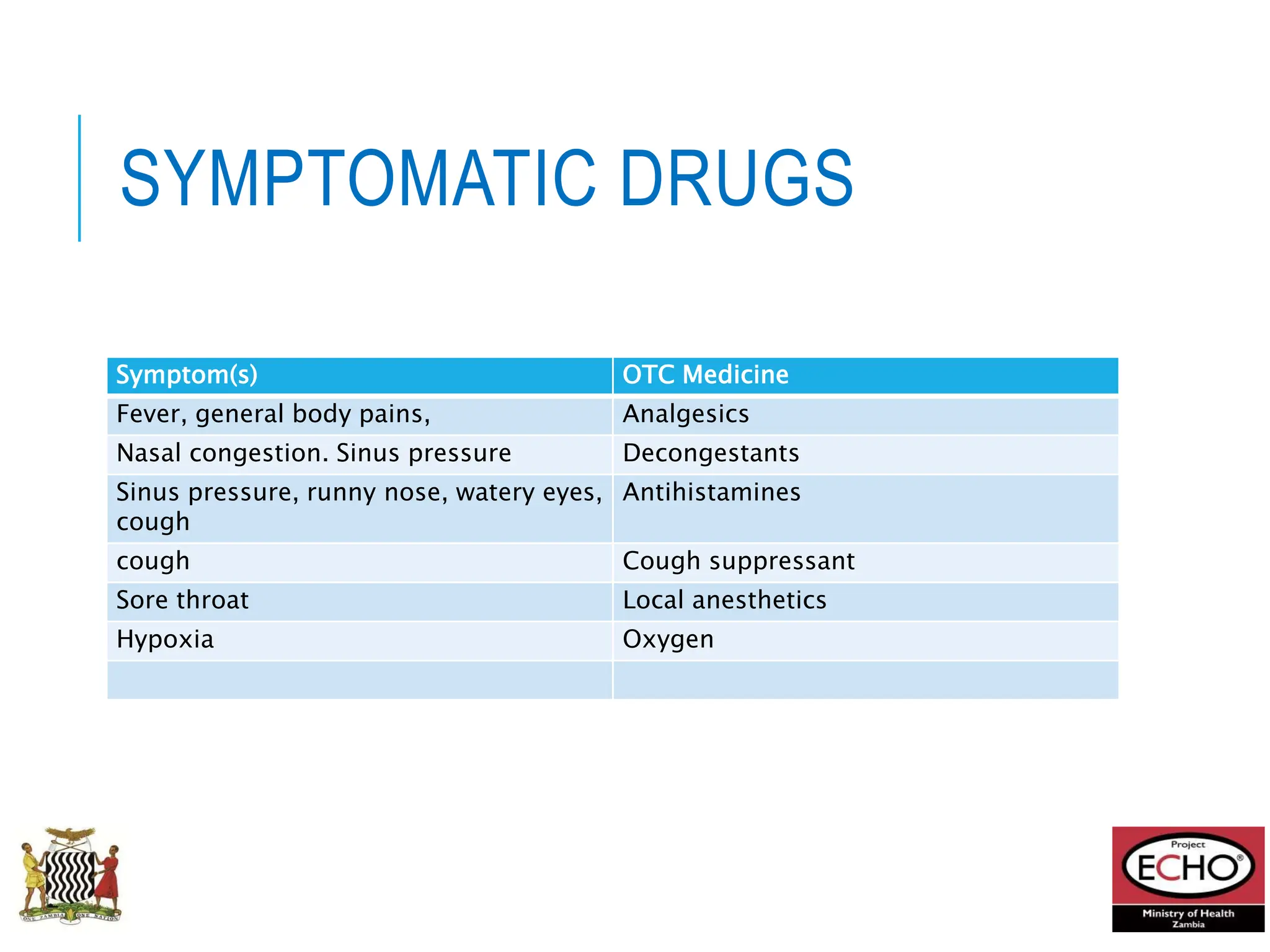
MOH HIV/TB ECHO
INFLUENZA
15TH July, 2024
Presenters:
Dr. Mwaka Monze
Dr Chitanika Chalomba
LEARNING OBJECTIVES
1. Discuss the Epidemiology of Influenza
2. Discuss Clinical Manifestations of Influenza
3. Discuss the Diagnosis of Influenza
4. Discuss the treatment of Influenza
The speaker has no significant financial conflicts of
interest to disclose.
POLL QUESTION 1
Which of the following types of Influenza have been
isolated in Zambia?
A. Influenza Type A
B. Influenza Type B
C. Both Influenza Type A and Type B
POLL QUESTION 2
Serology is a useful clinical tool for the diagnosis of
influenza
A. True
B. False
POLL QUESTION 3
Steroids are a mainstay of treatment in patients with
pneumonia due to influenza
A. True
B. False
Sentinel Surveillance for Influenza
in Severe Acute Respiratory Illnesses (SARI) and
Influenza-Like Illnesses (ILI)
Overview of SARI/ILI Surveillance in Zambia
Established: 2008/9
Target population:
General population (all ages) presenting to selected health facilities
(sentinel sites)
Case definitions :
ILI = fever + cough + ≤ 10 days duration
SARI = ILI + hospitalization
PUI (person under investigation) = SARI + exposure to Highly Pathogenic
Avian Influenza risk
Sampling:
5 ILI per day
All SARI cases
Lab testing:
Influenza and subtyping
A – H1N1 pandemic; H3N2; H5; H7 OR
B – Yamagata; Victoria
SARS Cov 2
Other respiratory pathogens as indicated
Reporting:
Weekly surveillance reports shared with stakeholders and uploaded to
FluNET/FluID
Health Facilities Serving as Sentinel Sites
1 UTH Pediatric Admission Ward SARI Paeds
2 UTH Adult Admission Ward SARI
3 Chipata First Level Hospital ILI
4 Arthur Davison Childrens Hospital SARI Paeds
5 Ndola Teaching Hospital SARI
6 New Masala Urban Health Center ILI
7 Nakonde District Hospital SARI
8 Nakonde Urban Health Center ILI
9 Livingstone Teaching Hospital SARI
10 Mahatma Ghandi Urban Health Center ILI
11 Solwezi General Hospital SARI
12 Solwezi Urban Health Center ILI
13 Chipata General Hospital SARI
14 Kapata Urban Health Center ILI
Lusaka, Lusaka
Ndola, Copperbelt
Nakonde, Muchinga
Livingstone, Southern
Solwezi, Northwestern
Chipata, Eastern
Surveillance Procedure
• Trained surveillance staff identify patients meeting a case definition:
• ILI – outpatient clinics
• SARI – admission wards
• Get verbal consent. Non-research determination
• Fill out a case investigation form (CIF): demographic information, case
classification information, illness severity, medical history, risk factors including
possible disease exposure, vaccination history, outcome
• Collect denominator data for each facility: total outpatient visits/admissions, total
respiratory visits/ admissions etc
• Collect, store and transport samples to the laboratory
• Laboratory testing using open PCR systems
• Results feedback, data analysis and weekly reporting
Lab Protocol
Reagents
from CDC
PCR Assays: CDC
protocols, reagents
distributed to NICS
through IRR
NGS: Currently using ONT
Table 1: Viruses detected in SARI/ ILI samples processed per epi-week (epi week 12 - epi week 27* of 2024)
WEEK
Total
Specimens
Received
Total
Specimens
Processed (%)
Influenza A Positives Influenza B Positives SARS
COV 2
Positives
(%)
A (H1N1
pandemic
2009) (%)
A (H3) (%)
A (H5)
(%)
A (Pending
subtyping) (%)
Total
influenza A
positive
(%)
B (Victoria)
(%)
B (Yamagata)
(%)
B (Pending
subtyping)
(%)
Total
influenza B
positive
(%)
12 110 110(100) 0 4 0 0 4 0 0 0 0 1
13 84 84(100) 0 4 0 0 4 0 0 0 0 0
14 66 66(100) 0 2 0 0 2 0 0 0 0 0
15 93 93(100) 0 2 0 0 2 1 0 0 1 0
16 115 115(100) 0 3 0 0 3 0 0 0 0 1
17 143 143(100) 0 6 0 0 6 0 0 0 0 0
18 86 86(100) 0 5 0 0 5 0 0 0 0 1
19 78 77(99) 0 5 0 0 5 0 0 0 0 0
20 108 108(100) 5 5 0 0 10 1 0 0 1 0
21 159 152(96) 12 2 0 0 14 1 0 0 1 0
22 98 98(100) 1 3 0 0 4 1 0 0 1 1
23 168 145(86) 9 8 0 0 17 2 0 0 2 0
24 111 94(85) 5 4 0 0 9 2 0 0 2 0
25 71 56(79) 3 4 0 0 7 1 0 0 1 0
26 23 17(74) 1 2 0 0 3 3 0 0 3 0
27 3 3(100) 1 0 0 0 1 1 0 0 1 0
Table 2: Number of specimens collected by districts per epi-week (epi week
24- 27*, 2024)
District
Total
Specimens
PCR Positive Results
A/H1N1
(pandemic)
A/H3N2 B SARS-CoV-2
Chipata 18 (8.7%) 1 (10.0%) 0 (0.0%) 3 (42.9%) 0 (-)
Livingstone 20 (9.6%) 6 (60.0%) 0 (0.0%) 2 (28.6%) 0 (-)
Lusaka 59 (28.4%) 2 (20.0%) 0 (0.0%) 2 (28.6%) 0 (-)
Nakonde 30 (14.4%) 0 (0.0%) 0 (0.0%) 0 (0.0%) 0 (-)
Ndola 51 (24.5%) 1 (10.0%) 10 (100.0%) 0 (0.0%) 0 (-)
Solwezi 30 (14.4%) 0 (0.0%) 0 (0.0%) 0 (0.0%) 0 (-)
Total 208 (100.0%) 10 (100.0%) 10 (100.0%) 7 (100.0%) 0 (-)
Percentage (%) of positive specimens by age per epi-week (epi week 24- 27*, 2024)
Age group
Total
Specimens
PCR Positive Results
A/H1N1
(pandemic)
A/H3N2 B
SARS-CoV-
2
<1 16 (7.7%) 0 (0.0%) 2 (20.0%) 0 (0.0%) 0 (-)
1-4 46 (22.1%) 4 (40.0%) 1 (10.0%) 1 (14.3%) 0 (-)
5-17 46 (22.1%) 1 (10.0%) 5 (50.0%) 2 (28.6%) 0 (-)
18-49 76 (36.5%) 3 (30.0%) 2 (20.0%) 4 (57.1%) 0 (-)
50+ 24 (11.5%) 2 (20.0%) 0 (0.0%) 0 (0.0%) 0 (-)
Total 208 (100.0%) 10 (100.0%)
10
(100.0%) 7 (100.0%) 0 (-)
ILI/SARI Threshold Monitoring for epi week 1 – 26 of 2024
Diagnosis
• Most cases of human influenza are clinically diagnosed especially in an
outbreak situation
• Rapid Diagnostic Tests (RDTs): Can be used in clinical settings but have
lower sensitivity
• During periods of low influenza activity or outside of epidemics situations,
further tests required to differentiate other causes of influenza-like illness
eg other respiratory viruses like SARS-CoV-2, rhinovirus, respiratory
syncytial virus, parainfluenza and adenovirus
• Nucleic acid tests – real time PCR is often used. These can help to further
characterize the virus eg subtype. Multiplex tests can give a one-step answer but
may be difficult to interpret.
• Direct antigenic detection
• Virus isolation in cell/tissue cultures or eggs is helpful to test for antiviral
sensitivity or to select vaccine viruses
• Sequencing or genotyping – used to enhance surveillance and to identify
significant changes or mutations in the virus
INTRODUCTION
Influenza is an acute respiratory illness caused by infection
with influenza viruses: commonly called “the flu”
Outbreaks of illness of variable extent and severity occur
nearly every year.
Such outbreaks result in significant morbidity rates in the
general population and in increased mortality rates among
certain high-risk patients, mainly as a result of pulmonary
complications.
The influenza virus comes from the Orthomyxoviridae
viruses
There are three viral sub-types, namely; type A, type B, Type
C
These viruses are antigenically distinct with no cross-
immunity
Influenza A and B viruses are responsible for epidemics of
disease throughout the world
18
ORTHOMYXOVIRUSES
M1 protein
helical nucleocapsid (RNA plus
NP protein)
HA - hemagglutinin
polymerase complex
lipid bilayer membrane
NA - neuraminidase
type A, B, C : NP, M1 protein
sub-types: HA or NA protein
The designation of influenza
viruses as type A, B, or C is based
on antigenic
characteristics of the
nucleoprotein (NP) and matrix (M)
protein
HISTORY
Spanish flu 1918 – 1919 (1/3 world population infected with about
50million deaths) – H1N1
1957: H2N2
1968: H3N2
Avian Flu (1997) H5N1
Swine Flu (2009) H1N1 17000 deaths by early 2010
- The exact timing of the onset, peak, and of influenza activity
vary, and cannot be predicted
- Peak activity most commonly occurs during the winter
- persons of all ages are susceptible to influenza
- Influenza incidence is difficult to quantify precisely, as many or
most of those infected may not seek medical attention and are
therefore not diagnosed.
TRANSMISSION DYNAMICS
AND CYCLE
Variable Description
Susceptible Elderly, Children without prior exposure,
DM, Immunosuppression, and Obesity
Infectiousness Peak Np viral RNA peak a day after onset
of symptoms
Infectious virus in 13% isolated after 8days
R0 was 1.3-1.7
Environment Household (secondary rates of 7.5%),
school, hospitals
Exposure Household, Children (more likely to
transmit)
Survival time <8 hours
MODE OF TRANSMISSION IN
HUMAN
The virus is spread
from person- to-
person through
respiratory secretions
either as droplets
(close contact) or as
airborne infection by
droplet nuclei
suspended in the air.
Incubation period 1-3
days
TRANSMISSION
AEROSOL
 100,000 TO 1,000,000 VIRIONS
PER DROPLET
SURFACES
- VIRUS CAN SURVIVE APPROX 2 TO
8 HRS
18-72 HR INCUBATION
Virus shedding generally stops
within 2–5 days after symptoms
first appear
Human Mobility is a key factor in
transmission
24
ANTIGENIC VARIATION
Influenza viruses tend to undergo changes from time to
time. There are two types of changes:
Antigenic variation may involve the hemagglutinin alone
or both the hemagglutinin and the neuraminidase. An
example of an antigenic shift involving both the
hemagglutinin and the neuraminidase is that of 1957,
when the predominant influenza A virus subtype shifted
from H1N1 to H2N2; this shift resulted in a severe
pandemic, with an estimated 70,000 excess deaths
(1) antigenic shift: Major antigenic variations, seen only
with influenza A viruses and may be associated with
pandemics.
(2) antigenic drift: minor changes
These changes in the antigenic characteristics of
influenza viruses determine the extent and severity of
influenza epidemics
PANDEMIC INFLUENZA
VIRUSES
Pandemic Subtype
1889 H2N?
1899 H3N8
1918 H1N1
1957 H2N2
1968 H3N2
1977 H1N1
WHERE DO “NEW” HA AND NA
COME FROM?
~16 types HA
~9 types NA
 all circulate in birds
pigs
 can be infected by
avian and human
influenza viruses
27
WHERE DO “NEW” HA AND NA COME
FROM?
28
WHERE DO “NEW” HA AND NA COME FROM
2009 PANDEMIC H1N1?
29
WHERE DO “NEW” HA AND NA COME FROM
- CAN ‘NEW’ BIRD FLU DIRECTLY INFECT
HUMANS?
30
Current “Bird flu” H5N1?
1918 influenza
H5N1 – IN BIRDS
Avian H5N1 has spread to humans
So far human cases in Asia and Africa
 442 cases (12-1-03 through 09-24-09)
 262 (59%) fatal
Have been a few instances where may have spread
human-to-human
So far no sustained spread in humans
Surveillance continues
31
2009 NOVEL H1N1 PANDEMIC
first novel H1N1 patient in the United States
confirmed by laboratory testing at CDC on April 15,
2009.
Quickly determined that the virus was spreading from
person-to-person.
By June 3, 2009, all 50 states in the United States and
the District of Columbia and Puerto Rico were
reporting cases of novel H1N1 infection.
32
http://www.cdc.gov/h1n1flu/update.htm
WHY DO WE NOT HAVE
INFLUENZA B PANDEMICS?
So far no shifts
have been
recorded
no animal
reservoir known
33
SYMPTOMS
FEVER
HEADACHE
MYALGIA
COUGH
RHINITIS
OCULAR SYMPTOMS
GI tract symptoms not typically seen
 but common with 2009 H1N1 influenza (‘swine flu’)
 vomiting, diarrhea
34
PATIENTS SUSEPTIBLE TO
COMPLICATIONS
 VERY YOUNG
 ELDERLY
 IMMUNO-COMPROMISED
 HEART OR LUNG DISEASE
35
PULMONARY COMPLICATIONS
CROUP (YOUNG CHILDREN)
PRIMARY INFLUENZA VIRUS PNEUMONIA
SECONDARY BACTERIAL INFECTION
 Streptococcus pneumoniae
 Staphlyococcus aureus
 Hemophilus influenzae
MIXED (VIRAL –BACTERIAL PNEUMONIA)
EXCERBATION OF CHRONIC PULMONARY DISEASES
(ASTHMA AND COPD)
36
PRIMARY VIRAL INFLUENZA
PNEUMONIA
Is the least common but most severe of the pneumonic complications
Risk factors are : cardiac and pulmonary disease (has been reported in healthy individuals)
It presents as acute influenza that does not resolve but instead progresses with fever,
dyspnea, and eventual cyanosis.
Sputum production is generally scanty, but the sputum can contain blood.
Few physical signs may be evident early in the illness. In more advanced cases, diffuse rale
may be noted,
imaging findings consistent with diffuse interstitial infiltrates and/or acute respiratory distress
syndrome may be present.
Arterial blood-gas determinations show marked hypoxia
histopathologic examination reveals marked inflammatory reaction of alveolar septa
edema and infiltration by lymphocytes,macrophages, occasional plasma cells, and variable
numbers of neutrophils.
Fibrin thrombi in alveolar capillaries, along with necrosis and hemorrhage, have also been
noted. Eosinophilic hyaline membranes can be found lining alveoli and alveolar ducts.
SECONDARY BACTERIAL
PNEUMONIA
Occurs most frequently in high-risk individuals with
chronic pulmonary and cardiac disease and in elderly
individuals
Improvement of the patient’s condition over 2–3 days s
followed by a reappearance of fever along with clinical
signs and symptoms of bacterial pneumonia
 Cough, production of purulent sputum, and physical and x-ray signs
of consolidation
Most common bacterial pathogens in this setting are
Streptococcus pneumoniae, Staphylococcus aureus, and
Haemophilus influenzae
Patients with secondary bacterial pneumonia often
respond to appropriate antibiotic therapy when it is
instituted promptly.
MIXED VIRAL AND BACTERIAL
PNEUMONIA
Most common pneumonic complications during outbreaks
Patients experience a gradual progression of their acute illness
or may show transient improvement followed by clinical
exacerbation.
Sputum cultures may contain both influenza A virus and one of
the bacterial pathogens described above.
Patchy infiltrates or areas of consolidation may be detected by
physical examination and chest x-ray.
Patients with mixed viral and bacterial pneumonia generally
have less widespread involvement of the lung than those with
primary viral pneumonia
Bacterial infections may respond to appropriate antibacterial
drugs.
Mixed viral and bacterial pneumonia occurs primarily in
patients with chronic cardiovascular and pulmonary diseases.
CHEST IMAGING FINDINGS
FROM A PATIENT WITH
PNEUMONIA THE CURRENT
OUTBREAK
Image courtesy of Levy Mwanawasa University Teaching Hospital department of
Radiology
CHEST IMAGING FINDINGS
FROM A PATIENT WITH
PNEUMONIA THE CURRENT
OUTBREAK
Image courtesy of Levy Mwanawasa University Teaching Hospital department of
Radiology
NON-PULMONARY
COMPLICATIONS
Myositis (rare, > in children, > with type B)
Cardiac complications
Encephalopathy
 2002/2003 season studies of patients younger than
21 yrs in Michigan - 8 cases (2 deaths)
Liver and CNS
 Reye’s syndrome
Peripheral nervous system
 Guillian-Barré syndrome
42
MORTALITY
MAJOR CAUSES OF INFLUENZA VIRUS- ASSOCIATED
DEATH
 BACTERIAL PNEUMONIA
 CARDIAC FAILURE
90% OF DEATHS IN THOSE OVER 65 YEARS OF AGE
43
DIAGNOSIS
ISOLATION OF VIRUS
 NOSE, THROAT SWAB
 GROW IN TISSUE CULTURE OR EGGS
SEROLOGY
PCR
RAPID TESTS
PROVISIONAL- clinical picture + outbreak
Other laboratory tests generally are not helpful in
dioagnosis
 Leukocyte counts are variable
44
DIFFERENTIAL DIAGNOSIS
During An outbreak ;clinical diagnosis can be made in
patients typical febrile
In the absence of an outbreak influenza may be difficult
to differentiate on clinical grounds
an acute respiratory illness can be caused by any of a
variety of respiratory viruses or by mycoplasma
pneumoniae
Severe streptococcal pharyngitis or early bacterial
pneumonia may mimic acute influenza,
Purulent sputum in which a bacterial pathogen can be
detected by Gram’s staining is an important diagnostic
feature in bacterial pneumonia.
PRINCIPLES OF TREATMENT
FOR INFLUENZA PNEUMONIA
Therapy for primary influenza pneumonia is directed at maintaining
oxygenation and with aggressive respiratory and hemodynamic support
as needed
Studies have suggested that treatment with oseltamivir may reduce the
frequency of lower respiratory complications and hospitalization.
Antibacterial drugs should be reserved for the treatment of bacterial
complications of acute influenza, such as secondary bacterial
pneumonia.
Choice of antibiotics should be guided by Gram’s staining and cultur
If the etiology of a case of bacterial pneumonia is unclear empirical
antibiotics effective against the most common bacterial pathogens in
this setting:
 S. pneumoniae, S. aureus, an H. influenzae
ANTI-VIRAL DRUGS
All anti-viral drugs inhibit viral replication, but they act in different
ways to achieve this. Drugs that are effective against influenza A
viruses: amantadine and rimantadine. Drugs that are effective
against influenza A viruses and influenza B viruses: zanamivir and
oseltamivir.
STEROIDS
Unlike COVID-19 , Corticosteroid treatment in influenza
is associated with increased mortality and hospital-acquired
infection
SYMPTOMATIC DRUGS
Symptom(s) OTC Medicine
Fever, general body pains, Analgesics
Nasal congestion. Sinus pressure Decongestants
Sinus pressure, runny nose, watery eyes,
cough
Antihistamines
cough Cough suppressant
Sore throat Local anesthetics
Hypoxia Oxygen
GOOD HEALTH HABITS FOR
PREVENTION
Avoid close contact
Stay at home when you are sick
Cover your mouth and nose
Clean your hands
Avoid touching your eyes, nose or mouth
Practice other good health habits: plenty of sleep,
manage your stress, drink plenty of fluids and eat
nutritious foods.
51
http://www.cdc.gov/flu/professionals/acip/flu_vax_children0910.htm#box1
52
http://www.cdc.gov/flu/professionals/acip/flu_vax_adults0910.htm#box2
INFLUENZA VERSUS COVID-19
ASPECT INFLUENZA COVID-19
CAUSATIVE AGENT Influenza virus (Types A, B, C, D) SARS-CoV-2 (a novel
coronavirus)
TRANSMISSION Respiratory droplets, contact Respiratory droplets,
contact, aerosol
INCUBATION
PERIOD
1-4 days 2-14 days
SYMPTOMS Fever, cough, sore throat,
muscle aches
Fever, cough, shortness
of breath, loss of
taste/smell, fatigue
SEVERITY Generally mild to moderate, can
be severe in high-risk
populations
Can range from mild to
severe, higher fatality
rate than flu
COMPLICATIONS Pneumonia, myocarditis,
encephalitis, exacerbation of
chronic diseases
Pneumonia, ARDS, blood
clots, multi-organ failure
SEASONALITY Predominantly in winter months Year-round, with varying
peaks
INFLUENZA VERSUS COVID-19
CNT’D
ASPECT INFLUENZA COVID-19
VACCINES Annual flu vaccines available COVID-19 vaccines available
(mRNA, vector, inactivated)
TREATMENT Antivirals (e.g., oseltamivir),
supportive care
Antivirals (e.g., remdesivir),
monoclonal antibodies,
supportive care
R0 (BASIC REPRODUCTION
NUMBER)
1.3-1.8 2-3.5 (varies with variants)
PREVENTIVE MEASURES Vaccination, hand hygiene,
mask-wearing, social
distancing
Vaccination, hand hygiene,
mask-wearing, social
distancing, ventilation
ASYMPTOMATIC
TRANSMISSION
Less common More common, significant
factor in spread
TESTING Rapid antigen tests, PCR tests Rapid antigen tests, PCR
tests, home testing kits
LONG-TERM EFFECTS Rare, post-infectious
syndromes can occur
Long COVID, with persistent
symptoms affecting various
organs
GLOBAL IMPACT Annual epidemics, occasional
pandemics (e.g., 1918
Spanish flu)
Global pandemic with
significant social and
economic impact since 2019
POLL QUESTION 1
Which of the following types of Influenza have been
isolated in Zambia?
A. Influenza Type A
B. Influenza Type B
C. Both Influenza Type A and Type B
POLL QUESTION 2
Serology is a useful clinical tool for the diagnosis of
influenza
A. True
B. False
POLL QUESTION 3
Steroids are a mainstay of treatment in patients with
pneumonia due to influenza
A. True
B. False
INFLUENZA VERSUS COVID-19
ASPECT INFLUENZA COVID-19
CAUSATIVE AGENT Influenza virus (Types A, B, C, D) SARS-CoV-2 (a novel
coronavirus)
TRANSMISSION Respiratory droplets, contact Respiratory droplets,
contact, aerosol
INCUBATION
PERIOD
1-4 days 2-14 days
SYMPTOMS Fever, cough, sore throat,
muscle aches
Fever, cough, shortness
of breath, loss of
taste/smell, fatigue
SEVERITY Generally mild to moderate, can
be severe in high-risk
populations
Can range from mild to
severe, higher fatality
rate than flu
COMPLICATIONS Pneumonia, myocarditis,
encephalitis, exacerbation of
chronic diseases
Pneumonia, ARDS, blood
clots, multi-organ failure
SEASONALITY Predominantly in winter months Year-round, with varying
peaks
THANK YOU

ECHO Influenza presentation 15-07-24.pptx

  • 1.
    MOH HIV/TB ECHO INFLUENZA 15THJuly, 2024 Presenters: Dr. Mwaka Monze Dr Chitanika Chalomba
  • 2.
    LEARNING OBJECTIVES 1. Discussthe Epidemiology of Influenza 2. Discuss Clinical Manifestations of Influenza 3. Discuss the Diagnosis of Influenza 4. Discuss the treatment of Influenza The speaker has no significant financial conflicts of interest to disclose.
  • 3.
    POLL QUESTION 1 Whichof the following types of Influenza have been isolated in Zambia? A. Influenza Type A B. Influenza Type B C. Both Influenza Type A and Type B
  • 4.
    POLL QUESTION 2 Serologyis a useful clinical tool for the diagnosis of influenza A. True B. False
  • 5.
    POLL QUESTION 3 Steroidsare a mainstay of treatment in patients with pneumonia due to influenza A. True B. False
  • 6.
    Sentinel Surveillance forInfluenza in Severe Acute Respiratory Illnesses (SARI) and Influenza-Like Illnesses (ILI)
  • 7.
    Overview of SARI/ILISurveillance in Zambia Established: 2008/9 Target population: General population (all ages) presenting to selected health facilities (sentinel sites) Case definitions : ILI = fever + cough + ≤ 10 days duration SARI = ILI + hospitalization PUI (person under investigation) = SARI + exposure to Highly Pathogenic Avian Influenza risk Sampling: 5 ILI per day All SARI cases Lab testing: Influenza and subtyping A – H1N1 pandemic; H3N2; H5; H7 OR B – Yamagata; Victoria SARS Cov 2 Other respiratory pathogens as indicated Reporting: Weekly surveillance reports shared with stakeholders and uploaded to FluNET/FluID
  • 8.
    Health Facilities Servingas Sentinel Sites 1 UTH Pediatric Admission Ward SARI Paeds 2 UTH Adult Admission Ward SARI 3 Chipata First Level Hospital ILI 4 Arthur Davison Childrens Hospital SARI Paeds 5 Ndola Teaching Hospital SARI 6 New Masala Urban Health Center ILI 7 Nakonde District Hospital SARI 8 Nakonde Urban Health Center ILI 9 Livingstone Teaching Hospital SARI 10 Mahatma Ghandi Urban Health Center ILI 11 Solwezi General Hospital SARI 12 Solwezi Urban Health Center ILI 13 Chipata General Hospital SARI 14 Kapata Urban Health Center ILI Lusaka, Lusaka Ndola, Copperbelt Nakonde, Muchinga Livingstone, Southern Solwezi, Northwestern Chipata, Eastern
  • 9.
    Surveillance Procedure • Trainedsurveillance staff identify patients meeting a case definition: • ILI – outpatient clinics • SARI – admission wards • Get verbal consent. Non-research determination • Fill out a case investigation form (CIF): demographic information, case classification information, illness severity, medical history, risk factors including possible disease exposure, vaccination history, outcome • Collect denominator data for each facility: total outpatient visits/admissions, total respiratory visits/ admissions etc • Collect, store and transport samples to the laboratory • Laboratory testing using open PCR systems • Results feedback, data analysis and weekly reporting
  • 10.
    Lab Protocol Reagents from CDC PCRAssays: CDC protocols, reagents distributed to NICS through IRR NGS: Currently using ONT
  • 11.
    Table 1: Virusesdetected in SARI/ ILI samples processed per epi-week (epi week 12 - epi week 27* of 2024) WEEK Total Specimens Received Total Specimens Processed (%) Influenza A Positives Influenza B Positives SARS COV 2 Positives (%) A (H1N1 pandemic 2009) (%) A (H3) (%) A (H5) (%) A (Pending subtyping) (%) Total influenza A positive (%) B (Victoria) (%) B (Yamagata) (%) B (Pending subtyping) (%) Total influenza B positive (%) 12 110 110(100) 0 4 0 0 4 0 0 0 0 1 13 84 84(100) 0 4 0 0 4 0 0 0 0 0 14 66 66(100) 0 2 0 0 2 0 0 0 0 0 15 93 93(100) 0 2 0 0 2 1 0 0 1 0 16 115 115(100) 0 3 0 0 3 0 0 0 0 1 17 143 143(100) 0 6 0 0 6 0 0 0 0 0 18 86 86(100) 0 5 0 0 5 0 0 0 0 1 19 78 77(99) 0 5 0 0 5 0 0 0 0 0 20 108 108(100) 5 5 0 0 10 1 0 0 1 0 21 159 152(96) 12 2 0 0 14 1 0 0 1 0 22 98 98(100) 1 3 0 0 4 1 0 0 1 1 23 168 145(86) 9 8 0 0 17 2 0 0 2 0 24 111 94(85) 5 4 0 0 9 2 0 0 2 0 25 71 56(79) 3 4 0 0 7 1 0 0 1 0 26 23 17(74) 1 2 0 0 3 3 0 0 3 0 27 3 3(100) 1 0 0 0 1 1 0 0 1 0
  • 13.
    Table 2: Numberof specimens collected by districts per epi-week (epi week 24- 27*, 2024) District Total Specimens PCR Positive Results A/H1N1 (pandemic) A/H3N2 B SARS-CoV-2 Chipata 18 (8.7%) 1 (10.0%) 0 (0.0%) 3 (42.9%) 0 (-) Livingstone 20 (9.6%) 6 (60.0%) 0 (0.0%) 2 (28.6%) 0 (-) Lusaka 59 (28.4%) 2 (20.0%) 0 (0.0%) 2 (28.6%) 0 (-) Nakonde 30 (14.4%) 0 (0.0%) 0 (0.0%) 0 (0.0%) 0 (-) Ndola 51 (24.5%) 1 (10.0%) 10 (100.0%) 0 (0.0%) 0 (-) Solwezi 30 (14.4%) 0 (0.0%) 0 (0.0%) 0 (0.0%) 0 (-) Total 208 (100.0%) 10 (100.0%) 10 (100.0%) 7 (100.0%) 0 (-)
  • 14.
    Percentage (%) ofpositive specimens by age per epi-week (epi week 24- 27*, 2024) Age group Total Specimens PCR Positive Results A/H1N1 (pandemic) A/H3N2 B SARS-CoV- 2 <1 16 (7.7%) 0 (0.0%) 2 (20.0%) 0 (0.0%) 0 (-) 1-4 46 (22.1%) 4 (40.0%) 1 (10.0%) 1 (14.3%) 0 (-) 5-17 46 (22.1%) 1 (10.0%) 5 (50.0%) 2 (28.6%) 0 (-) 18-49 76 (36.5%) 3 (30.0%) 2 (20.0%) 4 (57.1%) 0 (-) 50+ 24 (11.5%) 2 (20.0%) 0 (0.0%) 0 (0.0%) 0 (-) Total 208 (100.0%) 10 (100.0%) 10 (100.0%) 7 (100.0%) 0 (-)
  • 15.
    ILI/SARI Threshold Monitoringfor epi week 1 – 26 of 2024
  • 16.
    Diagnosis • Most casesof human influenza are clinically diagnosed especially in an outbreak situation • Rapid Diagnostic Tests (RDTs): Can be used in clinical settings but have lower sensitivity • During periods of low influenza activity or outside of epidemics situations, further tests required to differentiate other causes of influenza-like illness eg other respiratory viruses like SARS-CoV-2, rhinovirus, respiratory syncytial virus, parainfluenza and adenovirus • Nucleic acid tests – real time PCR is often used. These can help to further characterize the virus eg subtype. Multiplex tests can give a one-step answer but may be difficult to interpret. • Direct antigenic detection • Virus isolation in cell/tissue cultures or eggs is helpful to test for antiviral sensitivity or to select vaccine viruses • Sequencing or genotyping – used to enhance surveillance and to identify significant changes or mutations in the virus
  • 17.
    INTRODUCTION Influenza is anacute respiratory illness caused by infection with influenza viruses: commonly called “the flu” Outbreaks of illness of variable extent and severity occur nearly every year. Such outbreaks result in significant morbidity rates in the general population and in increased mortality rates among certain high-risk patients, mainly as a result of pulmonary complications. The influenza virus comes from the Orthomyxoviridae viruses There are three viral sub-types, namely; type A, type B, Type C These viruses are antigenically distinct with no cross- immunity Influenza A and B viruses are responsible for epidemics of disease throughout the world
  • 18.
    18 ORTHOMYXOVIRUSES M1 protein helical nucleocapsid(RNA plus NP protein) HA - hemagglutinin polymerase complex lipid bilayer membrane NA - neuraminidase type A, B, C : NP, M1 protein sub-types: HA or NA protein The designation of influenza viruses as type A, B, or C is based on antigenic characteristics of the nucleoprotein (NP) and matrix (M) protein
  • 20.
    HISTORY Spanish flu 1918– 1919 (1/3 world population infected with about 50million deaths) – H1N1 1957: H2N2 1968: H3N2 Avian Flu (1997) H5N1 Swine Flu (2009) H1N1 17000 deaths by early 2010 - The exact timing of the onset, peak, and of influenza activity vary, and cannot be predicted - Peak activity most commonly occurs during the winter - persons of all ages are susceptible to influenza - Influenza incidence is difficult to quantify precisely, as many or most of those infected may not seek medical attention and are therefore not diagnosed.
  • 22.
    TRANSMISSION DYNAMICS AND CYCLE VariableDescription Susceptible Elderly, Children without prior exposure, DM, Immunosuppression, and Obesity Infectiousness Peak Np viral RNA peak a day after onset of symptoms Infectious virus in 13% isolated after 8days R0 was 1.3-1.7 Environment Household (secondary rates of 7.5%), school, hospitals Exposure Household, Children (more likely to transmit) Survival time <8 hours
  • 23.
    MODE OF TRANSMISSIONIN HUMAN The virus is spread from person- to- person through respiratory secretions either as droplets (close contact) or as airborne infection by droplet nuclei suspended in the air. Incubation period 1-3 days
  • 24.
    TRANSMISSION AEROSOL  100,000 TO1,000,000 VIRIONS PER DROPLET SURFACES - VIRUS CAN SURVIVE APPROX 2 TO 8 HRS 18-72 HR INCUBATION Virus shedding generally stops within 2–5 days after symptoms first appear Human Mobility is a key factor in transmission 24
  • 25.
    ANTIGENIC VARIATION Influenza virusestend to undergo changes from time to time. There are two types of changes: Antigenic variation may involve the hemagglutinin alone or both the hemagglutinin and the neuraminidase. An example of an antigenic shift involving both the hemagglutinin and the neuraminidase is that of 1957, when the predominant influenza A virus subtype shifted from H1N1 to H2N2; this shift resulted in a severe pandemic, with an estimated 70,000 excess deaths (1) antigenic shift: Major antigenic variations, seen only with influenza A viruses and may be associated with pandemics. (2) antigenic drift: minor changes These changes in the antigenic characteristics of influenza viruses determine the extent and severity of influenza epidemics
  • 26.
    PANDEMIC INFLUENZA VIRUSES Pandemic Subtype 1889H2N? 1899 H3N8 1918 H1N1 1957 H2N2 1968 H3N2 1977 H1N1
  • 27.
    WHERE DO “NEW”HA AND NA COME FROM? ~16 types HA ~9 types NA  all circulate in birds pigs  can be infected by avian and human influenza viruses 27
  • 28.
    WHERE DO “NEW”HA AND NA COME FROM? 28
  • 29.
    WHERE DO “NEW”HA AND NA COME FROM 2009 PANDEMIC H1N1? 29
  • 30.
    WHERE DO “NEW”HA AND NA COME FROM - CAN ‘NEW’ BIRD FLU DIRECTLY INFECT HUMANS? 30 Current “Bird flu” H5N1? 1918 influenza
  • 31.
    H5N1 – INBIRDS Avian H5N1 has spread to humans So far human cases in Asia and Africa  442 cases (12-1-03 through 09-24-09)  262 (59%) fatal Have been a few instances where may have spread human-to-human So far no sustained spread in humans Surveillance continues 31
  • 32.
    2009 NOVEL H1N1PANDEMIC first novel H1N1 patient in the United States confirmed by laboratory testing at CDC on April 15, 2009. Quickly determined that the virus was spreading from person-to-person. By June 3, 2009, all 50 states in the United States and the District of Columbia and Puerto Rico were reporting cases of novel H1N1 infection. 32 http://www.cdc.gov/h1n1flu/update.htm
  • 33.
    WHY DO WENOT HAVE INFLUENZA B PANDEMICS? So far no shifts have been recorded no animal reservoir known 33
  • 34.
    SYMPTOMS FEVER HEADACHE MYALGIA COUGH RHINITIS OCULAR SYMPTOMS GI tractsymptoms not typically seen  but common with 2009 H1N1 influenza (‘swine flu’)  vomiting, diarrhea 34
  • 35.
    PATIENTS SUSEPTIBLE TO COMPLICATIONS VERY YOUNG  ELDERLY  IMMUNO-COMPROMISED  HEART OR LUNG DISEASE 35
  • 36.
    PULMONARY COMPLICATIONS CROUP (YOUNGCHILDREN) PRIMARY INFLUENZA VIRUS PNEUMONIA SECONDARY BACTERIAL INFECTION  Streptococcus pneumoniae  Staphlyococcus aureus  Hemophilus influenzae MIXED (VIRAL –BACTERIAL PNEUMONIA) EXCERBATION OF CHRONIC PULMONARY DISEASES (ASTHMA AND COPD) 36
  • 37.
    PRIMARY VIRAL INFLUENZA PNEUMONIA Isthe least common but most severe of the pneumonic complications Risk factors are : cardiac and pulmonary disease (has been reported in healthy individuals) It presents as acute influenza that does not resolve but instead progresses with fever, dyspnea, and eventual cyanosis. Sputum production is generally scanty, but the sputum can contain blood. Few physical signs may be evident early in the illness. In more advanced cases, diffuse rale may be noted, imaging findings consistent with diffuse interstitial infiltrates and/or acute respiratory distress syndrome may be present. Arterial blood-gas determinations show marked hypoxia histopathologic examination reveals marked inflammatory reaction of alveolar septa edema and infiltration by lymphocytes,macrophages, occasional plasma cells, and variable numbers of neutrophils. Fibrin thrombi in alveolar capillaries, along with necrosis and hemorrhage, have also been noted. Eosinophilic hyaline membranes can be found lining alveoli and alveolar ducts.
  • 38.
    SECONDARY BACTERIAL PNEUMONIA Occurs mostfrequently in high-risk individuals with chronic pulmonary and cardiac disease and in elderly individuals Improvement of the patient’s condition over 2–3 days s followed by a reappearance of fever along with clinical signs and symptoms of bacterial pneumonia  Cough, production of purulent sputum, and physical and x-ray signs of consolidation Most common bacterial pathogens in this setting are Streptococcus pneumoniae, Staphylococcus aureus, and Haemophilus influenzae Patients with secondary bacterial pneumonia often respond to appropriate antibiotic therapy when it is instituted promptly.
  • 39.
    MIXED VIRAL ANDBACTERIAL PNEUMONIA Most common pneumonic complications during outbreaks Patients experience a gradual progression of their acute illness or may show transient improvement followed by clinical exacerbation. Sputum cultures may contain both influenza A virus and one of the bacterial pathogens described above. Patchy infiltrates or areas of consolidation may be detected by physical examination and chest x-ray. Patients with mixed viral and bacterial pneumonia generally have less widespread involvement of the lung than those with primary viral pneumonia Bacterial infections may respond to appropriate antibacterial drugs. Mixed viral and bacterial pneumonia occurs primarily in patients with chronic cardiovascular and pulmonary diseases.
  • 40.
    CHEST IMAGING FINDINGS FROMA PATIENT WITH PNEUMONIA THE CURRENT OUTBREAK Image courtesy of Levy Mwanawasa University Teaching Hospital department of Radiology
  • 41.
    CHEST IMAGING FINDINGS FROMA PATIENT WITH PNEUMONIA THE CURRENT OUTBREAK Image courtesy of Levy Mwanawasa University Teaching Hospital department of Radiology
  • 42.
    NON-PULMONARY COMPLICATIONS Myositis (rare, >in children, > with type B) Cardiac complications Encephalopathy  2002/2003 season studies of patients younger than 21 yrs in Michigan - 8 cases (2 deaths) Liver and CNS  Reye’s syndrome Peripheral nervous system  Guillian-Barré syndrome 42
  • 43.
    MORTALITY MAJOR CAUSES OFINFLUENZA VIRUS- ASSOCIATED DEATH  BACTERIAL PNEUMONIA  CARDIAC FAILURE 90% OF DEATHS IN THOSE OVER 65 YEARS OF AGE 43
  • 44.
    DIAGNOSIS ISOLATION OF VIRUS NOSE, THROAT SWAB  GROW IN TISSUE CULTURE OR EGGS SEROLOGY PCR RAPID TESTS PROVISIONAL- clinical picture + outbreak Other laboratory tests generally are not helpful in dioagnosis  Leukocyte counts are variable 44
  • 45.
    DIFFERENTIAL DIAGNOSIS During Anoutbreak ;clinical diagnosis can be made in patients typical febrile In the absence of an outbreak influenza may be difficult to differentiate on clinical grounds an acute respiratory illness can be caused by any of a variety of respiratory viruses or by mycoplasma pneumoniae Severe streptococcal pharyngitis or early bacterial pneumonia may mimic acute influenza, Purulent sputum in which a bacterial pathogen can be detected by Gram’s staining is an important diagnostic feature in bacterial pneumonia.
  • 46.
    PRINCIPLES OF TREATMENT FORINFLUENZA PNEUMONIA Therapy for primary influenza pneumonia is directed at maintaining oxygenation and with aggressive respiratory and hemodynamic support as needed Studies have suggested that treatment with oseltamivir may reduce the frequency of lower respiratory complications and hospitalization. Antibacterial drugs should be reserved for the treatment of bacterial complications of acute influenza, such as secondary bacterial pneumonia. Choice of antibiotics should be guided by Gram’s staining and cultur If the etiology of a case of bacterial pneumonia is unclear empirical antibiotics effective against the most common bacterial pathogens in this setting:  S. pneumoniae, S. aureus, an H. influenzae
  • 47.
    ANTI-VIRAL DRUGS All anti-viraldrugs inhibit viral replication, but they act in different ways to achieve this. Drugs that are effective against influenza A viruses: amantadine and rimantadine. Drugs that are effective against influenza A viruses and influenza B viruses: zanamivir and oseltamivir.
  • 48.
    STEROIDS Unlike COVID-19 ,Corticosteroid treatment in influenza is associated with increased mortality and hospital-acquired infection
  • 49.
    SYMPTOMATIC DRUGS Symptom(s) OTCMedicine Fever, general body pains, Analgesics Nasal congestion. Sinus pressure Decongestants Sinus pressure, runny nose, watery eyes, cough Antihistamines cough Cough suppressant Sore throat Local anesthetics Hypoxia Oxygen
  • 50.
    GOOD HEALTH HABITSFOR PREVENTION Avoid close contact Stay at home when you are sick Cover your mouth and nose Clean your hands Avoid touching your eyes, nose or mouth Practice other good health habits: plenty of sleep, manage your stress, drink plenty of fluids and eat nutritious foods.
  • 51.
  • 52.
  • 53.
    INFLUENZA VERSUS COVID-19 ASPECTINFLUENZA COVID-19 CAUSATIVE AGENT Influenza virus (Types A, B, C, D) SARS-CoV-2 (a novel coronavirus) TRANSMISSION Respiratory droplets, contact Respiratory droplets, contact, aerosol INCUBATION PERIOD 1-4 days 2-14 days SYMPTOMS Fever, cough, sore throat, muscle aches Fever, cough, shortness of breath, loss of taste/smell, fatigue SEVERITY Generally mild to moderate, can be severe in high-risk populations Can range from mild to severe, higher fatality rate than flu COMPLICATIONS Pneumonia, myocarditis, encephalitis, exacerbation of chronic diseases Pneumonia, ARDS, blood clots, multi-organ failure SEASONALITY Predominantly in winter months Year-round, with varying peaks
  • 54.
    INFLUENZA VERSUS COVID-19 CNT’D ASPECTINFLUENZA COVID-19 VACCINES Annual flu vaccines available COVID-19 vaccines available (mRNA, vector, inactivated) TREATMENT Antivirals (e.g., oseltamivir), supportive care Antivirals (e.g., remdesivir), monoclonal antibodies, supportive care R0 (BASIC REPRODUCTION NUMBER) 1.3-1.8 2-3.5 (varies with variants) PREVENTIVE MEASURES Vaccination, hand hygiene, mask-wearing, social distancing Vaccination, hand hygiene, mask-wearing, social distancing, ventilation ASYMPTOMATIC TRANSMISSION Less common More common, significant factor in spread TESTING Rapid antigen tests, PCR tests Rapid antigen tests, PCR tests, home testing kits LONG-TERM EFFECTS Rare, post-infectious syndromes can occur Long COVID, with persistent symptoms affecting various organs GLOBAL IMPACT Annual epidemics, occasional pandemics (e.g., 1918 Spanish flu) Global pandemic with significant social and economic impact since 2019
  • 55.
    POLL QUESTION 1 Whichof the following types of Influenza have been isolated in Zambia? A. Influenza Type A B. Influenza Type B C. Both Influenza Type A and Type B
  • 56.
    POLL QUESTION 2 Serologyis a useful clinical tool for the diagnosis of influenza A. True B. False
  • 57.
    POLL QUESTION 3 Steroidsare a mainstay of treatment in patients with pneumonia due to influenza A. True B. False
  • 58.
    INFLUENZA VERSUS COVID-19 ASPECTINFLUENZA COVID-19 CAUSATIVE AGENT Influenza virus (Types A, B, C, D) SARS-CoV-2 (a novel coronavirus) TRANSMISSION Respiratory droplets, contact Respiratory droplets, contact, aerosol INCUBATION PERIOD 1-4 days 2-14 days SYMPTOMS Fever, cough, sore throat, muscle aches Fever, cough, shortness of breath, loss of taste/smell, fatigue SEVERITY Generally mild to moderate, can be severe in high-risk populations Can range from mild to severe, higher fatality rate than flu COMPLICATIONS Pneumonia, myocarditis, encephalitis, exacerbation of chronic diseases Pneumonia, ARDS, blood clots, multi-organ failure SEASONALITY Predominantly in winter months Year-round, with varying peaks
  • 59.