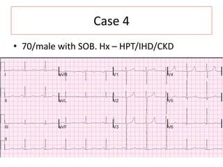
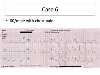
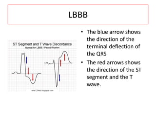
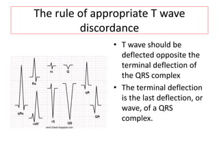
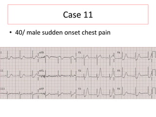
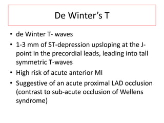
ECG
Dr Andey bin Rahman
Case 1
• 19/m/male, cough with pricking chest pain
BER
– Widespread concave ST elevation, most
prominent in the mid- to left precordial leads (V2-
5)
– Notching or slurring at the J-point
– Prominent, slightly asymmetrical T-waves
– No reciprocal
Case 2
• 40/lady found unconscious
STE @aVR
• Lead aVR is a very interesting lead in the ECG. aVR obtain
information from the
1) right upper portion of the heart, including the outflow tract of
the right ventricle and the basal portion of the septum
2) reciprocal information as to that which is derived from leads
aVL, II, V5, and V6
Important findings in aVR includes
1)ST-segment elevation as an indicator of significant left main
coronary artery(LMCA) involvement in acute coronary
syndromes
2)PR-segment elevation in acute cases of pericarditis
3)R' wave in tricyclic antidepressant (TCA) poisoning
STE @aVR
• STE in aVR indicate
1)Reciprocal STD from leads aVL, II, V5, and V6
Due to diffuse subendocardial ischemia indicates severe 3VD
- STE in aVR with diffuse STD in other leads usually more than 8 surface
leads
2)Theoretically infarction of the right upper portion of the heart,
including the outflow tract of the right ventricle and the basal
portion of the septum
The basal septum is supplied by the first septal perforator artery (a
very proximal branch of the LAD), so infarction of the basal septum
would imply involvement of the proximal LAD or LMCA.
Unless there is a concomitant anterior STEMI (high occlusion
resulting in STEMI of anterior and basal walls), STE in aVR is not a
STEMI in lead aVR; rather the STE in aVR is reciprocal to a leftward
and inferior ST depression axis caused by diffuse subendocardial
ischemia
Case 3
• 20/male with syncope
Brugada
• Type 1 (Coved ST segment elevation >2mm in
>1 of V1-V3 followed by a negative T wave) is
the only ECG abnormality that
is potentially diagnostic. This has been
referred to as Brugada sign.
Case 4
• 70/male with SOB. Hx – HPT/IHD/CKD
HyperK, hypoCa
• There are peaked T-waves
• There is a long ST segment, resulting in a long
QTc (490 ms).
Case 5
AIVR concordant
• AIVR
– No p wave
– Rate <100/min
• Concordant ST elevation in lead V4
• STEMI
Case 6
• 60/male with chest pain
LBBB
• The blue arrow shows
the direction of the
terminal deflection of
the QRS
• The red arrows shows
the direction of the ST
segment and the T
wave.
The rule of appropriate T wave
discordance
• T wave should be
deflected opposite the
terminal deflection of
the QRS complex
• The terminal deflection
is the last deflection, or
wave, of a QRS
complex.
Case 7
• 60/ female with chest pain
RBBB + LAFB + STEMI
• P wave
• LAFB
– LAD
– R in I & aVL
• STEMI
– Concordance STE V3-V4, possible I & aVL
Case 8
• 40/male with acute SOB
Mobitz 1 (wenkenbach)
• Prolong PR & drop beat
• STEMI
– Inferior
• Minimal horizontal STE
• Reciprocal @ aVL
– Possible posterior
Case 9
• 60/female with chest pain
Mobitz 2
• PR maintained
• Drop 2nd & 6th beat
Case 10
• 40/ male with intermittent chest pain 3/7
Wellen’s
• 2 type
– Biphasic T
– Deep symmetrical T
• indicative of reperfusion of the infarct-related vessel
– Spontaneously open
– Good collateral
• LAD occlusion
• Danger
– Pseudonormalization (reocclusion)
Case 11
• 40/ male sudden onset chest pain
De Winter’s T
• de Winter T- waves
• 1-3 mm of ST-depression upsloping at the J-
point in the precordial leads, leading into tall
symmetric T-waves
• High risk of acute anterior MI
• Suggestive of an acute proximal LAD occlusion
(contrast to sub-acute occlusion of Wellens
syndrome)
Case 12
• 40/M/Male
• C/o Palpitation
VT vs SVT with aberancy
RBBB like morphology
Qs wave in v6 -> VT
LBBB like morphology
Tq

Ecg intensive

  • 1.
  • 2.
    Case 1 • 19/m/male,cough with pricking chest pain
  • 3.
    BER – Widespread concaveST elevation, most prominent in the mid- to left precordial leads (V2- 5) – Notching or slurring at the J-point – Prominent, slightly asymmetrical T-waves – No reciprocal
  • 4.
    Case 2 • 40/ladyfound unconscious
  • 5.
    STE @aVR • LeadaVR is a very interesting lead in the ECG. aVR obtain information from the 1) right upper portion of the heart, including the outflow tract of the right ventricle and the basal portion of the septum 2) reciprocal information as to that which is derived from leads aVL, II, V5, and V6 Important findings in aVR includes 1)ST-segment elevation as an indicator of significant left main coronary artery(LMCA) involvement in acute coronary syndromes 2)PR-segment elevation in acute cases of pericarditis 3)R' wave in tricyclic antidepressant (TCA) poisoning
  • 6.
    STE @aVR • STEin aVR indicate 1)Reciprocal STD from leads aVL, II, V5, and V6 Due to diffuse subendocardial ischemia indicates severe 3VD - STE in aVR with diffuse STD in other leads usually more than 8 surface leads 2)Theoretically infarction of the right upper portion of the heart, including the outflow tract of the right ventricle and the basal portion of the septum The basal septum is supplied by the first septal perforator artery (a very proximal branch of the LAD), so infarction of the basal septum would imply involvement of the proximal LAD or LMCA. Unless there is a concomitant anterior STEMI (high occlusion resulting in STEMI of anterior and basal walls), STE in aVR is not a STEMI in lead aVR; rather the STE in aVR is reciprocal to a leftward and inferior ST depression axis caused by diffuse subendocardial ischemia
  • 7.
    Case 3 • 20/malewith syncope
  • 8.
    Brugada • Type 1(Coved ST segment elevation >2mm in >1 of V1-V3 followed by a negative T wave) is the only ECG abnormality that is potentially diagnostic. This has been referred to as Brugada sign.
  • 10.
    Case 4 • 70/malewith SOB. Hx – HPT/IHD/CKD
  • 11.
    HyperK, hypoCa • Thereare peaked T-waves • There is a long ST segment, resulting in a long QTc (490 ms).
  • 12.
  • 13.
    AIVR concordant • AIVR –No p wave – Rate <100/min • Concordant ST elevation in lead V4 • STEMI
  • 14.
    Case 6 • 60/malewith chest pain
  • 15.
    LBBB • The bluearrow shows the direction of the terminal deflection of the QRS • The red arrows shows the direction of the ST segment and the T wave.
  • 16.
    The rule ofappropriate T wave discordance • T wave should be deflected opposite the terminal deflection of the QRS complex • The terminal deflection is the last deflection, or wave, of a QRS complex.
  • 17.
    Case 7 • 60/female with chest pain
  • 18.
    RBBB + LAFB+ STEMI • P wave • LAFB – LAD – R in I & aVL • STEMI – Concordance STE V3-V4, possible I & aVL
  • 19.
    Case 8 • 40/malewith acute SOB
  • 20.
    Mobitz 1 (wenkenbach) •Prolong PR & drop beat • STEMI – Inferior • Minimal horizontal STE • Reciprocal @ aVL – Possible posterior
  • 21.
    Case 9 • 60/femalewith chest pain
  • 22.
    Mobitz 2 • PRmaintained • Drop 2nd & 6th beat
  • 23.
    Case 10 • 40/male with intermittent chest pain 3/7
  • 24.
    Wellen’s • 2 type –Biphasic T – Deep symmetrical T • indicative of reperfusion of the infarct-related vessel – Spontaneously open – Good collateral • LAD occlusion • Danger – Pseudonormalization (reocclusion)
  • 25.
    Case 11 • 40/male sudden onset chest pain
  • 26.
    De Winter’s T •de Winter T- waves • 1-3 mm of ST-depression upsloping at the J- point in the precordial leads, leading into tall symmetric T-waves • High risk of acute anterior MI • Suggestive of an acute proximal LAD occlusion (contrast to sub-acute occlusion of Wellens syndrome)
  • 27.
  • 28.
    VT vs SVTwith aberancy
  • 33.
    RBBB like morphology Qswave in v6 -> VT
  • 34.
  • 35.