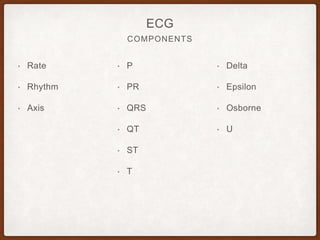
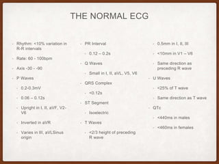
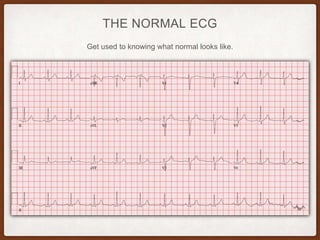
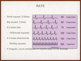
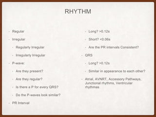
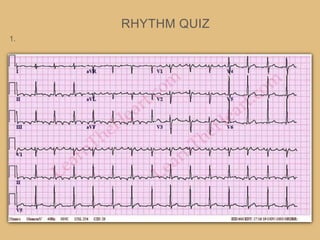
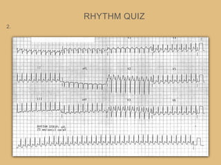
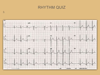
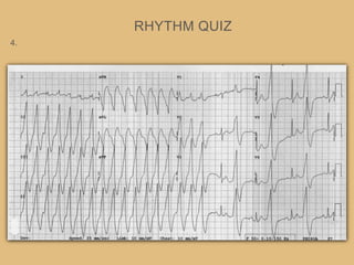
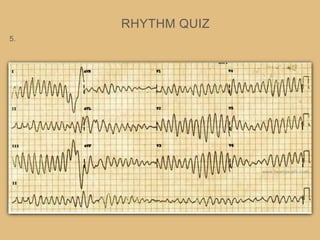
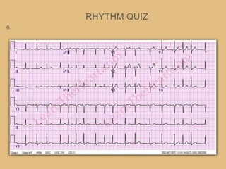
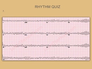
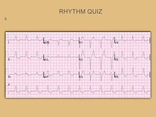
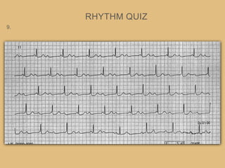
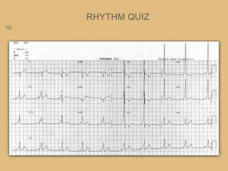
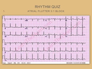
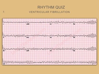
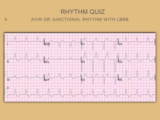
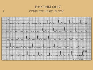
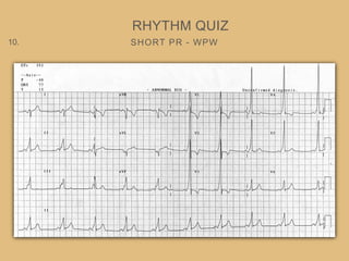
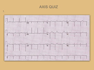
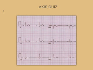
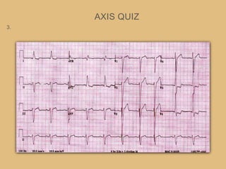
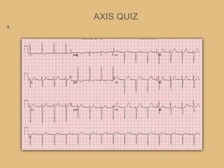
SYSTEMATIC
ECG ANALYSIS
REFERENCES
ECG
• previous CME
talks
• Life In The Fast
Lane
• Dr Smiths ECG
blog
• Google Image
Search
• Time on the Floor
COMPONENTS
ECG
• Rate
• Rhythm
• Axis
• P
• PR
• QRS
• QT
• ST
• T
• Delta
• Epsilon
• Osborne
• U
THE NORMAL ECG
• Rhythm: <10% variation in
R-R intervals
• Rate: 60 - 100bpm
• Axis -30 - -90
• P Waves
• 0.2-0.3mV
• 0.06 – 0.12s
• Upright in I, II, aVF, V2-
V6
• Inverted in aVR
• Varies in III, aVLSinus
origin
• PR Interval
• 0.12 – 0.2s
• Q Waves
• Small in I, II, aVL, V5, V6
• QRS Complex
• <0.12s
• ST Segment
• Isoelectric
• T Waves
• <2/3 height of preceding
R wave
• 0.5mm in I, II, III
• <10mm in V1 – V6
• Same direction as
preceding R wave
• U Waves
• <25% of T wave
• Same direction as T wave
• QTc
• <440ms in males
• <460ms in females
THE NORMAL ECG
Get used to knowing what normal looks like.
RATE
• Small square: 0.04sec
• Big square: 0.2sec
• If it looks fast
• 1500/small squares
• If it looks slow/normal
• 300/big squares
• If irregular, count complexes on rhythm strip x 6 for average
RHYTHM
• Regular
• Irregular
• Regularly Irregular
• Irregularly Irregular
• P-wave:
• Are they present?
• Are they regular?
• Is there a P for every QRS?
• Do the P-waves look similar?
• PR Interval
• Long? >0.12s
• Short? <0.06s
• Are the PR intervals Consistent?
• QRS
• Long? >0.12s
• Similar in appearance to each other?
• Atrial, AVNRT, Accessory Pathways,
Junctional rhythms, Ventricular
rhythmas
RHYTHM
Regular? P waves? PR interval? - long, short, consistent?P for every QRS?
QRS? Normal Sinus Rhythm
RHYTHM
Regular? P waves? PR interval? - long, short, consistent?P for every QRS?
QRS? 1st degree Heart Block
HEART BLOCK
RHYTHM
RHYTHM
Regular? P waves? PR interval? - long, short, consistent?P for every QRS?
QRS? Atrial Fibrillation
RHYTHM
Regular? P waves? PR interval? - long, short, consistent?P for every QRS?
QRS? Atrial Flutter 4:1 block
RHYTHM
Regular? P waves? PR interval? - long, short, consistent?P for every QRS?
QRS? Atrial Flutter
TO ILLUMINATE THE P WAVES
RHYTHM
CLASSIC LEAD PLACEMENT THEN WITH LEWIS LEADS
RHYTHM
RHYTHM
Regular? P waves? PR interval? - long, short, consistent?P for every QRS?
QRS? SVT - AVNRT
SUPRA VENTRICULAR TACHYCARDIAS
RHYTHM
• A tachydysrhythmia originating above the Bundle of His
• Sinus tachy, Atrial flutter, Atrial Fibrillation
• Atrio-Ventricular Re-Entry Tachycardia (AVRT)
• AV Nodal Re-Entry Tachycardia (AVNRT)
• Automatic Junctional Tachycardia
AVNRT VS AVRT
RHYTHM
• Left - AVNRT
• Right - AVRT (accessory pathway)
AVNRT
• Slow and fast pathway within
the AV Node
• If a PAC arrives whilst a
pathway is refractory, it can
allow the other pathway to set
up a re-entry pathway within
the AV node
ACCESSORY PATHWAYS
AVRT
• Orthodromic • Antidromic
RHYTHM
Regular? P waves? PR interval? - long, short, consistent?P for every QRS?
QRS? AVRT - Orthodromic
RHYTHM
Regular? P waves? PR interval? - long, short, consistent?P for every QRS?
QRS? AVRT - Antidromic
VT VS SVT WITH ABERRANT CONDUCTION
RHYTHM
• VT is more likely when
• absence of typical RBBB/LBBB
morphology
• Very broad complexes
(>160ms)
• AV dissociation
• Fusion beats - when sinus and
ventricular beat fuse to cause
hybrid complex
• Entirely positive or entirely
negative complexes throughout
V1-6
• Brugada’s - Distance from onset
of QRS to nadir of S-wave is
>100ms
• Josephson’s Sign - notching
near nadir of S-wave
• RSR complexes w/ Left Rabbit
Ear being taller. In contrast
Right Rabbit Ear is taller in
RBBB
VT VS SVT WITH ABERRANT CONDUCTION
RHYTHM
• Fusion then capture
• Positive concordance
VT VS SVT WITH ABERRANT CONDUCTION
RHYTHM
• Brugada
• Josephson’s
• RSR in V1
RHYTHM
Regular? P waves? PR interval? - long, short, consistent?P for every QRS?
QRS? Ventricular Fibrillation
RHYTHM
Regular? P waves? PR interval? - long, short, consistent?P for every QRS?
QRS? Polymorphic VT
QUIZ TIME!!!
RHYTHM QUIZ
1.
RHYTHM QUIZ
2.
RHYTHM QUIZ
3.
RHYTHM QUIZ
4.
RHYTHM QUIZ
5.
RHYTHM QUIZ
6.
RHYTHM QUIZ
7.
RHYTHM QUIZ
8.
RHYTHM QUIZ
9.
RHYTHM QUIZ
10.
WHAT IS A LICHTENBERG FIGURE?
RHYTHM QUIZ
11.
ATRIAL FLUTTER 3:1 BLOCK
RHYTHM QUIZ
1.
AVRT - ORTHODROMIC WPW
RHYTHM QUIZ
2.
MOBITZ 1 - WENCKEBACH
RHYTHM QUIZ
3.
VT THEN BIGEMINY
RHYTHM QUIZ
4.
TORSADES DE POINTES
RHYTHM QUIZ
5.
ATRIAL FIBRILLATION
RHYTHM QUIZ
6.
VENTRICULAR FIBRILLATION
RHYTHM QUIZ
7.
AIVR OR JUNCTIONAL RHYTHM WITH LBBB
RHYTHM QUIZ
8.
COMPLETE HEART BLOCK
RHYTHM QUIZ
9.
SHORT PR - WPW
RHYTHM QUIZ
10.
WHAT IS A LICHTENBERG FIGURE?
RHYTHM QUIZ
11.
They are branching electric discharges that sometimes appear on
the surface or in the interior of insulating materials.
AXIS
AXIS
• Normal Axis = -30 - +90
• Left Axis = <-30
• Right Axis = >+90
• Extreme Right Axis = -90 - 180
AXIS
• QRS Positive Deflection =
axis toward that lead
• QRS Negative Deflection =
axis away from the lead
• QRS Isoelectric = Axis
perpendicular to that lead
DIFFERENTIALS
AXIS
• RAD
• RVH
• RV Strain
• Lateral STEMI
• COPD
• HyperK+
• Na+ Blockade
• WPW
• Normal Paeds
ECG
• LAD
• LVH
• LBBB
• Inf. MI
• Ventricular
pacing
• WPW
• Extreme Axis
• HyperK+
• Ventricular
arrhythmias
• Severe RVH
QUIZ TIME!!!
AXIS QUIZ
1.
AXIS QUIZ
2.
AXIS QUIZ
3.
AXIS QUIZ
4.
AXIS QUIZ
5.
AXIS QUIZ
6.
WHAT IS COMMOTIO CORDIS?
AXIS QUIZ
7.
LEFT AXIS DEVIATION
AXIS QUIZ
1.
NORMAL AXIS
AXIS QUIZ
2.
LEFT AXIS DEVIATION - LVH
AXIS QUIZ
3.
LEFT AXIS DEVIATION
AXIS QUIZ
4.
RIGHT AXIS DEVIATION - RVH
AXIS QUIZ
5.
RIGHT AXIS DEVIATION
AXIS QUIZ
6.
WHAT IS COMMOTIO CORDIS?
AXIS QUIZ
7.
Commotio cordis (Latin, "agitation of the heart") is an often lethal disruption of heart
rhythm that occurs as a result of a blow to the area directly over the heart (the precordial
region), at a critical time during the cycle of a heart beat causing cardiac arrest. It is a
form of ventricular fibrillation (V-Fib), not mechanical damage to the heart muscle or
surrounding organs, and not the result of heart disease. The fatality rate is about 65%
even with prompt CPR and defibrillation, and more than 80% without.
THE P WAVE
• P Wave
• Width <120ms
• Amplitude
• <2.5mm limb leads
• <1.5mm Precordial
• V1 biphasic
• aVF inverted
THE P WAVE
THE PR INTERVAL
• PR Interval
• 120-200ms
• Long - any heart block
• Short - WPW, pre-excitation
• Elevation/Depression - Pericarditis
THE QRS COMPLEX
NEGATIVE DEFLECTION BEFORE THE R WAVE
THE Q WAVE
• They are pathological if:
• >40ms wide
• >2mm deep
• >0.25% QRS
• Present in leads V1-V3
• >2mm in leads III/avR can be a normal variant
NEGATIVE DEFLECTION BEFORE THE R WAVE
THE Q WAVE
• They are pathological if:
• >40ms wide
• >2mm deep
• >0.25% QRS
• Present in leads V1-V3
• >2mm in leads III/avR can
be a normal variant
FIRST POSITIVE DEFLECTION AFTER THE P-WAVE
THE R WAVE
• Causes of a dominant R wave in V1
• Normal in paediatric and young adults
• RVH
• RBBB
• Posterior STEMI
• WPW
• Dextrocardia
• HOCM
• Dystrophy
• Myotonic
• Duchennes
RVH
FIRST POSITIVE DEFLECTION AFTER THE P-WAVE
THE R WAVE
• Dominant R wave in aVR
• Poisoning - Na-Channel Blockers
• Dextrocardia
• Incorrect lead placement
• Commonly in VT
TCA poisoning
FIRST POSITIVE DEFLECTION AFTER THE P-WAVE
THE R WAVE
• Poor R Wave Progression - R wave <4mm in V3
• Prior anteroseptal MI
• LVH
• Inaccurate lead placement
• Can be normal variant
THE QRS
• LBBB - depolarisation
activated from RV via
right bundle then to LV
via left bundle
• RBBB - Right
ventricular
depolarisation is
delayed and so
depolarisation spreads
from left to right
BUNDLE BRANCH BLOCK
LEFT VENTRICULAR HYPERTROPHY
THE QRS
• Muscle wall thickens
• Increased S wave in Right
sided leads
• Increased R wave in Left sided
leads
• Prolonged depolarisation
• Repolarisation abnormalities in
lateral leads
• Causes
• HTN
• AR/AS
• MR
• HOCM
LEFT VENTRICULAR HYPERTROPHY
THE QRS
RIGHT VENTRICULAR HYPERTROPHY
THE QRS
• Right Axis
• Dominant R in V1 >7mm
• Dominant S in V6 >7mm
• RV Strain - ST depression in
V1-4 and inferiorly
• Causes
• PE
• Chronic Lung Dx
• Pulm. HTN
• Mitral Stenosis
• Congenital Heart Dx
RIGHT VENTRICULAR HYPERTROPHY
THE QRS
QUIZ TIME!!!
QRS QUIZ
1.
QRS QUIZ
2.
QRS QUIZ
3.
QRS QUIZ
4.
QRS QUIZ
5.
QRS QUIZ
6.
THE HEART OF A BLUE WHALE IS ABOUT AS BIG AS A…?
QRS QUIZ
7.
A. Volkswagen Beetle
B. Piano
C. Average Person
D. Bus
ANTERIOR Q WAVES
QRS QUIZ
1.
RBBB
QRS QUIZ
2.
LVH
QRS QUIZ
3.
POOR R WAVE PROGRESSION - LAD OCCLUSION
QRS QUIZ
4.
PATHOLOGICAL Q WAVES - AMI 1/52 PRIOR
QRS QUIZ
5.
LBBB
QRS QUIZ
6.
THE HEART OF A BLUE WHALE IS ABOUT AS BIG AS A…?
QRS QUIZ
7.
A. Volkswagen Beetle
B. Piano
C. Average Person
D. Bus
REPRESENTS INTERVAL BETWEEN VENTRICULAR
DEPOLARISATION AND REPOLARIZATION
THE ST SEGMENT
SGARBOSSA CRITERIA
ST SEGMENT
MODIFIED SGARBOSSA
ST SEGMENT
MODIFIED SGARBOSSA
ST SEGMENT
ST depression >0.3 height of R wave
QUIZ TIME!!!
DONDE ESTA LA STEMI?
ST QUIZ
1.
ST QUIZ
2.
ST QUIZ
3.
ST QUIZ
4.
ST QUIZ
5.
ST QUIZ
6.
ST QUIZ
7.
AN OCTOPUS HAS HOW MANY HEARTS?
ST QUIZ
8.
LATERAL STEMI
ST QUIZ
1.
INFERIOR STEMI
ST QUIZ
2.
PERICARDITIS
ST QUIZ
3.
POSTERIOR STEMI
ST QUIZ
4.
ANTERIOR STEMI
ST QUIZ
5.
INFERIOR STEMI
ST QUIZ
6.
ANTEROLATERAL STEMI
ST QUIZ
7.
AN OCTOPUS HAS HOW MANY HEARTS?
ST QUIZ
8.
3!!!
Two brachial hearts on either side of the body that oxygenate blood by pumping it through the gills
One systemic heart pumps blood from gills to the rest of the body
START OF THE Q TO THE END OF T
YOU QT
• Represents Ventricular de- and repolarization
• Inversely proportional to heart rate
• 440ms for males, 460 for females
• QT >500ms increases risk of TDP
VENTRICULAR REPOLARIZATION
T WAVES
• Can be inverted in V1 and aVR
• Flat, Biphasic, Inverted, Peaked, Tented
• Look for dynamic change
• Don’t miss Wellen’s Syndrome
• Hyperacute - early STEMI, prinzmetal Angina
• Inverted T - can be normal, MI, BBB, Hypertrophy, PE, HOCM
• Biphasic - Ischaemia, Hypokalaemia
WELLENS’ SYNDROME
T WAVES
• Inverted or Biphasic T-waves in V2-3 (in patients presenting with
ischaemic chest pain) highly specific for critical stenosis of LAD
Type A Type B
DE WINTERS - LAD OCCLUSION
T WAVES
• Peaked anterior T waves with ascending limb of T wave
commencing below isoelectric line - tall, prominent, symmetric
• Upsloping ST depression in precordial leads
DELTA WAVE
OTHER WAVES
• WPW - pre excitation
U WAVE
OTHER WAVES
• Thought to be delayed depolarisation of Purkinje Fibers
• Prominent U waves: Bradycardia, Hypo-Ca/Mg, Hypothermia, LVH,
HOCM, Digoxin, Class Ia, III antiarrhythmics
• Inverted U waves: CAD, HTN, Valvular HD, Congenital HD,
Hyperthyroid
OSBORNE WAVE
OTHER WAVES
• Positive deflection at J point
• Hypothermia, Normal Variant, Hypercalcaemia
Hypothermia Temp 32
QUIZ TIME!!!
CENTRAL CHEST PAIN
QUIZ TIME
1.
QUIZ TIME
2.
CENTRAL CHEST PAIN AND SHORTNESS OF BREATH
QUIZ TIME
3.
QUIZ TIME
4.
CHEST PAIN
QUIZ TIME
5.
CHEST PAIN
QUIZ TIME
6.
QUIZ TIME
7.
WHAT IS A SHARKS SIXTH SENSE?
BONUS POINTS: NAME THE ORGAN THAT DETECTS THIS
QUIZ TIME
8.
DE WINTERS T WAVES - LAD OCCLUSION
QUIZ TIME
1.
HYPERKALAEMIA K+ 9.0 - PEAKED T, WIDE QRS, SINE WAVE
APPEARANCE
QUIZ TIME
2.
MASSIVE PE - EXTREME RAD, S1Q3T3, RBBB, TWI V1-4
QUIZ TIME
3.
HYPOTHERMIA TEMP 26 - OSBORN WAVES
QUIZ TIME
4.
LBBB - CONCORDANT ST DEPRESSION V3 - STEMI
QUIZ TIME
5.
PERICARDITIS
QUIZ TIME
6.
WIDE QRS, TACHYCARDIA, POSITIVE R WAVE IN AVR - TCA
OVERDOSE
QUIZ TIME
7.
WHAT IS A SHARKS SIXTH SENSE?
BONUS POINTS: NAME THE ORGAN THAT DETECTS THIS
QUIZ TIME
8.
They can sense electricity!
Ampullae of Lorenzini
THANK YOU!

Systematic ECG analysis

Editor's Notes