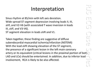
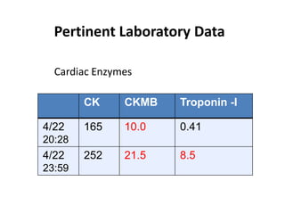
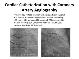
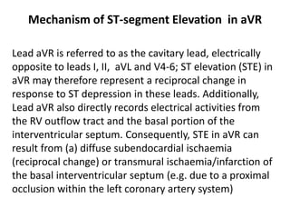
RJS ECG Case Series
Case 4
RJS ECG Case Series
                      Interpretation
Sinus rhythm at 85/min with left axis deviation.
Wide spread ST segment depression involving leads II, III,
aVF, and V2-V6 (with associated T wave inversion in leads
III, aVF, and V3-V6).
ST segment elevation in leads aVR and V1.

Taken together, these finding are suggestive of diffuse
subendocardial myocardial ischemia/infarction (NSTEMI)
With the lead aVR showing elevation of the ST segment,
the presence of a significant lesion in the left main coronary
artery or its equivalent (critical lesions in the proximal portion of both
LAD and LCx) should be entertained. In addition, due to inferior lead
involvement, RCA is likely to be also affected.
Brief Hx and Physical Findings
This 62-yr-old man was brought to the ER because of having severe
chest tightness of sudden onset, lasting for about 30 min. Reportedly,
he had experienced intermittent episodes of chest tightness like this
one not related to exertion for one year. About one week before this
admission, the frequency of these episodes had increased from 2-3 to
4-5/week.
Past medical Hx was significant for DM and hypertension for more
than 10 years and ESRD presumably due to diabetic nephropathy for
which he had been on peritoneal dialysis for one and a half years.
Additionally, he had one episode of TIA that had occurred
approximately one year prior. On PE, he was obese, in no respiratory
distress. BP measured 110/70 mmHg, PR 80/min and regular, and RR
16/min. The lungs were clear. The PMI of the heart was not visible.
There was a loud S4 but there were no murmurs.
Chest X-ray
There is no significant cardiomegaly.
The lung markings are slightly increased
in both lower lung fields.
Bilateral costophrenic angles are clearly
identified.
Pertinent Laboratory Data

  Cardiac Enzymes

        CK     CKMB   Troponin -I

4/22    165    10.0   0.41
20:28
4/22    252    21.5   8.5
23:59
Cardiac Catheterization with Coronary
         Artery Angiography
    Preserved LV systolic function without significant regional
    wall motion abnormality LM ostium: 30-40% narrowing;
    LAD-mid: 100% stenosis; LCX-proximal: 80% stenosis, LCx-
    m: 80% stenosis, LCx-OM1: 80% stenosis; RCA-m: 90%
    stenosis, RCA-PDA: 90% stenosis.
Mechanism of ST-segment Elevation in aVR

Lead aVR is referred to as the cavitary lead, electrically
opposite to leads I, II, aVL and V4-6; ST elevation (STE) in
aVR may therefore represent a reciprocal change in
response to ST depression in these leads. Additionally,
Lead aVR also directly records electrical activities from
the RV outflow tract and the basal portion of the
interventricular septum. Consequently, STE in aVR can
result from (a) diffuse subendocardial ischaemia
(reciprocal change) or transmural ischaemia/infarction of
the basal interventricular septum (e.g. due to a proximal
occlusion within the left coronary artery system)
Take-Home Message
• The ECG findings suggestive of significant LMCA are
  widespread horizontal ST depression, most prominent
  in leads I, II and V4-6; ST elevation in aVR ≥ 1mm; and
  ST elevation in aVR ≥ V1.
• However, ST elevation in aVR is not entirely specific to
  LMCA occlusion. It may also be seen with proximal
  LAD occlusion and severe triple vessel CAD.
• ST elevation in aVR ≥ 1 mm is a strong predictor of
  severe LMCA /triple vessel CAD requiring CABG.

    Kosuge M, et al. An early and simple predictor of severe left main and/or
    three-vessel disease in patients with non-ST-segment elevation
    acute coronary syndrome. Am J Cardiol. 107:495-500, 2011.
Acknolwledgment
The Case was provided by Int 王昇元 / VS 張盛
雄 at MacKay Memorial Hospital, Taipei
2012-04-28

Case 004

  • 1.
    RJS ECG CaseSeries Case 4
  • 2.
    RJS ECG CaseSeries Interpretation Sinus rhythm at 85/min with left axis deviation. Wide spread ST segment depression involving leads II, III, aVF, and V2-V6 (with associated T wave inversion in leads III, aVF, and V3-V6). ST segment elevation in leads aVR and V1. Taken together, these finding are suggestive of diffuse subendocardial myocardial ischemia/infarction (NSTEMI) With the lead aVR showing elevation of the ST segment, the presence of a significant lesion in the left main coronary artery or its equivalent (critical lesions in the proximal portion of both LAD and LCx) should be entertained. In addition, due to inferior lead involvement, RCA is likely to be also affected.
  • 3.
    Brief Hx andPhysical Findings This 62-yr-old man was brought to the ER because of having severe chest tightness of sudden onset, lasting for about 30 min. Reportedly, he had experienced intermittent episodes of chest tightness like this one not related to exertion for one year. About one week before this admission, the frequency of these episodes had increased from 2-3 to 4-5/week. Past medical Hx was significant for DM and hypertension for more than 10 years and ESRD presumably due to diabetic nephropathy for which he had been on peritoneal dialysis for one and a half years. Additionally, he had one episode of TIA that had occurred approximately one year prior. On PE, he was obese, in no respiratory distress. BP measured 110/70 mmHg, PR 80/min and regular, and RR 16/min. The lungs were clear. The PMI of the heart was not visible. There was a loud S4 but there were no murmurs.
  • 4.
    Chest X-ray There isno significant cardiomegaly. The lung markings are slightly increased in both lower lung fields. Bilateral costophrenic angles are clearly identified.
  • 5.
    Pertinent Laboratory Data Cardiac Enzymes CK CKMB Troponin -I 4/22 165 10.0 0.41 20:28 4/22 252 21.5 8.5 23:59
  • 6.
    Cardiac Catheterization withCoronary Artery Angiography Preserved LV systolic function without significant regional wall motion abnormality LM ostium: 30-40% narrowing; LAD-mid: 100% stenosis; LCX-proximal: 80% stenosis, LCx- m: 80% stenosis, LCx-OM1: 80% stenosis; RCA-m: 90% stenosis, RCA-PDA: 90% stenosis.
  • 7.
    Mechanism of ST-segmentElevation in aVR Lead aVR is referred to as the cavitary lead, electrically opposite to leads I, II, aVL and V4-6; ST elevation (STE) in aVR may therefore represent a reciprocal change in response to ST depression in these leads. Additionally, Lead aVR also directly records electrical activities from the RV outflow tract and the basal portion of the interventricular septum. Consequently, STE in aVR can result from (a) diffuse subendocardial ischaemia (reciprocal change) or transmural ischaemia/infarction of the basal interventricular septum (e.g. due to a proximal occlusion within the left coronary artery system)
  • 8.
    Take-Home Message • TheECG findings suggestive of significant LMCA are widespread horizontal ST depression, most prominent in leads I, II and V4-6; ST elevation in aVR ≥ 1mm; and ST elevation in aVR ≥ V1. • However, ST elevation in aVR is not entirely specific to LMCA occlusion. It may also be seen with proximal LAD occlusion and severe triple vessel CAD. • ST elevation in aVR ≥ 1 mm is a strong predictor of severe LMCA /triple vessel CAD requiring CABG. Kosuge M, et al. An early and simple predictor of severe left main and/or three-vessel disease in patients with non-ST-segment elevation acute coronary syndrome. Am J Cardiol. 107:495-500, 2011.
  • 9.
    Acknolwledgment The Case wasprovided by Int 王昇元 / VS 張盛 雄 at MacKay Memorial Hospital, Taipei 2012-04-28