
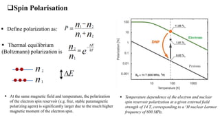
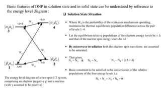
Downloaded 201 times

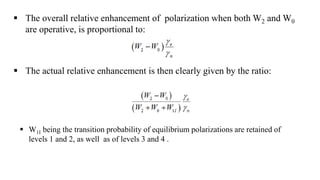
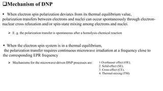
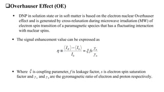
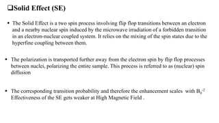
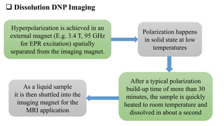

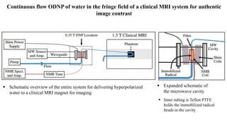
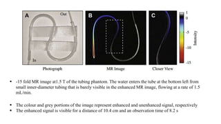
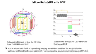
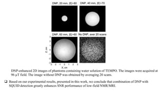
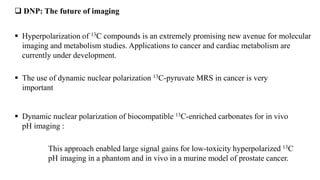
This document discusses dynamic nuclear polarization imaging (DNP). It begins by explaining the limitations of standard MRI, such as low sensitivity. DNP increases sensitivity by transferring polarization from unpaired electrons to nuclei. This can provide sensitivity enhancements over 10000x. There are several mechanisms by which DNP can occur, such as the Overhauser effect in liquids and the solid effect in solids. DNP can be used in dissolution mode, where polarization occurs in a separate polarizer, or in situ, directly in the imager. Applications include metabolic imaging with hyperpolarized 13C compounds and micro-Tesla MRI combined with DNP. In summary, DNP is a powerful technique for enhancing MRI sensitivity for various biomedical


![UV SPECTROSCOPY [ULTRA-VIOLET SPECTROSCOPY]](https://cdn.slidesharecdn.com/ss_thumbnails/40-191218142647-thumbnail.jpg?width=640&height=640&fit=bounds)


![ANIMAL_CELL_,_TISSUE_AND_ORGAN_CULTURE[1].pptx](https://cdn.slidesharecdn.com/ss_thumbnails/animalcelltissueandorganculture1-260204172026-4462b440-thumbnail.jpg?width=640&height=640&fit=bounds)

![Polymer [ बहुलक ] Chemistry Notes PDF - Irfanullah Mehar - JJ Sir Chemistry.pdf](https://cdn.slidesharecdn.com/ss_thumbnails/polymerchemistrynotespdf-irfanullahmehar-jjsirchemistry-260210172118-3f9b37f7-thumbnail.jpg?width=640&height=640&fit=bounds)


