Dr screening training for nurses definition of acceptable image quality-color
•
0 likes•24 views

Diabetic retinopathy-screening training program for nurses St John Eye Hospital - UNRWA

Recommended
Recommended
More Related Content
More from Riyad Banayot
More from Riyad Banayot (20)
Recently uploaded
Recently uploaded (20)
Dr screening training for nurses definition of acceptable image quality-color
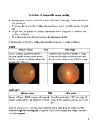
- 1. 1 Definition of acceptable image quality • Photographers should capture 2x nominal 45O fields per eye (1 x fovea centered, 1 x disc centered) • A combined assessment of field position and image quality should be made for each eye • Images must be graded for diabetic eye disease only if the grader is confident the quality is sufficient • All grading is to be performed by trained and accredited staff A combined assessment of field position and image quality is made as follows: GOOD Macular image AND Disc image Center of fovea ≤1DD from center of image & vessels clearly visible within 1DD of center of fovea & vessels visible across >90% of image Center of disc ≤1DD from center of image & fine vessels clearly visible on surface of disc & vessels visible across >90% of image ADEQUATE Macular image AND Disc image Center of fovea >2DD from edge of image & vessels visible within 1DD of center of fovea Complete optic disc >2DD from edge of image & fine vessels visible on surface of disc In some unusual cases (particularly in patients with a large disc), an image may fall within both good and adequate categories above. In such cases, the image should be classified as good
- 2. 2 INADEQUATE (ungradable) Failure to meet definition of adequate (above); UNLESS referable diabetic retinopathy (R2, R3, M1, unstable treated proliferative diabetic retinopathy) is visible anywhere in the eye. Definitions of disc, fovea, 1DD The image shown below is a perfectly aligned macular view of the right eye. The fovea lies at the center of the image and is marked “+”