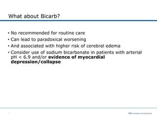
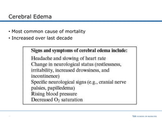
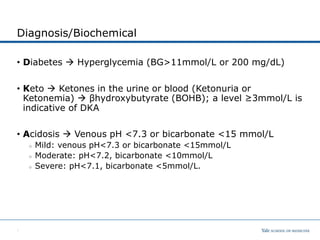
Objectives
• Recognize DKA
• Initialize therapy
• Recognize life-threatening complications
0
Epidemiology
• Has increased 1.54 per 1000 under age 20
• 40% present in DKA
○ Vague symptoms
○ Delay diagnosis
• DKA -- Most frequent cause of death in children with Type 1
DM (0.15% - 0.3%)
○ ~1 in 350 children by age 18 in US have DM
Pathophysiology
• Insulin deficiency has three main effects:
1. Loss of insulin-dependent glucose transport into peripheral
tissues
2. Increased gluconeogenesis in the liver
3. Increased breakdown of fat, protein, and glycogen
Thus, insulin deficiency results in hyperglycemia (from
increased hepatic glucose production and decreased peripheral
uptake) and acidosis (primarily derived from hepatic fatty acid
oxidation into ketoacids).
Common Cause: New onset, intercurrent illness, insulin pump
malfunction, or purposeful insulin omission
2
Diagnosis/Biochemical
• Diabetes  Hyperglycemia (BG>11mmol/L or 200 mg/dL)
• Keto  Ketones in the urine or blood (Ketonuria or
Ketonemia)  βhydroxybutyrate (BOHB); a level ≥3mmol/L is
indicative of DKA
• Acidosis  Venous pH <7.3 or bicarbonate <15 mmol/L
○ Mild: venous pH<7.3 or bicarbonate <15mmol/L
○ Moderate: pH<7.2, bicarbonate <10mmol/L
○ Severe: pH<7.1, bicarbonate <5mmol/L.
3
Clinical Signs
• Dehydration (polyuria)
• Tachycardia
• Tachypnea
• Deep, sighing(Kussmaul)
respiration
• Acetone breath
• Nausea, vomiting
• Abdominal pain that may
mimic an acute abdominal
condition
• Confusion, drowsiness,
progressive loss of
consciousness.
4
Risk Factors
• Very young children
• Lower social economic
Background
• Prior poor compliance
• Concomitant psychiatric
disease
• Adolescent girls
5
Complications of DKA
• Cerebral Edema
• Cardiac Arrhythmias
• Severe Electrolyte abnormalities
• Severe Dehydration
• Thrombosis (CVL related, Stroke)
• Pulmonary edema
• Renal Failure
• Pancreatitis
• Rhabdomyolysis
• Severe Infections/Sepsis
• Death
6
Laboratory Investigation
• Glucose, urine dip
• Blood gas (venous or arterial)
• Electrolytes and other labs
Na is typically low. For every 100 mg/dL glucose above 200 mg/dL,
the measure Na should be reduced by 1.6 mEq/L
Nacorrected = Nameasured + 1.6 x [Glucose] - 200
100
K+  total body depletion
Ca and phosp typically low
BUN/Cr typically elevated in dehydration
WBC and left shift  typically due to acute stress but also look
for infectious trigger process
7
Goals of Therapy
Correction of Dehydration, electrolyte deficits, hyperglycemia,
and acidosis.
A. Cardiovascular collapse
○ From dehydration
○ Treatment involves intravascular fluid expansion with
ISOTONIC fluids
B. Overwhelming acidosis
○ From ketoacid production and lactic acid accumulation
○ Volume expansion and tissue reperfusion to correct lactic acidosis
○ Prompt initiation of insulin to stop fatty acid oxidation and
ketone production
○ Consider use of sodium bicarbonate in patients with arterial pH <
6.9 and/or evidence of myocardial depression/collapse
• C. Hypokalemia
○ Insulin therapy is associated with rapid intracellular movement of
potassium
8
Treatment of DKA
9
Diagnose
• Hyperglycemia
• Ketoacidosis
Fluids
• Isotonic fluid bolus
• 1.5 to 2 X M (with NS)
Insulin
• 0.1 U/kg/hrstart after first fluid bolus
Dextrose
• Add when glucose drops below 300 mg/dL
K+
• Add if adequate renal function
What about Bicarb?
• No recommended for routine care
• Can lead to paradoxical worsening
• And associated with higher risk of cerebral edema
• Consider use of sodium bicarbonate in patients with arterial
pH < 6.9 and/or evidence of myocardial
depression/collapse
10
What about K?
• Potassium deplete
• Therapy (insulin and
correcting acidosis) cause
K to shift back into cells
• Check EKG/monitor
• Replete K if renal function
okay
11
Cerebral Edema
• Most common cause of mortality
• Increased over last decade
12
Cerebral Edema
• Hypertonic dehydration in DKA is associated with the
production of osmotically active particles in the brain that act
to prevent neuronal cellular dehydration.
• Correction of hyperosmolar state leads to fluid influx into the
brain. Rapid rates of rehydration or correction of
hyperosmolarity may lead to cerebral cellular swelling
and brain herniation.
• It usually occurs 6-18 hours into therapy, just as the patient appears to
be clinically and biochemically improving.
• This is the most common cause of mortality in children with DKA !!!
⎯ Mortality rate of 20% and morbidity rate of 25%
13
Cerebral Edema
• Much of the etiology and pathophysiology of cerebral edema is
still unknown, but the risk should be minimized by attention to
the following:
• Slow fluid replacement (over 48 hrs) with isotonic fluids
• Frequent monitoring and slow rise of calculated CORRECTED Na values
• Close neurologic surveillance for early signs of increased ICP
(headache, lethargy, slurred speech, obtundation) and rapid
evaluation & action
• Don’t exceed 4L/m2/24 hours
14
15
Treatment of Cerebral Edema
• Initiate treatment as soon as the condition is suspected.
• Reduce the rate of fluid administration.
• Give mannitol, 0.5–1g/kg IV over 10–15min, and repeat if there
is no initial response in 30min to 2h
• Hypertonic saline(3%),suggested dose 2.5–5mL/kg over 10–
15min, may be used as an alternative to mannitol.
• Elevate the head of the bed to 30◦.
• Intubation may be necessary for the patient with impending
respiratory failure.
• After treatment for cerebral edema has been started, cranial
imaging may be considered.
○ The primary concern is whether the patient has a lesion requiring
emergency neurosurgery (e.g., intracranial hemorrhage) or a lesion
that may necessitate anticoagulation (e.g., cerebrovascular
thrombosis).
16
The High-Risk Patient
• Age < 3 years
• Significantly altered or deteriorating mental status
• pH < 7.2
• Glucose > 900 mg/dl
• Na (calculated)>160 or any patient with falling (calculated) Na
• K < 3.5 mEq/L on admission
• Severe hyperosmolality (Sosm > 350 mOsm)
• Other organ system dysfunction that complicates treatment
17
18
Hyperglycemia Hyperosmolar State (HHS)
formerly hyperosmolar nonketotic coma
• Plasma glucose concentration >33.3mmol/L (600mg/dL)
• Arterial pH>7.30; venous pH>7.25
• Serum bicarbonate >15mmol/L
• Small ketonuria, absent to small ketonemia
• Effective serum osmolality >320mOsm/kg
• Obtundation, combativeness, or seizures(in approximately
50%).
• Primarily Type 2 diabetics but can overlap with Type 1
especially in those with severe dehydration or those with high
carbohydrate load before hand
• Volume Resuscitation and insulin (often less)
19

DKA-for-PEM-1.pptx

  • 1.
    Objectives • Recognize DKA •Initialize therapy • Recognize life-threatening complications 0
  • 2.
    Epidemiology • Has increased1.54 per 1000 under age 20 • 40% present in DKA ○ Vague symptoms ○ Delay diagnosis • DKA -- Most frequent cause of death in children with Type 1 DM (0.15% - 0.3%) ○ ~1 in 350 children by age 18 in US have DM
  • 3.
    Pathophysiology • Insulin deficiencyhas three main effects: 1. Loss of insulin-dependent glucose transport into peripheral tissues 2. Increased gluconeogenesis in the liver 3. Increased breakdown of fat, protein, and glycogen Thus, insulin deficiency results in hyperglycemia (from increased hepatic glucose production and decreased peripheral uptake) and acidosis (primarily derived from hepatic fatty acid oxidation into ketoacids). Common Cause: New onset, intercurrent illness, insulin pump malfunction, or purposeful insulin omission 2
  • 4.
    Diagnosis/Biochemical • Diabetes Hyperglycemia (BG>11mmol/L or 200 mg/dL) • Keto  Ketones in the urine or blood (Ketonuria or Ketonemia)  βhydroxybutyrate (BOHB); a level ≥3mmol/L is indicative of DKA • Acidosis  Venous pH <7.3 or bicarbonate <15 mmol/L ○ Mild: venous pH<7.3 or bicarbonate <15mmol/L ○ Moderate: pH<7.2, bicarbonate <10mmol/L ○ Severe: pH<7.1, bicarbonate <5mmol/L. 3
  • 5.
    Clinical Signs • Dehydration(polyuria) • Tachycardia • Tachypnea • Deep, sighing(Kussmaul) respiration • Acetone breath • Nausea, vomiting • Abdominal pain that may mimic an acute abdominal condition • Confusion, drowsiness, progressive loss of consciousness. 4
  • 6.
    Risk Factors • Veryyoung children • Lower social economic Background • Prior poor compliance • Concomitant psychiatric disease • Adolescent girls 5
  • 7.
    Complications of DKA •Cerebral Edema • Cardiac Arrhythmias • Severe Electrolyte abnormalities • Severe Dehydration • Thrombosis (CVL related, Stroke) • Pulmonary edema • Renal Failure • Pancreatitis • Rhabdomyolysis • Severe Infections/Sepsis • Death 6
  • 8.
    Laboratory Investigation • Glucose,urine dip • Blood gas (venous or arterial) • Electrolytes and other labs Na is typically low. For every 100 mg/dL glucose above 200 mg/dL, the measure Na should be reduced by 1.6 mEq/L Nacorrected = Nameasured + 1.6 x [Glucose] - 200 100 K+  total body depletion Ca and phosp typically low BUN/Cr typically elevated in dehydration WBC and left shift  typically due to acute stress but also look for infectious trigger process 7
  • 9.
    Goals of Therapy Correctionof Dehydration, electrolyte deficits, hyperglycemia, and acidosis. A. Cardiovascular collapse ○ From dehydration ○ Treatment involves intravascular fluid expansion with ISOTONIC fluids B. Overwhelming acidosis ○ From ketoacid production and lactic acid accumulation ○ Volume expansion and tissue reperfusion to correct lactic acidosis ○ Prompt initiation of insulin to stop fatty acid oxidation and ketone production ○ Consider use of sodium bicarbonate in patients with arterial pH < 6.9 and/or evidence of myocardial depression/collapse • C. Hypokalemia ○ Insulin therapy is associated with rapid intracellular movement of potassium 8
  • 10.
    Treatment of DKA 9 Diagnose •Hyperglycemia • Ketoacidosis Fluids • Isotonic fluid bolus • 1.5 to 2 X M (with NS) Insulin • 0.1 U/kg/hrstart after first fluid bolus Dextrose • Add when glucose drops below 300 mg/dL K+ • Add if adequate renal function
  • 11.
    What about Bicarb? •No recommended for routine care • Can lead to paradoxical worsening • And associated with higher risk of cerebral edema • Consider use of sodium bicarbonate in patients with arterial pH < 6.9 and/or evidence of myocardial depression/collapse 10
  • 12.
    What about K? •Potassium deplete • Therapy (insulin and correcting acidosis) cause K to shift back into cells • Check EKG/monitor • Replete K if renal function okay 11
  • 13.
    Cerebral Edema • Mostcommon cause of mortality • Increased over last decade 12
  • 14.
    Cerebral Edema • Hypertonicdehydration in DKA is associated with the production of osmotically active particles in the brain that act to prevent neuronal cellular dehydration. • Correction of hyperosmolar state leads to fluid influx into the brain. Rapid rates of rehydration or correction of hyperosmolarity may lead to cerebral cellular swelling and brain herniation. • It usually occurs 6-18 hours into therapy, just as the patient appears to be clinically and biochemically improving. • This is the most common cause of mortality in children with DKA !!! ⎯ Mortality rate of 20% and morbidity rate of 25% 13
  • 15.
    Cerebral Edema • Muchof the etiology and pathophysiology of cerebral edema is still unknown, but the risk should be minimized by attention to the following: • Slow fluid replacement (over 48 hrs) with isotonic fluids • Frequent monitoring and slow rise of calculated CORRECTED Na values • Close neurologic surveillance for early signs of increased ICP (headache, lethargy, slurred speech, obtundation) and rapid evaluation & action • Don’t exceed 4L/m2/24 hours 14
  • 16.
  • 17.
    Treatment of CerebralEdema • Initiate treatment as soon as the condition is suspected. • Reduce the rate of fluid administration. • Give mannitol, 0.5–1g/kg IV over 10–15min, and repeat if there is no initial response in 30min to 2h • Hypertonic saline(3%),suggested dose 2.5–5mL/kg over 10– 15min, may be used as an alternative to mannitol. • Elevate the head of the bed to 30◦. • Intubation may be necessary for the patient with impending respiratory failure. • After treatment for cerebral edema has been started, cranial imaging may be considered. ○ The primary concern is whether the patient has a lesion requiring emergency neurosurgery (e.g., intracranial hemorrhage) or a lesion that may necessitate anticoagulation (e.g., cerebrovascular thrombosis). 16
  • 18.
    The High-Risk Patient •Age < 3 years • Significantly altered or deteriorating mental status • pH < 7.2 • Glucose > 900 mg/dl • Na (calculated)>160 or any patient with falling (calculated) Na • K < 3.5 mEq/L on admission • Severe hyperosmolality (Sosm > 350 mOsm) • Other organ system dysfunction that complicates treatment 17
  • 19.
  • 20.
    Hyperglycemia Hyperosmolar State(HHS) formerly hyperosmolar nonketotic coma • Plasma glucose concentration >33.3mmol/L (600mg/dL) • Arterial pH>7.30; venous pH>7.25 • Serum bicarbonate >15mmol/L • Small ketonuria, absent to small ketonemia • Effective serum osmolality >320mOsm/kg • Obtundation, combativeness, or seizures(in approximately 50%). • Primarily Type 2 diabetics but can overlap with Type 1 especially in those with severe dehydration or those with high carbohydrate load before hand • Volume Resuscitation and insulin (often less) 19