SlideShare a Scribd company logo
1 of 10
Download to read offline
Major advances have been made in understanding the
mechanisms that are involved in the pathogenesis of
type 2 diabetes (T2D)1–5
. A decrease in insulin-stimulated
glucose uptake (insulin resistance) is associated with
obesity, ageing and inactivity. The pancreatic islets
respond to insulin resistance by enhancing their cell
mass and insulin secretory activity. However, when the
functional expansion of islet β-cells fails to compensate
for the degree of insulin resistance, insulin deficiency
and ultimately T2D develop. The onset of T2D leads in
turn to the development of its long-term consequences:
macrovascular complications (including atherosclerosis
and amputations) and microvascular complications
(including retinopathy, nephropathy and neuropathy).
Insulin resistance is typically present throughout the
progression from prediabetes to the later stages of
overt T2D. By contrast, the onset of T2D and its pro-
gression are largely determined by the progressive
failure of β-cells to produce sufficient levels of insu-
lin. Interestingly, many insulin-resistant individuals
do not become diabetic, because their β-cells are able
to compensate for the increased demand for insulin.
Only about one-third of obese, insulin-resistant indi-
viduals actually develop chronic hyperglycaemia and
T2D. The reasons for this heterogeneity are incompletely
understood, although genetics and epigenetics probably
have roles.
The leading hypothesized mechanisms to explain
insulin resistance and islet β-cell dysfunction in T2D
have been oxidative stress, endoplasmic reticulum stress
(ER stress), amyloid deposition in the pancreas, ectopic
lipid deposition in the muscle, liver and pancreas, and
lipotoxicity and glucotoxicity (BOX 1). All of these stresses
can be caused by overnutrition6–10
, although it has been
difficult to determine which mechanism is the most
important in each tissue and in each model or indi-
vidual with T2D. It is noteworthy, however, that each
of these cellular stresses is also thought to either induce
an inflammatory response or to be exacerbated by or
associated with inflammation11–15
.
This Review examines recent evidence that implicates
the pathological involvement of the immune system in
T2D, dissects potential underlying mechanisms and
concludes that obesity is associated with inflammation
and that the pathogenesis of T2D can be viewed as an
autoinflammatory disease. We also review the recent results
from clinical trials using anti-inflammatory drugs to
lower blood glucose levels in patients with T2D.
Evidence for T2D as an inflammatory disease
Circulating inflammatory factors in obesity and T2D.
Cross-sectional and prospective studies have described
elevated circulating levels of acute-phase proteins (such
as C-reactive protein (CRP), haptoglobin, fibrinogen,
plasminogen activator inhibitor and serum amyloid A)
and sialic acid, as well as cytokines and chemokines,
in patients with T2D16–19
. Furthermore, elevated levels
of interleukin-1β (IL-1β), IL-6 and CRP are predictive
of T2D17,20
. Similarly, the serum concentration of IL-1
receptor antagonist (IL-1RA) is elevated in obesity and
prediabetes21
, with an accelerated increase in IL-1RA
levels before the onset of T2D19,22,23
. The expression
*Clinic of Endocrinology,
Diabetes and Metabolism,
University Hospital Basel,
CH‑4031 Basel, Switzerland.
‡
Joslin Diabetes Center,
Harvard Medical School,
One Joslin Place, Boston,
Massachusetts 02215, USA.
e‑mails: MDonath@uhbs.ch;
steven.shoelson@joslin.
harvard.edu
doi:10.1038/nri2925
Published online
14 January 2011
Insulin resistance
A pathological condition in
which insulin becomes less
effective at lowering blood
glucose levels.
Endoplasmic reticulum
stress
(ER stress). A response by the
ER that results in the disruption
of protein folding and the
accumulation of unfolded
proteins in the ER.
Lipotoxicity
The toxic effects of elevated
levels of free fatty acids. These
detrimental effects may be
functional and reversible,
or may lead to cell death.
Type 2 diabetes as an inflammatory
disease
Marc Y. Donath* and Steven E. Shoelson‡
Abstract | Components of the immune system are altered in obesity and type 2 diabetes
(T2D), with the most apparent changes occurring in adipose tissue, the liver, pancreatic islets,
the vasculature and circulating leukocytes. These immunological changes include altered
levels of specific cytokines and chemokines, changes in the number and activation state of
various leukocyte populations and increased apoptosis and tissue fibrosis. Together, these
changes suggest that inflammation participates in the pathogenesis of T2D. Preliminary
results from clinical trials with salicylates and interleukin‑1 antagonists support this notion
and have opened the door for immunomodulatory strategies for the treatment of T2D
that simultaneously lower blood glucose levels and potentially reduce the severity and
prevalence of the associated complications of this disease.
REVIEWS
98 | FEBRuARy 2011 | VOLuME 11 www.nature.com/reviews/immunol
REVIEWS
© 2011 Macmillan Publishers Limited. All rights reserved
Glucotoxicity
The toxic effects of
hyperglycaemia. These
detrimental effects may be
functional and reversible, or
may lead to cell death.
Autoinflammatory disease
A disease resulting from an
attack by the innate immune
system on the body’s own
tissues. By contrast,
autoimmune diseases are
caused by the pathological
activation of adaptive immune
responses. Autoimmune and
autoinflammatory diseases
have some characteristics in
common, including shared
effector mechanisms.
M1‑type macrophage
A macrophage that is activated
by Toll-like receptor ligands
(such as lipopolysaccharide)
and interferon-γ, and that
expresses inducible nitric oxide
synthase, which generates
nitric oxide.
of IL-1RA is induced by IL-1β and reflects the body’s
response to counterbalance increased IL-1β activity.
Of particular interest is the increased CRP level, which
is currently the best epidemiological biomarker for
T2D-associated cardiovascular disease16–19
. Most pro-
inflammatory factors that are present at high levels in
the blood of patients with T2D are IL-1 dependent, and
blocking IL-1 activity has been shown to reduce their
concentrations24–27
(see below).
Elevated levels of circulating IL-1β, IL-6 and acute-
phase proteins in T2D may reflect the activation of
innate immune cells by increased nutrient concentra-
tions, but the levels of these inflammatory markers
may not necessarily reflect the degree of inflammation
in individual tissues. For example, the total volume of
the pancreatic islets is small compared with the blood
volume. Thus, even a high level of islet inflammation is
unlikely to demonstrably contribute to the circulating
levels of these inflammatory factors. By contrast, the
mass of adipose tissue in obese individuals is large, and
can make up over half of the body weight in morbid
obesity. The liver is also a relatively large organ and
is the site for IL-6-induced production of CRP. Thus,
the adipose tissue and the liver may disproportionately
contribute to the circulating levels of inflammatory
markers. Consistent with this, the circulating levels
of inflammatory factors in obese individuals with
prediabetes are similar to the levels in those with overt
diabetes. Furthermore, the levels of circulating CRP or
IL-6 do not predict the efficacy of anti-inflammatory
treatments directed towards insulin secretion or insu-
lin resistance25,28
. In summary, degrees of inflammation
vary within individuals and between tissues, and circu-
lating levels of inflammatory factors may not reflect the
severity of inflammation within a specific tissue.
Evidence for inflammation in insulin-sensitive tissues
and islets. The production of tumour necrosis factor
(TNF) by cells in the adipose tissue of obese rodents
provided early evidence of tissue inflammation in the
pathogenesis of insulin resistance and T2D29
(FIG. 1).
Some animal studies30
and several clinical trials using
TNF blockade have failed to demonstrate beneficial
effects on glucose metabolism31–36
(see below). However,
a few small studies conducted with obese individuals or
patients being treated for alternative conditions suggest
that TNF blockers may alter insulin sensitivity or gly-
caemic parameters, indicating that further prospective
studies may be warranted37–40
.
Despite the ongoing controversy over whether TNF
blockade improves glycaemic parameters in patients
with T2D, the identification of adipose tissue-derived
TNF has been highly instructive. The source of TNF
in adipose tissue was originally thought to be the adi-
pocytes themselves in response to obesity. However,
this notion has been revised by the discovery of macro-
phages in adipose tissue, and the finding that obes-
ity results in increased numbers of macrophages and
changes in the activation status of these cells. We now
appreciate that adipose tissue macrophages produce
a significant proportion of the inflammatory factors
that are upregulated by obesity41,42
. The increase in
the number of macrophages in adipose tissue largely
correlates with the degree of obesity.
Initial studies are beginning to characterize the
macrophage subtypes in the adipose tissue under dif-
ferent conditions, including in lean or obese animals
and individuals43,44
, following rapid weight loss45
, and
in lipodystrophy (a condition of adipose tissue loss that
is paradoxically associated with insulin resistance and
T2D)46
. Similar to resident macrophages in other tissues,
adipose tissue macrophages adapt to their environment;
for example, their genomic and proteomic expression
profiles are highly distinct from those of resident mac-
rophages in other tissues (H. Shapiro, J. Lee and S.E.S.,
unpublished observations). Furthermore, the genomic
profile of adipose tissue macrophages from lean mice
differed from the profile of macrophages that had been
recentlyrecruitedtoadiposetissueduringtheinductionof
diet-induced obesity. The recently recruited macrophages
have a classically activated, pro-inflammatory phenotype
(M1-type macrophages; expressing TNF and inducible
nitric oxide synthase) compared with the alternatively
activated phenotype (M2-type macrophages; expressing
yM1 (also known as CHI3L3), arginase 1 and IL-10)
of the resident adipose tissue macrophages from lean
mice43
. The authors proposed that during the progression
to obesity, adipose tissue is associated with a phenotypic
switch in macrophages from a M2 to a M1 phenotype
and that these M1-type macrophages contribute to the
Box 1 | Potential pathogenic mechanisms in type 2 diabetes
Several mechanisms have been described to explain impaired insulin secretion and
function in type 2 diabetes (T2D). Interestingly, each of these mechanisms, except for
amyloid deposition, is thought to have a role in both insulin resistance and islet β‑cell
failure. Although listed separately, these mechanisms are strongly linked and
contribute to tissue inflammation.
Glucotoxicity. Hyperglycaemia per se impairs insulin secretion116,117
and induces β‑cell
death81
. Of note, small changes in glucose concentrations, which are apparent years
before overt T2D, are toxic for β‑cells7
. In vivo studies performed in patients with type 1
diabetes118
and in rat models of the disease119
have demonstrated that chronic
hyperglycaemia also promotes insulin resistance.
Lipotoxicity. Similar to glucose, long‑chain free fatty acid levels in the plasma are often
increased in states of insulin resistance, impairing β‑cell secretory function120,121
and
inducing β‑cell apoptosis122,123
and insulin resistance124
. Interestingly, saturated fatty
acids seem to be particularly toxic, whereas mono‑unsaturated fatty acids are
protective, and the combination of elevated glucose and free fatty acids has a
potentiating effect on T2D (glucolipotoxicity)125
. Lipotoxicity may act through the
circulation or locally by ectopic tissue lipid deposition126
.
Oxidative stress. Several cell stressors (including glucose in particular) lead to the
generation of reactive oxygen species127
. β‑cells have very low levels of antioxidative
enzymes and are therefore particularly vulnerable to oxidative stress. Oxidative stress is
also central to the development of insulin resistance128,129
.
Endoplasmic reticulum stress. In response to insulin resistance, β‑cells dramatically
increase insulin production. The flux of proteins through the endoplasmic reticulum
(ER) of β‑cells is quite high under physiological conditions and any further increase is
expected to tilt the balance towards ER stress10,130,131
. ER stress is also thought to have a
role in insulin resistance132
.
Amyloid deposition. Islet amyloid deposits are found in the islets of most patients with
T2D. However, it remains unclear whether aggregation of human islet amyloid
polypeptide is a cause or consequence of β‑cell failure133
.
REVIEWS
NATuRE REVIEWS | Immunology VOLuME 11 | FEBRuARy 2011 | 99
focuS on MEtabolISM and IMMunology
© 2011 Macmillan Publishers Limited. All rights reserved
0CVWTG4GXKGYU^+OOWPQNQI[
(TQOICUVTQKPVGUVKPCNVTCEV
6EGNN
r)NWEQUG
r(TGGHCVV[CEKFU
2TQKPȯCOOCVQT[
E[VQMKPGUCPFEJGOQMKPGU
+.β60(%%.%%.%:%.
%%.%%.
%:%.
+.β
↓+.4#
↑+.β
1DGUGCFKRQUGVKUUWG
2CPETGCVKEKUNGVU
βEGNN
Mast cell
Adipocyte
Macrophage
M2‑type macrophage
A macrophage that is
stimulated by interleukin-4
(IL-4) or IL-13 and that
expresses arginase 1, the
mannose receptor CD206 and
the IL-4 receptor α-chain.
KitW–sh/W–sh
mice
The KitW–sh
(or sash) mutation
abolishes KIT expression in
mast cells, and the mutant
mice are deficient in mast cells.
Insulitis
Inflammation of the pancreatic
islets during the progression of
diabetes. Insulitis in type 1
diabetes is caused by
autoimmunity and in type 2
diabetes by metabolic
stressors such as
hyperglycaemia and elevated
levels of free fatty acids.
development of insulin resistance47
. But by comparing
resident adipose tissue macrophages from lean mice with
recently recruited immature macrophages from obese
mice, these studies compared kinetically distinct popula-
tions, and this alone might account for their conclusions.
A recent study by Shaul et al.44
concluded that resident
CD11c+
adipose tissue macrophages in the fat pads of
obese mice have a mixed M1/M2 phenotype that did
not seem to be pro-inflammatory. Therefore, the pre-
cise phenotype of adipose tissue macrophages remains
to be clarified, and most importantly, we need to know
which macrophage phenotype (if any) is related to the
development of insulin resistance.
Although macrophages are the most abundant leuko-
cyte population in expanding adipose tissue, other
immune cell types are present and their numbers and
activities may change during the transition from lean to
obese. For example, mast cells were shown to accumulate
in subcutaneous adipose tissue during the induction of
obesity in mice48
. Moreover, obese mast-cell-deficient
KitW–sh/W–sh
mice and obese mice treated with ketotifen,
which blocks mast cell function, had improved insulin
resistance compared with wild-type mice or untreated
control mice, respectively48
. However, the pathological
role of mast cells in obesity and T2D remains to be clari-
fied, as both of these approaches led to substantial weight
loss relative to the control mice, which makes it difficult
to distinguish the potential mechanisms, as weight loss
itself promotes insulin sensitivity.
Cells of the adaptive immune system are also present
in adipose tissue and may contribute to metabolic dis-
ruption. T cells generally accumulate in obese adipose
tissue in parallel with macrophages49
, although changes
in the relative numbers and activities of CD4+
and CD8+
T cells and of T helper 1 (TH
1), TH
2 and forkhead box P3
(FOXP3)+
regulatory T (TReg
) cells occur asynchronously
and with distinct kinetics. In general, CD8+
T cells and
TH
1 cells are thought to contribute to the insulin resist-
ance phenotype, whereas TReg
cells and TH
2 cells tend to
counter it50,51
. In this scenario, the macrophages would
be the effector cells under the control of the T cells. One
of the more interesting of the unanswered questions is
whether the T cells recognize antigens that are present in
the adipose tissue and, if so, what these antigens are.
TReg
cells are of special interest owing to their impor-
tant role in the maintenance of self-tolerance and the sup-
pression of potentially autoreactive T cells; through these
functions they prevent the development of autoimmunity
inexperimentalmodelsinbothmiceandhumans52,53
.The
number of TReg
cells in the adipose tissue of lean mice is
unusually high at ~50% of the CD4+
T cell compartment,
butthisnumberdecreasesdramaticallyinproportionwith
increasing obesity50
. This contrasts with the increase in
macrophage number that accompanies obesity, and sug-
gests a potential relationship between these two cell popu-
lations in adipose tissue. Furthermore, adipose tissue TReg
cellsexpress(andarethoughttosecrete)anunusuallyhigh
amount of the anti-inflammatory cytokine IL-10, which
in lean mice could help to suppress adipose tissue inflam-
mation50
. Targeted induction of TReg
cells improves circu-
lating glucose levels and insulin sensitivity in obese mice,
reduces macrophage numbers and TNF levels in adipose
tissue, and decreases pancreatic islet hyperplasia50,51
.
Tissue inflammation has also been detected in the
islets of patients with T2D, along with increased levels
of cytokines and chemokines54–56
. Of note, patients with
T2D and every animal model of T2D investigated to date
display immune cell infiltration of the islets56
. Islet tis-
sue sections from patients with T2D also show fibrosis,
which is found in conjunction with amyloid deposits,
and this also argues for an inflammatory response in
islets, as fibrosis is a hallmark of chronic inflammation.
An interesting recent report shows that human islet amy-
loid polypeptide (IAPP) induces the secretion of IL-1β
by bone marrow-derived macrophages, suggesting that
IAPP may contribute to islet inflammation15
.
Although the concept of insulitis in T2D is recent14
, it
is well established in type 1 diabetes and is considered
to be a characteristic of the disease. The precise aetiol-
ogy of the insulitis in both types of diabetes remains to
be fully understood, but differences are known to exist;
for example, the insulitis in type 1 diabetes is driven by
an autoimmune-mediated process, whereas in T2D it is
now thought to be due to autoinflammation. However,
a common final effector pathway involving IL-1β seems
to be activated in both types of diabetes12
(see below).
Furthermore, additional overlap exists between both dis-
eases (TABLE 1) and in many cases a clear classification is
not feasible, arguing for an involvement of the immune
system not only in type 1 diabetes but also in T2D.
Figure 1 | Development of inflammation in type 2 diabetes. Excessive levels of
nutrients, including glucose and free fatty acids, will stress the pancreatic islets and
insulin‑sensitive tissues such as adipose tissue (and the liver and muscle, not shown),
leading to the local production and release of cytokines and chemokines. These factors
include interleukin‑1β (IL‑1β), tumour necrosis factor (TNF), CC‑chemokine ligand 2
(CCL2), CCL3 and CXC‑chemokine ligand 8 (CXCL8). Furthermore, production of IL‑1
receptor antagonist (IL‑1RA) by β‑cells is decreased. As a result, immune cells will
be recruited and contribute to tissue inflammation. The release of cytokines and
chemokines from the adipose tissues into the circulation promotes inflammation in
other tissues, including the islets.
REVIEWS
100 | FEBRuARy 2011 | VOLuME 11 www.nature.com/reviews/immunol
REVIEWS
© 2011 Macmillan Publishers Limited. All rights reserved
Ischaemia
A condition in which the flow of
blood to a tissue or organs is
less than normal, and which
results in injury to that tissue or
organ.
Inflammatory mechanisms in T2D
The studies discussed above support the hypothesis that
inflammation has a role in the pathogenesis of T2D.
Here, we discuss some of potential mechanisms involved
in the inflammatory response in this disease.
Hypoxia. It has been proposed that hypoxia in expand-
ing adipose tissue may induce an inflammatory response.
It is well established in oncology that rapidly growing tis-
sue can expand faster than the vasculature that supports
its oxygen and nutrient requirements. Hypoxia ensues
as oxygen supplies become limited, and compensatory
angiogenesisisinducedthroughtheproductionofvarious
angiogenic factors in an attempt to restore the required
levels of oxygen and nutrient delivery57
. Hypoxia and
neovascularization are also seen in rodent models
of obesity in which the fat mass is rapidly expanding;
for example during high-fat feeding58,59
. Furthermore,
hypoxia has been observed in human adipose tissue and
contributes to adipose tissue dysfunction60,61
.
Macrophages accumulate at sites of hypoxia or
ischaemia, providing a pathological link between adipose
tissue expansion and the induction of inflammation. The
recruitment of macrophages to hypoxic or ischaemic tis-
sues has been studied in greater detail in tumour growth,
wounds and infections, atherogenesis and arthritis62
, but
the principles seem to be similar for expanding adipose
tissue. Hypoxia induces the expression of numerous
pro-angiogenic and pro-inflammatory genes in macro-
phages63
, suggesting that the recruited macrophages have
an important role in resolving hypoxia, possibly in an
attempt to repair damaged tissue.
Cell death. Adipocyte expansion beyond oxygen and
nutrient requirements also seems to lead to adipocyte
cell death. This is readily apparent in mice fed a high-fat
diet, as dead adipocytes are located throughout their
fat pads64,65
, whereas this is not observed in the fat pads
of mice fed a normal diet. The most distinguishing fea-
ture of the dead adipocytes is that they are located indi-
vidually and sporadically throughout the fat pads and are
surrounded by macrophages to form what are referred to
as ‘crown-like structures’64,65
. There are higher numbers
of crown-like structures in the gonadal white adipose
tissue of male mice than in the subcutaneous white
adipose tissue. The high proportions of macrophages
that are found in crown-like structures suggest that
many of the monocytes that are recruited to expanding
adipose tissue in obesity are there to remove cellular
debris. However, it does not establish that recruited
macrophages are causally linked to the development of
insulin resistance.
In contrast to adipose tissue macrophages, islet mac-
rophages were not detected in the vicinity of necrotic or
apoptotic cells56
. Furthermore, islet inflammation is an
early event in the development of T2D and is apparent
in mice after 8 weeks of high-fat feeding56
, during which
time β-cell function declines but β-cell mass increases
without an increase in islet cell death. Therefore, it is
unlikely that cell death has an important role in the
recruitment of macrophages to the islets. This recruit-
ment may instead be a consequence of islet-derived
chemokines that are produced in response to metabolic
stress (see below).
The NF-κB and JNK pathways. Many of the metabolic
stresses that promote insulin resistance and T2D also
activate the inflammation- and stress-induced kinases
IκB kinase-β (IKKβ) and JuN N-terminal kinase
(JNK)1,66,67
, suggesting that these kinases may have key
roles in the pathogenesis of these conditions. Indeed,
IKKβ activates the transcription factor nuclear factor-κB
(NF-κB), and obesity induces the expression of NF-κB
target genes, such as pro-inflammatory cytokines, in the
liver and adipose tissue1,66,67
. These cytokines, including
TNF, IL-6, and IL-1β, may promote insulin resistance
in the tissues where they are produced, such as the
liver and adipose tissue, and may also be transported
through the circulation to affect more distant sites,
including the vessel walls, skeletal and cardiac muscle, the
kidneys and circulating leukocytes. The other potentially
important kinase, JNK, activates transcription factors
such as ELK1, ATF2 (activating transcription factor 2)
and JuN, although the potential roles of these JNK-
responsive transcription factors in obesity are not well
established68
. Nevertheless, bone marrow transplant
and selective genetic ablation experiments have pro-
vided ample evidence to support a role for JNK in the
inflammatory response to obesity and the development
of insulin resistance (see below).
Table 1 | Comparison of characteristics associated with type 1 and type 2 diabetes*
Type 1 diabetes Type 2 diabetes Refs
Age of onset Mainly young but can occur
at all ages
Usually associated with ageing but prevalence is
increasing in younger individuals
134–136
Insulin deficiency Absolute Relative to the prevailing resistance to insulin 137
Risk factors Genetics‡
, obesity, insulin
resistance
Genetics‡
, obesity, insulin resistance 135,
138–140
Insulitis Autoimmune Autoinflammatory 2,12
Autoantibodies Present in 85–90% May be present 134–136
Treatment Insulin Diet and exercise, oral agents such as metformin;
insulin recommended early in the treatment
141
*No single clinical feature or diagnostic parameter completely discriminates the two diseases142
. ‡
Genetics are relevant to both
type 1 and 2 diabetes, but different susceptibility genes have been identified in different families.
REVIEWS
NATuRE REVIEWS | Immunology VOLuME 11 | FEBRuARy 2011 | 101
focuS on MEtabolISM and IMMunology
© 2011 Macmillan Publishers Limited. All rights reserved
Cachexia
Severe weight loss, muscle
wasting and debility caused by
prolonged disease. It is thought
to be mediated through neuro-
immunoendocrine interactions.
Leptin
A protein hormone that
regulates energy intake and
expenditure. It is one of the
most important
adipose-derived hormones and
its production correlates with
the mass of adipose tissue.
Inflammasome
A molecular complex of several
proteins that, when activated,
results in the production of
active caspase 1, which cleaves
pro-interleukin-1β (pro-IL-1β)
andpro-IL-18 to produce the
active cytokines.
In fact, many of the same cytokines that are produced
inresponsetoNF-κBactivationalsoactivatebothJNKand
NF-κB in a feed-forward manner. This includes TNF and
IL-1β,whichactivateJNKandNF-κBthroughtheengage-
ment of their specific cellular receptors. Other stimuli that
promote insulin resistance and T2D, including free fatty
acids (FFAs) and advanced glycation end-products, also
actthroughspecificcellsurfacereceptors,suchasToll-like
receptors(TLRs)andreceptorforadvancedglycationend-
products(RAGE)69
.Alltheseextracellularstimulibindcell
surface receptors and activate intracellular pathways that
converge on both IKKβ–NF-κB and JNK signalling.
Bone marrow transplant and selective genetic ablation
methods have been used to assess the relative contribu-
tion of the JNK and IKKβ–NF-κB signalling pathways in
haematopoietic and non-haematopoietic parenchymal
cells in obesity-induced insulin resistance, and to identify
the main tissue sites involved. The liver and adipose tissue
are important sites for the activation of both pathways. In
the liver, this activation occurs in both hepatocytes and
myeloid cells such as macrophages, and upregulates the
production of pro-inflammatory cytokines, including
TNF, IL-6 and IL-1β66,67,70–72
. Although these pathways are
activated in both haematopoietic and non-haematopoi-
etic cells, it is the leukocytes that account for most of the
local production of pro- and anti-inflammatory cytokines
in the liver and adipose tissue. However, in muscle cells,
the activation of IKKβ and NF-κB results in wasting
and cachexia through the activation of the E3 ubiquitin
ligase TRIM63 (also known as MuRF1)73
, whereas in the
hypothalamus it seems that the IKKβ–NF-κB pathway
affects feeding behaviour and the leptin signalling axis74
.
Therefore, IKKβ–NF-κB activation in these tissues affects
insulin resistance indirectly, through changes in body
weight, as opposed to the more direct effects on insulin
resistance that result from the activation of this pathway
in the liver, adipose tissue and leukocytes.
NF-κB is also activated in islet β-cells through the
actions of glucose and IL-1β, and inhibition of NF-κB
seems to protect β-cells from various insults, including
from the effects of glucotoxicity or multiple treatments
with low-dose streptozotocin (a natural chemical that
is particularly toxic to β-cells)55,75
. The NF-κB and JNK
pathways are thus activated in multiple tissues in obes-
ity and T2D, and have central roles in promoting tissue
inflammation. Accordingly, reducing the activity of these
pathways may be of therapeutic benefit (see below).
IL-6 and insulin resistance. The roles of IL-6 signal-
ling in insulin resistance have been controversial and at
times paradoxical76,77
. Concentrations of circulating IL-6
and CRP (the hepatic expression of which is induced by
IL-6) are increased in obesity and predict the incidence
of T2D in predisposed individuals20
. Hepatic and adipose
production of IL-6 are thought to promote insulin resist-
ance67,76,78
, whereas production of IL-6by skeletal muscle,
especially during intense exercise, is thought to be benefi-
cial77
. Analysis of hepatocyte-specific deletion of the IL-6
receptor in mice has added to the controversy, as these
mice seem to be protected from both local and systemic
insulin resistance79,80
.
The IL-1 system as a sensor of metabolic stress. The ear-
liest evidence for an inflammatory process in pancreatic
islets arose from the observation that hyperglycaemia
induces β-cell apoptosis81
. By examining the underlying
mechanism, it was shown that high glucose concentra-
tions induce the expression of the pro-apoptotic receptor
FAS (also known as CD95) on β-cells82
, which is further
upregulated by glucose-induced IL-1β production by
β-cells55
. Therefore, IL-1β and FAS contribute to both the
glucose-induced impairment of β-cell secretory function
and apoptosis55,83
.
Additional mechanisms regulate IL-1β expression in
islets (FIG. 2). FFAs (such as oleate, palmitate and stearate)
stimulate IL-1β secretion and the production of IL-1β-
dependent pro-inflammatory molecules in cultured
human and rodent islets84–86
. A combination of moder-
ately increased glucose levels and FFAs was shown to
induce an even stronger increase in cytokine production
than just FFAs alone85
.
The underlying mechanisms of ‘nutrient’ (that is, glu-
cose and FFA)-induced activation of IL-1β are complex.
FFAs may stimulate the production pro-inflammatory
molecules by direct activation of TLR2 and TLR4, which
can sense lipids, or indirectly through FFA metabolites
such as ceramide86–89
. Glucose-induced IL-β produc-
tion is thought to involve the NOD-, LRR- and pyrin
domain-containing 3 (NLRP3; also known as NALP3)
inflammasome. High concentrations of glucose induce the
dissociation of thioredoxin-interacting protein (TXNIP)
from thioredoxin under the influence of reactive oxy-
gen species, allowing binding of TXNIP to the NLRP3
inflammasome. This leads to the activation of caspase 1
and the subsequent processing of pro-IL-1β and release
of mature IL-1β90
. Whether reactive oxygen species are
indispensable in this process remains unclear. β-cells
have very low levels of antioxidative enzymes and are
therefore particularly vulnerable to oxidative stress;
however, activation of the inflammasome in the absence
of reactive oxygen species has been shown in patients
with chronic granulomatous disease91,92
.
Interestingly, deposition of amyloid in the islets is a
hallmark of T2D, and human IAPP seems to contribute
to the induction of IL-1β production in the islets through
the NLRP3 inflammasome15
. However, the induction of
IL-1β secretion by IAPP has only been shown in mac-
rophages, and in vivo amyloid deposition requires pro-
longed high-fat feeding (for a period of 1 year), whereas
the first signs of islet inflammation are apparent after
8 weeks56
, indicating that IAPP-mediated IL-1β secretion
may be a late event in islet inflammation. It remains pos-
sible that the inflammasome may act as a sensor of met-
abolic danger93
, resulting in IL-1β production and the
induction of numerous cytokines and chemokines24,94,95
.
Therefore, activation of the inflammasome may con-
tribute to the recruitment of immune cells, which can
mediate a broad inflammatory response.
These initial mechanisms of IL-1β induction may
be amplified by a cycle of autoinflammation. Indeed,
human islets, particularly purified human β-cells, are
very sensitive to IL-1β autostimulation84
. This is prob-
ably a consequence of the abundant expression of IL-1
REVIEWS
102 | FEBRuARy 2011 | VOLuME 11 www.nature.com/reviews/immunol
REVIEWS
© 2011 Macmillan Publishers Limited. All rights reserved
0CVWTG4GXKGYU^+OOWPQNQI[
Inflammasome
Inflammasome
Pro-caspase 1
Active
caspase 1
IL-1β
Pro-IL-1β
IL-1β
ASC
NLRP3
FFAs
Glucose
TLR2 or TLR4
MYD88
MYD88
IL-1R1
CCL2, CCL3
CXCL8
Recruitment
0(κ$
βEGNN
/CETQRJCIG
TXNIP
TXNIP
TXR TXR
Amyloid
receptor type 1 (IL-1R1) by these cells. Analysis of IL-1R1
expression in numerous tissues showed that the highest
levels were expressed in mouse islets and by the insulin-
producing cell line MIN6 compared with 20 other mouse
tissues, including immune tissues such as the spleen and
thymus85
. IL-1β autostimulation of islets can be pre-
vented by reducing NF-κB activity or by blocking IL-1R1
signalling (with IL-1RA, by ligand neutralization or by
the genetic elimination of the IL-1R1-associated signal-
ling protein myeloid differentiation primary-response
protein 88 (MyD88))84,85
. Blocking IL-1R1 signalling
also inhibits FFA- and glucose-induced upregulation
of IL-1β84,85
.
Another factor that promotes islet inflammation in
T2D is a defect in an anti-inflammatory mechanism.
IL-1RA is highly expressed in the endocrine pancreas
of non-diabetic individuals but is decreased in the islets
of patients with T2D, and this enhances the suscepti-
bility of the β-cells to IL-1β54
. The precise mechanisms
responsible for this decrease remain to be elucidated
but the adipose tissue-derived hormone leptin might be
involved, as it decreases IL-1RA expression in human
islets in vitro54
.
Therefore, the IL-1 system is an integral part of the
response to metabolic disturbance and IL-1 antagonism
has therapeutic potential (see below).
Chemokines. Adipocytes may secrete chemokines such as
CC-chemokine ligand 2 (CCL2; also known as MCP1),
which recruits monocytes. Consistent with this hypoth-
esis, the expression of CCL2 is increased in the adipose
tissue of obese rodents and humans96–99
. Mice with a tar-
geteddeletionofeitherCcl2oritsreceptorCC-chemokine
receptor 2 (Ccr2) have decreased numbers of macro-
phages in adipose tissue41,98
, whereas transgenic upregu-
lation of Ccl2 expression in adipocytes results in increased
macrophage numbers100
. However, the metabolic
consequences of diminished signalling through the
CCL2–CCR2 axis are relatively small, possibly owing
to the redundancy between chemokines that recruit
monocytes101
. In addition to CCL2, the expression of
CCL3, CCL6, CCL7, CCL8 and CCL9 is increased in
adipocytes from mice fed a high-fat diet compared with
mice fed a normal diet, suggesting that these chemo-
kines could also have a role in monocyte recruitment101
.
These findings are consistent with a potential increase
in chemokine-mediated recruitment of monocytes to
expandingadiposetissue,althoughotherchemoattractants
such as leukotrienes could also be involved.
Islet cells can also produce a wide range of chemo-
kines in the context of T2D. In vitro treatment of islets
with high concentrations of glucose and the satu-
rated fatty acid palmitate increases the production of
Figure 2 | Interleukin‑1β‑induced inflammation in islets of patients with type 2 diabetes. High concentrations
of glucose promote β‑cell production of interleukin‑1β (IL‑1β) through the dissociation of thioredoxin‑interacting
protein (TXNIP) from its inhibitor thioredoxin (TXR), resulting in activation of the NOD‑, LRR‑ and pyrin domain‑
containing 3 (NLRP3) inflammasome, activation of caspase 1 and processing of pro‑IL‑1β into its mature form. IL‑1β
induces the production of a wide range of cytokines and chemokines such as CC‑chemokine ligand 2 (CCL2), CCL3
and CXC‑chemokine ligand 8 (CXCL8) through nuclear factor‑κB (NF‑κB) activation. This is enhanced by free fatty acid
(FFA)‑induced activation of Toll‑like receptor 2 (TLR2) or TLR4 and leads to the recruitment of macrophages. FFAs may
also directly activate the NLRP3 inflammasome. Islet‑derived amyloid can activate the recruited macrophages through
the NLRP3 inflammasome, increasing IL‑1β production and the vicious cycle of IL‑1β autostimulation through IL‑1
receptor type 1 (IL‑1R1). ASC, apoptosis‑associated speck‑like protein containing a CARD; MYD88, myeloid
differentiation primary‑response protein 88.
REVIEWS
NATuRE REVIEWS | Immunology VOLuME 11 | FEBRuARy 2011 | 103
focuS on MEtabolISM and IMMunology
© 2011 Macmillan Publishers Limited. All rights reserved
Salsalate
A prodrug form of salicylic acid
that has fewer side effects than
sodium salicylate. Salsalate is
approved for use in humans as
a source of salicylic acid.
several biologically active chemotactic factors (CXC-
chemokine ligand 8 (CXCL8) and CCL3 in human
islets; CXCL1 in mouse islets)56,85
. Islets isolated from
rodent models of T2D (Goto–Kakizaki rats, high-fat-
fed mice and Zucker rats) also show increased produc-
tion of various chemokines, including CXCL1, CCL2
and CCL3 (REFS 26,102). Importantly, the relevance of
these findings for humans is supported by evidence
for the upregulation of various chemokines in laser-
captured nearly pure β-cells from patients with T2D103
.
Although most chemokines are produced by β-cells
in the islets, some (for example, CXCL8) may also be
produced by pancreatic α-cells56
.
The precise functions of the various chemokines
remain to be clarified; however, they have a crucial role
in tissue infiltration by immune cells in T2D.
Adipokines. Adipokines are hormones that are produced
mainly or exclusively by adipocytes. Examples include
leptin and adiponectin, both of which have potential
immunomodulatory effects. Genetically obese ob/ob
mice, which produce a mutated, non-functional form
of leptin, show many of the same inflammatory changes
as other models of obesity (including diet-induced obese
mice), and ob/ob mice become both insulin resistant
and diabetic. These results indicate that leptin may not
have a particularly important role in obesity-induced
inflammation. Adiponectin is considered to be an anti-
inflammatory and cardioprotective protein. It may
exert these effects in several ways; for example, by
inducing anti-inflammatory cytokines such as IL-10
and IL-1RA104
, through vascular mechanisms includ-
ing enhancement of nitric oxide bioavailability105
, or by
reducing endothelial cell–leukocyte adhesion106
.
In summary, multiple mechanisms may contribute
to inflammation in T2D, some of which are general and
others are tissue specific. Thus in the pancreatic islet
cells, inflammation may be initiated by direct sensing of
excess nutrients, leading to activation of the IL-1 system,
whereas in adipose tissue, excess storage of fat causes
hypoxia and inflammation. Common downstream
mechanisms include the activation of NF-κB and JNK
pathways and cytokine and chemokine release, leading
to the recruitment of immune cells.
Clinical trials and implications
Further evidence for roles of inflammation in T2D comes
from clinical studies using either small molecule anti-
inflammatory approaches or biological agents that target
specific pro-inflammatory cytokine pathways to improve
parameters of glucose control, such as glycated haemo-
globin levels. To date, the most promising approaches
include the selective blockade of IL-1R1 activation with
either IL-1RA or specific antibodies, and inhibition of
the NF-κB pathway with salicylate derivatives such as
salsalate. Both approaches seem to lower blood glucose
levels and improve β-cell secretory function and insu-
lin sensitivity, as well as reducing evidence of systemic
inflammation25,107
. Of note, the improvement in insulin
secretion lasted 39 weeks following the withdrawal of
IL-1RA treatment108
. Similarly, 3 months after a single
injection with an IL-1β-specific antibody, individuals
with T2D showed sustained reductions in glycated hae-
moglobin levels and an improvement in insulin secre-
tion by β-cells27
. This probably reflects the interruption
of IL-1β autoinduction84
.
These proof-of-concept studies validate the poten-
tial approach of targeting of inflammatory mediators
as a treatment for T2D and support a causative role for
inflammation in the pathogenesis of this disease. They
pave the way for new therapeutic approaches that could
be disease modifying as opposed to palliative. This offers
the opportunity to simultaneously target several features
of the disease (including defective insulin secretion by
β-cells, insulin resistance in adipose tissue, and micro-
vascular and macrovascular complications) with anti-
inflammatory drugs (either alone or in combination) to
alter the course of the disease.
Based on preclinical studies, three anti-inflammatory
approaches have been clinically tested: TNF antagonism,
IL-1β antagonism and salsalate treatment (TABLE 2). In
contrast to IL-1β antagonism and salsalate treatment,
TNF antagonism has thus far failed to improve blood
glucose levels in patients with T2D32–36
. Improvements
in glucose metabolism have been observed in patients
being treated with TNF blockers for rheumatoid arthri-
tis37,39,40,109–111
, and a marginal effect of TNF blockers
on fasting glucose levels was observed in obese indi-
viduals in a recent report38
. Based on these latest find-
ings additional clinical trials may aim to block TNF
signalling, either alone or in conjunction with other
cytokine-blocking approaches.
Current anti-inflammatory approaches to treat-
ing T2D focus on salsalate and IL-1β antagonism.
Mechanistically these approaches may have similari-
ties, including the modulation of IL-1R1 and NF-κB
pathways1,55,67,112
. IL-1 antagonists are large proteins that
must be injected and have effects that may last for sev-
eral weeks to months. By contrast, salsalate is an orally
administered small molecule with a short half-life that
requires more than once-a-day dosing. IL-1 antagonists
are also designed to be highly specific for their targets,
whereas salsalate and other non-acetylated forms of sali-
cylate may have broader molecular actions. Thus in addi-
tion to the inhibition of NF-κB, they may inhibit other
kinases113
, upregulate the expression of heat shock factor
protein 1 (REF. 114) and inhibit insulin clearance115
.
In addition to having apparent efficacy and dura-
bility in lowering glucose levels, it is encouraging
that both approaches also seem to have high mar-
gins of safety. Salicylates such as salsalate have been
used to treat joint pain in millions of patients over
many decades, and many of these patients may also
have diabetes, cardiovascular disease or other meta-
bolic conditions. Although not as broadly used, IL-1
antagonists have been used for several indications,
including in more than 100,000 patients with rheu-
matoid arthritis. It is also encouraging that the rate
and severity of infections are unaffected, and rare or
unusual infections have not been reported for indi-
viduals taking either drug, in contrast to certain other
immunomodulatory therapies.
REVIEWS
104 | FEBRuARy 2011 | VOLuME 11 www.nature.com/reviews/immunol
REVIEWS
© 2011 Macmillan Publishers Limited. All rights reserved
1. Shoelson, S. E., Lee, J.  Goldfine, A. B.
Inflammation and insulin resistance. J. Clin. Invest.
116, 1793–1801 (2006).
2. Donath, M. Y., Boni‑Schnetzler, M., Ellingsgaard, H. 
Ehses, J. A. Islet inflammation impairs the pancreatic
β‑cell in type 2 diabetes. Physiology 24, 325–331
(2009).
3. Bonner‑Weir, S. Islet growth and development in the
adult. J. Mol. Endocrinol. 24, 297–302 (2000).
4. Kahn, B. B. Type 2 diabetes: when insulin secretion
fails to compensate for insulin resistance. Cell 92,
593–596 (1998).
5. Rhodes, C. J. Type 2 diabetes‑a matter of β‑cell life
and death? Science 307, 380–384 (2005).
6. Robertson, R. P., Harmon, J., Tran, P. O.  Poitout, V.
β‑cell glucose toxicity, lipotoxicity, and chronic
oxidative stress in type 2 diabetes. Diabetes 53,
S119–S124 (2004).
7. Weir, G. C.  Bonner‑Weir, S. Five stages of evolving
β‑cell dysfunction during progression to diabetes.
Diabetes 53, S16–S21 (2004).
8. Prentki, M.  Nolan, C. J. Islet β cell failure in
type 2 diabetes. J. Clin. Invest. 116, 1802–1812
(2006).
9. Hull, R. L., Westermark, G. T., Westermark, P. 
Kahn, S. E. Islet amyloid: a critical entity in the
pathogenesis of type 2 diabetes. J. Clin. Endocrinol.
Metab. 89, 3629–3643 (2004).
10. Harding, H. P.  Ron, D. Endoplasmic reticulum stress
and the development of diabetes: a review. Diabetes
51, S455–S461 (2002).
11. Hotamisligil, G. S.  Erbay, E. Nutrient sensing and
inflammation in metabolic diseases. Nature Rev.
Immunol. 8, 923–934 (2008).
12. Donath, M. Y., Storling, J., Maedler, K.  Mandrup‑
Poulsen, T. Inflammatory mediators and islet β‑cell
failure: a link between type 1 and type 2 diabetes.
J. Mol. Med. 81, 455–470 (2003).
13. Ehses, J. A., Ellingsgaard, H., Boni‑Schnetzler, M. 
Donath, M. Y. Pancreatic islet inflammation in type 2
diabetes: from α and β cell compensation to dysfunction.
Arch. Physiol. Biochem. 115, 240–247 (2009).
Outstanding questions and future directions
Increasing data suggest a potential role for inflamma-
tion in the pathogenesis of T2D. This is supported by the
results of both preclinical studies and new clinical trials
using anti-inflammatory approaches to treat the disease.
But these are early days and there are many unanswered
questions. What is the relative contribution of inflamma-
tion to the development of T2D? How efficacious are the
anti-inflammatory approaches at improving glycaemia
and T2D complications, and how durable will the effects
be? What will be the best therapeutic modality: life-long
treatment or short-term interventions aiming at breaking
inflammatory flares? How do drugs such as salsalate and
IL-1 blockers really work in T2D? Do anti-inflammatory
strategies target the underlying mechanisms of the dis-
ease, and if so, would starting these therapies early pre-
vent progression or even the overt manifestation of the
disease? The early studies suggest that these strategies are
well tolerated with few serious side effects and with lit-
tle evidence of immunosuppression. From the numerous
ongoing preclinical and clinical studies (TABLE 2), some of
these questions should be addressed in the near future.
Table 2 | Clinical studies using anti-inflammatory approaches to treat type 2 diabetes or prediabetes
mechanism Drug Trial
Phase
number of
subjects
Treatment
duration (weeks)
main findings Refs
IL‑1 receptor blockade Anakinra (Kineret;
Amgen/Biovitrum)
II 69 13 ↓ Glycated haemoglobin, ↓ CRP,
↑ insulin production
25
IKKβ–NF‑κB inhibition Salsalate II 20 4 ↓ FBG, ↓ CRP, ↑ insulin sensitivity,
↑ adiponectin
107
IKKβ–NF‑κB inhibition Salsalate II 16 2–4 ↓ FBG, ↓ FFA, ↓ triglycerides,
↓ CRP, ↑ adiponectin
143
IKKβ–NF‑κB inhibition Salsalate II 40 1 ↓ FBG, ↑ insulin 144
IKKβ–NF‑κB inhibition Salsalate IIb 104 12 ↓ Glycated haemoglobin, ↓ FBG,
↓ triglycerides, ↑ adiponectin
28
IL‑1β‑specific antibody XOMA 052 (Xoma) I 98 Single injection ↓ Glycated haemoglobin, ↓ CRP,
↑ insulin production
27
IL‑1 receptor blockade Anakinra (Kineret;
Amgen/Biovitrum)
II 12 4 Ongoing, closed for recruitment NCT00928876*
IL‑1β‑specific antibody ACZ885 (canakinumab;
Novartis)
II 231 Unknown Ongoing, closed for recruitment NCT00605475*
IL‑1β‑specific antibody ACZ885 (canakinumab;
Novartis)
II 140 48 Ongoing NCT00995930*
IL‑1β‑specific antibody ACZ885 (canakinumab;
Novartis)
II 232 4 Ongoing, closed for recruitment NCT01068860*
IL‑1β‑specific antibody ACZ885 (canakinumab;
Novartis)
II‑III 600 17 Ongoing, closed for recruitment NCT00900146*
IKKβ–NF‑κB inhibition Salsalate III 284 48 Ongoing, closed for recruitment NCT00799643*
IKKβ–NF‑κB inhibition Salsalate II 80 12 Ongoing, closed for recruitment NCT00330733*
IL‑1β‑specific antibody XOMA 052 (Xoma) II 325 26 Ongoing, closed for recruitment NCT01066715*
IL‑1β‑specific antibody XOMA 052 (Xoma) II 80 48 Ongoing, closed for recruitment NCT01144975*
IL‑1β‑specific antibody LY2189102 (Lilly) II 80 12 Ongoing, closed for recruitment NCT00942188*
IL‑1β‑specific vaccine CYT013‑IL1bQb
(Cytos Biotech.)
I 32 Unknown Ongoing NCT00924105*
Trials with tumour necrosis factor (TNF) antagonists31–40
are not listed owing to the lack of effects in patients with type 2 diabetes. CRP, C‑reactive protein; FBG,
fasting blood glucose; FFA, free fatty acid; IKKβ, IκB kinase‑β; IL‑1, interleukin‑1; NF‑κB, nuclear factor‑κB. *ClinicalTrials.gov identifier.
REVIEWS
NATuRE REVIEWS | Immunology VOLuME 11 | FEBRuARy 2011 | 105
focuS on MEtabolISM and IMMunology
© 2011 Macmillan Publishers Limited. All rights reserved
14. Donath, M. Y. et al. Islet inflammation in type 2
diabetes: from metabolic stress to therapy. Diabetes
Care 31, S161–S164 (2008).
15. Masters, S. L. et al. Activation of the NLRP3
inflammasome by islet amyloid polypeptide provides
a mechanism for enhanced IL‑1β in type 2 diabetes.
Nature Immunol. 11, 897–904 (2010).
16. Pickup, J. C., Mattock, M. B., Chusney, G. D.  Burt, D.
NIDDM as a disease of the innate immune system:
association of acute‑phase reactants and interleukin‑6
with metabolic syndrome X. Diabetologia 40,
1286–1292 (1997).
17. Spranger, J. et al. Inflammatory cytokines and the risk
to develop type 2 diabetes: results of the prospective
population‑based European prospective investigation
into cancer and nutrition (EPIC)‑potsdam study.
Diabetes 52, 812–817 (2003).
18. Herder, C. et al. Inflammation and type 2 diabetes:
results from KORA Augsburg. Gesundheitswesen 67,
S115–S121 (2005).
19. Herder, C. et al. Elevated levels of the anti‑
inflammatory interleukin‑1 receptor antagonist
precede the onset of type 2 diabetes: the Whitehall II
study. Diabetes Care 32, 421–423 (2009).
20. Pradhan, A. D., Manson, J. E., Rifai, N., Buring, J. E.
 Ridker, P. M. C‑reactive protein, interleukin 6, and
risk of developing type 2 diabetes mellitus. JAMA
286, 327–334 (2001).
21. Meier, C. A. et al. IL‑1 receptor antagonist serum
levels are increased in human obesity: a possible link
to the resistance to leptin? J. Clin. Endocrinol. Metab.
87, 1184–1188 (2002).
22. Carstensen, M. et al. Accelerated increase in serum
interleukin‑1 receptor antagonist starts 6 years before
diagnosis of type 2 diabetes: Whitehall II prospective
cohort study. Diabetes 59, 1222–1227 (2010).
23. Marculescu, R. et al. Interleukin‑1 receptor antagonist
genotype is associated with coronary atherosclerosis
in patients with type 2 diabetes. Diabetes 51,
3582–3585 (2002).
24. Dinarello, C. A. The role of the interleukin‑1‑receptor
antagonist in blocking inflammation mediated by
interleukin‑1.N. Engl. J. Med. 343, 732–734
(2000).
25. Larsen, C. M. et al. Interleukin‑1‑receptor antagonist
in type 2 diabetes mellitus. N. Engl. J. Med. 356,
1517–1526 (2007).
A proof-of-concept clinical study demonstrating
the potential of immunomodulation with an IL-1
antagonist in T2D.
26. Ehses, J. A. et al. IL‑1 antagonism reduces
hyperglycemia and tissue inflammation in the type 2
diabetic GK rat. Proc. Natl Acad. Sci. USA 106,
13998–14003 (2009).
27. Donath, M. Y. et al. XOMA 052, an anti‑IL‑1β
antibody, in a double‑blind, placebo‑controlled, dose
escalation study of the safety and pharmacokinetics in
patients with type 2 diabetes mellitus – a new
approach to therapy. Diabetologia 51, S7 (2008).
28. Goldfine, A. B. et al. The effects of salsalate on glycemic
control in patients with type 2 diabetes: a randomized
trial. Ann. Intern. Med. 152, 346–357 (2010).
This clinical trial showed that salsalate improves
circulating glucose and lipid levels in patients with
T2D.
29. Hotamisligil, G. S., Shargill, N. S.  Spiegelman, B. M.
Adipose expression of tumor necrosis factor‑α: direct
role in obesity‑linked insulin resistance. Science 259,
87–91 (1993).
This early study suggested that TNF could cause
insulin resistance.
30. Schreyer, S. A., Chua, S. C. Jr  LeBoeuf, R. C.
Obesity and diabetes in TNF‑α receptor‑ deficient
mice. J. Clin. Invest. 102, 402–411 (1998).
31. Bernstein, L. E., Berry, J., Kim, S., Canavan, B. 
Grinspoon, S. K. Effects of etanercept in patients with
the metabolic syndrome. Arch. Intern. Med. 166,
902–908 (2006).
32. Dominguez, H. et al. Metabolic and vascular effects of
tumor necrosis factor‑α blockade with etanercept in
obese patients with type 2 diabetes. J. Vasc. Res. 42,
517–525 (2005).
33. Lo, J. et al. Effects of TNF‑α neutralization on
adipocytokines and skeletal muscle adiposity in the
metabolic syndrome. Am. J. Physiol. Endocrinol.
Metab. 293, E102–E109 (2007).
34. Ofei, F., Hurel, S., Newkirk, J., Sopwith, M. 
Taylor, R. Effects of an engineered human anti‑TNF‑α
antibody (CDP571) on insulin sensitivity and glycemic
control in patients with NIDDM. Diabetes 45,
881–885 (1996).
35. Paquot, N., Castillo, M. J., Lefebvre, P. J.  Scheen,
A. J. No increased insulin sensitivity after a single
intravenous administration of a recombinant human
tumor necrosis factor receptor: Fc fusion protein in
obese insulin‑resistant patients. J. Clin. Endocrinol.
Metab. 85, 1316–1319 (2000).
36. Rosenvinge, A., Krogh‑Madsen, R., Baslund, B. 
Pedersen, B. K. Insulin resistance in patients with
rheumatoid arthritis: effect of anti‑TNFα therapy.
Scand. J. Rheumatol. 36, 91–96 (2007).
37. Kiortsis, D. N., Mavridis, A. K., Vasakos, S., Nikas,
S. N.  Drosos, A. A. Effects of infliximab treatment
on insulin resistance in patients with rheumatoid
arthritis and ankylosing spondylitis. Ann. Rheum. Dis.
64, 765–766 (2005).
38. Stanley, T. L. et al. TNF‑α antagonism with etanercept
decreases glucose and increases the proportion of
high molecular weight adiponectin in obese subjects
with features of the metabolic syndrome. J. Clin.
Endocrinol. Metab. 3 Nov 2010 (doi:10.1210/
jc.2010‑1170).
39. Yazdani‑Biuki, B. et al. Relapse of diabetes after
interruption of chronic administration of anti‑tumor
necrosis factor‑α antibody infliximab: a case
observation. Diabetes Care 29, 1712–1713 (2006).
40. Yazdani‑Biuki, B. et al. Improvement of insulin
sensitivity in insulin resistant subjects during
prolonged treatment with the anti‑TNF‑α antibody
infliximab. Eur. J. Clin. Invest. 34, 641–642 (2004).
41. Weisberg, S. P. et al. Obesity is associated with
macrophage accumulation in adipose tissue.
J. Clin. Invest. 112, 1796–1808 (2003).
42. Xu, H. et al. Chronic inflammation in fat plays a
crucial role in the development of obesity‑related
insulin resistance. J. Clin. Invest. 112, 1821–1830
(2003).
References 41 and 42 identified macrophages in
adipose tissue and showed that their numbers
increased with obesity.
43. Lumeng, C. N., Deyoung, S. M., Bodzin, J. L. 
Saltiel, A. R. Increased inflammatory properties of
adipose tissue macrophages recruited during diet‑
induced obesity. Diabetes 56, 16–23 (2007).
44. Shaul, M. E., Bennett, G., Strissel, K. J.,
Greenberg, A. S.  Obin, M. S. Dynamic, M2‑like
remodeling phenotypes of CD11c+
adipose tissue
macrophages during high‑fat diet–induced obesity in
mice. Diabetes 59, 1171–1181 (2010).
45. Kosteli, A. et al. Weight loss and lipolysis promote a
dynamic immune response in murine adipose tissue.
J. Clin. Invest. 120, 3466–3479 (2010).
46. Herrero, L., Shapiro, H., Nayer, A., Lee, J. 
Shoelson, S. E. Inflammation and adipose tissue
macrophages in lipodystrophic mice. Proc. Natl Acad.
Sci. USA 107, 240–245 (2010).
47. Lumeng, C. N., Bodzin, J. L.  Saltiel, A. R.
Obesity induces a phenotypic switch in adipose tissue
macrophage polarization. J. Clin. Invest. 117,
175–184 (2007).
48. Liu, J. et al. Genetic deficiency and pharmacological
stabilization of mast cells reduce diet‑induced obesity
and diabetes in mice. Nature Med. 15, 940–945
(2009).
49. Wu, H. et al. T‑cell accumulation and regulated on
activation, normal T cell expressed and secreted
upregulation in adipose tissue in obesity. Circulation
115, 1029–1038 (2007).
50. Feuerer, M. et al. Lean, but not obese, fat is enriched
for a unique population of regulatory T cells that affect
metabolic parameters. Nature Med. 15, 930–939
(2009).
51. Ilan, Y. et al. Induction of regulatory T cells decreases
adipose inflammation and alleviates insulin resistance
in ob/ob mice. Proc. Natl Acad. Sci. USA 107,
9765–9770 (2010).
52. Baecher‑Allan, C.  Hafler, D. A. Human regulatory
T cells and their role in autoimmune disease. Immunol.
Rev. 212, 203–216 (2006).
53. Roncarolo, M. G.  Battaglia, M. Regulatory T‑cell
immunotherapy for tolerance to self antigens and
alloantigens in humans. Nature Rev. Immunol. 7,
585–598 (2007).
54. Maedler, K. et al. Leptin modulates β cell expression
of IL‑1 receptor antagonist and release of IL‑1β in
human islets. Proc. Natl Acad. Sci. USA 101,
8138–8143 (2004).
55. Maedler, K. et al. Glucose‑induced β‑cell production of
interleukin‑1β contributes to glucotoxicity in human
pancreatic islets. J. Clin. Invest. 110, 851–860
(2002).
The first description of the role of IL-1β in T2D.
56. Ehses, J. A. et al. Increased number of islet‑associated
macrophages in type 2 diabetes. Diabetes 56,
2356–2370 (2007).
The original description of macrophage infiltration
in islets of patients with T2D.
57. Carmeliet, P. Angiogenesis in life, disease and
medicine. Nature 438, 932–936 (2005).
58. Hosogai, N. et al. Adipose tissue hypoxia in obesity
and its impact on adipocytokine dysregulation.
Diabetes 56, 901–911 (2007).
59. Yin, J. et al. Role of hypoxia in obesity‑induced
disorders of glucose and lipid metabolism in adipose
tissue. Am. J. Physiol. Endocrinol. Metab. 296,
E333–E342 (2009).
60. Pasarica, M. et al. Reduced oxygenation in human
obese adipose tissue is associated with impaired
insulin suppression of lipolysis. J. Clin. Endocrinol.
Metab. 95, 4052–4055 (2010).
61. Pasarica, M. et al. Reduced adipose tissue oxygenation
in human obesity: evidence for rarefaction, macrophage
chemotaxis, and inflammation without an angiogenic
response. Diabetes 58, 718–725 (2009).
62. Murdoch, C., Muthana, M.  Lewis, C. E.
Hypoxia regulates macrophage functions in
inflammation. J. Immunol. 175, 6257–6263 (2005).
63. Burke, B. et al. Hypoxia‑induced gene expression in
human macrophages: implications for ischemic tissues
and hypoxia‑regulated gene therapy. Am. J. Pathol.
163, 1233–1243 (2003).
64. Cinti, S. et al. Adipocyte death defines macrophage
localization and function in adipose tissue of obese
mice and humans. J. Lipid Res. 46, 2347–2355
(2005).
65. Strissel, K. J. et al. Adipocyte death, adipose tissue
remodeling, and obesity complications. Diabetes 56,
2910–2918 (2007).
66. Arkan, M. C. et al. IKK‑β links inflammation to obesity‑
induced insulin resistance. Nature Med. 11, 191–198
(2005).
67. Cai, D. et al. Local and systemic insulin resistance
resulting from hepatic activation of IKK‑β and NF‑κB.
Nature Med. 11, 183–190 (2005).
68. Solinas, G.  Karin, M. JNK1 and IKKβ: molecular
links between obesity and metabolic dysfunction.
FASEB J. 24, 2596–2611 (2010).
69. Shi, H. et al. TLR4 links innate immunity and fatty
acid‑induced insulin resistance. J. Clin. Invest. 116,
3015–3025 (2006).
70. Sabio, G. et al. A stress signaling pathway in adipose
tissue regulates hepatic insulin resistance. Science
322, 1539–1543 (2008).
71. Solinas, G. et al. JNK1 in hematopoietically derived
cells contributes to diet‑induced inflammation and
insulin resistance without affecting obesity.
Cell. Metab. 6, 386–397 (2007).
72. Tuncman, G. et al. Functional in vivo interactions
between JNK1 and JNK2 isoforms in obesity and
insulin resistance. Proc. Natl Acad. Sci. USA 103,
10741–10746 (2006).
73. Cai, D. et al. IKKβ/NF‑κB activation causes severe
muscle wasting in mice. Cell 119, 285–298 (2004).
74. Zhang, X. et al. Hypothalamic IKKβ/NF‑κB and ER
stress link overnutrition to energy imbalance and
obesity. Cell 135, 61–73 (2008).
75. Eldor, R. et al. Conditional and specific NF‑κB
blockade protects pancreatic β cells from diabetogenic
agents. Proc. Natl Acad. Sci. USA 103, 5072–5077
(2006).
76. Mooney, R. A. Counterpoint: interleukin‑6 does not
have a beneficial role in insulin sensitivity and glucose
homeostasis. J. Appl. Physiol. 102, 816–818 (2007).
77. Pedersen, B. K.  Febbraio, M. A. Point: interleukin‑6
does have a beneficial role in insulin sensitivity and
glucose homeostasis. J. Appl. Physiol. 102, 814–816
(2007).
78. Fried, S. K., Bunkin, D. A.  Greenberg, A. S.
Omental and subcutaneous adipose tissues of obese
subjects release interleukin‑6: depot difference and
regulation by glucocorticoid. J. Clin. Endocrinol.
Metab. 83, 847–850 (1998).
79. Wunderlich, F. T. et al. Interleukin‑6 signaling in liver‑
parenchymal cells suppresses hepatic inflammation
and improves systemic insulin action. Cell. Metab. 12,
237–249 (2010).
80. Matthews, V. B. et al. Interleukin‑6‑deficient mice
develop hepatic inflammation and systemic insulin
resistance. Diabetologia 53, 2431–2441 (2010).
81. Donath, M. Y., Gross, D. J., Cerasi, E.  Kaiser, N.
Hyperglycemia‑induced β‑cell apoptosis in pancreatic
islets of Psammomys obesus during development of
diabetes. Diabetes 48, 738–744 (1999).
REVIEWS
106 | FEBRuARy 2011 | VOLuME 11 www.nature.com/reviews/immunol
REVIEWS
© 2011 Macmillan Publishers Limited. All rights reserved
82. Maedler, K. et al. Glucose induces β‑cell apoptosis via
upregulation of the Fas‑receptor in human islets.
Diabetes 50, 1683–1690 (2001).
83. Schumann, D. M. et al. The Fas pathway is involved in
pancreatic β cell secretory function. Proc. Natl Acad.
Sci. USA 104, 2861–2866 (2007).
84. Boni‑Schnetzler, M. et al. Increased interleukin (IL)‑1β
messenger ribonucleic acid expression in β‑cells of
individuals with type 2 diabetes and regulation of
IL‑1β in human islets by glucose and autostimulation.
J. Clin. Endocrinol. Metab. 93, 4065–4074 (2008).
85. Boni‑Schnetzler, M. et al. Free fatty acids induce a
proinflammatory response in islets via the abundantly
expressed interleukin‑1 receptor I. Endocrinology
150, 5218–5229 (2009).
86. Ehses, J. A. et al. Toll‑like receptor 2‑deficient mice
are protected from insulin resistance and β cell
dysfunction induced by a high‑fat diet. Diabetologia
53, 1795–1806 (2010).
87. Haversen, L., Danielsson, K. N., Fogelstrand, L. 
Wiklund, O. Induction of proinflammatory cytokines
by long‑chain saturated fatty acids in human
macrophages. Atherosclerosis 202, 382–393
(2009).
88. Lee, J. Y., Sohn, K. H., Rhee, S. H.  Hwang, D.
Saturated fatty acids, but not unsaturated fatty acids,
induce the expression of cyclooxygenase‑2 mediated
through Toll‑like receptor 4. J. Biol. Chem. 276,
16683–16689 (2001).
89. Lee, J. Y. et al. Saturated fatty acid activates but
polyunsaturated fatty acid inhibits Toll‑like receptor 2
dimerized with Toll‑like receptor 6 or 1. J. Biol. Chem.
279, 16971–16979 (2004).
90. Zhou, R., Tardivel, A., Thorens, B., Choi, I.  Tschopp, J.
Thioredoxin‑interacting protein links oxidative stress
to inflammasome activation. Nature Immunol. 11,
136–140 (2010).
A study that describes the mechanism of glucose-
induced IL-1β production in pancreatic islets.
91. van de Veerdonk, F. L. et al. Reactive oxygen species‑
independent activation of the IL‑1β inflammasome in
cells from patients with chronic granulomatous
disease. Proc. Natl Acad. Sci. USA 107, 3030–3033
(2010).
92. Meissner, F. et al. Inflammasome activation in NADPH
oxidase defective mononuclear phagocytes from
patients with chronic granulomatous disease.
Blood 116, 1570–1573 (2010).
93. Schroder, K., Zhou, R.  Tschopp, J. The NLRP3
inflammasome: a sensor for metabolic danger?
Science 327, 296–300 (2010).
94. Dinarello, C. A. Biologic basis for interleukin‑1 in
disease. Blood 87, 2095–2147 (1996).
95. Ehses, J. A. et al. IL‑1 antagonism reduces
hyperglycemia and tissue inflammation in the type 2
diabetic GK rat. Proc. Natl Acad. Sci. USA 106,
13998–14003 (2009).
96. Bruun, J. M., Lihn, A. S., Pedersen, S. B.  Richelsen, B.
Monocyte chemoattractant protein‑1 release is
higher in visceral than subcutaneous human adipose
tissue (AT): implication of macrophages resident in
the AT. J. Clin. Endocrinol. Metab. 90, 2282–2289
(2005).
97. Harman‑Boehm, I. et al. Macrophage infiltration into
omental versus subcutaneous fat across different
populations: effect of regional adiposity and the
comorbidities of obesity. J. Clin. Endocrinol. Metab.
92, 2240–2247 (2007).
98. Kanda, H. et al. MCP‑1 contributes to macrophage
infiltration into adipose tissue, insulin resistance, and
hepatic steatosis in obesity. J. Clin. Invest. 116,
1494–1505 (2006).
99. Sartipy, P.  Loskutoff, D. J. Monocyte
chemoattractant protein 1 in obesity and insulin
resistance. Proc. Natl Acad. Sci. USA 100,
7265–7270 (2003).
100. Kamei, N. et al. Overexpression of monocyte
chemoattractant protein‑1 in adipose tissues causes
macrophage recruitment and insulin resistance.
J. Biol. Chem. 281, 26602–26614 (2006).
101. Jiao, P. et al. Obesity‑related upregulation of
monocyte chemotactic factors in adipocytes:
involvement of nuclear factor‑κB and c‑Jun
NH2‑terminal kinase pathways. Diabetes 58,
104–115 (2009).
102. Ehses, J. A. et al. IL‑1β‑MyD88 signaling is central to
islet chemokine secretion in response to metabolic
stress: evidence from a spontaneous model of type 2
diabetes, the GK rat. Diabetologia 50 S177 (2007).
103. Marselli, L. et al. Evidence of inflammatory markers in
β cells of type 2 diabetic subjects. Diabetologia 50,
S178–S179 (2007).
104. Wolf, A. M., Wolf, D., Rumpold, H., Enrich, B.  Tilg, H.
Adiponectin induces the anti‑inflammatory cytokines
IL‑10 and IL‑1RA in human leukocytes. Biochem.
Biophys. Res. Commun. 323, 630–635 (2004).
105. Greenstein, A. S. et al. Local inflammation and
hypoxia abolish the protective anticontractile
properties of perivascular fat in obese patients.
Circulation 119, 1661–1670 (2009).
106. Rutkowski, J. M., Davis, K. E.  Scherer, P. E.
Mechanisms of obesity and related pathologies:
the macro‑ and microcirculation of adipose tissue.
FEBS J. 276, 5738–5746 (2009).
107. Fleischman, A., Shoelson, S. E., Bernier, R. 
Goldfine, A. B. Salsalate improves glycemia and
inflammatory parameters in obese young adults.
Diabetes Care 31, 289–294 (2008).
108. Larsen, C. M. et al. Sustained effects of interleukin‑1
receptor antagonist treatment in type 2 diabetes.
Diabetes Care 32, 1663–1668 (2009).
109. Gonzalez‑Gay, M. A. et al. Anti‑tumor necrosis factor‑α
blockade improves insulin resistance in patients with
rheumatoid arthritis. Clin. Exp. Rheumatol. 24,
83–86 (2006).
110. Gonzalez‑Gay, M. A. et al. Insulin resistance in
rheumatoid arthritis: the impact of the anti‑TNF‑α
therapy. Ann. NY Acad. Sci. 1193, 153–159 (2010).
111. Huvers, F. C., Popa, C., Netea, M. G.,
van den Hoogen, F. H.  Tack, C. J. Improved insulin
sensitivity by anti‑TNFα antibody treatment in
patients with rheumatic diseases. Ann. Rheum. Dis.
66, 558–559 (2007).
112. Yuan, M. et al. Reversal of obesity‑ and diet‑induced
insulin resistance with salicylates or targeted
disruption of IKKβ. Science 293, 1673–1677
(2001).
This study initially identified a potential role for
NF-κB in T2D and showed that salicylates improved
blood glucose levels in rodent models.
113. Frantz, B.  O’Neill, E. A. The effect of sodium
salicylate and aspirin on NF‑κB. Science 270,
2017–2019 (1995).
114. Jurivich, D. A., Sistonen, L., Kroes, R. A. 
Morimoto, R. I. Effect of sodium salicylate on the
human heat shock response. Science 255,
1243–1245 (1992).
115. Hundal, R. S. et al. Mechanism by which high‑dose
aspirin improves glucose metabolism in type 2
diabetes. J. Clin. Invest. 109, 1321–1326 (2002).
116. Bonner‑Weir, S., Trent, D. F.  Weir, G. C. Partial
pancreatectomy in the rat and subsequent defect in
glucose‑induced insulin release. J. Clin. Invest. 71,
1544–1553 (1983).
117. Leahy, J. L., Cooper, H. E., Deal, D. A.  Weir, G. C.
Chronic hyperglycemia is associated with impaired
glucose influence on insulin secretion. A study in
normal rats using chronic in vivo glucose infusions.
J. Clin. Invest. 77, 908–915 (1986).
118. Yki‑Jarvinen, H., Helve, E.  Koivisto, V. A.
Hyperglycemia decreases glucose uptake in type I
diabetes. Diabetes 36, 892–896 (1987).
119. Rossetti, L., Smith, D., Shulman, G. I., Papachristou, D.
 DeFronzo, R. A. Correction of hyperglycemia with
phlorizin normalizes tissue sensitivity to insulin in
diabetic rats. J. Clin. Invest. 79, 1510–1515 (1987).
120. Reaven, G. M., Hollenbeck, C., Jeng, C. Y., Wu, M. S. 
Chen, Y. D. Measurement of plasma glucose, free fatty
acid, lactate, and insulin for 24h in patients with
NIDDM. Diabetes 37, 1020–1024 (1988).
121. Walker, K. Z. et al. Body fat distribution and non‑insulin‑
dependent diabetes: comparison of a fiber‑rich, high‑
carbohydrate, low‑fat (23%) diet and a 35% fat diet
high in monounsaturated fat. Am. J. Clin. Nutr. 63,
254–260 (1996).
122. Maedler, K., Oberholzer, J., Bucher, P., Spinas, G. A. 
Donath, M. Y. Monounsaturated fatty acids prevent
the deleterious effects of palmitate and high glucose
on human pancreatic β‑cell turnover and function.
Diabetes 52, 726–733 (2003).
123. Maedler, K. et al. Distinct effects of saturated and
monounsaturated fatty acids on β‑cell turnover
and function. Diabetes 50, 69–76 (2001).
124. Unger, R. H. Lipotoxicity in the pathogenesis of
obesity‑dependent NIDDM. Genetic and clinical
implications. Diabetes 44, 863–870 (1995).
125. Prentki, M.  Corkey, B. E. Are the β‑cell signaling
molecules malonyl‑CoA and cystolic long‑chain acyl‑
CoA implicated in multiple tissue defects of obesity
and NIDDM? Diabetes 45, 273–283 (1996).
126. Unger, R. H., Clark, G. O., Scherer, P. E.  Orci, L.
Lipid homeostasis, lipotoxicity and the metabolic
syndrome. Biochim. Biophys. Acta 1801, 209–214
(2010).
127. Poitout, V.  Robertson, R. P. Glucolipotoxicity:
fuel excess and β‑cell dysfunction. Endocr. Rev. 29,
351–366 (2008).
128. Evans, J. L., Goldfine, I. D., Maddux, B. A. 
Grodsky, G. M. Oxidative stress and stress‑activated
signaling pathways: a unifying hypothesis of type 2
diabetes. Endocr. Rev. 23, 599–622 (2002).
129. Evans, J. L., Goldfine, I. D., Maddux, B. A. 
Grodsky, G. M. Are oxidative stress‑activated
signaling pathways mediators of insulin resistance
and β‑cell dysfunction? Diabetes 52, 1–8 (2003).
130. Araki, E., Oyadomari, S.  Mori, M. Endoplasmic
reticulum stress and diabetes mellitus. Intern. Med.
42, 7–14 (2003).
131. Izumi, T. et al. Dominant negative pathogenesis by
mutant proinsulin in the Akita diabetic mouse.
Diabetes 52, 409–416 (2003).
132. Hotamisligil, G. S. Endoplasmic reticulum stress and
the inflammatory basis of metabolic disease. Cell
140, 900–917 (2010).
133. Zraika, S. et al. Toxic oligomers and islet β cell death:
guilty by association or convicted by circumstantial
evidence? Diabetologia 53, 1046–1056 (2010).
134. American Diabetes Association. Diagnosis and
classification of diabetes mellitus. Diabetes Care 33,
S62–S69 (2010).
135. Kibirige, M., Metcalf, B., Renuka, R.  Wilkin, T. J.
Testing the accelerator hypothesis: the relationship
between body mass and age at diagnosis of type 1
diabetes. Diabetes Care 26, 2865–2870 (2003).
136. Wilkin, T. J. The accelerator hypothesis: weight gain as
the missing link between type I and type II diabetes.
Diabetologia 44, 914–922 (2001).
137. Donath, M. Y.  Halban, P. A. Decreased β‑cell
mass in diabetes: significance, mechanisms and
therapeutic implications. Diabetologia 47, 581–589
(2004).
138. Hypponen, E., Virtanen, S. M., Kenward, M. G.,
Knip, M.  Akerblom, H. K. Obesity, increased linear
growth, and risk of type 1 diabetes in children.
Diabetes Care 23, 1755–1760 (2000).
139. Libman, I. M., Pietropaolo, M., Arslanian, S. A.,
LaPorte, R. E.  Becker, D. J. Changing prevalence
of overweight children and adolescents at onset of
insulin‑treated diabetes. Diabetes Care 26,
2871–2875 (2003).
140. Fourlanos, S., Narendran, P., Byrnes, G. B.,
Colman, P. G.  Harrison, L. C. Insulin resistance
is a risk factor for progression to type 1 diabetes.
Diabetologia 47, 1661–1667 (2004).
141. Nathan, D. M. et al. Medical management of
hyperglycaemia in type 2 diabetes mellitus: a
consensus algorithm for the initiation and adjustment
of therapy. Diabetes Care 32, 193–203 (2009).
142. Donath, M. Y.  Ehses, J. A. Type 1, type 1.5, and
type 2 diabetes: NOD the diabetes we thought it was.
Proc. Natl Acad. Sci. USA 103, 12217–12218
(2006).
143. Goldfine, A. B. et al. Use of salsalate to target
inflammation in the treatment of insulin resistance
and type 2 diabetes. Clin. Transl. Sci. 1, 36–43
(2008).
144. Koska, J. et al. The effect of salsalate on insulin action
and glucose tolerance in obese non‑diabetic patients:
results of a randomised double‑blind placebo‑
controlled study. Diabetologia 52, 385–393 (2009).
Acknowledgements
The authors wish to thank their scientific collaborators who
have contributed so much to these studies, in particular
A. Goldfine, J. Lee, D. Mathis, K. Maedler, P. Halban,
T. Mandrup‑Poulsen, J. Ehses and M. Boni‑Schnetzler.
Competing interests statement
The authors declare competing financial interests: see web
version for details.
FURTHER INFORMATION
clinicaltrials.gov: http://clinicaltrials.gov
All lInks ARe AcTIve In The onlIne PDf
REVIEWS
NATuRE REVIEWS | Immunology VOLuME 11 | FEBRuARy 2011 | 107
focuS on MEtabolISM and IMMunology
© 2011 Macmillan Publishers Limited. All rights reserved

More Related Content

Similar to Diabetes y su asociación con la inflamación

Management of Diabetes Mellitus A Review
Management of Diabetes Mellitus A ReviewManagement of Diabetes Mellitus A Review
Management of Diabetes Mellitus A Reviewijtsrd
 
Metabolic syndrome & its complications
Metabolic syndrome & its complicationsMetabolic syndrome & its complications
Metabolic syndrome & its complicationsPradeep Singh Narwat
 
Concept note presentation.pptx
Concept note presentation.pptxConcept note presentation.pptx
Concept note presentation.pptxhailuhenock
 
Insulin and non insulin dependent glut4 trafficking regulation by the tug pro...
Insulin and non insulin dependent glut4 trafficking regulation by the tug pro...Insulin and non insulin dependent glut4 trafficking regulation by the tug pro...
Insulin and non insulin dependent glut4 trafficking regulation by the tug pro...TÀI LIỆU NGÀNH MAY
 
Alterations of Hepcidin and Interleukin in Diabetics
Alterations of Hepcidin and Interleukin in DiabeticsAlterations of Hepcidin and Interleukin in Diabetics
Alterations of Hepcidin and Interleukin in Diabeticsasclepiuspdfs
 
Emad hamed.insulin resistance idf
Emad hamed.insulin resistance idfEmad hamed.insulin resistance idf
Emad hamed.insulin resistance idfEmad Hamed
 
Seaman, Palombo, Metabolic Syndrome, JCM 2014
Seaman, Palombo, Metabolic Syndrome, JCM 2014Seaman, Palombo, Metabolic Syndrome, JCM 2014
Seaman, Palombo, Metabolic Syndrome, JCM 2014Adam Palombo DC, ICCSP
 
Hiperuricemia diabetes y sindrome metabolico2
Hiperuricemia diabetes y sindrome metabolico2Hiperuricemia diabetes y sindrome metabolico2
Hiperuricemia diabetes y sindrome metabolico2Residentes1hun
 
Impact of glucose lowering drugs on
Impact of glucose lowering drugs onImpact of glucose lowering drugs on
Impact of glucose lowering drugs onJose Mejias Melendez
 
Do worms protect against MetS (4).pptx
Do worms protect against MetS (4).pptxDo worms protect against MetS (4).pptx
Do worms protect against MetS (4).pptxsaraso888
 
DIABETES MELLITUS.ppt
DIABETES MELLITUS.pptDIABETES MELLITUS.ppt
DIABETES MELLITUS.pptmalti19
 
Insulin resistance idf
Insulin resistance idfInsulin resistance idf
Insulin resistance idfEmad Hamed
 
Herbal therapies for type 2 diabetes mellitus
Herbal therapies for type 2 diabetes mellitusHerbal therapies for type 2 diabetes mellitus
Herbal therapies for type 2 diabetes mellitusDwie Craph
 
Adipokines in Insulin Resistance Current Updates.pptx
Adipokines in Insulin Resistance Current Updates.pptxAdipokines in Insulin Resistance Current Updates.pptx
Adipokines in Insulin Resistance Current Updates.pptxMoustafa Rezk
 
Influence of systemic conditions on the periodontium
Influence of systemic conditions on the periodontiumInfluence of systemic conditions on the periodontium
Influence of systemic conditions on the periodontiumDR. OINAM MONICA DEVI
 

Similar to Diabetes y su asociación con la inflamación (20)

Management of Diabetes Mellitus A Review
Management of Diabetes Mellitus A ReviewManagement of Diabetes Mellitus A Review
Management of Diabetes Mellitus A Review
 
Metabolic syndrome & its complications
Metabolic syndrome & its complicationsMetabolic syndrome & its complications
Metabolic syndrome & its complications
 
Concept note presentation.pptx
Concept note presentation.pptxConcept note presentation.pptx
Concept note presentation.pptx
 
Insulin and non insulin dependent glut4 trafficking regulation by the tug pro...
Insulin and non insulin dependent glut4 trafficking regulation by the tug pro...Insulin and non insulin dependent glut4 trafficking regulation by the tug pro...
Insulin and non insulin dependent glut4 trafficking regulation by the tug pro...
 
Alterations of Hepcidin and Interleukin in Diabetics
Alterations of Hepcidin and Interleukin in DiabeticsAlterations of Hepcidin and Interleukin in Diabetics
Alterations of Hepcidin and Interleukin in Diabetics
 
Emad hamed.insulin resistance idf
Emad hamed.insulin resistance idfEmad hamed.insulin resistance idf
Emad hamed.insulin resistance idf
 
Diabetes mellitus
Diabetes mellitusDiabetes mellitus
Diabetes mellitus
 
DKA FINAL
DKA FINALDKA FINAL
DKA FINAL
 
Seaman, Palombo, Metabolic Syndrome, JCM 2014
Seaman, Palombo, Metabolic Syndrome, JCM 2014Seaman, Palombo, Metabolic Syndrome, JCM 2014
Seaman, Palombo, Metabolic Syndrome, JCM 2014
 
Hiperuricemia diabetes y sindrome metabolico2
Hiperuricemia diabetes y sindrome metabolico2Hiperuricemia diabetes y sindrome metabolico2
Hiperuricemia diabetes y sindrome metabolico2
 
Impact of glucose lowering drugs on
Impact of glucose lowering drugs onImpact of glucose lowering drugs on
Impact of glucose lowering drugs on
 
Do worms protect against MetS (4).pptx
Do worms protect against MetS (4).pptxDo worms protect against MetS (4).pptx
Do worms protect against MetS (4).pptx
 
Diabetes care
Diabetes careDiabetes care
Diabetes care
 
DIABETES MELLITUS.ppt
DIABETES MELLITUS.pptDIABETES MELLITUS.ppt
DIABETES MELLITUS.ppt
 
diabetes mellitus.pptx
diabetes mellitus.pptxdiabetes mellitus.pptx
diabetes mellitus.pptx
 
Insulin resistance idf
Insulin resistance idfInsulin resistance idf
Insulin resistance idf
 
Herbal therapies for type 2 diabetes mellitus
Herbal therapies for type 2 diabetes mellitusHerbal therapies for type 2 diabetes mellitus
Herbal therapies for type 2 diabetes mellitus
 
Adipokines in Insulin Resistance Current Updates.pptx
Adipokines in Insulin Resistance Current Updates.pptxAdipokines in Insulin Resistance Current Updates.pptx
Adipokines in Insulin Resistance Current Updates.pptx
 
Metabolic Syndrome.pptx
Metabolic Syndrome.pptxMetabolic Syndrome.pptx
Metabolic Syndrome.pptx
 
Influence of systemic conditions on the periodontium
Influence of systemic conditions on the periodontiumInfluence of systemic conditions on the periodontium
Influence of systemic conditions on the periodontium
 

Recently uploaded

Call Girl Indore Vrinda 9907093804 Independent Escort Service Indore
Call Girl Indore Vrinda 9907093804 Independent Escort Service IndoreCall Girl Indore Vrinda 9907093804 Independent Escort Service Indore
Call Girl Indore Vrinda 9907093804 Independent Escort Service IndoreRiya Pathan
 
Russian Call Girls Chickpet - 7001305949 Booking and charges genuine rate for...
Russian Call Girls Chickpet - 7001305949 Booking and charges genuine rate for...Russian Call Girls Chickpet - 7001305949 Booking and charges genuine rate for...
Russian Call Girls Chickpet - 7001305949 Booking and charges genuine rate for...narwatsonia7
 
Hi,Fi Call Girl In Mysore Road - 7001305949 | 24x7 Service Available Near Me
Hi,Fi Call Girl In Mysore Road - 7001305949 | 24x7 Service Available Near MeHi,Fi Call Girl In Mysore Road - 7001305949 | 24x7 Service Available Near Me
Hi,Fi Call Girl In Mysore Road - 7001305949 | 24x7 Service Available Near Menarwatsonia7
 
Ahmedabad Call Girls CG Road 🔝9907093804 Short 1500 💋 Night 6000
Ahmedabad Call Girls CG Road 🔝9907093804  Short 1500  💋 Night 6000Ahmedabad Call Girls CG Road 🔝9907093804  Short 1500  💋 Night 6000
Ahmedabad Call Girls CG Road 🔝9907093804 Short 1500 💋 Night 6000aliya bhat
 
Call Girl Surat Madhuri 7001305949 Independent Escort Service Surat
Call Girl Surat Madhuri 7001305949 Independent Escort Service SuratCall Girl Surat Madhuri 7001305949 Independent Escort Service Surat
Call Girl Surat Madhuri 7001305949 Independent Escort Service Suratnarwatsonia7
 
Call Girls ITPL Just Call 7001305949 Top Class Call Girl Service Available
Call Girls ITPL Just Call 7001305949 Top Class Call Girl Service AvailableCall Girls ITPL Just Call 7001305949 Top Class Call Girl Service Available
Call Girls ITPL Just Call 7001305949 Top Class Call Girl Service Availablenarwatsonia7
 
Call Girls Hsr Layout Just Call 7001305949 Top Class Call Girl Service Available
Call Girls Hsr Layout Just Call 7001305949 Top Class Call Girl Service AvailableCall Girls Hsr Layout Just Call 7001305949 Top Class Call Girl Service Available
Call Girls Hsr Layout Just Call 7001305949 Top Class Call Girl Service Availablenarwatsonia7
 
Call Girls In Andheri East Call 9920874524 Book Hot And Sexy Girls
Call Girls In Andheri East Call 9920874524 Book Hot And Sexy GirlsCall Girls In Andheri East Call 9920874524 Book Hot And Sexy Girls
Call Girls In Andheri East Call 9920874524 Book Hot And Sexy Girlsnehamumbai
 
Vip Call Girls Anna Salai Chennai 👉 8250192130 ❣️💯 Top Class Girls Available
Vip Call Girls Anna Salai Chennai 👉 8250192130 ❣️💯 Top Class Girls AvailableVip Call Girls Anna Salai Chennai 👉 8250192130 ❣️💯 Top Class Girls Available
Vip Call Girls Anna Salai Chennai 👉 8250192130 ❣️💯 Top Class Girls AvailableNehru place Escorts
 
Low Rate Call Girls Pune Esha 9907093804 Short 1500 Night 6000 Best call girl...
Low Rate Call Girls Pune Esha 9907093804 Short 1500 Night 6000 Best call girl...Low Rate Call Girls Pune Esha 9907093804 Short 1500 Night 6000 Best call girl...
Low Rate Call Girls Pune Esha 9907093804 Short 1500 Night 6000 Best call girl...Miss joya
 
Call Girls Electronic City Just Call 7001305949 Top Class Call Girl Service A...
Call Girls Electronic City Just Call 7001305949 Top Class Call Girl Service A...Call Girls Electronic City Just Call 7001305949 Top Class Call Girl Service A...
Call Girls Electronic City Just Call 7001305949 Top Class Call Girl Service A...narwatsonia7
 
VIP Call Girls Pune Vrinda 9907093804 Short 1500 Night 6000 Best call girls S...
VIP Call Girls Pune Vrinda 9907093804 Short 1500 Night 6000 Best call girls S...VIP Call Girls Pune Vrinda 9907093804 Short 1500 Night 6000 Best call girls S...
VIP Call Girls Pune Vrinda 9907093804 Short 1500 Night 6000 Best call girls S...Miss joya
 
Housewife Call Girls Hoskote | 7001305949 At Low Cost Cash Payment Booking
Housewife Call Girls Hoskote | 7001305949 At Low Cost Cash Payment BookingHousewife Call Girls Hoskote | 7001305949 At Low Cost Cash Payment Booking
Housewife Call Girls Hoskote | 7001305949 At Low Cost Cash Payment Bookingnarwatsonia7
 
Call Girls Jp Nagar Just Call 7001305949 Top Class Call Girl Service Available
Call Girls Jp Nagar Just Call 7001305949 Top Class Call Girl Service AvailableCall Girls Jp Nagar Just Call 7001305949 Top Class Call Girl Service Available
Call Girls Jp Nagar Just Call 7001305949 Top Class Call Girl Service Availablenarwatsonia7
 
Asthma Review - GINA guidelines summary 2024
Asthma Review - GINA guidelines summary 2024Asthma Review - GINA guidelines summary 2024
Asthma Review - GINA guidelines summary 2024Gabriel Guevara MD
 
Sonagachi Call Girls Services 9907093804 @24x7 High Class Babes Here Call Now
Sonagachi Call Girls Services 9907093804 @24x7 High Class Babes Here Call NowSonagachi Call Girls Services 9907093804 @24x7 High Class Babes Here Call Now
Sonagachi Call Girls Services 9907093804 @24x7 High Class Babes Here Call NowRiya Pathan
 
Artifacts in Nuclear Medicine with Identifying and resolving artifacts.
Artifacts in Nuclear Medicine with Identifying and resolving artifacts.Artifacts in Nuclear Medicine with Identifying and resolving artifacts.
Artifacts in Nuclear Medicine with Identifying and resolving artifacts.MiadAlsulami
 
Russian Call Girls in Pune Riya 9907093804 Short 1500 Night 6000 Best call gi...
Russian Call Girls in Pune Riya 9907093804 Short 1500 Night 6000 Best call gi...Russian Call Girls in Pune Riya 9907093804 Short 1500 Night 6000 Best call gi...
Russian Call Girls in Pune Riya 9907093804 Short 1500 Night 6000 Best call gi...Miss joya
 
VIP Call Girls Mumbai Arpita 9910780858 Independent Escort Service Mumbai
VIP Call Girls Mumbai Arpita 9910780858 Independent Escort Service MumbaiVIP Call Girls Mumbai Arpita 9910780858 Independent Escort Service Mumbai
VIP Call Girls Mumbai Arpita 9910780858 Independent Escort Service Mumbaisonalikaur4
 
Call Girl Koramangala | 7001305949 At Low Cost Cash Payment Booking
Call Girl Koramangala | 7001305949 At Low Cost Cash Payment BookingCall Girl Koramangala | 7001305949 At Low Cost Cash Payment Booking
Call Girl Koramangala | 7001305949 At Low Cost Cash Payment Bookingnarwatsonia7
 

Recently uploaded (20)

Call Girl Indore Vrinda 9907093804 Independent Escort Service Indore
Call Girl Indore Vrinda 9907093804 Independent Escort Service IndoreCall Girl Indore Vrinda 9907093804 Independent Escort Service Indore
Call Girl Indore Vrinda 9907093804 Independent Escort Service Indore
 
Russian Call Girls Chickpet - 7001305949 Booking and charges genuine rate for...
Russian Call Girls Chickpet - 7001305949 Booking and charges genuine rate for...Russian Call Girls Chickpet - 7001305949 Booking and charges genuine rate for...
Russian Call Girls Chickpet - 7001305949 Booking and charges genuine rate for...
 
Hi,Fi Call Girl In Mysore Road - 7001305949 | 24x7 Service Available Near Me
Hi,Fi Call Girl In Mysore Road - 7001305949 | 24x7 Service Available Near MeHi,Fi Call Girl In Mysore Road - 7001305949 | 24x7 Service Available Near Me
Hi,Fi Call Girl In Mysore Road - 7001305949 | 24x7 Service Available Near Me
 
Ahmedabad Call Girls CG Road 🔝9907093804 Short 1500 💋 Night 6000
Ahmedabad Call Girls CG Road 🔝9907093804  Short 1500  💋 Night 6000Ahmedabad Call Girls CG Road 🔝9907093804  Short 1500  💋 Night 6000
Ahmedabad Call Girls CG Road 🔝9907093804 Short 1500 💋 Night 6000
 
Call Girl Surat Madhuri 7001305949 Independent Escort Service Surat
Call Girl Surat Madhuri 7001305949 Independent Escort Service SuratCall Girl Surat Madhuri 7001305949 Independent Escort Service Surat
Call Girl Surat Madhuri 7001305949 Independent Escort Service Surat
 
Call Girls ITPL Just Call 7001305949 Top Class Call Girl Service Available
Call Girls ITPL Just Call 7001305949 Top Class Call Girl Service AvailableCall Girls ITPL Just Call 7001305949 Top Class Call Girl Service Available
Call Girls ITPL Just Call 7001305949 Top Class Call Girl Service Available
 
Call Girls Hsr Layout Just Call 7001305949 Top Class Call Girl Service Available
Call Girls Hsr Layout Just Call 7001305949 Top Class Call Girl Service AvailableCall Girls Hsr Layout Just Call 7001305949 Top Class Call Girl Service Available
Call Girls Hsr Layout Just Call 7001305949 Top Class Call Girl Service Available
 
Call Girls In Andheri East Call 9920874524 Book Hot And Sexy Girls
Call Girls In Andheri East Call 9920874524 Book Hot And Sexy GirlsCall Girls In Andheri East Call 9920874524 Book Hot And Sexy Girls
Call Girls In Andheri East Call 9920874524 Book Hot And Sexy Girls
 
Vip Call Girls Anna Salai Chennai 👉 8250192130 ❣️💯 Top Class Girls Available
Vip Call Girls Anna Salai Chennai 👉 8250192130 ❣️💯 Top Class Girls AvailableVip Call Girls Anna Salai Chennai 👉 8250192130 ❣️💯 Top Class Girls Available
Vip Call Girls Anna Salai Chennai 👉 8250192130 ❣️💯 Top Class Girls Available
 
Low Rate Call Girls Pune Esha 9907093804 Short 1500 Night 6000 Best call girl...
Low Rate Call Girls Pune Esha 9907093804 Short 1500 Night 6000 Best call girl...Low Rate Call Girls Pune Esha 9907093804 Short 1500 Night 6000 Best call girl...
Low Rate Call Girls Pune Esha 9907093804 Short 1500 Night 6000 Best call girl...
 
Call Girls Electronic City Just Call 7001305949 Top Class Call Girl Service A...
Call Girls Electronic City Just Call 7001305949 Top Class Call Girl Service A...Call Girls Electronic City Just Call 7001305949 Top Class Call Girl Service A...
Call Girls Electronic City Just Call 7001305949 Top Class Call Girl Service A...
 
VIP Call Girls Pune Vrinda 9907093804 Short 1500 Night 6000 Best call girls S...
VIP Call Girls Pune Vrinda 9907093804 Short 1500 Night 6000 Best call girls S...VIP Call Girls Pune Vrinda 9907093804 Short 1500 Night 6000 Best call girls S...
VIP Call Girls Pune Vrinda 9907093804 Short 1500 Night 6000 Best call girls S...
 
Housewife Call Girls Hoskote | 7001305949 At Low Cost Cash Payment Booking
Housewife Call Girls Hoskote | 7001305949 At Low Cost Cash Payment BookingHousewife Call Girls Hoskote | 7001305949 At Low Cost Cash Payment Booking
Housewife Call Girls Hoskote | 7001305949 At Low Cost Cash Payment Booking
 
Call Girls Jp Nagar Just Call 7001305949 Top Class Call Girl Service Available
Call Girls Jp Nagar Just Call 7001305949 Top Class Call Girl Service AvailableCall Girls Jp Nagar Just Call 7001305949 Top Class Call Girl Service Available
Call Girls Jp Nagar Just Call 7001305949 Top Class Call Girl Service Available
 
Asthma Review - GINA guidelines summary 2024
Asthma Review - GINA guidelines summary 2024Asthma Review - GINA guidelines summary 2024
Asthma Review - GINA guidelines summary 2024
 
Sonagachi Call Girls Services 9907093804 @24x7 High Class Babes Here Call Now
Sonagachi Call Girls Services 9907093804 @24x7 High Class Babes Here Call NowSonagachi Call Girls Services 9907093804 @24x7 High Class Babes Here Call Now
Sonagachi Call Girls Services 9907093804 @24x7 High Class Babes Here Call Now
 
Artifacts in Nuclear Medicine with Identifying and resolving artifacts.
Artifacts in Nuclear Medicine with Identifying and resolving artifacts.Artifacts in Nuclear Medicine with Identifying and resolving artifacts.
Artifacts in Nuclear Medicine with Identifying and resolving artifacts.
 
Russian Call Girls in Pune Riya 9907093804 Short 1500 Night 6000 Best call gi...
Russian Call Girls in Pune Riya 9907093804 Short 1500 Night 6000 Best call gi...Russian Call Girls in Pune Riya 9907093804 Short 1500 Night 6000 Best call gi...
Russian Call Girls in Pune Riya 9907093804 Short 1500 Night 6000 Best call gi...
 
VIP Call Girls Mumbai Arpita 9910780858 Independent Escort Service Mumbai
VIP Call Girls Mumbai Arpita 9910780858 Independent Escort Service MumbaiVIP Call Girls Mumbai Arpita 9910780858 Independent Escort Service Mumbai
VIP Call Girls Mumbai Arpita 9910780858 Independent Escort Service Mumbai
 
Call Girl Koramangala | 7001305949 At Low Cost Cash Payment Booking
Call Girl Koramangala | 7001305949 At Low Cost Cash Payment BookingCall Girl Koramangala | 7001305949 At Low Cost Cash Payment Booking
Call Girl Koramangala | 7001305949 At Low Cost Cash Payment Booking
 

Diabetes y su asociación con la inflamación

  • 1. Major advances have been made in understanding the mechanisms that are involved in the pathogenesis of type 2 diabetes (T2D)1–5 . A decrease in insulin-stimulated glucose uptake (insulin resistance) is associated with obesity, ageing and inactivity. The pancreatic islets respond to insulin resistance by enhancing their cell mass and insulin secretory activity. However, when the functional expansion of islet β-cells fails to compensate for the degree of insulin resistance, insulin deficiency and ultimately T2D develop. The onset of T2D leads in turn to the development of its long-term consequences: macrovascular complications (including atherosclerosis and amputations) and microvascular complications (including retinopathy, nephropathy and neuropathy). Insulin resistance is typically present throughout the progression from prediabetes to the later stages of overt T2D. By contrast, the onset of T2D and its pro- gression are largely determined by the progressive failure of β-cells to produce sufficient levels of insu- lin. Interestingly, many insulin-resistant individuals do not become diabetic, because their β-cells are able to compensate for the increased demand for insulin. Only about one-third of obese, insulin-resistant indi- viduals actually develop chronic hyperglycaemia and T2D. The reasons for this heterogeneity are incompletely understood, although genetics and epigenetics probably have roles. The leading hypothesized mechanisms to explain insulin resistance and islet β-cell dysfunction in T2D have been oxidative stress, endoplasmic reticulum stress (ER stress), amyloid deposition in the pancreas, ectopic lipid deposition in the muscle, liver and pancreas, and lipotoxicity and glucotoxicity (BOX 1). All of these stresses can be caused by overnutrition6–10 , although it has been difficult to determine which mechanism is the most important in each tissue and in each model or indi- vidual with T2D. It is noteworthy, however, that each of these cellular stresses is also thought to either induce an inflammatory response or to be exacerbated by or associated with inflammation11–15 . This Review examines recent evidence that implicates the pathological involvement of the immune system in T2D, dissects potential underlying mechanisms and concludes that obesity is associated with inflammation and that the pathogenesis of T2D can be viewed as an autoinflammatory disease. We also review the recent results from clinical trials using anti-inflammatory drugs to lower blood glucose levels in patients with T2D. Evidence for T2D as an inflammatory disease Circulating inflammatory factors in obesity and T2D. Cross-sectional and prospective studies have described elevated circulating levels of acute-phase proteins (such as C-reactive protein (CRP), haptoglobin, fibrinogen, plasminogen activator inhibitor and serum amyloid A) and sialic acid, as well as cytokines and chemokines, in patients with T2D16–19 . Furthermore, elevated levels of interleukin-1β (IL-1β), IL-6 and CRP are predictive of T2D17,20 . Similarly, the serum concentration of IL-1 receptor antagonist (IL-1RA) is elevated in obesity and prediabetes21 , with an accelerated increase in IL-1RA levels before the onset of T2D19,22,23 . The expression *Clinic of Endocrinology, Diabetes and Metabolism, University Hospital Basel, CH‑4031 Basel, Switzerland. ‡ Joslin Diabetes Center, Harvard Medical School, One Joslin Place, Boston, Massachusetts 02215, USA. e‑mails: MDonath@uhbs.ch; steven.shoelson@joslin. harvard.edu doi:10.1038/nri2925 Published online 14 January 2011 Insulin resistance A pathological condition in which insulin becomes less effective at lowering blood glucose levels. Endoplasmic reticulum stress (ER stress). A response by the ER that results in the disruption of protein folding and the accumulation of unfolded proteins in the ER. Lipotoxicity The toxic effects of elevated levels of free fatty acids. These detrimental effects may be functional and reversible, or may lead to cell death. Type 2 diabetes as an inflammatory disease Marc Y. Donath* and Steven E. Shoelson‡ Abstract | Components of the immune system are altered in obesity and type 2 diabetes (T2D), with the most apparent changes occurring in adipose tissue, the liver, pancreatic islets, the vasculature and circulating leukocytes. These immunological changes include altered levels of specific cytokines and chemokines, changes in the number and activation state of various leukocyte populations and increased apoptosis and tissue fibrosis. Together, these changes suggest that inflammation participates in the pathogenesis of T2D. Preliminary results from clinical trials with salicylates and interleukin‑1 antagonists support this notion and have opened the door for immunomodulatory strategies for the treatment of T2D that simultaneously lower blood glucose levels and potentially reduce the severity and prevalence of the associated complications of this disease. REVIEWS 98 | FEBRuARy 2011 | VOLuME 11 www.nature.com/reviews/immunol REVIEWS © 2011 Macmillan Publishers Limited. All rights reserved
  • 2. Glucotoxicity The toxic effects of hyperglycaemia. These detrimental effects may be functional and reversible, or may lead to cell death. Autoinflammatory disease A disease resulting from an attack by the innate immune system on the body’s own tissues. By contrast, autoimmune diseases are caused by the pathological activation of adaptive immune responses. Autoimmune and autoinflammatory diseases have some characteristics in common, including shared effector mechanisms. M1‑type macrophage A macrophage that is activated by Toll-like receptor ligands (such as lipopolysaccharide) and interferon-γ, and that expresses inducible nitric oxide synthase, which generates nitric oxide. of IL-1RA is induced by IL-1β and reflects the body’s response to counterbalance increased IL-1β activity. Of particular interest is the increased CRP level, which is currently the best epidemiological biomarker for T2D-associated cardiovascular disease16–19 . Most pro- inflammatory factors that are present at high levels in the blood of patients with T2D are IL-1 dependent, and blocking IL-1 activity has been shown to reduce their concentrations24–27 (see below). Elevated levels of circulating IL-1β, IL-6 and acute- phase proteins in T2D may reflect the activation of innate immune cells by increased nutrient concentra- tions, but the levels of these inflammatory markers may not necessarily reflect the degree of inflammation in individual tissues. For example, the total volume of the pancreatic islets is small compared with the blood volume. Thus, even a high level of islet inflammation is unlikely to demonstrably contribute to the circulating levels of these inflammatory factors. By contrast, the mass of adipose tissue in obese individuals is large, and can make up over half of the body weight in morbid obesity. The liver is also a relatively large organ and is the site for IL-6-induced production of CRP. Thus, the adipose tissue and the liver may disproportionately contribute to the circulating levels of inflammatory markers. Consistent with this, the circulating levels of inflammatory factors in obese individuals with prediabetes are similar to the levels in those with overt diabetes. Furthermore, the levels of circulating CRP or IL-6 do not predict the efficacy of anti-inflammatory treatments directed towards insulin secretion or insu- lin resistance25,28 . In summary, degrees of inflammation vary within individuals and between tissues, and circu- lating levels of inflammatory factors may not reflect the severity of inflammation within a specific tissue. Evidence for inflammation in insulin-sensitive tissues and islets. The production of tumour necrosis factor (TNF) by cells in the adipose tissue of obese rodents provided early evidence of tissue inflammation in the pathogenesis of insulin resistance and T2D29 (FIG. 1). Some animal studies30 and several clinical trials using TNF blockade have failed to demonstrate beneficial effects on glucose metabolism31–36 (see below). However, a few small studies conducted with obese individuals or patients being treated for alternative conditions suggest that TNF blockers may alter insulin sensitivity or gly- caemic parameters, indicating that further prospective studies may be warranted37–40 . Despite the ongoing controversy over whether TNF blockade improves glycaemic parameters in patients with T2D, the identification of adipose tissue-derived TNF has been highly instructive. The source of TNF in adipose tissue was originally thought to be the adi- pocytes themselves in response to obesity. However, this notion has been revised by the discovery of macro- phages in adipose tissue, and the finding that obes- ity results in increased numbers of macrophages and changes in the activation status of these cells. We now appreciate that adipose tissue macrophages produce a significant proportion of the inflammatory factors that are upregulated by obesity41,42 . The increase in the number of macrophages in adipose tissue largely correlates with the degree of obesity. Initial studies are beginning to characterize the macrophage subtypes in the adipose tissue under dif- ferent conditions, including in lean or obese animals and individuals43,44 , following rapid weight loss45 , and in lipodystrophy (a condition of adipose tissue loss that is paradoxically associated with insulin resistance and T2D)46 . Similar to resident macrophages in other tissues, adipose tissue macrophages adapt to their environment; for example, their genomic and proteomic expression profiles are highly distinct from those of resident mac- rophages in other tissues (H. Shapiro, J. Lee and S.E.S., unpublished observations). Furthermore, the genomic profile of adipose tissue macrophages from lean mice differed from the profile of macrophages that had been recentlyrecruitedtoadiposetissueduringtheinductionof diet-induced obesity. The recently recruited macrophages have a classically activated, pro-inflammatory phenotype (M1-type macrophages; expressing TNF and inducible nitric oxide synthase) compared with the alternatively activated phenotype (M2-type macrophages; expressing yM1 (also known as CHI3L3), arginase 1 and IL-10) of the resident adipose tissue macrophages from lean mice43 . The authors proposed that during the progression to obesity, adipose tissue is associated with a phenotypic switch in macrophages from a M2 to a M1 phenotype and that these M1-type macrophages contribute to the Box 1 | Potential pathogenic mechanisms in type 2 diabetes Several mechanisms have been described to explain impaired insulin secretion and function in type 2 diabetes (T2D). Interestingly, each of these mechanisms, except for amyloid deposition, is thought to have a role in both insulin resistance and islet β‑cell failure. Although listed separately, these mechanisms are strongly linked and contribute to tissue inflammation. Glucotoxicity. Hyperglycaemia per se impairs insulin secretion116,117 and induces β‑cell death81 . Of note, small changes in glucose concentrations, which are apparent years before overt T2D, are toxic for β‑cells7 . In vivo studies performed in patients with type 1 diabetes118 and in rat models of the disease119 have demonstrated that chronic hyperglycaemia also promotes insulin resistance. Lipotoxicity. Similar to glucose, long‑chain free fatty acid levels in the plasma are often increased in states of insulin resistance, impairing β‑cell secretory function120,121 and inducing β‑cell apoptosis122,123 and insulin resistance124 . Interestingly, saturated fatty acids seem to be particularly toxic, whereas mono‑unsaturated fatty acids are protective, and the combination of elevated glucose and free fatty acids has a potentiating effect on T2D (glucolipotoxicity)125 . Lipotoxicity may act through the circulation or locally by ectopic tissue lipid deposition126 . Oxidative stress. Several cell stressors (including glucose in particular) lead to the generation of reactive oxygen species127 . β‑cells have very low levels of antioxidative enzymes and are therefore particularly vulnerable to oxidative stress. Oxidative stress is also central to the development of insulin resistance128,129 . Endoplasmic reticulum stress. In response to insulin resistance, β‑cells dramatically increase insulin production. The flux of proteins through the endoplasmic reticulum (ER) of β‑cells is quite high under physiological conditions and any further increase is expected to tilt the balance towards ER stress10,130,131 . ER stress is also thought to have a role in insulin resistance132 . Amyloid deposition. Islet amyloid deposits are found in the islets of most patients with T2D. However, it remains unclear whether aggregation of human islet amyloid polypeptide is a cause or consequence of β‑cell failure133 . REVIEWS NATuRE REVIEWS | Immunology VOLuME 11 | FEBRuARy 2011 | 99 focuS on MEtabolISM and IMMunology © 2011 Macmillan Publishers Limited. All rights reserved
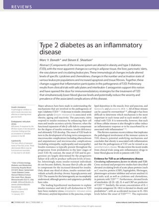
  • 3. 0CVWTG4GXKGYU^+OOWPQNQI[ (TQOICUVTQKPVGUVKPCNVTCEV 6EGNN r)NWEQUG r(TGGHCVV[CEKFU 2TQKPȯCOOCVQT[ E[VQMKPGUCPFEJGOQMKPGU +.β60(%%.%%.%:%. %%.%%. %:%. +.β ↓+.4# ↑+.β 1DGUGCFKRQUGVKUUWG 2CPETGCVKEKUNGVU βEGNN Mast cell Adipocyte Macrophage M2‑type macrophage A macrophage that is stimulated by interleukin-4 (IL-4) or IL-13 and that expresses arginase 1, the mannose receptor CD206 and the IL-4 receptor α-chain. KitW–sh/W–sh mice The KitW–sh (or sash) mutation abolishes KIT expression in mast cells, and the mutant mice are deficient in mast cells. Insulitis Inflammation of the pancreatic islets during the progression of diabetes. Insulitis in type 1 diabetes is caused by autoimmunity and in type 2 diabetes by metabolic stressors such as hyperglycaemia and elevated levels of free fatty acids. development of insulin resistance47 . But by comparing resident adipose tissue macrophages from lean mice with recently recruited immature macrophages from obese mice, these studies compared kinetically distinct popula- tions, and this alone might account for their conclusions. A recent study by Shaul et al.44 concluded that resident CD11c+ adipose tissue macrophages in the fat pads of obese mice have a mixed M1/M2 phenotype that did not seem to be pro-inflammatory. Therefore, the pre- cise phenotype of adipose tissue macrophages remains to be clarified, and most importantly, we need to know which macrophage phenotype (if any) is related to the development of insulin resistance. Although macrophages are the most abundant leuko- cyte population in expanding adipose tissue, other immune cell types are present and their numbers and activities may change during the transition from lean to obese. For example, mast cells were shown to accumulate in subcutaneous adipose tissue during the induction of obesity in mice48 . Moreover, obese mast-cell-deficient KitW–sh/W–sh mice and obese mice treated with ketotifen, which blocks mast cell function, had improved insulin resistance compared with wild-type mice or untreated control mice, respectively48 . However, the pathological role of mast cells in obesity and T2D remains to be clari- fied, as both of these approaches led to substantial weight loss relative to the control mice, which makes it difficult to distinguish the potential mechanisms, as weight loss itself promotes insulin sensitivity. Cells of the adaptive immune system are also present in adipose tissue and may contribute to metabolic dis- ruption. T cells generally accumulate in obese adipose tissue in parallel with macrophages49 , although changes in the relative numbers and activities of CD4+ and CD8+ T cells and of T helper 1 (TH 1), TH 2 and forkhead box P3 (FOXP3)+ regulatory T (TReg ) cells occur asynchronously and with distinct kinetics. In general, CD8+ T cells and TH 1 cells are thought to contribute to the insulin resist- ance phenotype, whereas TReg cells and TH 2 cells tend to counter it50,51 . In this scenario, the macrophages would be the effector cells under the control of the T cells. One of the more interesting of the unanswered questions is whether the T cells recognize antigens that are present in the adipose tissue and, if so, what these antigens are. TReg cells are of special interest owing to their impor- tant role in the maintenance of self-tolerance and the sup- pression of potentially autoreactive T cells; through these functions they prevent the development of autoimmunity inexperimentalmodelsinbothmiceandhumans52,53 .The number of TReg cells in the adipose tissue of lean mice is unusually high at ~50% of the CD4+ T cell compartment, butthisnumberdecreasesdramaticallyinproportionwith increasing obesity50 . This contrasts with the increase in macrophage number that accompanies obesity, and sug- gests a potential relationship between these two cell popu- lations in adipose tissue. Furthermore, adipose tissue TReg cellsexpress(andarethoughttosecrete)anunusuallyhigh amount of the anti-inflammatory cytokine IL-10, which in lean mice could help to suppress adipose tissue inflam- mation50 . Targeted induction of TReg cells improves circu- lating glucose levels and insulin sensitivity in obese mice, reduces macrophage numbers and TNF levels in adipose tissue, and decreases pancreatic islet hyperplasia50,51 . Tissue inflammation has also been detected in the islets of patients with T2D, along with increased levels of cytokines and chemokines54–56 . Of note, patients with T2D and every animal model of T2D investigated to date display immune cell infiltration of the islets56 . Islet tis- sue sections from patients with T2D also show fibrosis, which is found in conjunction with amyloid deposits, and this also argues for an inflammatory response in islets, as fibrosis is a hallmark of chronic inflammation. An interesting recent report shows that human islet amy- loid polypeptide (IAPP) induces the secretion of IL-1β by bone marrow-derived macrophages, suggesting that IAPP may contribute to islet inflammation15 . Although the concept of insulitis in T2D is recent14 , it is well established in type 1 diabetes and is considered to be a characteristic of the disease. The precise aetiol- ogy of the insulitis in both types of diabetes remains to be fully understood, but differences are known to exist; for example, the insulitis in type 1 diabetes is driven by an autoimmune-mediated process, whereas in T2D it is now thought to be due to autoinflammation. However, a common final effector pathway involving IL-1β seems to be activated in both types of diabetes12 (see below). Furthermore, additional overlap exists between both dis- eases (TABLE 1) and in many cases a clear classification is not feasible, arguing for an involvement of the immune system not only in type 1 diabetes but also in T2D. Figure 1 | Development of inflammation in type 2 diabetes. Excessive levels of nutrients, including glucose and free fatty acids, will stress the pancreatic islets and insulin‑sensitive tissues such as adipose tissue (and the liver and muscle, not shown), leading to the local production and release of cytokines and chemokines. These factors include interleukin‑1β (IL‑1β), tumour necrosis factor (TNF), CC‑chemokine ligand 2 (CCL2), CCL3 and CXC‑chemokine ligand 8 (CXCL8). Furthermore, production of IL‑1 receptor antagonist (IL‑1RA) by β‑cells is decreased. As a result, immune cells will be recruited and contribute to tissue inflammation. The release of cytokines and chemokines from the adipose tissues into the circulation promotes inflammation in other tissues, including the islets. REVIEWS 100 | FEBRuARy 2011 | VOLuME 11 www.nature.com/reviews/immunol REVIEWS © 2011 Macmillan Publishers Limited. All rights reserved
  • 4. Ischaemia A condition in which the flow of blood to a tissue or organs is less than normal, and which results in injury to that tissue or organ. Inflammatory mechanisms in T2D The studies discussed above support the hypothesis that inflammation has a role in the pathogenesis of T2D. Here, we discuss some of potential mechanisms involved in the inflammatory response in this disease. Hypoxia. It has been proposed that hypoxia in expand- ing adipose tissue may induce an inflammatory response. It is well established in oncology that rapidly growing tis- sue can expand faster than the vasculature that supports its oxygen and nutrient requirements. Hypoxia ensues as oxygen supplies become limited, and compensatory angiogenesisisinducedthroughtheproductionofvarious angiogenic factors in an attempt to restore the required levels of oxygen and nutrient delivery57 . Hypoxia and neovascularization are also seen in rodent models of obesity in which the fat mass is rapidly expanding; for example during high-fat feeding58,59 . Furthermore, hypoxia has been observed in human adipose tissue and contributes to adipose tissue dysfunction60,61 . Macrophages accumulate at sites of hypoxia or ischaemia, providing a pathological link between adipose tissue expansion and the induction of inflammation. The recruitment of macrophages to hypoxic or ischaemic tis- sues has been studied in greater detail in tumour growth, wounds and infections, atherogenesis and arthritis62 , but the principles seem to be similar for expanding adipose tissue. Hypoxia induces the expression of numerous pro-angiogenic and pro-inflammatory genes in macro- phages63 , suggesting that the recruited macrophages have an important role in resolving hypoxia, possibly in an attempt to repair damaged tissue. Cell death. Adipocyte expansion beyond oxygen and nutrient requirements also seems to lead to adipocyte cell death. This is readily apparent in mice fed a high-fat diet, as dead adipocytes are located throughout their fat pads64,65 , whereas this is not observed in the fat pads of mice fed a normal diet. The most distinguishing fea- ture of the dead adipocytes is that they are located indi- vidually and sporadically throughout the fat pads and are surrounded by macrophages to form what are referred to as ‘crown-like structures’64,65 . There are higher numbers of crown-like structures in the gonadal white adipose tissue of male mice than in the subcutaneous white adipose tissue. The high proportions of macrophages that are found in crown-like structures suggest that many of the monocytes that are recruited to expanding adipose tissue in obesity are there to remove cellular debris. However, it does not establish that recruited macrophages are causally linked to the development of insulin resistance. In contrast to adipose tissue macrophages, islet mac- rophages were not detected in the vicinity of necrotic or apoptotic cells56 . Furthermore, islet inflammation is an early event in the development of T2D and is apparent in mice after 8 weeks of high-fat feeding56 , during which time β-cell function declines but β-cell mass increases without an increase in islet cell death. Therefore, it is unlikely that cell death has an important role in the recruitment of macrophages to the islets. This recruit- ment may instead be a consequence of islet-derived chemokines that are produced in response to metabolic stress (see below). The NF-κB and JNK pathways. Many of the metabolic stresses that promote insulin resistance and T2D also activate the inflammation- and stress-induced kinases IκB kinase-β (IKKβ) and JuN N-terminal kinase (JNK)1,66,67 , suggesting that these kinases may have key roles in the pathogenesis of these conditions. Indeed, IKKβ activates the transcription factor nuclear factor-κB (NF-κB), and obesity induces the expression of NF-κB target genes, such as pro-inflammatory cytokines, in the liver and adipose tissue1,66,67 . These cytokines, including TNF, IL-6, and IL-1β, may promote insulin resistance in the tissues where they are produced, such as the liver and adipose tissue, and may also be transported through the circulation to affect more distant sites, including the vessel walls, skeletal and cardiac muscle, the kidneys and circulating leukocytes. The other potentially important kinase, JNK, activates transcription factors such as ELK1, ATF2 (activating transcription factor 2) and JuN, although the potential roles of these JNK- responsive transcription factors in obesity are not well established68 . Nevertheless, bone marrow transplant and selective genetic ablation experiments have pro- vided ample evidence to support a role for JNK in the inflammatory response to obesity and the development of insulin resistance (see below). Table 1 | Comparison of characteristics associated with type 1 and type 2 diabetes* Type 1 diabetes Type 2 diabetes Refs Age of onset Mainly young but can occur at all ages Usually associated with ageing but prevalence is increasing in younger individuals 134–136 Insulin deficiency Absolute Relative to the prevailing resistance to insulin 137 Risk factors Genetics‡ , obesity, insulin resistance Genetics‡ , obesity, insulin resistance 135, 138–140 Insulitis Autoimmune Autoinflammatory 2,12 Autoantibodies Present in 85–90% May be present 134–136 Treatment Insulin Diet and exercise, oral agents such as metformin; insulin recommended early in the treatment 141 *No single clinical feature or diagnostic parameter completely discriminates the two diseases142 . ‡ Genetics are relevant to both type 1 and 2 diabetes, but different susceptibility genes have been identified in different families. REVIEWS NATuRE REVIEWS | Immunology VOLuME 11 | FEBRuARy 2011 | 101 focuS on MEtabolISM and IMMunology © 2011 Macmillan Publishers Limited. All rights reserved
  • 5. Cachexia Severe weight loss, muscle wasting and debility caused by prolonged disease. It is thought to be mediated through neuro- immunoendocrine interactions. Leptin A protein hormone that regulates energy intake and expenditure. It is one of the most important adipose-derived hormones and its production correlates with the mass of adipose tissue. Inflammasome A molecular complex of several proteins that, when activated, results in the production of active caspase 1, which cleaves pro-interleukin-1β (pro-IL-1β) andpro-IL-18 to produce the active cytokines. In fact, many of the same cytokines that are produced inresponsetoNF-κBactivationalsoactivatebothJNKand NF-κB in a feed-forward manner. This includes TNF and IL-1β,whichactivateJNKandNF-κBthroughtheengage- ment of their specific cellular receptors. Other stimuli that promote insulin resistance and T2D, including free fatty acids (FFAs) and advanced glycation end-products, also actthroughspecificcellsurfacereceptors,suchasToll-like receptors(TLRs)andreceptorforadvancedglycationend- products(RAGE)69 .Alltheseextracellularstimulibindcell surface receptors and activate intracellular pathways that converge on both IKKβ–NF-κB and JNK signalling. Bone marrow transplant and selective genetic ablation methods have been used to assess the relative contribu- tion of the JNK and IKKβ–NF-κB signalling pathways in haematopoietic and non-haematopoietic parenchymal cells in obesity-induced insulin resistance, and to identify the main tissue sites involved. The liver and adipose tissue are important sites for the activation of both pathways. In the liver, this activation occurs in both hepatocytes and myeloid cells such as macrophages, and upregulates the production of pro-inflammatory cytokines, including TNF, IL-6 and IL-1β66,67,70–72 . Although these pathways are activated in both haematopoietic and non-haematopoi- etic cells, it is the leukocytes that account for most of the local production of pro- and anti-inflammatory cytokines in the liver and adipose tissue. However, in muscle cells, the activation of IKKβ and NF-κB results in wasting and cachexia through the activation of the E3 ubiquitin ligase TRIM63 (also known as MuRF1)73 , whereas in the hypothalamus it seems that the IKKβ–NF-κB pathway affects feeding behaviour and the leptin signalling axis74 . Therefore, IKKβ–NF-κB activation in these tissues affects insulin resistance indirectly, through changes in body weight, as opposed to the more direct effects on insulin resistance that result from the activation of this pathway in the liver, adipose tissue and leukocytes. NF-κB is also activated in islet β-cells through the actions of glucose and IL-1β, and inhibition of NF-κB seems to protect β-cells from various insults, including from the effects of glucotoxicity or multiple treatments with low-dose streptozotocin (a natural chemical that is particularly toxic to β-cells)55,75 . The NF-κB and JNK pathways are thus activated in multiple tissues in obes- ity and T2D, and have central roles in promoting tissue inflammation. Accordingly, reducing the activity of these pathways may be of therapeutic benefit (see below). IL-6 and insulin resistance. The roles of IL-6 signal- ling in insulin resistance have been controversial and at times paradoxical76,77 . Concentrations of circulating IL-6 and CRP (the hepatic expression of which is induced by IL-6) are increased in obesity and predict the incidence of T2D in predisposed individuals20 . Hepatic and adipose production of IL-6 are thought to promote insulin resist- ance67,76,78 , whereas production of IL-6by skeletal muscle, especially during intense exercise, is thought to be benefi- cial77 . Analysis of hepatocyte-specific deletion of the IL-6 receptor in mice has added to the controversy, as these mice seem to be protected from both local and systemic insulin resistance79,80 . The IL-1 system as a sensor of metabolic stress. The ear- liest evidence for an inflammatory process in pancreatic islets arose from the observation that hyperglycaemia induces β-cell apoptosis81 . By examining the underlying mechanism, it was shown that high glucose concentra- tions induce the expression of the pro-apoptotic receptor FAS (also known as CD95) on β-cells82 , which is further upregulated by glucose-induced IL-1β production by β-cells55 . Therefore, IL-1β and FAS contribute to both the glucose-induced impairment of β-cell secretory function and apoptosis55,83 . Additional mechanisms regulate IL-1β expression in islets (FIG. 2). FFAs (such as oleate, palmitate and stearate) stimulate IL-1β secretion and the production of IL-1β- dependent pro-inflammatory molecules in cultured human and rodent islets84–86 . A combination of moder- ately increased glucose levels and FFAs was shown to induce an even stronger increase in cytokine production than just FFAs alone85 . The underlying mechanisms of ‘nutrient’ (that is, glu- cose and FFA)-induced activation of IL-1β are complex. FFAs may stimulate the production pro-inflammatory molecules by direct activation of TLR2 and TLR4, which can sense lipids, or indirectly through FFA metabolites such as ceramide86–89 . Glucose-induced IL-β produc- tion is thought to involve the NOD-, LRR- and pyrin domain-containing 3 (NLRP3; also known as NALP3) inflammasome. High concentrations of glucose induce the dissociation of thioredoxin-interacting protein (TXNIP) from thioredoxin under the influence of reactive oxy- gen species, allowing binding of TXNIP to the NLRP3 inflammasome. This leads to the activation of caspase 1 and the subsequent processing of pro-IL-1β and release of mature IL-1β90 . Whether reactive oxygen species are indispensable in this process remains unclear. β-cells have very low levels of antioxidative enzymes and are therefore particularly vulnerable to oxidative stress; however, activation of the inflammasome in the absence of reactive oxygen species has been shown in patients with chronic granulomatous disease91,92 . Interestingly, deposition of amyloid in the islets is a hallmark of T2D, and human IAPP seems to contribute to the induction of IL-1β production in the islets through the NLRP3 inflammasome15 . However, the induction of IL-1β secretion by IAPP has only been shown in mac- rophages, and in vivo amyloid deposition requires pro- longed high-fat feeding (for a period of 1 year), whereas the first signs of islet inflammation are apparent after 8 weeks56 , indicating that IAPP-mediated IL-1β secretion may be a late event in islet inflammation. It remains pos- sible that the inflammasome may act as a sensor of met- abolic danger93 , resulting in IL-1β production and the induction of numerous cytokines and chemokines24,94,95 . Therefore, activation of the inflammasome may con- tribute to the recruitment of immune cells, which can mediate a broad inflammatory response. These initial mechanisms of IL-1β induction may be amplified by a cycle of autoinflammation. Indeed, human islets, particularly purified human β-cells, are very sensitive to IL-1β autostimulation84 . This is prob- ably a consequence of the abundant expression of IL-1 REVIEWS 102 | FEBRuARy 2011 | VOLuME 11 www.nature.com/reviews/immunol REVIEWS © 2011 Macmillan Publishers Limited. All rights reserved
  • 6. 0CVWTG4GXKGYU^+OOWPQNQI[ Inflammasome Inflammasome Pro-caspase 1 Active caspase 1 IL-1β Pro-IL-1β IL-1β ASC NLRP3 FFAs Glucose TLR2 or TLR4 MYD88 MYD88 IL-1R1 CCL2, CCL3 CXCL8 Recruitment 0(κ$ βEGNN /CETQRJCIG TXNIP TXNIP TXR TXR Amyloid receptor type 1 (IL-1R1) by these cells. Analysis of IL-1R1 expression in numerous tissues showed that the highest levels were expressed in mouse islets and by the insulin- producing cell line MIN6 compared with 20 other mouse tissues, including immune tissues such as the spleen and thymus85 . IL-1β autostimulation of islets can be pre- vented by reducing NF-κB activity or by blocking IL-1R1 signalling (with IL-1RA, by ligand neutralization or by the genetic elimination of the IL-1R1-associated signal- ling protein myeloid differentiation primary-response protein 88 (MyD88))84,85 . Blocking IL-1R1 signalling also inhibits FFA- and glucose-induced upregulation of IL-1β84,85 . Another factor that promotes islet inflammation in T2D is a defect in an anti-inflammatory mechanism. IL-1RA is highly expressed in the endocrine pancreas of non-diabetic individuals but is decreased in the islets of patients with T2D, and this enhances the suscepti- bility of the β-cells to IL-1β54 . The precise mechanisms responsible for this decrease remain to be elucidated but the adipose tissue-derived hormone leptin might be involved, as it decreases IL-1RA expression in human islets in vitro54 . Therefore, the IL-1 system is an integral part of the response to metabolic disturbance and IL-1 antagonism has therapeutic potential (see below). Chemokines. Adipocytes may secrete chemokines such as CC-chemokine ligand 2 (CCL2; also known as MCP1), which recruits monocytes. Consistent with this hypoth- esis, the expression of CCL2 is increased in the adipose tissue of obese rodents and humans96–99 . Mice with a tar- geteddeletionofeitherCcl2oritsreceptorCC-chemokine receptor 2 (Ccr2) have decreased numbers of macro- phages in adipose tissue41,98 , whereas transgenic upregu- lation of Ccl2 expression in adipocytes results in increased macrophage numbers100 . However, the metabolic consequences of diminished signalling through the CCL2–CCR2 axis are relatively small, possibly owing to the redundancy between chemokines that recruit monocytes101 . In addition to CCL2, the expression of CCL3, CCL6, CCL7, CCL8 and CCL9 is increased in adipocytes from mice fed a high-fat diet compared with mice fed a normal diet, suggesting that these chemo- kines could also have a role in monocyte recruitment101 . These findings are consistent with a potential increase in chemokine-mediated recruitment of monocytes to expandingadiposetissue,althoughotherchemoattractants such as leukotrienes could also be involved. Islet cells can also produce a wide range of chemo- kines in the context of T2D. In vitro treatment of islets with high concentrations of glucose and the satu- rated fatty acid palmitate increases the production of Figure 2 | Interleukin‑1β‑induced inflammation in islets of patients with type 2 diabetes. High concentrations of glucose promote β‑cell production of interleukin‑1β (IL‑1β) through the dissociation of thioredoxin‑interacting protein (TXNIP) from its inhibitor thioredoxin (TXR), resulting in activation of the NOD‑, LRR‑ and pyrin domain‑ containing 3 (NLRP3) inflammasome, activation of caspase 1 and processing of pro‑IL‑1β into its mature form. IL‑1β induces the production of a wide range of cytokines and chemokines such as CC‑chemokine ligand 2 (CCL2), CCL3 and CXC‑chemokine ligand 8 (CXCL8) through nuclear factor‑κB (NF‑κB) activation. This is enhanced by free fatty acid (FFA)‑induced activation of Toll‑like receptor 2 (TLR2) or TLR4 and leads to the recruitment of macrophages. FFAs may also directly activate the NLRP3 inflammasome. Islet‑derived amyloid can activate the recruited macrophages through the NLRP3 inflammasome, increasing IL‑1β production and the vicious cycle of IL‑1β autostimulation through IL‑1 receptor type 1 (IL‑1R1). ASC, apoptosis‑associated speck‑like protein containing a CARD; MYD88, myeloid differentiation primary‑response protein 88. REVIEWS NATuRE REVIEWS | Immunology VOLuME 11 | FEBRuARy 2011 | 103 focuS on MEtabolISM and IMMunology © 2011 Macmillan Publishers Limited. All rights reserved
  • 7. Salsalate A prodrug form of salicylic acid that has fewer side effects than sodium salicylate. Salsalate is approved for use in humans as a source of salicylic acid. several biologically active chemotactic factors (CXC- chemokine ligand 8 (CXCL8) and CCL3 in human islets; CXCL1 in mouse islets)56,85 . Islets isolated from rodent models of T2D (Goto–Kakizaki rats, high-fat- fed mice and Zucker rats) also show increased produc- tion of various chemokines, including CXCL1, CCL2 and CCL3 (REFS 26,102). Importantly, the relevance of these findings for humans is supported by evidence for the upregulation of various chemokines in laser- captured nearly pure β-cells from patients with T2D103 . Although most chemokines are produced by β-cells in the islets, some (for example, CXCL8) may also be produced by pancreatic α-cells56 . The precise functions of the various chemokines remain to be clarified; however, they have a crucial role in tissue infiltration by immune cells in T2D. Adipokines. Adipokines are hormones that are produced mainly or exclusively by adipocytes. Examples include leptin and adiponectin, both of which have potential immunomodulatory effects. Genetically obese ob/ob mice, which produce a mutated, non-functional form of leptin, show many of the same inflammatory changes as other models of obesity (including diet-induced obese mice), and ob/ob mice become both insulin resistant and diabetic. These results indicate that leptin may not have a particularly important role in obesity-induced inflammation. Adiponectin is considered to be an anti- inflammatory and cardioprotective protein. It may exert these effects in several ways; for example, by inducing anti-inflammatory cytokines such as IL-10 and IL-1RA104 , through vascular mechanisms includ- ing enhancement of nitric oxide bioavailability105 , or by reducing endothelial cell–leukocyte adhesion106 . In summary, multiple mechanisms may contribute to inflammation in T2D, some of which are general and others are tissue specific. Thus in the pancreatic islet cells, inflammation may be initiated by direct sensing of excess nutrients, leading to activation of the IL-1 system, whereas in adipose tissue, excess storage of fat causes hypoxia and inflammation. Common downstream mechanisms include the activation of NF-κB and JNK pathways and cytokine and chemokine release, leading to the recruitment of immune cells. Clinical trials and implications Further evidence for roles of inflammation in T2D comes from clinical studies using either small molecule anti- inflammatory approaches or biological agents that target specific pro-inflammatory cytokine pathways to improve parameters of glucose control, such as glycated haemo- globin levels. To date, the most promising approaches include the selective blockade of IL-1R1 activation with either IL-1RA or specific antibodies, and inhibition of the NF-κB pathway with salicylate derivatives such as salsalate. Both approaches seem to lower blood glucose levels and improve β-cell secretory function and insu- lin sensitivity, as well as reducing evidence of systemic inflammation25,107 . Of note, the improvement in insulin secretion lasted 39 weeks following the withdrawal of IL-1RA treatment108 . Similarly, 3 months after a single injection with an IL-1β-specific antibody, individuals with T2D showed sustained reductions in glycated hae- moglobin levels and an improvement in insulin secre- tion by β-cells27 . This probably reflects the interruption of IL-1β autoinduction84 . These proof-of-concept studies validate the poten- tial approach of targeting of inflammatory mediators as a treatment for T2D and support a causative role for inflammation in the pathogenesis of this disease. They pave the way for new therapeutic approaches that could be disease modifying as opposed to palliative. This offers the opportunity to simultaneously target several features of the disease (including defective insulin secretion by β-cells, insulin resistance in adipose tissue, and micro- vascular and macrovascular complications) with anti- inflammatory drugs (either alone or in combination) to alter the course of the disease. Based on preclinical studies, three anti-inflammatory approaches have been clinically tested: TNF antagonism, IL-1β antagonism and salsalate treatment (TABLE 2). In contrast to IL-1β antagonism and salsalate treatment, TNF antagonism has thus far failed to improve blood glucose levels in patients with T2D32–36 . Improvements in glucose metabolism have been observed in patients being treated with TNF blockers for rheumatoid arthri- tis37,39,40,109–111 , and a marginal effect of TNF blockers on fasting glucose levels was observed in obese indi- viduals in a recent report38 . Based on these latest find- ings additional clinical trials may aim to block TNF signalling, either alone or in conjunction with other cytokine-blocking approaches. Current anti-inflammatory approaches to treat- ing T2D focus on salsalate and IL-1β antagonism. Mechanistically these approaches may have similari- ties, including the modulation of IL-1R1 and NF-κB pathways1,55,67,112 . IL-1 antagonists are large proteins that must be injected and have effects that may last for sev- eral weeks to months. By contrast, salsalate is an orally administered small molecule with a short half-life that requires more than once-a-day dosing. IL-1 antagonists are also designed to be highly specific for their targets, whereas salsalate and other non-acetylated forms of sali- cylate may have broader molecular actions. Thus in addi- tion to the inhibition of NF-κB, they may inhibit other kinases113 , upregulate the expression of heat shock factor protein 1 (REF. 114) and inhibit insulin clearance115 . In addition to having apparent efficacy and dura- bility in lowering glucose levels, it is encouraging that both approaches also seem to have high mar- gins of safety. Salicylates such as salsalate have been used to treat joint pain in millions of patients over many decades, and many of these patients may also have diabetes, cardiovascular disease or other meta- bolic conditions. Although not as broadly used, IL-1 antagonists have been used for several indications, including in more than 100,000 patients with rheu- matoid arthritis. It is also encouraging that the rate and severity of infections are unaffected, and rare or unusual infections have not been reported for indi- viduals taking either drug, in contrast to certain other immunomodulatory therapies. REVIEWS 104 | FEBRuARy 2011 | VOLuME 11 www.nature.com/reviews/immunol REVIEWS © 2011 Macmillan Publishers Limited. All rights reserved
  • 8. 1. Shoelson, S. E., Lee, J. Goldfine, A. B. Inflammation and insulin resistance. J. Clin. Invest. 116, 1793–1801 (2006). 2. Donath, M. Y., Boni‑Schnetzler, M., Ellingsgaard, H. Ehses, J. A. Islet inflammation impairs the pancreatic β‑cell in type 2 diabetes. Physiology 24, 325–331 (2009). 3. Bonner‑Weir, S. Islet growth and development in the adult. J. Mol. Endocrinol. 24, 297–302 (2000). 4. Kahn, B. B. Type 2 diabetes: when insulin secretion fails to compensate for insulin resistance. Cell 92, 593–596 (1998). 5. Rhodes, C. J. Type 2 diabetes‑a matter of β‑cell life and death? Science 307, 380–384 (2005). 6. Robertson, R. P., Harmon, J., Tran, P. O. Poitout, V. β‑cell glucose toxicity, lipotoxicity, and chronic oxidative stress in type 2 diabetes. Diabetes 53, S119–S124 (2004). 7. Weir, G. C. Bonner‑Weir, S. Five stages of evolving β‑cell dysfunction during progression to diabetes. Diabetes 53, S16–S21 (2004). 8. Prentki, M. Nolan, C. J. Islet β cell failure in type 2 diabetes. J. Clin. Invest. 116, 1802–1812 (2006). 9. Hull, R. L., Westermark, G. T., Westermark, P. Kahn, S. E. Islet amyloid: a critical entity in the pathogenesis of type 2 diabetes. J. Clin. Endocrinol. Metab. 89, 3629–3643 (2004). 10. Harding, H. P. Ron, D. Endoplasmic reticulum stress and the development of diabetes: a review. Diabetes 51, S455–S461 (2002). 11. Hotamisligil, G. S. Erbay, E. Nutrient sensing and inflammation in metabolic diseases. Nature Rev. Immunol. 8, 923–934 (2008). 12. Donath, M. Y., Storling, J., Maedler, K. Mandrup‑ Poulsen, T. Inflammatory mediators and islet β‑cell failure: a link between type 1 and type 2 diabetes. J. Mol. Med. 81, 455–470 (2003). 13. Ehses, J. A., Ellingsgaard, H., Boni‑Schnetzler, M. Donath, M. Y. Pancreatic islet inflammation in type 2 diabetes: from α and β cell compensation to dysfunction. Arch. Physiol. Biochem. 115, 240–247 (2009). Outstanding questions and future directions Increasing data suggest a potential role for inflamma- tion in the pathogenesis of T2D. This is supported by the results of both preclinical studies and new clinical trials using anti-inflammatory approaches to treat the disease. But these are early days and there are many unanswered questions. What is the relative contribution of inflamma- tion to the development of T2D? How efficacious are the anti-inflammatory approaches at improving glycaemia and T2D complications, and how durable will the effects be? What will be the best therapeutic modality: life-long treatment or short-term interventions aiming at breaking inflammatory flares? How do drugs such as salsalate and IL-1 blockers really work in T2D? Do anti-inflammatory strategies target the underlying mechanisms of the dis- ease, and if so, would starting these therapies early pre- vent progression or even the overt manifestation of the disease? The early studies suggest that these strategies are well tolerated with few serious side effects and with lit- tle evidence of immunosuppression. From the numerous ongoing preclinical and clinical studies (TABLE 2), some of these questions should be addressed in the near future. Table 2 | Clinical studies using anti-inflammatory approaches to treat type 2 diabetes or prediabetes mechanism Drug Trial Phase number of subjects Treatment duration (weeks) main findings Refs IL‑1 receptor blockade Anakinra (Kineret; Amgen/Biovitrum) II 69 13 ↓ Glycated haemoglobin, ↓ CRP, ↑ insulin production 25 IKKβ–NF‑κB inhibition Salsalate II 20 4 ↓ FBG, ↓ CRP, ↑ insulin sensitivity, ↑ adiponectin 107 IKKβ–NF‑κB inhibition Salsalate II 16 2–4 ↓ FBG, ↓ FFA, ↓ triglycerides, ↓ CRP, ↑ adiponectin 143 IKKβ–NF‑κB inhibition Salsalate II 40 1 ↓ FBG, ↑ insulin 144 IKKβ–NF‑κB inhibition Salsalate IIb 104 12 ↓ Glycated haemoglobin, ↓ FBG, ↓ triglycerides, ↑ adiponectin 28 IL‑1β‑specific antibody XOMA 052 (Xoma) I 98 Single injection ↓ Glycated haemoglobin, ↓ CRP, ↑ insulin production 27 IL‑1 receptor blockade Anakinra (Kineret; Amgen/Biovitrum) II 12 4 Ongoing, closed for recruitment NCT00928876* IL‑1β‑specific antibody ACZ885 (canakinumab; Novartis) II 231 Unknown Ongoing, closed for recruitment NCT00605475* IL‑1β‑specific antibody ACZ885 (canakinumab; Novartis) II 140 48 Ongoing NCT00995930* IL‑1β‑specific antibody ACZ885 (canakinumab; Novartis) II 232 4 Ongoing, closed for recruitment NCT01068860* IL‑1β‑specific antibody ACZ885 (canakinumab; Novartis) II‑III 600 17 Ongoing, closed for recruitment NCT00900146* IKKβ–NF‑κB inhibition Salsalate III 284 48 Ongoing, closed for recruitment NCT00799643* IKKβ–NF‑κB inhibition Salsalate II 80 12 Ongoing, closed for recruitment NCT00330733* IL‑1β‑specific antibody XOMA 052 (Xoma) II 325 26 Ongoing, closed for recruitment NCT01066715* IL‑1β‑specific antibody XOMA 052 (Xoma) II 80 48 Ongoing, closed for recruitment NCT01144975* IL‑1β‑specific antibody LY2189102 (Lilly) II 80 12 Ongoing, closed for recruitment NCT00942188* IL‑1β‑specific vaccine CYT013‑IL1bQb (Cytos Biotech.) I 32 Unknown Ongoing NCT00924105* Trials with tumour necrosis factor (TNF) antagonists31–40 are not listed owing to the lack of effects in patients with type 2 diabetes. CRP, C‑reactive protein; FBG, fasting blood glucose; FFA, free fatty acid; IKKβ, IκB kinase‑β; IL‑1, interleukin‑1; NF‑κB, nuclear factor‑κB. *ClinicalTrials.gov identifier. REVIEWS NATuRE REVIEWS | Immunology VOLuME 11 | FEBRuARy 2011 | 105 focuS on MEtabolISM and IMMunology © 2011 Macmillan Publishers Limited. All rights reserved
  • 9. 14. Donath, M. Y. et al. Islet inflammation in type 2 diabetes: from metabolic stress to therapy. Diabetes Care 31, S161–S164 (2008). 15. Masters, S. L. et al. Activation of the NLRP3 inflammasome by islet amyloid polypeptide provides a mechanism for enhanced IL‑1β in type 2 diabetes. Nature Immunol. 11, 897–904 (2010). 16. Pickup, J. C., Mattock, M. B., Chusney, G. D. Burt, D. NIDDM as a disease of the innate immune system: association of acute‑phase reactants and interleukin‑6 with metabolic syndrome X. Diabetologia 40, 1286–1292 (1997). 17. Spranger, J. et al. Inflammatory cytokines and the risk to develop type 2 diabetes: results of the prospective population‑based European prospective investigation into cancer and nutrition (EPIC)‑potsdam study. Diabetes 52, 812–817 (2003). 18. Herder, C. et al. Inflammation and type 2 diabetes: results from KORA Augsburg. Gesundheitswesen 67, S115–S121 (2005). 19. Herder, C. et al. Elevated levels of the anti‑ inflammatory interleukin‑1 receptor antagonist precede the onset of type 2 diabetes: the Whitehall II study. Diabetes Care 32, 421–423 (2009). 20. Pradhan, A. D., Manson, J. E., Rifai, N., Buring, J. E. Ridker, P. M. C‑reactive protein, interleukin 6, and risk of developing type 2 diabetes mellitus. JAMA 286, 327–334 (2001). 21. Meier, C. A. et al. IL‑1 receptor antagonist serum levels are increased in human obesity: a possible link to the resistance to leptin? J. Clin. Endocrinol. Metab. 87, 1184–1188 (2002). 22. Carstensen, M. et al. Accelerated increase in serum interleukin‑1 receptor antagonist starts 6 years before diagnosis of type 2 diabetes: Whitehall II prospective cohort study. Diabetes 59, 1222–1227 (2010). 23. Marculescu, R. et al. Interleukin‑1 receptor antagonist genotype is associated with coronary atherosclerosis in patients with type 2 diabetes. Diabetes 51, 3582–3585 (2002). 24. Dinarello, C. A. The role of the interleukin‑1‑receptor antagonist in blocking inflammation mediated by interleukin‑1.N. Engl. J. Med. 343, 732–734 (2000). 25. Larsen, C. M. et al. Interleukin‑1‑receptor antagonist in type 2 diabetes mellitus. N. Engl. J. Med. 356, 1517–1526 (2007). A proof-of-concept clinical study demonstrating the potential of immunomodulation with an IL-1 antagonist in T2D. 26. Ehses, J. A. et al. IL‑1 antagonism reduces hyperglycemia and tissue inflammation in the type 2 diabetic GK rat. Proc. Natl Acad. Sci. USA 106, 13998–14003 (2009). 27. Donath, M. Y. et al. XOMA 052, an anti‑IL‑1β antibody, in a double‑blind, placebo‑controlled, dose escalation study of the safety and pharmacokinetics in patients with type 2 diabetes mellitus – a new approach to therapy. Diabetologia 51, S7 (2008). 28. Goldfine, A. B. et al. The effects of salsalate on glycemic control in patients with type 2 diabetes: a randomized trial. Ann. Intern. Med. 152, 346–357 (2010). This clinical trial showed that salsalate improves circulating glucose and lipid levels in patients with T2D. 29. Hotamisligil, G. S., Shargill, N. S. Spiegelman, B. M. Adipose expression of tumor necrosis factor‑α: direct role in obesity‑linked insulin resistance. Science 259, 87–91 (1993). This early study suggested that TNF could cause insulin resistance. 30. Schreyer, S. A., Chua, S. C. Jr LeBoeuf, R. C. Obesity and diabetes in TNF‑α receptor‑ deficient mice. J. Clin. Invest. 102, 402–411 (1998). 31. Bernstein, L. E., Berry, J., Kim, S., Canavan, B. Grinspoon, S. K. Effects of etanercept in patients with the metabolic syndrome. Arch. Intern. Med. 166, 902–908 (2006). 32. Dominguez, H. et al. Metabolic and vascular effects of tumor necrosis factor‑α blockade with etanercept in obese patients with type 2 diabetes. J. Vasc. Res. 42, 517–525 (2005). 33. Lo, J. et al. Effects of TNF‑α neutralization on adipocytokines and skeletal muscle adiposity in the metabolic syndrome. Am. J. Physiol. Endocrinol. Metab. 293, E102–E109 (2007). 34. Ofei, F., Hurel, S., Newkirk, J., Sopwith, M. Taylor, R. Effects of an engineered human anti‑TNF‑α antibody (CDP571) on insulin sensitivity and glycemic control in patients with NIDDM. Diabetes 45, 881–885 (1996). 35. Paquot, N., Castillo, M. J., Lefebvre, P. J. Scheen, A. J. No increased insulin sensitivity after a single intravenous administration of a recombinant human tumor necrosis factor receptor: Fc fusion protein in obese insulin‑resistant patients. J. Clin. Endocrinol. Metab. 85, 1316–1319 (2000). 36. Rosenvinge, A., Krogh‑Madsen, R., Baslund, B. Pedersen, B. K. Insulin resistance in patients with rheumatoid arthritis: effect of anti‑TNFα therapy. Scand. J. Rheumatol. 36, 91–96 (2007). 37. Kiortsis, D. N., Mavridis, A. K., Vasakos, S., Nikas, S. N. Drosos, A. A. Effects of infliximab treatment on insulin resistance in patients with rheumatoid arthritis and ankylosing spondylitis. Ann. Rheum. Dis. 64, 765–766 (2005). 38. Stanley, T. L. et al. TNF‑α antagonism with etanercept decreases glucose and increases the proportion of high molecular weight adiponectin in obese subjects with features of the metabolic syndrome. J. Clin. Endocrinol. Metab. 3 Nov 2010 (doi:10.1210/ jc.2010‑1170). 39. Yazdani‑Biuki, B. et al. Relapse of diabetes after interruption of chronic administration of anti‑tumor necrosis factor‑α antibody infliximab: a case observation. Diabetes Care 29, 1712–1713 (2006). 40. Yazdani‑Biuki, B. et al. Improvement of insulin sensitivity in insulin resistant subjects during prolonged treatment with the anti‑TNF‑α antibody infliximab. Eur. J. Clin. Invest. 34, 641–642 (2004). 41. Weisberg, S. P. et al. Obesity is associated with macrophage accumulation in adipose tissue. J. Clin. Invest. 112, 1796–1808 (2003). 42. Xu, H. et al. Chronic inflammation in fat plays a crucial role in the development of obesity‑related insulin resistance. J. Clin. Invest. 112, 1821–1830 (2003). References 41 and 42 identified macrophages in adipose tissue and showed that their numbers increased with obesity. 43. Lumeng, C. N., Deyoung, S. M., Bodzin, J. L. Saltiel, A. R. Increased inflammatory properties of adipose tissue macrophages recruited during diet‑ induced obesity. Diabetes 56, 16–23 (2007). 44. Shaul, M. E., Bennett, G., Strissel, K. J., Greenberg, A. S. Obin, M. S. Dynamic, M2‑like remodeling phenotypes of CD11c+ adipose tissue macrophages during high‑fat diet–induced obesity in mice. Diabetes 59, 1171–1181 (2010). 45. Kosteli, A. et al. Weight loss and lipolysis promote a dynamic immune response in murine adipose tissue. J. Clin. Invest. 120, 3466–3479 (2010). 46. Herrero, L., Shapiro, H., Nayer, A., Lee, J. Shoelson, S. E. Inflammation and adipose tissue macrophages in lipodystrophic mice. Proc. Natl Acad. Sci. USA 107, 240–245 (2010). 47. Lumeng, C. N., Bodzin, J. L. Saltiel, A. R. Obesity induces a phenotypic switch in adipose tissue macrophage polarization. J. Clin. Invest. 117, 175–184 (2007). 48. Liu, J. et al. Genetic deficiency and pharmacological stabilization of mast cells reduce diet‑induced obesity and diabetes in mice. Nature Med. 15, 940–945 (2009). 49. Wu, H. et al. T‑cell accumulation and regulated on activation, normal T cell expressed and secreted upregulation in adipose tissue in obesity. Circulation 115, 1029–1038 (2007). 50. Feuerer, M. et al. Lean, but not obese, fat is enriched for a unique population of regulatory T cells that affect metabolic parameters. Nature Med. 15, 930–939 (2009). 51. Ilan, Y. et al. Induction of regulatory T cells decreases adipose inflammation and alleviates insulin resistance in ob/ob mice. Proc. Natl Acad. Sci. USA 107, 9765–9770 (2010). 52. Baecher‑Allan, C. Hafler, D. A. Human regulatory T cells and their role in autoimmune disease. Immunol. Rev. 212, 203–216 (2006). 53. Roncarolo, M. G. Battaglia, M. Regulatory T‑cell immunotherapy for tolerance to self antigens and alloantigens in humans. Nature Rev. Immunol. 7, 585–598 (2007). 54. Maedler, K. et al. Leptin modulates β cell expression of IL‑1 receptor antagonist and release of IL‑1β in human islets. Proc. Natl Acad. Sci. USA 101, 8138–8143 (2004). 55. Maedler, K. et al. Glucose‑induced β‑cell production of interleukin‑1β contributes to glucotoxicity in human pancreatic islets. J. Clin. Invest. 110, 851–860 (2002). The first description of the role of IL-1β in T2D. 56. Ehses, J. A. et al. Increased number of islet‑associated macrophages in type 2 diabetes. Diabetes 56, 2356–2370 (2007). The original description of macrophage infiltration in islets of patients with T2D. 57. Carmeliet, P. Angiogenesis in life, disease and medicine. Nature 438, 932–936 (2005). 58. Hosogai, N. et al. Adipose tissue hypoxia in obesity and its impact on adipocytokine dysregulation. Diabetes 56, 901–911 (2007). 59. Yin, J. et al. Role of hypoxia in obesity‑induced disorders of glucose and lipid metabolism in adipose tissue. Am. J. Physiol. Endocrinol. Metab. 296, E333–E342 (2009). 60. Pasarica, M. et al. Reduced oxygenation in human obese adipose tissue is associated with impaired insulin suppression of lipolysis. J. Clin. Endocrinol. Metab. 95, 4052–4055 (2010). 61. Pasarica, M. et al. Reduced adipose tissue oxygenation in human obesity: evidence for rarefaction, macrophage chemotaxis, and inflammation without an angiogenic response. Diabetes 58, 718–725 (2009). 62. Murdoch, C., Muthana, M. Lewis, C. E. Hypoxia regulates macrophage functions in inflammation. J. Immunol. 175, 6257–6263 (2005). 63. Burke, B. et al. Hypoxia‑induced gene expression in human macrophages: implications for ischemic tissues and hypoxia‑regulated gene therapy. Am. J. Pathol. 163, 1233–1243 (2003). 64. Cinti, S. et al. Adipocyte death defines macrophage localization and function in adipose tissue of obese mice and humans. J. Lipid Res. 46, 2347–2355 (2005). 65. Strissel, K. J. et al. Adipocyte death, adipose tissue remodeling, and obesity complications. Diabetes 56, 2910–2918 (2007). 66. Arkan, M. C. et al. IKK‑β links inflammation to obesity‑ induced insulin resistance. Nature Med. 11, 191–198 (2005). 67. Cai, D. et al. Local and systemic insulin resistance resulting from hepatic activation of IKK‑β and NF‑κB. Nature Med. 11, 183–190 (2005). 68. Solinas, G. Karin, M. JNK1 and IKKβ: molecular links between obesity and metabolic dysfunction. FASEB J. 24, 2596–2611 (2010). 69. Shi, H. et al. TLR4 links innate immunity and fatty acid‑induced insulin resistance. J. Clin. Invest. 116, 3015–3025 (2006). 70. Sabio, G. et al. A stress signaling pathway in adipose tissue regulates hepatic insulin resistance. Science 322, 1539–1543 (2008). 71. Solinas, G. et al. JNK1 in hematopoietically derived cells contributes to diet‑induced inflammation and insulin resistance without affecting obesity. Cell. Metab. 6, 386–397 (2007). 72. Tuncman, G. et al. Functional in vivo interactions between JNK1 and JNK2 isoforms in obesity and insulin resistance. Proc. Natl Acad. Sci. USA 103, 10741–10746 (2006). 73. Cai, D. et al. IKKβ/NF‑κB activation causes severe muscle wasting in mice. Cell 119, 285–298 (2004). 74. Zhang, X. et al. Hypothalamic IKKβ/NF‑κB and ER stress link overnutrition to energy imbalance and obesity. Cell 135, 61–73 (2008). 75. Eldor, R. et al. Conditional and specific NF‑κB blockade protects pancreatic β cells from diabetogenic agents. Proc. Natl Acad. Sci. USA 103, 5072–5077 (2006). 76. Mooney, R. A. Counterpoint: interleukin‑6 does not have a beneficial role in insulin sensitivity and glucose homeostasis. J. Appl. Physiol. 102, 816–818 (2007). 77. Pedersen, B. K. Febbraio, M. A. Point: interleukin‑6 does have a beneficial role in insulin sensitivity and glucose homeostasis. J. Appl. Physiol. 102, 814–816 (2007). 78. Fried, S. K., Bunkin, D. A. Greenberg, A. S. Omental and subcutaneous adipose tissues of obese subjects release interleukin‑6: depot difference and regulation by glucocorticoid. J. Clin. Endocrinol. Metab. 83, 847–850 (1998). 79. Wunderlich, F. T. et al. Interleukin‑6 signaling in liver‑ parenchymal cells suppresses hepatic inflammation and improves systemic insulin action. Cell. Metab. 12, 237–249 (2010). 80. Matthews, V. B. et al. Interleukin‑6‑deficient mice develop hepatic inflammation and systemic insulin resistance. Diabetologia 53, 2431–2441 (2010). 81. Donath, M. Y., Gross, D. J., Cerasi, E. Kaiser, N. Hyperglycemia‑induced β‑cell apoptosis in pancreatic islets of Psammomys obesus during development of diabetes. Diabetes 48, 738–744 (1999). REVIEWS 106 | FEBRuARy 2011 | VOLuME 11 www.nature.com/reviews/immunol REVIEWS © 2011 Macmillan Publishers Limited. All rights reserved
  • 10. 82. Maedler, K. et al. Glucose induces β‑cell apoptosis via upregulation of the Fas‑receptor in human islets. Diabetes 50, 1683–1690 (2001). 83. Schumann, D. M. et al. The Fas pathway is involved in pancreatic β cell secretory function. Proc. Natl Acad. Sci. USA 104, 2861–2866 (2007). 84. Boni‑Schnetzler, M. et al. Increased interleukin (IL)‑1β messenger ribonucleic acid expression in β‑cells of individuals with type 2 diabetes and regulation of IL‑1β in human islets by glucose and autostimulation. J. Clin. Endocrinol. Metab. 93, 4065–4074 (2008). 85. Boni‑Schnetzler, M. et al. Free fatty acids induce a proinflammatory response in islets via the abundantly expressed interleukin‑1 receptor I. Endocrinology 150, 5218–5229 (2009). 86. Ehses, J. A. et al. Toll‑like receptor 2‑deficient mice are protected from insulin resistance and β cell dysfunction induced by a high‑fat diet. Diabetologia 53, 1795–1806 (2010). 87. Haversen, L., Danielsson, K. N., Fogelstrand, L. Wiklund, O. Induction of proinflammatory cytokines by long‑chain saturated fatty acids in human macrophages. Atherosclerosis 202, 382–393 (2009). 88. Lee, J. Y., Sohn, K. H., Rhee, S. H. Hwang, D. Saturated fatty acids, but not unsaturated fatty acids, induce the expression of cyclooxygenase‑2 mediated through Toll‑like receptor 4. J. Biol. Chem. 276, 16683–16689 (2001). 89. Lee, J. Y. et al. Saturated fatty acid activates but polyunsaturated fatty acid inhibits Toll‑like receptor 2 dimerized with Toll‑like receptor 6 or 1. J. Biol. Chem. 279, 16971–16979 (2004). 90. Zhou, R., Tardivel, A., Thorens, B., Choi, I. Tschopp, J. Thioredoxin‑interacting protein links oxidative stress to inflammasome activation. Nature Immunol. 11, 136–140 (2010). A study that describes the mechanism of glucose- induced IL-1β production in pancreatic islets. 91. van de Veerdonk, F. L. et al. Reactive oxygen species‑ independent activation of the IL‑1β inflammasome in cells from patients with chronic granulomatous disease. Proc. Natl Acad. Sci. USA 107, 3030–3033 (2010). 92. Meissner, F. et al. Inflammasome activation in NADPH oxidase defective mononuclear phagocytes from patients with chronic granulomatous disease. Blood 116, 1570–1573 (2010). 93. Schroder, K., Zhou, R. Tschopp, J. The NLRP3 inflammasome: a sensor for metabolic danger? Science 327, 296–300 (2010). 94. Dinarello, C. A. Biologic basis for interleukin‑1 in disease. Blood 87, 2095–2147 (1996). 95. Ehses, J. A. et al. IL‑1 antagonism reduces hyperglycemia and tissue inflammation in the type 2 diabetic GK rat. Proc. Natl Acad. Sci. USA 106, 13998–14003 (2009). 96. Bruun, J. M., Lihn, A. S., Pedersen, S. B. Richelsen, B. Monocyte chemoattractant protein‑1 release is higher in visceral than subcutaneous human adipose tissue (AT): implication of macrophages resident in the AT. J. Clin. Endocrinol. Metab. 90, 2282–2289 (2005). 97. Harman‑Boehm, I. et al. Macrophage infiltration into omental versus subcutaneous fat across different populations: effect of regional adiposity and the comorbidities of obesity. J. Clin. Endocrinol. Metab. 92, 2240–2247 (2007). 98. Kanda, H. et al. MCP‑1 contributes to macrophage infiltration into adipose tissue, insulin resistance, and hepatic steatosis in obesity. J. Clin. Invest. 116, 1494–1505 (2006). 99. Sartipy, P. Loskutoff, D. J. Monocyte chemoattractant protein 1 in obesity and insulin resistance. Proc. Natl Acad. Sci. USA 100, 7265–7270 (2003). 100. Kamei, N. et al. Overexpression of monocyte chemoattractant protein‑1 in adipose tissues causes macrophage recruitment and insulin resistance. J. Biol. Chem. 281, 26602–26614 (2006). 101. Jiao, P. et al. Obesity‑related upregulation of monocyte chemotactic factors in adipocytes: involvement of nuclear factor‑κB and c‑Jun NH2‑terminal kinase pathways. Diabetes 58, 104–115 (2009). 102. Ehses, J. A. et al. IL‑1β‑MyD88 signaling is central to islet chemokine secretion in response to metabolic stress: evidence from a spontaneous model of type 2 diabetes, the GK rat. Diabetologia 50 S177 (2007). 103. Marselli, L. et al. Evidence of inflammatory markers in β cells of type 2 diabetic subjects. Diabetologia 50, S178–S179 (2007). 104. Wolf, A. M., Wolf, D., Rumpold, H., Enrich, B. Tilg, H. Adiponectin induces the anti‑inflammatory cytokines IL‑10 and IL‑1RA in human leukocytes. Biochem. Biophys. Res. Commun. 323, 630–635 (2004). 105. Greenstein, A. S. et al. Local inflammation and hypoxia abolish the protective anticontractile properties of perivascular fat in obese patients. Circulation 119, 1661–1670 (2009). 106. Rutkowski, J. M., Davis, K. E. Scherer, P. E. Mechanisms of obesity and related pathologies: the macro‑ and microcirculation of adipose tissue. FEBS J. 276, 5738–5746 (2009). 107. Fleischman, A., Shoelson, S. E., Bernier, R. Goldfine, A. B. Salsalate improves glycemia and inflammatory parameters in obese young adults. Diabetes Care 31, 289–294 (2008). 108. Larsen, C. M. et al. Sustained effects of interleukin‑1 receptor antagonist treatment in type 2 diabetes. Diabetes Care 32, 1663–1668 (2009). 109. Gonzalez‑Gay, M. A. et al. Anti‑tumor necrosis factor‑α blockade improves insulin resistance in patients with rheumatoid arthritis. Clin. Exp. Rheumatol. 24, 83–86 (2006). 110. Gonzalez‑Gay, M. A. et al. Insulin resistance in rheumatoid arthritis: the impact of the anti‑TNF‑α therapy. Ann. NY Acad. Sci. 1193, 153–159 (2010). 111. Huvers, F. C., Popa, C., Netea, M. G., van den Hoogen, F. H. Tack, C. J. Improved insulin sensitivity by anti‑TNFα antibody treatment in patients with rheumatic diseases. Ann. Rheum. Dis. 66, 558–559 (2007). 112. Yuan, M. et al. Reversal of obesity‑ and diet‑induced insulin resistance with salicylates or targeted disruption of IKKβ. Science 293, 1673–1677 (2001). This study initially identified a potential role for NF-κB in T2D and showed that salicylates improved blood glucose levels in rodent models. 113. Frantz, B. O’Neill, E. A. The effect of sodium salicylate and aspirin on NF‑κB. Science 270, 2017–2019 (1995). 114. Jurivich, D. A., Sistonen, L., Kroes, R. A. Morimoto, R. I. Effect of sodium salicylate on the human heat shock response. Science 255, 1243–1245 (1992). 115. Hundal, R. S. et al. Mechanism by which high‑dose aspirin improves glucose metabolism in type 2 diabetes. J. Clin. Invest. 109, 1321–1326 (2002). 116. Bonner‑Weir, S., Trent, D. F. Weir, G. C. Partial pancreatectomy in the rat and subsequent defect in glucose‑induced insulin release. J. Clin. Invest. 71, 1544–1553 (1983). 117. Leahy, J. L., Cooper, H. E., Deal, D. A. Weir, G. C. Chronic hyperglycemia is associated with impaired glucose influence on insulin secretion. A study in normal rats using chronic in vivo glucose infusions. J. Clin. Invest. 77, 908–915 (1986). 118. Yki‑Jarvinen, H., Helve, E. Koivisto, V. A. Hyperglycemia decreases glucose uptake in type I diabetes. Diabetes 36, 892–896 (1987). 119. Rossetti, L., Smith, D., Shulman, G. I., Papachristou, D. DeFronzo, R. A. Correction of hyperglycemia with phlorizin normalizes tissue sensitivity to insulin in diabetic rats. J. Clin. Invest. 79, 1510–1515 (1987). 120. Reaven, G. M., Hollenbeck, C., Jeng, C. Y., Wu, M. S. Chen, Y. D. Measurement of plasma glucose, free fatty acid, lactate, and insulin for 24h in patients with NIDDM. Diabetes 37, 1020–1024 (1988). 121. Walker, K. Z. et al. Body fat distribution and non‑insulin‑ dependent diabetes: comparison of a fiber‑rich, high‑ carbohydrate, low‑fat (23%) diet and a 35% fat diet high in monounsaturated fat. Am. J. Clin. Nutr. 63, 254–260 (1996). 122. Maedler, K., Oberholzer, J., Bucher, P., Spinas, G. A. Donath, M. Y. Monounsaturated fatty acids prevent the deleterious effects of palmitate and high glucose on human pancreatic β‑cell turnover and function. Diabetes 52, 726–733 (2003). 123. Maedler, K. et al. Distinct effects of saturated and monounsaturated fatty acids on β‑cell turnover and function. Diabetes 50, 69–76 (2001). 124. Unger, R. H. Lipotoxicity in the pathogenesis of obesity‑dependent NIDDM. Genetic and clinical implications. Diabetes 44, 863–870 (1995). 125. Prentki, M. Corkey, B. E. Are the β‑cell signaling molecules malonyl‑CoA and cystolic long‑chain acyl‑ CoA implicated in multiple tissue defects of obesity and NIDDM? Diabetes 45, 273–283 (1996). 126. Unger, R. H., Clark, G. O., Scherer, P. E. Orci, L. Lipid homeostasis, lipotoxicity and the metabolic syndrome. Biochim. Biophys. Acta 1801, 209–214 (2010). 127. Poitout, V. Robertson, R. P. Glucolipotoxicity: fuel excess and β‑cell dysfunction. Endocr. Rev. 29, 351–366 (2008). 128. Evans, J. L., Goldfine, I. D., Maddux, B. A. Grodsky, G. M. Oxidative stress and stress‑activated signaling pathways: a unifying hypothesis of type 2 diabetes. Endocr. Rev. 23, 599–622 (2002). 129. Evans, J. L., Goldfine, I. D., Maddux, B. A. Grodsky, G. M. Are oxidative stress‑activated signaling pathways mediators of insulin resistance and β‑cell dysfunction? Diabetes 52, 1–8 (2003). 130. Araki, E., Oyadomari, S. Mori, M. Endoplasmic reticulum stress and diabetes mellitus. Intern. Med. 42, 7–14 (2003). 131. Izumi, T. et al. Dominant negative pathogenesis by mutant proinsulin in the Akita diabetic mouse. Diabetes 52, 409–416 (2003). 132. Hotamisligil, G. S. Endoplasmic reticulum stress and the inflammatory basis of metabolic disease. Cell 140, 900–917 (2010). 133. Zraika, S. et al. Toxic oligomers and islet β cell death: guilty by association or convicted by circumstantial evidence? Diabetologia 53, 1046–1056 (2010). 134. American Diabetes Association. Diagnosis and classification of diabetes mellitus. Diabetes Care 33, S62–S69 (2010). 135. Kibirige, M., Metcalf, B., Renuka, R. Wilkin, T. J. Testing the accelerator hypothesis: the relationship between body mass and age at diagnosis of type 1 diabetes. Diabetes Care 26, 2865–2870 (2003). 136. Wilkin, T. J. The accelerator hypothesis: weight gain as the missing link between type I and type II diabetes. Diabetologia 44, 914–922 (2001). 137. Donath, M. Y. Halban, P. A. Decreased β‑cell mass in diabetes: significance, mechanisms and therapeutic implications. Diabetologia 47, 581–589 (2004). 138. Hypponen, E., Virtanen, S. M., Kenward, M. G., Knip, M. Akerblom, H. K. Obesity, increased linear growth, and risk of type 1 diabetes in children. Diabetes Care 23, 1755–1760 (2000). 139. Libman, I. M., Pietropaolo, M., Arslanian, S. A., LaPorte, R. E. Becker, D. J. Changing prevalence of overweight children and adolescents at onset of insulin‑treated diabetes. Diabetes Care 26, 2871–2875 (2003). 140. Fourlanos, S., Narendran, P., Byrnes, G. B., Colman, P. G. Harrison, L. C. Insulin resistance is a risk factor for progression to type 1 diabetes. Diabetologia 47, 1661–1667 (2004). 141. Nathan, D. M. et al. Medical management of hyperglycaemia in type 2 diabetes mellitus: a consensus algorithm for the initiation and adjustment of therapy. Diabetes Care 32, 193–203 (2009). 142. Donath, M. Y. Ehses, J. A. Type 1, type 1.5, and type 2 diabetes: NOD the diabetes we thought it was. Proc. Natl Acad. Sci. USA 103, 12217–12218 (2006). 143. Goldfine, A. B. et al. Use of salsalate to target inflammation in the treatment of insulin resistance and type 2 diabetes. Clin. Transl. Sci. 1, 36–43 (2008). 144. Koska, J. et al. The effect of salsalate on insulin action and glucose tolerance in obese non‑diabetic patients: results of a randomised double‑blind placebo‑ controlled study. Diabetologia 52, 385–393 (2009). Acknowledgements The authors wish to thank their scientific collaborators who have contributed so much to these studies, in particular A. Goldfine, J. Lee, D. Mathis, K. Maedler, P. Halban, T. Mandrup‑Poulsen, J. Ehses and M. Boni‑Schnetzler. Competing interests statement The authors declare competing financial interests: see web version for details. FURTHER INFORMATION clinicaltrials.gov: http://clinicaltrials.gov All lInks ARe AcTIve In The onlIne PDf REVIEWS NATuRE REVIEWS | Immunology VOLuME 11 | FEBRuARy 2011 | 107 focuS on MEtabolISM and IMMunology © 2011 Macmillan Publishers Limited. All rights reserved